T
T
h
h
e
e
r
r
a
a
n
n
o
o
s
s
t
t
i
i
c
c
s
s
2019; 9(7): 1923-1951. doi: 10.7150/thno.30787
Review
Berberine in Cardiovascular and Metabolic Diseases:
From Mechanisms to Therapeutics
Xiaojun Feng1, Antoni Sureda2, Samineh Jafari3, Zahra Memariani4, Devesh Tewari5, Giuseppe
Annunziata6, Luigi Barrea7, Sherif T.S. Hassan8, Karel Šmejkal8, Milan Malaník8, Alice Sychrová8, Davide
Barreca9, Lovro Ziberna10, Mohamad Fawzi Mahomoodally11, Gokhan Zengin12, Suowen Xu13, Seyed
Mohammad Nabavi14, Ai-Zong Shen1
1. The First Affiliated Hospital of USTC, Division of Life Sciences and Medicine, University of Science and Technology of China, Hefei, Anhui, 230001, P.R. China
2. Research Group on Community Nutrition and Oxidative Stress, University of the Balearic Islands, Palma de Mallorca, Spain and CIBEROBN (Physiopathology of Obesity and Nutrition CB12/03/30038), Instituto de Salud Carlos III, Madrid, Spain.
3. Department of Pharmacognosy, School of Pharmacy, Zanjan University of Medical Sciences, Zanjan, Iran.
4. Traditional Medicine and History of Medical Sciences Research Center, Health Research Institute, Babol University of Medical Sciences, Babol, Iran 5. Department of Pharmaceutical Sciences, Faculty of Technology, Bhimtal Campus, Kumaun University, Nainital, Uttarakhand, India.
6. Department of Pharmacy, University of Naples Federico II, Naples, Italy.
7. Dipartimento di Medicina Clinica e Chirurgia, Sezione di Endocrinologia, Università Federico II di Napoli, Naples, Italy.
8. Department of Natural Drugs, Faculty of Pharmacy, University of Veterinary and Pharmaceutical Sciences Brno, Palackého tř. 1946/1, 612 42 Brno, Czech Republic.
9. Department of Chemical, Biological, Pharmaceutical and Environmental Sciences, University of Messina, 98168, Messina, Italy. 10. Institute of Pharmacology and Experimental Toxicology, Faculty of Medicine, University of Ljubljana, SI-1000 Ljubljana, Slovenia. 11. Department of Health Sciences, Faculty of Science, University of Mauritius, Réduit, Mauritius.
12. Department of Biology, Science Faculty, Selcuk University, Campus, Konya, Turkey.
13. Aab Cardiovascular Research Institute, Department of Medicine, University of Rochester, School of Medicine and Dentistry, Rochester, NY, USA. 14. Applied Biotechnology Research Center, Baqiyatallah University of Medical Sciences, Tehran 14359-16471, Iran.
Corresponding authors: Ai-Zong Shen ([email protected]); Suowen Xu ([email protected]); Seyed Mohammad Nabavi ([email protected])
© Ivyspring International Publisher. This is an open access article distributed under the terms of the Creative Commons Attribution (CC BY-NC) license (https://creativecommons.org/licenses/by-nc/4.0/). See http://ivyspring.com/terms for full terms and conditions.
Received: 2018.10.18; Accepted: 2019.02.05; Published: 2019.03.16
Abstract
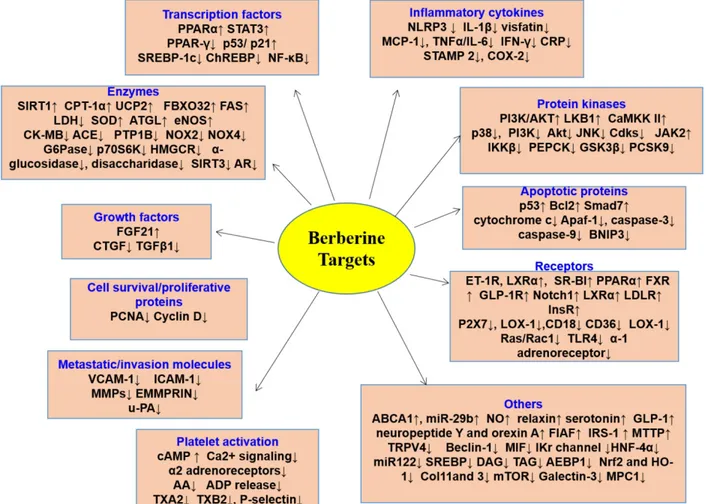
Cardiovascular and metabolic diseases (CVMD) are the leading causes of death worldwide, underscoring the urgent necessity to develop new pharmacotherapies. Berberine (BBR) is an eminent component of traditional Chinese and Ayurvedic medicine for more than 2000 years. Recently, BBR has attracted much interest for its pharmacological actions in treating and/or managing CVMD. Recent discoveries of basic, translational and clinical studies have identified many novel molecular targets of BBR (such as AMPK, SIRT1, LDLR, PCSK9, and PTP1B) and provided novel evidences supporting the promising therapeutic potential of BBR to combat CVMD. Thus, this review provides a timely overview of the pharmacological properties and therapeutic application of BBR in CVMD, and underlines recent pharmacological advances which validate BBR as a promising lead drug against CVMD.
Key words: berberine, cardiovascular diseases, metabolic diseases, targets, therapeutics
Introduction
Cardiovascular diseases (including atherosclero-sis, myocardial infarction, hypertension, cardiac hyp-ertrophy and heart failure), and metabolic diseases (including diabesity, obesity, and non-alcoholic fatty liver disease), are the leading causes of death worldwide [1-8]. These diseases are caused by the combined effects of multiple pathological factors, and their pathogenesis has not been fully clarified yet
[1-8]. Although the prevention and treatment of cardiovascular diseases and metabolic diseases (CVMD) have made great progress in the past 20 years, the morbidity and mortality arising from CVMD are still very high [1-8]. Western medicine is still the mainstream therapy of CVMD [9-12]. For example, hypoglycemic agents, statins, anticoagula-nts, beta receptor blockers, nitrates, and anti-
Ivyspring
International Publisherthrombotic drugs are widely used in patients with CVMD [9-11, 13, 14]. Despite widespread evidences showing that these drugs are effective in a treatment of CVMD, potentially serious adverse consequences remain key challenges [9-14]. Therefore, there is an urgent need to identify alternative and complemen-tary therapies to better manage CVMD. Berberine (BBR) is widely used in Asian countries (mainly in China) due to its good clinical and safety profile [15-17]. With the advances of pharmacological resear-ch, BBR is considered to be one of the most promising natural product-derived drug for the treatment of CVMD (Figure 1). To this end, we provide a timely and insightful overview of the therapeutic potential and molecular targets of BBR in treating CVMD.
BBR – basic characteristics and history of
use
BBR is the principal bioactive ingredient of
Rhizoma coptidis (also named 'Huang Lian' in Chinese),
a common traditional Chinese medicinal herb used for the therapy of inflammatory disorders and diabetes mellitus (DM) [18, 19]. The earliest record of the use of Rhizoma coptidis as a medication is dated in A.D. 200 in the book of ‘Shen Nong Ben Cao Jing’ [18, 19]. For the first time, the anti-diabetic effect of
Rhizoma coptidis was recorded in the book Note of Elite Physicians in about 1500 years ago by Hongjing Tao
[20].
BBR (C20H18NO4) is a quaternary ammonium salt derived from isoquinoline alkaloid, with a molar weight of 336.36 g/mol [21, 22]. BBR is a yellow powder that is odorless and has characteristic alkaloid bitterness [21]. It is sparingly soluble in water, slightly soluble in ethanol or methanol; however, the salt form is relatively water-soluble [21, 22]. BBR can be easily obtained from medicinal plants or through total synthesis [23, 24].
Bioavailability and metabolism of BBR
Chloride or sulfate of BBR are commonly used
for clinical purposes [15, 17]. Nevertheless, pharma-cokinetic data in rodents and humans have revealed poor absorption from the gut and rapid metabolism in the body that caused its low oral bioavailability [21]. For example, BBR is converted to ionic form under physiological conditions and self-aggregates at low pH values [25-27]. Self-aggregation of BBR reduces its solubility in the gastrointestinal tract and its ability to permeate the gut wall [26, 27]. P-glycoprotein (P-gp) is located in the epithelial cell membrane and can efflux many drugs (including BBR), thereby limiting their oral bioavailability [26]. P-gp inhibitors, including D-tocopheryl polyethylene glycol 1000 succinate, are common adjuvants to increase the oral bioavailability of BBR [25]. In addition, penetration enhancers and lipid particle delivery systems can also increase the bioavailability of BBR [27]. BBR is metabolized by oxidative demethylation and glucur-onidation to berberrubine, thalifendine, demethylene-berberine and jatrorrhizine and their corresponding glucuronides in the liver [21, 28] (Figure 2). CYP2D6 is the major cytochrome P450 (CYPs) for BBR metabol-ism, followed by CYP1A2, 3A4, 2E1 and CYP2C19. Finally, BBR metabolites are excreted through bile, feces, and urine [21, 28].
Although plasma concentration of BBR is low, the tissue concentrations of BBR and its metabolites are high [29]. BBR and its metabolites are widely distributed in the liver, kidney, muscle, lung, brain, heart, pancreas and adipose tissue [29-31]. BBR can also penetrate the blood-brain barrier [32]. Specifically, the rapid clearance of BBR from plasma as compared to the hippocampus indicates that BBR may have an important effect on hippocampal neurons [31]. Moreover, infusion of BBR (2 μg/h, 28d) by bilateral hypothalamic paraventricular nucleus (PVN) through an osmotic minipump can reduce hypertension and sympathoexcitation in two-kidney, one-clip (2K1C) renovascular hypertensive rats by ROS/ERK1/2 (extracellular-signal regulated kinase 1/2)/ inducible nitric oxide (iNOS) pathway [33].
Figure 1. Therapeutic potential of BBR in cardiometabolic diseases. Current researches support that BBR may play a therapeutic role in the treatment of cardiovascular disease (including atherosclerosis, heart failure, myocardial infarction, arrhythmia, abdominal aortic aneurysm, stroke) and metabolic diseases (including nonalcoholic fatty liver, obesity, diabetes and its cardiovascular complications).
Figure 2. Selected metabolites of BBR in human. BBR is metabolized in the body by metabolic pathways (such as demethylation, glucuronidation etc) to thalifendin, berberrubine, jatrorrhizin, demethyleneberberin.
Emerging studies have shown that BBR is almost safe at conventional doses, with a relatively low incidence of adverse reactions, such as gastrointes-tinal discomfort, and transient increases in plasma bilirubin levels [27, 34]. Although the safety of BBR is relatively high, it should be taken carefully to avoid adverse reactions in specific cases. For example, BBR replaces bilirubin in binding to albumin (in nearly 10 times greater effect compared to phenylbutazone), so any BBR containing herbs should be avoided in jaundice in pregnant women and infants [35]. BBR interacts with macrolides and it may lead to potentially dangerous arrhythmias [36]. BBR in combination with statins increases cardiotoxicity by inhibiting CYP3A4 and human ether-a-go-go related genes (hERG) potassium channels [37]. On the other hand, BBR can prevent toxic reactions in different tissues caused by antitumor drugs such as cisplatin [38], cyclophosphamide [39], doxorubicin [40] and bleomycin [41] as well as side effects of analgesics (e.g. acetaminophen [42]).
BBR in the treatment of cardiovascular
diseases
Atherosclerosis
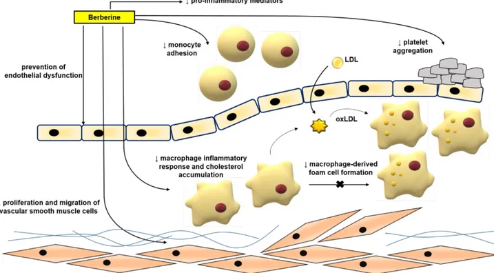
Atherosclerosis is mainly a lipid metabolic disorder which underlies multiple cardio- and cerebro-vascular diseases [43-46]. Atherosclerosis commences with endothelial dysfunction, followed by neointima formation, lipid accumulation, foam cell
formation, and plaque rupture [43, 44, 46-48]. BBR exerts protective effects against atherosclerosis by modulating various pro-atherogenic cellular events (Figure 3).
Mechanism of BBR in protecting against atherosclerosis in vitro
Normalizing endothelial function
Endothelial dysfunction and its related complications are typically specified by reduced local bioavailability of nitric oxide (NO) and excessive oxidative stress; the increased NADH/NADPH and xanthine-oxidase activity are important factors for oxidative stress in endothelial cells [49-51]. Enhanced adhesion of leukocytes to the endothelium plays an initiating role in inflammation [3, 7, 52, 53]. Studies have proven the ameliorative effects of BBR in endothelial dysfunction via regulating reactive oxygen species (ROS)/NO balance [49, 52, 54]. BBR treatment is capable to reduce oxidized low density lipoprotein (oxLDL)-stimulated production of ROS in human umbilical vein endothelial cells (HUVECs) [54]. BBR could inhibit oxLDL-stimulated monocyte adhesion to HUVECs via the mechanism associated with the suppression of vascular cell adhesion molecule-1 (VCAM-1) and intercellular adhesion molecule-1 (ICAM-1) [52]. BBR has been shown to reduce the tumor necrosis factor α (TNFα)-stimulated expression of the pro-inflammatory monocyte chemotactic protein-1 (MCP-1) and ICAM-1 in human amniotic epithelial cells (HAECs) and to suppress the
activation of nuclear factor kappa B (NF-κB) in HAECs involving AMP protein kinase (AMPK)- dependent pathway [55]. The exposure of HUVECs to oxLDL or TNFα significantly increased the expression of lectin-like oxidized low density lipoprotein receptor-1 (LOX-1) and the production of intracellular ROS [56-58]. Treatment with BBR reduced ERK1/2 signaling pathway, the expression of nicotinamide adenine dinucleotide phosphate-oxidase 2 (NOX2) and the expression of VCAM-1 and ICAM-1 [56]. BBR protected against palmitate-induced endothelial dysfunction via increasing NO level, and endothelial nitric oxide synthase (eNOS) expression and down- regulating the expression of NOX4 in HUVECs [59]. BBR also contributed to the inhibition of the vascular inflammation via preventing angiotensin II (Ang II)-induced adhesion of monocytes to endothelial cells and reduced ROS and MCP-1 expression in HUVECs [60]. Berberrubine, the active metabolite of BBR, has recently been shown to decrease xanthine oxidase activity and reduce TNFα-induced ICAM-1 expre-ssion in HUVECs [61].
BBR also dose-dependently inhibited the proliferation of HUVECs induced by oxLDL; its anti-proliferative and anti-inflammatory properties are exerted through decreasing the expression of NF-кB, LOX-1, proliferating cell nuclear antigen (PCNA) and inhibiting the phosphorylation of the phosphoinositide 3-kinase (PI3K)/Akt, ERK1/2, p38
mitogen activated protein kinases (MAPKs) signaling pathways [62]. In HUVECs, BBR treatment showed protective effects also against oxLDL-induced endothelial injury and apoptosis through cytochrome c-mediated caspase activation pathway [54].
Inhibiting migration and proliferation of vascular smooth muscle cells (VSMCs)
The enhancement of VSMCs proliferation and migration is an important factor for causing of neointimal hyperplasia and luminal stenosis after vascular injury [63]. Components of extracellular matrix, cytokines, shear stress, ROS, and other factors can significantly reduce the expression of markers of the differentiation of VSMCs, increase VSMCs proliferation and migration, and also a capacity for the synthesis of the extracellular matrix and promo-tion of neointimal formapromo-tion [64]. BBR had a beneficial effect on vascular remodeling [65, 66]. BBR inhibited fetal bovine serum-induced proliferation of VSMCs by up-regulation of peroxisome proliferator-activated receptor α (PPARα) expression and NO concentration [67]. BBR inhibited the migration of human aortic VSMCs by down-regulating matrix metalloproteinase (MMP)-2 and MMP-9, as well as urokinase-type plasminogen activator (u-PA) [68]. Similarly, BBR attenuated Chlamydia pneumoniae infection-induced VSMCs migration by inhibiting PI3K and down- regulating the expression of MMP-3 and MMP-9 [69].
Figure 3. Anti-atherosclerotic effects of BBR. The role of BBR in inhibiting atherosclerosis includes improving endothelial dysfunction; inhibiting smooth muscle cell proliferation and migration; reducing monocyte adhesion, macrophage inflammation and cholesterol aggregation, foam cell formation, and platelet aggregation. Abbreviations: low density lipoprotein (LDL); oxidized LDL (oxLDL).
In addition to its effect on the extracellular matrix, BBR reduced platelet-derived growth factor (PDGF)-induced proliferation of VSMCs by activating AMPK/p53/p21 signaling, while simultaneously inactivating Ras/Rac1/Cyclin D/Cdks, and attenua-ted PDGF-induced migration by inhibiting Rac1 and Cdc42 [70]. BBR limited the proliferation and migration of lysophosphatidylcholine (lysoPC)- stimulated VSMCs by decreasing ROS level and subsequent activation of the ERK1/2 pathway [71]. BBR also significantly decreased the proliferation and migration of VSMCs stimulated by Ang II and heparin binding epidermal growth factor (HB-EGF) by inhibiting the activation of Akt pathway [65]. In norepinephrine (NE)-induced pulmonary arterial VSMC proliferation and migration models, BBR can reverse the effect of NE by increasing protein phosphatase 2A (PP2A) signaling, suggesting that BBR may have a therapeutic effect in pulmonary hypertension [72]. Furthermore, BBR induced the cell cycle arrest of A7r5 cells by disrupting the combination of p27, p21 and Skp2, inhibited the proliferation of A7r5 induced by PDGF-BB and enhanced the anti-proliferative effect of adrenal steroid dehydroepiandrosterone sulfate on A7r5 [73]. BBR blocked the cell cycle progression of VSMCs at G(1) phase by down-regulating the expression of cyclin D1, without affecting G(2)/M phase [74]. Inhibiting of macrophage-derived foam cell formation, inflammation, and inflammasome activation
The uptake of oxLDL into macrophages is mediated by diverse scavenger receptors (SRs), such as scavenger receptor A (SR-A)I/II, scavenger receptor class B type I (SR-BI), LOX-1, and the cluster of differentiation 36 (CD36)[75]. When oxLDL is endocytosed into macrophages, they develop into foam cells that form the core of atherosclerotic lesion [76, 77]. BBR reduced oxLDL uptake by inhibiting CD36 and LOX-1, and promoted cholesterol efflux by suppressing the expression of adipocyte enhancer- binding protein 1 (AEBP1) in macrophages [78]. BBR also inhibited the effect of oxLDL on macrophage- derived foam cell formation by inhibiting LOX-1, while up-regulating SR-BI expression [79]. Moreover, it has been reported that BBR minimized the accumulation of lipids in human macrophages exposed to hypercholesterolemic serum by reducing the process of micropinocytosis and also decreased the secretion of MCP-1 [80]. Furthermore, BBR eliminated foam cells by enhancing liver X receptor α (LXRα) and ATP-binding membrane cassette transport protein A1 (ABCA1)-stimulated cholesterol efflux [81]. The combination of atorvastatin and BBR suppressed the foam cells formation more effectively
than atorvastatin alone [82]. BBR suppressed the formation of foam cells from THP-1-derived macrophages by increasing the expression of AMPK and silent information regulator 1 (SIRT1), by decreasing the expression of PPARγ, and reducing the uptake of oxLDL [82]. In addition, BBR combined with atorvastatin diminished the levels of serum triglyceride (TG), TC, and low-density lipoprotein cholesterol (LDL-C) and inflammation and oxidative stress markers [83]. BBR combined with atorvastatin down-regulated the expression of LOX-1 through endothelin 1 receptor (ET-1R) in monocytes/ macrophages [83], and in macrophages-derived foam cells, BBR-mediated sonodynamic therapy enhanced cholesterol efflux by promoting the production of ROS, and enhanced autophagy through the PI3K/Akt/mammalian target of rapamycin (mTOR) signaling pathway [84]. However, it was shown that BBR induced the foam cell formation and promoted atherosclerosis by inducing the expression of SR-A in macrophages and increasing the uptake of modified low-density lipoprotein (LDL) [85]. Although the vast majority of above mentioned studies show that BBR can suppress foam cell formation and the progression of atherosclerosis, this conclusion is still controversial. BBR also exerted anti-inflammatory effects in macrophages stimulated with oxLDL. BBR down- regulated lipopolysaccharide (LPS)-induced IL-1β, IL-6, iNOS, MCP-1, cyclooxygenase (COX)-2 and MMP-9 expression, and p-MAPKs (including p38, c-Jun N-terminal kinase (JNK), and ERK) by activating AMPK [86]. BBR reduced the production of inflammatory markers and induced autophagy in oxLDL-exposed J774A.1 cells [87]. The mechanism of effect was associated with the activation of AMPK/mTOR signaling pathway [87]. Furthermore, BBR inhibited the activation of the Nod-like receptor family pyrin domain containing-3 (NLRP3) inflamm-asome: BBR exposure reduced ROS-dependent activation of NLRP3 inflammasome in macrophages and inhibited the expression and release of IL-1β by inhibition of NF-κB [88]. BBR can also reduce NLRP3 inflammatory expression in PMA-stimulated macro-phages by inhibiting Toll-like receptor 4 (TLR4)/ myeloid differentiation factor 88 (MyD88)/NF-κB signaling pathway [89]. BBR decreased sodium uric acid crystal induced inflammasome activation by suppressing the expression of NLRP3 and IL-1β [90]. In addition, in adipose tissue-derived macrophages, BBR suppressed NLRP3 inflammasome activation and IL-1β production stimulated by saturated fatty acid (palmitate), by activating AMPK-dependent autophagy [91]. BBR also inhibited the activation of the NLRP3 inflammasome pathway, and then improved liver injury in mice by inhibiting the
purinoreceptor P2X7 [92]. TNFα and LPS stimulated the expression of miR-23a in RAW 264.7 macrophages, resulting in TLR4/TNF receptor-associated factor 2 (TRAF2)-mediated activation of apoptosis-signaling kinase 1 (ASK1) and phosphorylation of p38, which mediate inflammatory reaction [93]. BBR treatment inhibited LPS and TNFα-induced inflammatory responses by up-regulating miR-23a, ameliorating TLR4, TRAF2, TNFα, IL-6 and IL-23 gene expression, and inhibiting TLR4/TRAF2-mediated ASK1/p38 phosphorylation [93]. After percutaneous coronary intervention, macrophage activation plays an essential role in neovascular atherosclerosis and in-stent restenosis [94]. Galectin-3, which was mainly expressed on macrophages, is an important mediator of inflammation. BBR down-regulated oxLDL-induced macrophage activation by down-regulating Galectin-3
via NF-κB and AMPK signaling pathways [94].
Inhibiting platelet activation
BBR has been shown to effectively prevent thrombus formation in arteries [95], and assisted thrombolysis induced by the plasminogen activators urokinase and streptokinase [96]. The anti-platelet aggregation effects of BBR have been demonstrated in platelets isolated from healthy subjects, patients with platelet hyperaggregability, and also patients with atherosclerotic cerebral infarction [95]. Chu et al. well reviewed the effect of BBR on platelets in 1996 [97]. BBR in vitro could inhibit platelet aggregation stimulated by adenosine diphosphate (ADP), colla-gen, adrenaline, arachidonic acid (AA), and calcium ionophore A23187 [97]. It also had inhibitory effect on clot retraction induced by ADP [98]. BBR inhibited platelet activation by modulating the effect on cAMP level in platelets, antagonistic activity against Ca2+, partial and competitive binding with α2 -adreno-receptors on platelet membrane, affecting AA metabolism and ADP release from platelets [97].
BBR chloride decreased plasmatic levels of thromboxane B2 and P-selectin in mice with dextran sulphate sodium-stimulated colitis [99]. BBR chloride also inhibited platelet activation via down-regulation of P-selectin expression and inhibiting fibrinogen binding to platelet GP IIb/IIIa integrin receptor on the surface of platelets [100]. Shah et al. [101] reported that BBR inhibited collagen-induced platelet aggregation without affecting the platelet responses to the other platelet agonists including platelet-activating factor (PAF), AA and adrenaline. Its anti-platelet activity has been suggested to be exerted via suppressing with the collagen-induced adhesion process without any effect on cAMP, or A23187-induced platelet aggregation [101]. While in one animal study investigating the
effect of BBR on GPVI (glycoprotein receptor for collagen, expressed in platelets) BBR could not statistically inhibit the expression of GPVI [102], another study has proven the protective effects of BBR in combination with ligustrazine (alkylpyrazine from
Ligusticum chuanxiong) in a rat model of coronary
microembolization [103]. In this study, ADP-induced platelet activation, von Willebrand factor, ET-1, and levels of TNFα, IL-1β, and ICAM-1 in serum and heart tissues were decreased by BBR/ligustrazine combina-tion [103]. Tissue factor (TF) is closely related to coag-ulation; its expression by vascular cells surrounding blood vessels is stimulated by pro-inflammatory substances such as LPS or TNFα [104]. In this regard, BBR attenuated LPS-stimulated TF protein expression by suppressing NF-κB, Akt and MAPK signaling pathways in THP-1 cells [105]. However, another study revealed that BBR could increase TNFα- stimulated TF expression in HAECs, and could decrease the expression of TF pathway inhibitor in HAECs and in arteries of apolipoprotein E-deficient mice (ApoE-/-) mice, what might promote thrombosis [104]. BBR therefore might act bidirectionally on TF expression in different cell types [105].
Anti-atherosclerotic effects of BBR in vivo
Diverse investigations have reported the therapeutic potential of BBR to inhibit atherosclerosis in mouse models of atherosclerosis. For example, BBR treatment (150 mg/kg/d, p.o., 12wk) obviously reduced the atherosclerotic plaque area and displayed suppression of inflammatory and oxidative markers [106]. Specifically, BBR diminished the serum levels of IL-1β and TNFα, and decreased the expression of ICAM-1, iNOS, and IL-6 in the aorta. Furthermore, the treatment with BBR reduced the translocation of NF-κB to the nucleus [106]. In homocysteine thiolactone (HTL)-fed ApoE-/- mice, BBR (1.0 g/kg/d,
p.o., 8wk) increased atherosclerotic plaque stability in
the carotid artery and normalized the redox state. These protective effects on vascular function might be ascribed to an activation of PPARγ, which leads to a down-regulation of oxidative stress [107]. BBR also abrogated the HTL–induced oxidative stress in HUVECs by activating PPARγ [107].
Visfatin is a pro-inflammatory adipokine, which is expressed in visceral fat and induces endothelial dysfunction by promoting the production of inflammatory and adhesion molecules [108]. Oral administration of BBR (5 mg/kg/d, p.o., 12wk) in ApoE-/- mice lowered the serum expression of visfatin and inflammatory cytokines (TNFα and IL-6), as well as it reduced the distribution of visfatin in the atherosclerotic plaques [108]. BBR pretreatment also reversed the visfatin-induced increase of IL-6 and
TNFα in HUVECs [108]. The amelioration of visfatin- induced endothelial dysfunction was associated with the inhibition of MAPK signaling pathway (p38 and JNK) [108]. Moreover, BBR suppressed the increase of p-p38 MAPK, p-JNK and Bcl2- associated X protein (Bax) in visfatin-induced endothelial dysfunction in HUVECs [108]. Furthermore, it was suggested that the protective effects of BBR (in drinking water (0.5
g/L), 14wk) in ApoE-/- mice can be related to the
modulation of gut microbiota, specifically an up-regulation in Akkermansia spp. abundance which has been known to regulate inflammation, metabolic endotoxemia, and gut barrier integrity [109]. A recent study showed that BBR (50 mg/kg, p.o., twice weekly, 12wk) inhibition of HFD-induced atherosclerosis in ApoE-/- mice is associated with changes in gut micro-biota composition, such as BBR treatment altering an occurrence of Firmicutes and Verrucomicrobia [110].
Increased circulating endothelial microparticles in the peripheral circulation show the state of endothelial damage and endothelial cell activation, apoptosis and injury [111]. BBR treatment (1.2 g/d,
p.o.) in healthy humans led to a decrease in serum
malondialdehyde and circulating CD31+/CD4- micro-particles (as markers of endothelial injury) and the improvement of flow-mediated vasodilation. BBR contributed to an improvement of endothelial function via up-regulating eNOS expression and down-regulating NOX4 expression, and ROS production in HUVECs [112-114].
The MyD88 dependent TLR4 signaling pathway plays a critical role in hypertension-induce endothe-lial damage. TLR4 activation could activate NF-κB, leading to endothelial cell injury via an increased production of inflammatory enzymes and mediators (including COX-2, IL-1, and IL-6) [115]. An increased level of TNFα initiates apoptosis and endothelial cell turnover, resulting in the formation of atherosclerotic plaques [115]. BBR treatment inhibited the apoptosis, decreased the expression of NF-κB as well as the levels of TNFα and IL-6 in aortic endothelial cells isolated from spontaneously hypertensive rats (SHR) by decreasing the expression of TLR4 and MyD88 [116]. Endothelium-dependent contractions (EDCs) are also involved in endothelial dysfunction, and EDCs are caused by contracting factors such as endothelial COX-produced prostanoids or augmented level of ROS [117]. EDCs of the carotid arteries of SHR were reduced by BBR administration [118]. This effect has been reported to be caused by triggering of activated AMPK, inhibition of endoplasmic reticulum (ER) stress, reduction of ROS and expression of COX-2 [118]. BBR (5, 10 mg/kg, i.p.) has also been reported to inhibit NF-κB activity and decrease VCAM-1 production in lung of LPS-stimulated rats
[119].
Cardiac hypertrophy and heart failure
Cardiac remodeling is associated with progress-ive cardiac hypertrophy, fibrosis, and the eventual occurrence of heart failure (HF) [120-123]. Interven-tions in cardiac remodeling are important strategies for the treatment of HF [124]. The effect of BBR on HF is shown in Figure 4.
In animal models, BBR can reduce the degree of HF. For example, in ischemic left ventricular heart failure dogs, intravenous injection of BBR (1 mg/kg, within 3 min) and then repeated infusion (0.2 mg/kg/ min, 30 min) for 10 consecutive days, increased the cardiac output and peak rate of rise of left ventricular pressure (+dp/dt) while left ventricular end-diastolic pressure (LVEDP), diastolic blood pressure (DBP), and systemic vascular resistance were decreased[125]. The total saponins of Panax ginseng (20 mg/kg/d) combined with BBR (20 mg/kg/d), and captopril were administered for 12 consecutive days to rats in a model of HF induced by injecting of high dose of isoproterenol (s.c.), and a similar anti-HF effect was observed in both groups [126]. BBR (63 mg/kg/d, p.o., 4wk) further proved to be a potential drug for
improving HF symptoms by inhibiting the Ca2+
overload in cardiomyocytes [127].
Pathological cardiac hypertrophy is a critical intermediate step toward HF and other cardiac diseases [128]. BBR (10 mg/kg/d, p.o., 8wk) prevented the development of left ventricular hypertrophy in rats caused by pressure overload; +dp/dt increased, while the weight of the whole heart and left ventricle decreased, and the increase of left ventricular myocardial cell size was significantly reduced [129, 130]. Mechanistic studies have shown that BBR can up-regulate F-Box protein 32 (FBXO32), an E3 ubiqui-tin ligase, and down-regulated mTOR, ERK1/2, as well as p38 phosphorylation, which enhances auto-phagy and thus inhibits cardiac hypertrophy [128, 131]. Cardiac fibrosis is an important pathological change in the development of HF, arrhythmia and cardiac arrest [132]. BBR can restrict cardiac fibrosis by up-regulating the expression of relaxin in vitro [133]. BBR administration (5, 10 mg/kg/d, p.o., 4wk) also obviously reduced cardiac fibrosis in two-kidney, two-clip (2K2C) rats via increased levels of NO and cAMP in left ventricular tissue[134].
Abdominal aortic aneurysm
Abdominal aortic aneurysm is characterized by dilatation of the abdominal aorta with an etiology not completely understood [135]. The presence of aneurysm leads to the weakening of the aortic wall, followed by a progressive dilatation and potential risk
of aortic rupture [136]. To date, only very limited studies investigating the effects of BBR against abdominal aortic aneurysm were published. One of the potential causes of formation of aneurysms is the increase in the ratio of collagen to elastin [137]. It has been evidenced that BBR significantly reduced collagen synthesis in cardiac fibroblasts subjected to Ang II [138]. The activation of MMPs is also a mechanism that participates in the etiology of aneurysms since it weakens the aortic media [139]. In this sense, it was reported that BBR obviously decreased the activity and expression of MMP-9 as well as the expression of extracellular MMP inducer (EMMPRIN) in stimulated macrophages [140]. In addition, BBR treatment ameliorated vascular remodeling in a rat model of metabolic syndrome by reducing the aortic levels of MMP-2 [141].
It is well established that the formation of abdominal aortic aneurysm is related to abnormal wall stiffness [142]. A clinical study investigated the effects of the two months consumption of 200 mg red yeast rice (RYR), 500 mg BBR and 10 mg policosanols in 70 hypercholesterolemic patients [143]. The intervention significantly improved aortic stiffness by reducing lipid levels and increasing aortic pulse wave velocity [143]. Furthermore, in aged mice, it has been evidenced that BBR induced the relaxation of VSMCs with decreasing of blood pressure and also reduced vascular stiffness via suppression of the activity of Ca2+ channel transient receptor potential vanilloid 4 (TRPV4) [144]. The effect of BBR on abdominal aortic aneurysm was summarized in Figure 4.
Ischemia-reperfusion injury
Ischemic heart disease (ISHD) is primarily caused by coronary atherosclerosis and its complications [145, 146]. Myocardial infarction is the most severe ISHD with high mortality [145]. Reperfusion is beneficial for ischemia, but it can also cause severe cardiac damage [145]. Thus, studying the underlying mechanisms of myocardial ischemia- reperfusion (I/R), contributes to the treatment of I/R injury in myocardial tissue [145, 146]. The effect of BBR on myocardial ischemia was shown in Figure 4.
Experiments on isolated cardiomyocytes in hypoxia-reoxygenation model showed that pretreat-ment with BBR resulted in reduction in lactate dehydrogenase (LDH) and malondialdehyde (MDA) release, which are biomarkers of the extent of I/R injury [147]. BBR inhibited I/R injury by the following mechanisms: i) direct and/or indirect antioxidant activity [148]; ii) post-ischemic anti-inflammatory activity [149]; iii) vasodilation of coronary arteries [150]; iv) anti-apoptotic activity on cardiomyocytes [151]; v) suppressing an activation of autophagy
during I/R[152]; vi) promoting angiogenesis in heart after I/R injury[153].
Antioxidant activity of BBR
BBR showed direct antioxidant activity in several cell-free systems, such as DPPH radical scavenging assay [154, 155]. Moreover, antioxidant activity was observed in cultured cells subject to oxidative stress [148, 156]. BBR inhibited oxidative stress by increasing super oxide dismutase (SOD), uncoupling protein 2 (UCP2) and decreasing NOX expression, via SIRT1/FoxO (forkhead box O) or AMPK signaling pathways [157].
Anti-inflammatory effects of BBR
BBR is anti-inflammatory active by decreasing the secretion of an array of pro-inflammatory cytokines/mediators (IL-6, IL-1β, and TNFα) in myocardial tissue and serum by suppressing PI3K/Akt signaling pathway [157-159]. BBR in cardiomyocytes in the mouse model of I/R injury inhibited inflammatory responses through the suppression of NF-κB signaling pathway [149]. Another study showed that BBR can also activate SIRT1 signaling, and thus further reduce myocardial inflammatory response [160].
Hypotensive effects of BBR
BBR has hypotensive effects by functioning as a vasodilator in isolated blood vessels [161, 162]. Vasodilation of coronary arteries and thus coronary flow is of key importance during the I/R injury of the heart [163]. In this context, BBR enhanced coronary blood flow in isolated guinea pig hearts with ventricular fibrillation [150]. Moreover, BBR can stimulate cardiac contractility (positive inotropic activity) by increasing intracellular calcium levels in addition to lowering peripheral vascular resistance and blood pressure [127]. Several mechanisms are proposed for vasodilation and/or hypotensive effect of BBR: i) the antagonism on α1-adrenoreceptors on VSMCs [164, 165]; ii) the enhancement of the hypotensive effect of acetylcholine (ACh) on nervus
vagus, and thus the inhibition of carotid sinus pressor
reflex [166]; iii) the endothelium-dependent release of NO [167, 168]; iv) via angiotensin-converting enzyme (ACE) inhibition of the NO-cGMP axis [169]; v) the direct activation of K+ channels in arterial VSMCs leading to hyperpolarization, thereby the inhibition of calcium influx leading to smooth muscle relaxation [168, 170]; vi) the inhibition of L- and T-type voltage-gated Ca2+ currents in ventricular myocytes [171].
Anti-apoptotic effects of BBR
protecting against I/R injury is the inhibition of apoptosis of cardiomyocytes. For example, in a mouse model of myocardial I/R, BBR inhibited the activation of caspase-3, caspase-9 and apoptotic protease- activating factor 1 (Apaf-1) in cardiomyocytes, while increased the expression of Bcl-2-like protein 1 and p53 [149]. In the rat hearts subjected to I/R injury, BBR treatment decreased AMPK and Bcl-2-like protein 1 in the myocardial risk areas, while increased AMPK in the non-ischemia areas compared to the control hearts [172]. In neonatal rat cardiomyocytes, BBR decreased hypoxia-reoxygenation-stimulated cardiomyocytes apoptosis, increased Bcl-2/Bax ratio, activated PI3K-Akt, AMPK and eNOS phosphor-ylation, while decreased caspase-3 expression [173, 174]. Recent studies on hypoxia-reoxygenation injury on H9C2 rat cardiac myoblasts proposed that BBR prevents apoptosis by modulating Notch1/Hes1- PTEN (phosphatase and tensin homolog)/Akt signaling pathway [175], as well as Smad7 (Mothers against decapentaplegic homolog 7) activation [151]. BBR-mediated activation of Smad7 pathway resulted in the suppression of caspase-3 expression and activity. Indeed, the Smad7 knockdown confirmed this hypothesis [151]. Importantly, BBR also activated the Janus kinase 2 (JAK2)/ signal transducer and activator of transcription 3 (STAT3) pathway, and thus decreased the endoplasmic reticulum (ER) stress [176].
Effect of BBR on autophagy and angiogenesis
Inhibition of activation of autophagy is an additional mechanism underlying the protective effects of BBR [152]. Incubation of the cells with BBR or treatment with BBR increased survival of cells,
decreased the infarct zone in mice hearts, and suppressed autophagy, which was confirmed by the decreased expression levels of autophagy-related proteins such as SIRT1, BNIP3, and Beclin-1 [152]. BBR can also be useful after I/R injury, since BBR can promote angiogenesis of the small vascular beds in coronary arteries [153]. Post-ischemic application of BBR for 14 days reduced the infract zone, improved the heart function, as well as up-regulated miR-29b expression, and enhanced cell proliferation and migration in endothelial cells from left anterior descending (LAD) coronary artery [153].
Importantly, similar protective activities of BBR against I/R injury were observed also in non-cardiac tissues [177]. In neuronal cells during ischemia, BBR reduced p53 and cyclin D1, enhanced phosphory-lation of Bad and decreased cleavage of caspase-3, thereby inducing cell cycle arrest and inhibiting apoptosis [177]. During the reperfusion phase, BBR protected from cerebral ischemia injury via activating the PI3K/Akt signaling pathway [177]. A pretreat-ment with BBR significantly preserved cells in
response to a strong oxidant (CoCl2) [178]. BBR
injections were given prior to I/R surgery in rats and led to increased survival of neurons due to anti- apoptotic effects mediated by PI3K/Akt signaling pathway [179]. Moreover, in a mouse model of cere-bral ischemia, BBR decreased the neuronal apoptosis
via activating PI3K/Akt signaling pathway [180].
Protective activity was not only BBR-specific but it can be related to the other members of structural class of isoquinoline alkaloids, as both coptisine [181] and palmatine [182] showed similar protective effects against I/R injury on hearts of rats.
Figure 4. Effects of BBR on heart failure, arrhythmia, myocardial ischemia and abdominal aortic aneurysm. Specifically, BBR prevents ischemia/reperfusion injury via its positive inotropic activity, increased phosphorylation of Bad, decreased production of pro-inflammatory mediators (IL-6, IL-1β, and TNFα), reducing oxidative stress, blood-pressure lowering, anti-apoptotic effects and protective effects against endoplasmic reticulum stress. BBR prevents heart failure by increasing cardiac output, and decreasing LVEDP and DBP. BBR prevents arrhythmia by reducing ventricular premature beats and tachycardia. BBR prevents abdominal aortic aneurysm by reducing vascular remodeling and pressure, reducing aortic stiffness, modulating lipid level, and increase aortic pulse wave velocity. ↑indicates increase or activation, and ↓indicates decrease or suppression. Abbreviations: Bad, Bcl-2-associated death promoter; diastolic blood pressure (DBP), interleukin-1β (IL-1β), interleukin-6 (IL-6), left ventricular end-diastolic pressure (LVEDP) , peak rate of rise of left ventricular pressure (+dp/dt), tumor necrosis factor α (TNFα).
Furthermore, administration of BBR (100 mg/ kg/d, p.o., 14d) in rats prior to I/R injury (a surgical occlusion of LAD coronary artery) resulted in a reduced size of infarction and protection from arrhythmias [172]. BBR prevented the occurrence and duration of isolated premature beats, ventricular fibrillation and ventricular tachycardia [172]. In another model of acute myocardial infarction (induc-tion by isoproterenol injec(induc-tion), BBR (30, 60 mg/kg/ d, p.o., 14d) decreased the ST elevation stimulated by acute myocardial ischemia, and also down-regulated serum levels of ischemic biomarkers muscle/brain (MB) isoenzyme of creatine kinase (CK-MB), IL-6, TNFα and LDH [183]. Moreover, ex vivo experiments evaluating I/R on isolated hearts showed that hearts pre-treated with BBR before ischemia had improved preservation of left ventricular developed pressure and LVEDP when compared to the non-treated group [172]. Importantly, BBR had no influence on these parameters per se (on hearts without I/R injury) [172]. In addition, a solid dispersion of BBR and sodium citrate (HGSD) improved BBR bioavailability and membrane permeability, and HGSD treatment (12.5, 25, 50 mg/kg/d, p.o., 7d) protected the rat heart from I/R injury by attenuating NF-κB and JNK signaling pathways [184].
Stroke
Similar to myocardial infarction, stroke also results from vascular or microvascular diseases which cause an interruption of cerebral blood supply and consequently brain dysfunction [185]. To date, the reperfusion is the merely approved treatment for acute ischemic stroke [186]. Consistently, thromboly-tic/anti-platelet agents and surgery are the only available options [187]. However, due to the fact that multiple mechanisms are involved in stroke progression, the therapeutic effect of current regimens is limited and agents that possess multiple pharmacologic actions have attracted much attention. Inflammation, apoptosis, and oxidation are three major mechanisms for the pathogenic proceeding of stroke [188].
In addition to the routine treatment, the admini-stration of BBR 300 mg (t.i.d., p.o.) significantly redu-ced serum levels of macrophage migration inhibitory factor (MIF) and IL-6 in patients with acute cerebral ischemic stroke and to a certain extent decreased the carotid atherosclerosis and neurological deficit [189].
The right carotid artery of rats was ligated (ischemic injury) and BBR solution (0.2‒2 mg/kg) was intraperitoneally injected. After 30 min, the rats were subjected to hypoxic conditions by breathing in air with 10% oxygen and 90% nitrogen (a model of hypoxic damage). BBR dose-dependently reduced
cerebral ischemia-hypoxic injury [190]. BBR (0.2, 0.5, 1 or 2 mg/kg, i.p. or 0.2, 0.02, 0.002 mg/kg, i.v.) inhibited ischemia-induced apoptosis via enhancing the PI3K/Akt signaling pathway and exerted its neuroprotective effects in vivo and in vitro [179, 191]. Additional mechanisms of BBR in preventing ischemic brain damage include reducing intracellular ROS levels and subsequently inhibiting of the mitochondrial apoptotic pathway [192]. Moreover, BBR (10, 40 mg/kg) can reduce the ischemic brain injury in rats by promoting the activation of Akt/ GSK3β (glycogen synthase kinase 3β) signaling pathway and claudin-5, while down-regulating the NF-κB expression [32]. A similar study showed that BBR (25, 50 mg/kg/d, p.o., 14d) pretreatment dose- dependently reduced infarct size, neurological deficits, and cerebral edema in model of cortical tissue ischemia performed by transient middle cerebral artery occlusion [193]. Its mechanism may depend on its anti-inflammatory effects, including an inhibition of nuclear translocation of high-mobility group box1 (HMGB1) and NF-κB, as well as TLR4 expression [193]. In addition, BBR can promote angiogenesis in rats suffering from cerebral ischemia-reperfusion injury, which may be related to increased expression of hypoxia-inducible factor 1α (HIF-1α) and its downstream gene-vascular endothelial growth factor (VEGF) [194].
BBR can protect neurons during focal cerebral ischemia by up-regulating anti-inflammatory cytoki-nes and down-regulating pro-inflammatory cytokicytoki-nes [195]. The combination of BBR and other drugs has been tested to improve cerebral ischemia. For example, rats were reperfused after bilateral common carotid occlusion to induce a transient global cerebral ischemia model. Co-administration of BBR (5, 10, 20 mg/kg/d, 19d) and verapamil (2mg/kg/d, 19d) enhanced brain uptake of BBR and improved brain functions in rats [196]. Berberis koreana extract (containing 7.39±0.78 mg/g of BBR) significantly ameliorated nerve injury induced by ischemic stroke in gerbils, by inhibiting COX-2 expression and PGE2 production [197]. The main components of Huang-Lian-Jie-Du-Decoction (HLJDD) are BBR, baicalin and jasminoidin, which are commonly used in traditional Chinese medicine to treat ischemic stroke [198]. HLJDD administration ameliorated ischemic stroke in rats by improving abnormal metabolism and regulating oxidative stress, neuronal autophagy, and inflammatory responses [198]. A study evaluating the Yi-qi-jie-du formula (YJ) with the main constituents ginsenosides (G), BBR and jasminoidin (J) (in a ratio 3 (G): 2 (BBR): 0.5 (J)) showed the improvement of state in the focal cerebral ischemia in rats [199].
Arrhythmia
The anti-arrhythmic effect of BBR was firstly
reported by Huang et al. in 1989 [200]. The authors induced ischemic ventricular arrhythmias in canines through occluding the LAD coronary artery and showed that BBR can prevent total ventricular prema-ture beats and ventricular tachycardia [200]. Recently, BBR (2 mg/kg, i.v.) hindered ACh-induced atrial fibrillation in rabbits via extension of action potential (AP) and effective refractory period in atrial myocytes [201]. In addition, BBR (at a concentration of 300 mmol/L) prevented stretch-induced arrhythmia in isolated myocardial infracted hearts of Wistar rats [202].
Early studies proposed that BBR has class III anti-arrhythmic effects (by blocking K+ channel) [203]. However, further studies revealed that BBR targets several types of channels such as the cardiac slow (IKs)
and rapid (IKr) delayed rectifier K+ channels,
ATP-sensitive K+ channel (KATP), inwardly-rectifying K+ channel (IKl), L-type Ca2+ (ICa) [166] and human hyperpolarization-activated cyclic nucleotide-gated 4 (hHCN4) channels [204]. BBR (10, 20 mg/kg, p.o.) suppressed expression of IKr channel in rat ventricular tissues [205]. Moreover, BBR inhibited the expression of hERG channel in hERG-transfected HEK293 cells [205]. HERG channel encoded by hERG is an important subunit of Ikr channel [206]. It crucially contributes to cardiac repolarization and its defect leads to long QT syndrome with manifestations such as delayed repolarization and prolonged QT interval [206]. BBR (27.1 mg/kg, p.o..) induced extension of the AP duration and the QT interval in guinea pigs [207]. BBR caused mature hERG reduction through inducing defect in channel trafficking, which led to an immature hERG response (unfolded protein respo-nse), and defective hERG underwent degradation in lysosomes and proteasomes [207]. Fexofenadine and resveratrol, as two drugs preventing hERG trafficking defect, can reverse BBR-induced prolongation of AP duration [208]. BBR can also block hERG channel directly via interacting with residues V625, Y652, and F656, respectively, in the central cavity of the channel [209]. The effect of BBR on arrhythmia was summarized in Figure 4.
BBR in treating metabolic diseases
BBR in diabetes mellitus and its cardiovascular complications
Diabetes mellitus (DM) is linked to chronic hyperglycemia and abnormal metabolism of carbohy-drates, lipids, and lipoproteins, which are caused by an inadequate production of insulin and/or insulin action [210, 211]. DM is increasingly being recognized
as a significant cause of mortality and morbidity, and profoundly contributes to the global health burden and death toll [210, 211]. Based on the latest disease statistical report (The International Diabetes Federation, https://www.idf.org/): approximately 425 million people were diabetics in 2017 and this number is anticipated to increase to 629 million by 2045. There are two categories of DM: Type 1 diabetes (T1DM) and Type 2 diabetes (T2DM) [212]. T1DM is characterized by the absolute deficiency of insulin secretion, while T2DM, the most common one, has features of insulin resistance (IR) and an insufficient compensatory insulin secretory response [212]. Therapeutic effects of BBR on diabetes mellitus and its cardiovascular complications were summarized in Figure 5.
Effects of BBR on T1DM and T2DM
Physiologically, blood glucose level is regulated by the liver (gluconeogenesis and glycogenolysis) and glucose utilization by the peripheral tissues. An increase in the hepatic glucose production due to an inadequate insulin secretion/action is the major cause of hyperglycemia in DM patients [210, 211]. DM can lead to many complications such as hyperlipidemia, hypertension, atherosclerosis, hyperinsulinemia, retinopathy, nephropathy and peripheral neuropathy [213, 214]. Approved oral hyperglycemic drugs such as derivatives of sulfonylurea, metformin, and thiazolidinediones are reported to induce some side effects [9-11]. In addition to the adverse effects, the conventional medicines are expensive and not always satisfactory in maintaining normal level of blood glucose [215]. Many herbal medicines with potential of anti-diabetic effect have been widely used for treating DM in various traditional systems of medicine worldwide since the immemorial time [216]. Nowadays, the market for natural anti-diabetic drugs is booming as they are preferred for their effectiveness, lower occurrence of side effects and relative low costs [216].
Recently, a plethora of investigations has shown that BBR is a promising hypoglycemic, and anti- hyperlipidemic compound by modulating various signaling pathways [217-221]. It diminished body mass in DM patients [217-219]. The exact mechanism of action underlying the hypoglycemic effect of BBR is not fully elucidated. In vitro and in vivo studies have highlighted that BBR contributed to its therapeutic activities via variety of mechanisms and signaling pathways, including improving IR, activating AMPK, modulating gut microbiota, reducing gluconeogenesis in liver and enhancing glycolysis in peripheral tissues [217, 222-224]. Although many studies have shown that BBR is an AMPK activator [30, 225, 226], in
HepG2 and C2C12 cells BBR promoted glucose metabolism by stimulating glycolysis, and this effect may be independent of AMPK activity [227]. A recent study has shown that BBR (150 mg/kg/d, p.o., 7d) promoted glucose uptake and restrict gluconeogen-esis in rats by inhibiting SIRT3 and promoting ubiquitination of phosphoenol pyruvate carboxykin-ase 1 (PEPCK1) [228]. The entry of pyruvate into the mitochondria via the mitochondrial pyruvate carrier (MPC) is a central step in hepatic gluconeogenesis [229]. BBR limited the gluconeogenesis of mitochon-drial pyruvate by inhibiting the deacetylation of MPC1 by SIRT3 [229]. This can be a therapeutic strategy to prevent excessive hepatic glucose production [229]. The hypoglycemic effect of BBR was also partially mediated by an anti-inflammatory and antioxidative mechanism [157, 230, 231]. It is challenging to justify the clinical efficacy of BBR due to its low oral bioavailability, although it is clinically active, and it exerted therapeutic effects through different mechanisms [232, 233].
Effects of BBR on insulin resistance (IR)
BBR has been shown to ameliorate IR, which is the main metabolic abnormality culminating not only
to T2DM, but also to metabolic syndrome [234]. It can be defined as a state in which normal or increased insulin level produces an attenuated biological response [235]. Several researchers have reported that BBR is effective in mitigating the IR through different pathways. Kong et al. [236] have investigated the molecular mechanism of BBR against IR and BBR was found to reduce the fasting blood glucose (FBG) and fasting serum insulin (FSI), increase insulin receptor (InsR) mRNA and insulin sensitivity as well as protein kinase C (PKC) activity both in vitro and in T2DM rats. Mahmoud et al. [237] have demonstrated that treating rats, with IR syndrome, with 50 mg/kg/day of BBR for 2 weeks was effective against IR syndrome by improving IR, lipid profile, antioxidant enzymes, pro-inflammatory cytokines, and IFN-γ. Liu et al. [238, 239] have also revealed the effectiveness of BBR against IR. Increased branched- chain amino acids (BCAAs) are involved in obesity and IR as Yue et al. [240] have reported that BBR (200 mg/kg/d, p.o., 10wk) can improve IR in HFD-fed mice and diabetic patients by altering the intestinal microbiota of BCAAs biosynthesis and BCAAs catabolism in liver and adipose tissue.
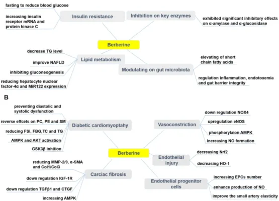
Figure 5. Pharmacological effects of berberine in treating diabetes (A) and its cardiovascular complications (B). BBR exerts protective effects in diabetes by ameliorating insulin resistance, modulating lipid metabolism and gut microbiota, inhibiting α-amylase and α-glucosidase activity. BBR also prevents cardiovascular complications associated with diabetes, such as diabetic cardiomyopathy, cardiac fibrosis, endothelial injury, endothelial progenitor cell dysfunction, and vasoconstriction.↑indicates increase or activation, and ↓indicates decrease or suppression. Abbreviations: AMP protein kinase (AMPK), α-smooth muscle actin (α-SMA), cholesterol (TC), collagen (Col), connective tissue growth factor (CTGF), dinucleotide phosphate-oxidase (NOX), endothelial nitric oxide synthase (eNOS), endothelial progenitor cells (EPCs), fasting blood glucose(FBG), fasting serum insulin (FSI), glycogen synthase kinase 3β (GSK3β), heme oxygenase-1 (HO-1), insulin-like growth factor-1 receptor (IGF-1R), matrix metalloproteinase (MMP), non-alcoholic fatty liver disease (NAFLD), nitric oxide (NO), nuclear factor erythroid 2-related factor 2 (Nrf2), phosphatidylcholine (PC), phosphatidylethanolamine (PE), protein kinase B (Akt), sphingolipid (SM),triglyceride (TG),transforming growth factor β1 (TGFβ1).
Effects of BBR on lipid metabolism
BBR is well-known to improve glucose and lipid metabolic disorders. BBR was an approved compo-nent of nutraceuticals for treatment of hyperlipidemia in many countries [241]. Kong et al. [23] have described BBR as “a new lipid-lowering drug” after they observed the efficacy of BBR in lowering lipid level in vitro and in vivo, which was comparable to that of statins. Zhao et al. [242] have reported that BBR (150 mg/kg/d, p.o., 16wk) improved NAFLD, a critical hepatic manifestation of metabolic syndrome, by inhibiting gluconeogenesis and regulating lipid metabolism. BBR (40, 60 mg/kg/d, p.o., 4wk) regulated hepatic gluconeogenesis and lipid metabolism in T2DM mice via reducing the expression of hepatocyte nuclear factor-4α and miR122 [243]. The main metabolite of BBR in vivo, berberrubine (M3), showed the most potential hypolipidemic effect by up-regulating LDLR expression in HepG2 cells [244]. Nine berberrubine analogs modified at the C9 position were tested for their lipid-lowering activity. The results showed that berberrubine and hydroxypropyl-berberine can regulate the expression of liver LDLR and PCSK9 through ERK signaling pathway. Hydroxypropyl-berberine showing a greater effect, which may indicate it as a candidate drug for anti-hyperlipidemia [244].
Effect of BBR on modulating gut microbiota
Human gut microbiota plays a vital role in mediating obesity-related metabolic dysfunction, including T2DM [245]. For example, Fei et al. [246] have revealed that an endotoxin-producing
Entero-bacter cloacae B29, obtained from the gut of obese
subjects, induced IR and obesity in germ-free mice. Another study has shown that endotoxin produced by the pathogen in the gut, such as Escherichia coli, caused obesity and IR when it was subcutaneously administered into mice [247]. As BBR has been known for treating intestinal infection-related diarrhea, Han et al. [248] hypothesized that gut microbiota regulation can be one of the anti-diabetic mechanisms of BBR. Recently, Zhu et al. [109] have reported that BBR (in drinking water (0.5 g/L), 14wk) treatment significantly reduced atherosclerosis in HFD treated mice by up-regulating the population of
Akkermansia spp. which was confirmed to regulate
inflammation, endotoxemia and gut barrier integrity. The study carried by Zhang et al. [233] showed that BBR (100 mg/kg/d, p.o., 18wk) modulated gut microbiota, and it is particularly important that the observed elevating of short chain fatty acids (SCFAs) levels in the intestine, may contribute to its therapeutic effect against DM, obesity and other
metabolic diseases.
BBR as an inhibitor of α-amylase and α-glucosidase Inhibition of key carbohydrate hydrolyzing enzymes, in clinic relevant α-amylase and α-glucosi-dase, has been acknowledged as one of the most effective approaches for managing of DM and delaying postprandial hyperglycemia [249, 250]. Enzymes α-amylase and α-glucosidase are present in the small intestine and its brush border and are responsible for hydrolytic breakdown of complex oligosaccharides and disaccharides into glucose and further monosaccharides suitable for absorption [249, 250]. Inhibition of these enzymes delayed carbohydr-ate digestion, resulting in a down-regulation in the rate of intestinal glucose absorption and subsequent reduction of the post-prandial glucose levels [249, 250]. Interestingly, BBR has been found to exhibit significant inhibitory potency against both α-amylase and α-glucosidase [223, 249, 251, 252].
Effect of BBR on DM-induced cardiovascular disease and complications
Chronic exposure to hyperglycemic conditions (accompanying DM), can cause damages to various tissues, as well as induces vascular injury and cardiomyopathy [253, 254]. High glucose level plays an important role for endothelial injury. BBR (100 mg/kg/d, p.o., 8wk) ameliorated impaired endothelium-dependent vasorelaxation of aorta in T2DM rats by down-regulating NOX4 expression and up-regulating eNOS expression [255]. Normal vascular tone is sustained by various dilatory and constrictory agents where NO is the major vasodilator. A main feature of endothelial dysfunction is impaired endothelium-dependent relaxation [256]. An effect of BBR on vascular tone has been reported in different studies [168, 169, 257]. BBR caused vasorelaxation in isolated rat aorta cells via phosphorylation of AMPK and eNOS [257]. BBR has been evidenced to exert its protective effects on vascular endothelium via increasing NO formation [168, 169]. In addition, it has been shown that BBR (50, 100, 200 mg/kg/d, p.o., 4wk) might contribute to the protection against endothelial injuries in retinal tissue of DM rats via decreasing the expressions of nuclear factor erythroid 2-related factor 2 (Nrf2) and heme oxygenase-1 (HO-1) [258]. Both the endothelium and the underlying VSMCs could be affected by BBR to induce relaxation [168]. BBR, as a sensitizer of the insulin signal in HUVECs, also improved insulin-mediated vasodilatation in diabetic rats involving PI3K/Akt and AMPK activation via the up-regulation of the InsR [259]. The vasodilator effect of BBR (200 mg/kg/d, p.o., 4wk) also improved
ACh-induced mesenteric artery vasodilation in diabetic rats [259]. There are findings indicating that the decrease in UCP2 expression and mitochondrial ROS accumulation are related to vascular damage [260]. UCP2 has been found to reduce high glucose-induced apoptosis in HUVECs by increasing Bcl-2 while decreasing caspase-3 and cytochrome c [261]. Concurrently, BBR promoted the mitochondrial biogenesis and enhance UCP2 mRNA and protein expression in cultured HUVECs [262]. BBR exerted this pharmacological effect through AMPK-depen-dent manner, which led to reduction in oxidative stress and vascular inflammation [262]. Increased atherothrombotic events due to platelet activation and apoptosis are the leading cause of high mortality and morbidity during DM [263]. Platelet hyperresponsi-veness and apoptosis during DM are ROS accumula-tion caused by activaaccumula-tion of aldose reductase (AR) and NOX. BBR protects platelets by suppressing AR and NOX activity in high glucose treated platelets [263].
Endothelial progenitor cells (EPCs) play a significant role in endothelial function [264]. The reduced number and impaired function of circulating EPCs are markers of vascular diseases, and contribute to impaired arterial elasticity and endothelial dysfunction, particularly in cardiovascular diseases [264]. BBR (1.2 g/d, p.o., 30d) has been confirmed to increase the number of circulating EPCs [265, 266], enhanced the production of NO[265] and improved the small artery elasticity [266] in healthy volunteers. However, in one randomized trial there were no changes in circulating EPCs in individuals with dyslipidemia after treatment with a nutraceutical formulation containing BBR [267]. Oppositely, another trial concluded the improvement of endothelial function after treatment by combination of BBR, RYR and policosanols, through increasing the endothelial-dependent flow-mediated dilation (FMD) in patients with hypercholesterolemia [268].
Diabetic cardiomyopathy (DCM) is considered to be a clinical pathology independent of concomitant vascular diseases and can be primarily caused by disturbances in the energy substrate [269]. In the high-sucrose and HFD/streptozotocin (STZ)-induced rat DCM model, BBR (10, 30 mg/kg/d, p.o., 16wk) significantly prevented diastolic and systolic dysfunc-tion, and cardiac hypertrophy [269]. Phosphatidyl-choline (PC), phosphatidylethanolamine (PE) and sphingolipid (SM) are potential biomarkers of DCM, and the protective effect of BBR on DCM can be through reversal of PC, PE and SM metabolic disturbances [269]. BBR treatment (100 mg/kg/d, p.o., 16wk) of diabetic rats can partially improve cardiac function and significantly reduce FSI, FBG, total TC and TG levels. The underlying mechanism can be that
BBR activated cardiac AMPK and Akt in diabetic rats and inhibited GSK3β activity [270]. Similarly, in the palmitate-induced H9C2 cell hypertrophy model, BBR up-regulated alpha-myosin heavy chain (α-MHC) expression, down-regulated beta-myosin heavy chain (β-MHC) expression and thus inhibited H9C2 cell hypertrophy [270]. Moreover, BBR also enhanced AMPK and Akt activation in H9C2 cells and inhibited GSK3β activity [270, 271]. Diabetic hearts are more sensitive to I/R injury, and BBR treatment protected the heart of diabetic rats from I/R damage [272]. This protective effect of BBR was achieved by activating AMPK and Akt activity, while inhibiting GSK3β activity in non-ischemic regions of the diabetic rat hearts [272].
Diabetic cardiac fibrosis causes ventricular stiffness and leads to diastolic dysfunction [273]. In a diabetic rat model, BBR treatment (100 mg/kg/d, p.o., 4wk) improved cardiac fibrosis and dysfunction by down-regulating insulin-like growth factor-1 receptor (IGF-1R) expression in cardiac fibroblasts, specifically by reducing expression of MMP-2/9, α-smooth muscle actin (α-SMA) and type 1 collagen (Col1) in diabetic hearts. BBR also exerted a similar effect in cardiac fibroblasts exposed to high levels of glucose [273]. Furthermore, BBR (50, 100, 150 mg/kg/d, p.o., 12wk) reduced cardiac fibrosis in DM rats by down-regulating the expression of transforming growth factor β1 (TGFβ1) and connective tissue growth factor (CTGF), reducing the expression of Col1 and Col3 [274]. In addition, BBR treatment promoted glucose depletion and glucose uptake as well as improved IR in H9C2 cells, at least partially by increasing AMPK activity [222]. Further, BBR (100 mg/kg/day, p.o., 7d) ameliorated arrhythmias induced by ischemia (LAD ligature) in diabetic rats [275]. Researchers have shown that BBR reversed the down-regulation of the transient outward K+ current
(Ito) and ICa [275]. In a similar study, BBR (60
mg/kg/day, p.o., 14d) led to the restoration of diminished inwardly rectifying potassium channel Kir2.1 to nearly normal levels [276]. The effect of BBR on Kir2.1, the main subunit of IK1, was associated with recovering of resting membrane potential and alleviating of ischemia-induced arrhythmias in diabetic rats [276].
In conclusion, BBR is a promising candidate for therapy of T2DM and metabolic syndrome. However, further investigations of the mechanisms of BBR are required for its appropriate application in clinical settings.
Obesity
Obesity is a complex metabolic disease occurring in both developed and developing countries [277].
Obesity contributes to the development of several other health problems, such as cardiovascular diseases, DM, cancer, chronic obstructive pulmonary disease, and depression [277]. Etiology of obesity is multi-factorial and is associated with the energy input exceeding energy output [278]. Most commonly, it is induced by excessive food intake, imbalanced nutrition, physical inactivity, and genetic predispose-itions. Other causes of obesity include endocrine dysfunction, mental illnesses, or iatrogenic etiologies [278].
BBR reduced food intake in the mice fed with HFD, and decreased their weight gain [279]. Such an effect led to the question as to how BBR affects the appetite. BBR can improve lipid dysregulation by controlling both peripheral and central AMPK activity [280]. Although BBR cannot cross the cerebrovascular barrier, studies have shown that an application of BBR intraperitoneally caused a 47% up-regulation in serotonin levels in the brain [281]. The expression of glucagon-like peptide 1 receptor (GLP-1R), orexin A and neuropeptide Y in brain of tested animals were found to be up-regulated [282]. Together with changes observed in hypothalamus, this can be a factor causing the decrease of body weight, enhanced lipolysis, and decrease of IR in obese experimental animals and even humans.
It is clear, that anti-obesity effects of BBR are associated with its anti-diabetic activity [283]. Many studies identified BBR as an activator of AMPK, which is, besides other effects, responsible for triggering of the glucose uptake and fatty acid oxidation in skeletal muscles, and regulation of insulin secretion by pancreatic β-secretory cells [30, 225, 226]. Effects of BBR on an increase of glucagon-like peptide-1 (GLP-1) via enhancing GLP-1 secretion and GLP-1 biosynthesis in enteroendocrine L-cells and some neurons were also observed, and BBR stimulated GLP-1 secretion can trigger the feeling of satiety and decrease the food intake [284]. Mitochondrial stress responses (e.g., mitochondrial swelling and membrane rupture) and GLP-1 expression were decreased in the colon of diet-induced obese mice [285]. Administration of BBR (100 mg/kg/d, p.o., 8wk) up-regulated GLP-1 expression in mice and prevented mitochondrial stress response [285]. All these pharmacological effects of BBR can translate to improved outcomes in DM and obesity.
Several studies have shown altered intestinal architecture as a result of a shift in gut microbiota composition to the phyla Bacteroidetes and Firmicutes [286]. The pivotal barrier-protecting function conferred by BBR was mediated by elevated levels of SCFAs [233]. Gene profiling analysis showed that BBR
can regulate gut microbiota diversity. In the group of BBR-treated rats (100 mg/kg/d, p.o., 18wk), the shift to bacteria producing SCFAs was shown to be significant [233]. On the other hand, fasting-induced adipose factor (FIAF), which inhibits circulating lipoprotein lipase (LPL), is a regulator as a putative mediator of microbial modulation of energy storage, and fat mobilization [287]. BBR (200 mg/kg/d, p.o., 6wk) up-regulated FIAF expression in adipose tissues and intestinal and also changed the abundance of fecal Bacteroidetes and Firmicutes in total bacteria in the gut of mice fed with HFD [288]. Additionally, Chae et
al.[289] showed modulation of growth of gut
microbiota, especially Lactobacillus and Bifidobacterium by BBR.
Non-alcoholic fatty liver disease (NAFLD)
NAFLD is manifested as an accumulation of TG in liver [290]. NAFLD is closely related to the obesity and related metabolic risk factors (T2DM and dyslipidemia). Untreated NAFLD can progress to non-alcoholic steatohepatitis (NASH), characterized by persistent hepatocyte inflammation and injury with or without fibrosis. NASH can progress to cirrhosis and liver cancer [291, 292].
According to the main characteristics of NAFLD, the first therapeutic option is the restoration of dysregulated lipid metabolism, which leads to the fat reduction and weight loss [290]. BBR (5 mg/kg/d,
p.o., 3wk) improved fatty liver in obese mice by
regulating neural signaling from the central nervous system and peripheral AMPK signaling [293]. Sterol regulatory element binding protein 1c (SREBP1c) can be regulated by AMPK via direct phosphorylation [294]. AMPK activation suppresses SREBP-1c activity and lowers hepatic and plasma levels of TG and TC [294]. BBR acted as a potent inhibitor of SREBP-1c, possibly through AMPK pathway [295, 296]. BBR (100 mg/kg) can down-regulate SREBP-1c and ameliorate lipid profile in rats [297]. The combination of BBR (50 mg/kg/d, p.o., 8wk) and curcumin (50 mg/kg/d, p.o., 8wk) is more effective than lovastatin (100 mg/kg/d,
p.o., 8wk) [298]. BBR can reverse the disorder of lipid
metabolism in 3T3-L1 cells induced by olanzapine, a second-generation antipsychotic drug, by inhibiting SREBP and activating AMPKα [299]. Overexpression of SREBP-1c leads to the down-regulation of insulin receptor substrate 2 (IRS-2) mRNA levels which is closely linked to the hepatic IR [300]. BBR reduced liver TC levels by down-regulation of sterol carrier protein 2 expression and inhibiting COX-2 induced prostaglandin production[301]. The improvement of IR by increasing IRS-2 mRNA expression is one of the possible mechanisms in the treatment of NAFLD with BBR [302].