R E S E A R C H A R T I C L E
Open Access
Correlation of EGFR, pEGFR and p16
INK4
expressions
and high risk HPV infection in HIV/AIDS-related
squamous cell carcinoma of conjunctiva
Anthony Mwololo
1, Joshua Nyagol
1,3*, Emily Rogena
1, Willis Ochuk
1, Mary Kimani
1, Noel Onyango
1,
Lorenzo Pacenti
2, Rosa Santopietro
2, Lorenzo Leoncini
2and Walter Mwanda
1Abstract
Background: Squamous cell carcinoma of conjunctiva has increased tenfold in the era of HIV/AIDS. The disease pattern has also changed in Africa, affecting young persons, with peak age-specific incidence of 30-39 years, similar to that of Kaposi sarcoma, a well known HIV/AIDS defining neoplasm. In addition, the disease has assumed more aggressive clinical course. The contributing role of exposure to high risk HPV in the development of SCCC is still emerging.
Objective: The present study aimed to investigate if immunohistochemical expressions of EGFR, pEGFR and p16, could predict infection with high risk HPV in HIV-related SCCC.
Methods: FFPE tissue blocks of fifty-eight cases diagnosed on hematoxylin and eosin with SCCC between 2005-2011, and subsequently confirmed from medical records to be HIV positive at the department of human pathology, UoN/KNH, were used for the study. Immunohistochemistry was performed to assess the expressions of p16INK4A, EGFR and pEGFR. This was followed with semi-nested PCR based detection and sequencing of HPV genotypes. The sequences were compared with the GenBank database, and data analyzed for significant statistical correlations using SPSS 16.0. Ethical approval to conduct the study was obtained from KNH-ERC.
Results: Out of the fifty-eight cases of SCCC analyzed, twenty-nine (50%) had well differentiated (grade 1), twenty one (36.2%) moderately differentiated (grade 2) while eight (13.8%) had poorly differentiated (grade 3) tumours. Immunohistochemistry assay was done in all the fifty eight studied cases, of which thirty nine cases (67.2%) were positive for p16INK4A staining, forty eight cases (82.8%) for EGFR and fifty one cases (87.9%) showed positivity for p-EGFR. HPV DNA was detected in 4 out of 40 SCCC cases (10%) in which PCR was performed, with HPV16 being the only HPV sub-type detected. Significant statistical association was found between HPV detection and p16INK4 (p=0.000, at 99% C.I) and EGFR (p=0.028, at 95% C.I) expressions, but not pEGFR. In addition, the expressions of these biomarkers did not show any significant association with tumor grades.
Conclusion: This study points to an association of high risk HPV with over expressions of p16INK4A and EGFR proteins in AIDS-associated SCCC.
Keywords: SCCC, Biomarkers, HPV, HIV/AIDS
* Correspondence:[email protected]
1
Department of Human Pathology, University of Nairobi, Nairobi, Kenya
3Department of Human Pathology, Unit of Immunology, School of Medicine,
College of Health Sciences, University of Nairobi, P.O. Box 19676-00202, KNH (Off Ngong Road), Nairobi, Kenya
Full list of author information is available at the end of the article
© 2014 Mwololo et al.; licensee BioMed Central Ltd. This is an Open Access article distributed under the terms of the Creative Commons Attribution License (http://creativecommons.org/licenses/by/2.0), which permits unrestricted use, distribution, and reproduction in any medium, provided the original work is properly credited. The Creative Commons Public Domain Dedication waiver (http://creativecommons.org/publicdomain/zero/1.0/) applies to the data made available in this article, unless otherwise stated.
Background
Squamous cell carcinoma of conjunctiva (SCCC) is a rare, slow-growing tumor of a spectrum of conditions collect-ively known as ‘ocular surface epithelial dysplasias’. These range from benign dysplasias, to carcinomain situ and ul-timately to invasive carcinoma [1,2]. It is also considered the most common neoplasm of the conjunctiva, normally affecting elderly men of around 70 years [3,4]. The etiology of SCCC has been previously viewed as multifactorial, with ultraviolet light implicated as the major risk factor for these tumors, and more common in equatorial Africa than in Europe or North America [5].
In Africa, however, the incidence of SCCC has risen rapidly in the recent past, with the peak age-specific incidence reported to be 30-39 years, similar to that of HIV/AIDS pandemics in the tropics. The disease is also more aggressive, with a mean history of three months at presentation [4,5]. An association between human immunodeficiency virus (HIV) infection and squamous cell carcinoma of the conjunctiva was first reported in the mid-1990s. Subsequently, other studies also reported that since the advent of HIV/AIDS in 1980s, the number of patients presenting with SCCC had been increasing exponentially [6-8]. In 1995, Ateenyi-Agaba observed that a high incidence of these tumors in Uganda appeared to be related to HIV infection [9]. Parallel studies by Waddell and colleagues also suggested that HIV infection is strongly associated with an increase in the incidence of conjunctival carcinoma in Africa [10].
Although the natural history of the SCCC appears to be unique in this region of the world with etiologic mechanism unclear and therapeutic options limited, im-munosuppression from HIV has been thought to facilitate the activity of other infective agents that induce the carcin-oma. This has been supported by many studies that have documented the presence of high risk HPV genotypes 16 and 18 DNA in a proportion of SCCC [11]. The oncopro-teins E6 and E7 encoded by the high risk HPV genotypes are well documented to play a critical role in pathogenesis of anogenital carcinomas by deregulation of cell cycle control proteins p53 and pRb2/p130, respectively [12-18].
Another cell cycle regulatory protein whose increased expression is reported to be predictive of, and an inde-pendent prognostic marker in high risk HPV infection is p16INK4A[19-22]. However, in our previous study to investigate the roles played by HIV-1 Tat and high risk HPV E6/E7 proteins in promoting carcinogenesis in cervical cancers, expressions of p16INK4Awas found to be reduced in HIV-related squamous cell carcinoma of the uterine cervix, a correlation which has also been supported by other studies [23,24].
Few authors have also proposed involvement of high risk HPVs in the oncogenesis through altered expression of other key molecules involved in tyrosine kinase pathways,
in which inverse expressions of epidermal growth factor receptor (EGFR) and its phosphorylated form (pEGFR) have been reported to contribute to the pathogenesis of HIV/AIDS–associated SCCC, and correlates with poor prognosis [25-27]. Altogether, expressions of p16INKA4, EGFR and its phosphorylated form pEGFR have been proposed to reflect infection with high risk HPV in squamous cell carcinoma of conjunctiva. However, HPV detection in conjunctival neoplasm has been largely contro-versial, and the pathogenic mechanism not well elucidated. Some of these studies have reported a heterogeneous preva-lence of high-risk HPV genotypes, suggesting that only a subset of cases can be attributed to these viruses. This vari-ation in the reported HPV infection rates in conjunctival squamous cell carcinoma could result from differences in detection methods and the studied populations [28-31].
The present study therefore compared PCR-based detec-tion of HPV with immunohistochemical expressions of p16INK4A, EGFR and pEGFR; and the potential value of use of these biomarkers as indicators of HPV positivity in selected cases of HIV/AIDS-related SCCC.
Results
Patient’s demographics and histology
Out of the 58 samples evaluated, the ratio of females to males was 1:1. The age ranged from 23 to 73 years, with a mean of 41.6 years and median of 39 years. Fifty six cases were classified as squamous cell carcinomas and the remaining two as carcinoma in situ. Tumor site, tumor size and tumor grades were as shown in Table 1 and Figure 1a-c.
Expressions of p16INK4A, EGFR and pEGFR
Immunohistochemistry assay was done in all the fifty eight studied cases, of which Out of the fifty eight cases, thirty nine (67.2%) were positive for p16INK4A staining, forty eight cases (82.8%) for EGFR and fifty one cases (87.9%) showed positivity for p-EGFR (Figure 2a-c).
The patterns of p16INK4A, EGFR and pEGFR expressions were semi-quantitatively scored for both intensity and proportion of staining in the cell nucleus, cytoplasm and membrane as reported previously [32,33]. Intensity of the staining were scored as 0 (no staining), 1 (weak positivity), 2 (moderate positivity), or 3 (strong positivity). The pro-portions of the staining were also evaluated and scored as 0 (1%-10% of cells stained), 1 (11%-50%), 2 (51%-80%) or 3 (81%-100%). Sections scored as 0 or 1 for intensity was defined as negative; whereas those scored 2 or 3 were defined as positive.
HPV 16 is the predominant genotype in AIDS- related SCCC
From the forty cases where DNA was extracted, four cases (10%) showed positivity for HPV 16 genotype, and no
other genotypes were detected. Although this is in tandem with other studies that have documented detection ranges from 8-10% of high risk HPV in head and neck squamous cell carcinomas, this low rate of detection suggests that other mechanisms could underlie pathogenesis of HIV/ AIDS-related squamous cell carcinoma of conjunctiva independently from the active role of high risk HPV.
Correlations of HPV detection with p16INK4A, EGFR expressions
For the cases in which high risk HPV were positive, statis-tical significant association was found between HPV detec-tion and p16INK4expressions, p = 0.000, at 99% confidence interval and EGFR expression, p = 0.028, at 95% C.I, but not with pEGFR.
Discussion
A causal relationship between the epidemic of HIV/AIDS in the Sub-Saharan region and increased incidence of squamous cell carcinoma of conjunctiva (SCCC) has been hypothesized, with the prevalence of the neoplasm re-ported in Kenya alone to be 8% [34,35]. Although immune suppression has been known to play a critical role in the pathogenesis, a number of epidemiological studies have postulated other etiological agents for the development of squamous cell carcinoma of conjunctiva, such as ultraviolet
(UV) light, history of pterygium, and high risk human papillomavirus infection (HPV) [36-39].
Detection of high risk HPV associated with squamous cell carcinoma has, however, been controversial, with dif-ferent authors reporting varied percentages of positivity
Figure 1 Haematoxylin and eosin staining. a. Grade I, SCCC X 10 Neoplasm comprising of infiltrating cords of mild to moderately pleomorphic malignant squamous cells and of keratin pearls, interspersed with a chronic inflammatory cell infiltrate). b. Grade II SCCC X 10 (Neoplasm comprising of infiltrating cords of moderately pleomorphic malignant squamous cells). c. Grade III SCCC X 20 (Neoplasm disposed in a diffuse architecture, comprising of moderately to markedly pleomorphic malignant squamous cells).
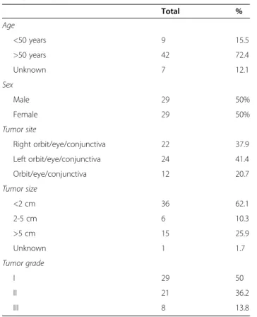
Table 1 n = 58 Total % Age <50 years 9 15.5 >50 years 42 72.4 Unknown 7 12.1 Sex Male 29 50% Female 29 50% Tumor site Right orbit/eye/conjunctiva 22 37.9 Left orbit/eye/conjunctiva 24 41.4 Orbit/eye/conjunctiva 12 20.7 Tumor size <2 cm 36 62.1 2-5 cm 6 10.3 >5 cm 15 25.9 Unknown 1 1.7 Tumor grade I 29 50 II 21 36.2 III 8 13.8
and prevalence [40-42]. This could be attributed to vari-ous techniques currently in use, ranging from consensus and type-specific end-point PCR methods followed with sequencing, real-time PCR assays for quantification of viral load,in-situ hybridization, detection of serum antibodies directed against HPV epitopes, to immunohistochemical detection of surrogate biomarkers such as p16INK4A, a cyc-lin dependent kinase inhibitor protein [42-45]. In our study, a prevalence of 10% of high risk HPV 16 genotype detec-tion was realized, which is consistent with the findings of other studies. Many infectious oncogenic viruses yet to be discovered can use the same mechanism as of high risk
HPV in a state of immunosuppression, but the low prevalence in our study could also be a factor attributed to poor tissue processing and thus DNA degradation in a number of the studied cases.
A number of studies have therefore demonstrated that p16INK4Aprotein over-expression may serve as a surro-gate biomarker for biologically and clinically relevant high risk HPV infection in squamous cell carcinoma of the conjunctiva, as well as squamous cell carcinoma and glandular epithelial dysplasia of the uterine cervix [44,45]. Besides, over expression of p16INK4Ahas also been shown to correlate with the degree and behavioral characteristics
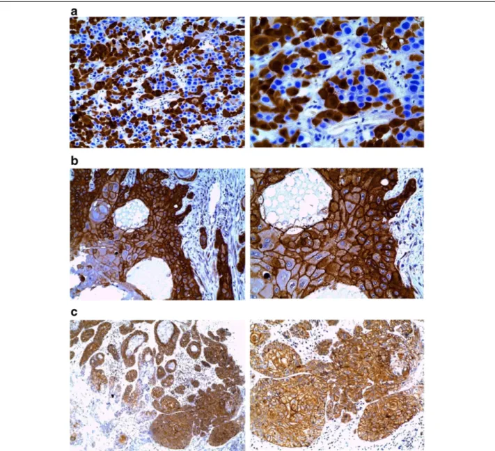
Figure 2 a1 (X20) and a2 (X40). Staining for P16INK4Ain majority of the neoplastic cells showed intense nuclear staining, which reflects the functional role of p16 in control of cell cycle prior to the S-phase. In some cells, cytoplasmic positivity appeared prominent. b1 (X20) and b2 (X40). EGFR staining was observed to be brown membrane and cytoplasmic, uniformly distributed in all the squamous epithelial cells. c1 (X10) and c2 (X20). Staining for pEGFR showed complete brown membrane and cytoplasmic reaction in all epithelial tumors cells.
of both cervical as well as head and neck neoplasm [46,47].
In addition to over expression of p16INK4Aas potential surrogate marker of HPV-derived squamous cell carcin-omas, the development of SCCC in HIV positive patients has been documented to involve activation of epidermal growth factor receptor (EGFR) and its phosphorylated form (p-EGFR) [48-50]. The relationship between HPV status and levels of EGFR protein expression with clinical outcome has been reported in several studies, indicating that the best outcomes are observed in patients with HPV-positive tumors with low EGFR expression [50-52]. However, an inverse relationship between HPV status and EGFR gene amplification has also been reported.
Several studies have discussed about the optimal method for determining HPV status on FFPE tumor sections (5, 34, 35), and in the current study, we evaluated im-munohistochemical expressions of p16INKA, EGFR and p-EGFR IHC as markers of HPV status and utilized HPV PCR in a subset of cases to correlate the results. Immunohistochemistry was performed in all the fifty eight studied cases, and semi-quantitative scores of the bio-markers enumerated. However, 18 cases were excluded from HPV detection by PCR technique, followed with sequencing, because tumor tissue was either inadequate or had extensive DNA degradation due to poor tissue processing. A prevalence of 10% of high risk HPV 16 genotype detection was realized, which is consistent with the findings of other studies. However, the low prevalence of HPV could be associated with poor tissue processing.
In this study p16INK4Aexpression in the epithelium was characterized by variable, weak to strong, diffuse nuclear and cytoplasmic staining. Thirty nine cases (67.2%) were positive for p16INK4Astaining and showed significant statis-tical correlation between HPV positivity and p16INK4A ex-pression (correlation coefficient -0.502, p = 0.000, at 99% confidence interval (C.I)). This was in agreement with similar findings from other authors who demonstrated that p16INK4Aexpression was significantly associated with the presence of HPV-16 [20].
The expression of EGFR and its activated form, p-EGFR, were also independently enumerated in all of the fifty-eight cases, from which the results demonstrated that 82.8% of the cases expressed EGFR while 87.9% expressed p-EGFR as brown membranous and/or cytoplasmic immunostain-ing, similar to the findings from other studies [53].
Once EGFR is activated into p-EGFR, it undergoes internalization, resulting in a marked decrease in the non-activated membrane-bound EGFR [54]. For correl-ation with HPV positive cases, significant statistical corre-lations was obtained with EGFR expression (correlation coefficient -.252, p = .028 at 95% confidence interval). There was, however, no statistical significant correlation between HPV status and p-EGFR at both 95% and 99% confidence
Intervals. Equally, no biomarker showed significant statis-tical correlation with tumor grades.
Therefore, both immunohistochemical assay and PCR-based techniques showed close agreement in the use of p16INK4Aand EGFR but not p-EGFR as clinically useful surrogate biomarkers for high risk HPV infection in HIV/ AIDS-associated squamous cell carcinoma of conjunctiva.
Conclusion
Although the exact role played by high risk human papil-lomavirus in carcinogenesis is not well known, to the best of our knowledge, this is the first study to show an associ-ation of high risk HPV infection with p16INK4Aand EGFR over expressions in HIV/AIDS-associated squamous cell carcinoma of conjunctiva.
Materials and methods
Selection of the study cases
Fifty-eight HIV positive SCCC formalin fixed paraffin embedded (FFPE) tissue blocks were retrieved from the archives at pathology department, UON/KNH for analysis. The criteria for inclusion as HIV positive case included in-formation from the clinician in the patient’s histological request form indicating on follow-up at the comprehen-sive care centre, on HAART treatment, ARV treatment, immunnosuppression, p24 marker reactive, retrovirus dis-ease and HIV positive. Sections (4 μm) from the tissue blocks were stained withhematoxylin and eosin to confirm the previous diagnoses and establish the tumor grades according to the WHO guidelines. Ethical approval to conduct the study was obtained from KNH-ERC.
Immunohistochemistry tests for p16INK4A, EGFR and pEGFR
Immunohistochemistry was performed as described pre-viously by Russo G,et al. [55]. Briefly, 4 μm sections of the FFPE tissue blocks were mounted on positively charged slides and incubated overnight incubation at 65 º C. Sections were subsequently de-waxed through a series of xylene, rehydrated in graded series of alcohol (absolute, 90% and 75%), and taken to water. Antigen retrieval was done by immersing the slides in Tris buffer pH 9 contain-ing 10 mmol/l EDTA and 15 mmol/l sodium azide (NaN3)
(for p16INK4A) or citrate buffer, pH 6.0 (for EGFR and pEGFR) and subsequently heated in a microwave at 750 W for 20 minutes, with 5 minutes intervals. All the slides were then allowed to cool for 20 minutes, and then rinsed with phosphate buffered saline (PBS) wash buffer. The sections were quenched in 3% hydrogen peroxide (H2O2) containing 15 mmol/l sodium azide (NaN3) for
10 minutes to block endogenous peroxidase activity, followed with a rinse in PBS wash buffer. Ten microlitres of ready to use primary monoclonal mouse anti-human antibodies, each of p16INK4A, EGFR (clone EGFR-384-R-7-CE; Leica, Germany), pEGFR (clone EP774Y, Biocare
Medical, USA) were applied, and incubated for 30 minutes at room temperature, and according to manufacturers’ instructions (Table 1). The sections were rinsed with wash buffer, followed with incubation in a visualization reagent for 30 minutes. This was followed with rinsing and incubation in a substrate-chromogen solution for 10 minutes to facilitate visualization. After the final rinse with PBS buffer, sections were counterstained with Mayer’s hematoxylin, dehydrated and mounted. For each run, normal skin, normal placenta and colon carcinoma specimens were used as positive controls for p16INK4, EGFR and pEGFR, respectively.
DNA extraction from tissue blocks
DNA was extracted from 20 sections (5μm) of the FFPE tissue blocks using digestion buffer, 50 mM Tris-pH 8.5, 1 mM EDTA, pH 8.0 and 0.5% Tween 20 (Sigma) and proteinase-K (Roche) at a final concentration of 500μg/ml. The lysates were purified using EZ1 DNA Tissue Kit (Qiagen) and 1 μl of DNA were used in a reaction to verify DNA integrity, using beta globin and as described previously in other studies [56-59]. DNA extraction was successful in forty out of the fifty eight study cases, most probably due to poor tissue processing that resulted in DNA degradation.
PCR testing for HPV and genotyping
For the detection of human papillomavirus (HPV) and the genotypes, PCR was performed, followed with DNA sequencing on ABI PRISM 310 Genetic Analyzer. The sequences compared with the GenBank database. In brief, the PCR reaction was performed in a final volume of 50 μl. Each PCR mixture contained 25 μl of AmpliTaq Gold PCR Master Mix 2X (Applied Biosystems, NJ USA), 30 pmol of primers (A1 5'- TTGGATCCATGTTAATWS AGCCWCCAAAATT –3' A2 5'- TTGGATCCTTATCA WATGCCCAYTGTACCAT–3'), 2 μl of MgCl2(25 mM)
1μl of either DNA or water. The cycling conditions were as follows: 10 minutes at 95°C activation step for Taq DNA polymerase, followed by 40 cycles each for 40 sec-onds at 95°C, 50 secsec-onds at 55°C and 40 secsec-onds at 72°C. The last cycle was followed by a final extension step of 10 minutes at 72°C. The PCR products were analyzed by electrophoresis on 2% agarose gel stained with eth-idium bromide using the protocol described by Gleissner et al. [60].
Positive samples were typed using cut and purified DNA bands from agarose gel. The DNA were sequenced with an ABI PRISM 310 Genetic Analyzer (Applied Biosystems, Weiterstadt, Germany), with the Big Dye Terminator V1.1 cycle sequencing kit (Applied Biosystems, NJ, USA) using primer (A1). Sequences were then compared with the GenBank database as described by Sguegliaet al. [61].
Statistical analysis
Data was then analysed for significant statistical co-relations using SPSS Version 16. One-tailedp < 0.05 at 95% CI was considered statistically significant.
Competing interest
The authors declare that they have no competing interests. Authors’ contributions
Contributions: AM, JN, WO, MK, LP and NO, performed the experiments; ER and JN designed the overall study; WM, coordinated the work; RS, ER and LL contributed their expertise in the field of pathology; AM and JN wrote the work. All authors read and approved the final manuscript.
Acknowledgements
This work was supported by ACSR-Kenya (Prof. Ayers Leona). Author details
1Department of Human Pathology, University of Nairobi, Nairobi, Kenya. 2Department of Anatomical Pathology and Human Oncology, University of
Siena, Siena, Italy.3Department of Human Pathology, Unit of Immunology,
School of Medicine, College of Health Sciences, University of Nairobi, P.O. Box 19676-00202, KNH (Off Ngong Road), Nairobi, Kenya.
Received: 11 November 2013 Accepted: 11 February 2014 Published: 26 February 2014
References
1. Margo C, Mack W, Guffey JM: Squamous cell carcinoma of the conjunctiva and human immunodeficiency virus infection. Arch Ophthalmol 1996, 114(3):349.
2. Ogun OA, Ogun GO, Bekibele CO, Akang EE: Squamous papillomas of the conjunctiva: a retrospective clinicopathological study. Niger J Clin Pract 2012, 15(1):89–92.
3. Chokunonga E, Borok MZ, Chirenje ZM, Nyakabau AM, Parkin DM:“Trends in the incidence of cancer in the black population of Harare, Zimbabwe 1991-2010”. Int J Canc 2013, 133(3):721–729.
4. Sasco AJ, Jaquet A, Boidin E, Ekouevi DK, Thouillot F, Lemabec T, Forstin M-A, Renaudier P, N’dom P, Malvy D, Dabis F: “The challenge of AIDS-related malignancies in sub-Saharan Africa”. PLoS One 2010, 5(1):e8621.
5. Sun EC, Fears TR, Goedert JJ: Epidemiology of squamous cell conjunctival cancer. Canc Epidemiol Biomark Prev Publ Am Assoc Canc Res Cosponsored Am Soc Prev Oncol 1997, 6(2):73–77.
6. Guramatunhu S:“Squamous cell carcinoma in HIV/AIDS”. Community Eye Health 2003, 16(47):37.
7. Newton R: A review of the aetiology of squamous cell carcinoma of the conjunctiva. Br J Canc 1996, 74(10):1511–1513.
8. Margo Ce Guffey Jm MW:“Squamous cell carcinoma of the conjunctiva and human immunodeficiency virus infection”. Arch Ophthalmol 1996, 114(3):349.
9. Ateenyi-Agaba C: Conjunctival squamous-cell carcinoma associated with HIV infection in Kampala, Uganda. Lancet 1995, 345(8951):695–696. 10. Tornesello ML, Duraturo ML, Waddell KM, Biryahwaho B, Downing R, Balinandi
S, Lucas SB, Buonaguro L, Buonaguro FM: Evaluating the role of human papillomaviruses in conjunctival neoplasia. Br J Canc 2006, 94(3):446–449. 11. Tulvatana W, Bhattarakosol P, Sansopha L, Sipiyarak W, Kowitdamrong E,
Paisuntornsug T, Karnsawai S: Risk factors for conjunctival squamous cell neoplasia: a matched case-control study. Br J Ophthalmol 2003, 87(4):396–398.
12. Maciag PC, Villa LL: Genetic susceptibility to HPV infection and cervical cancer. Braz J Med Biol Res 1999, 32:915–922.
13. Coleman DV, Richman PI:“Human papillomavirus infection and cancer of the uterine cervix”. J Pathol 1987, 145(3):430–432. 23.
14. Zhang WH, Wu AR, Li JX, Lin YC: Relation between human papillomavirus (HPV) and cancer of uterine cervix. Zhonghua Zhong Liu Za Zhi Chin J Oncol 1987, 9(6):433–435.
15. Brisson J, Bairati I, Morin C, Fortier M, Bouchard C, Christen A, Bernard P, Roy M, Meisels A: Detection of human papillomavirus DNA and expression of p16, Rb, and p53 proteins in small cell carcinomas of the uterine cervix. Am J Surg Pathol 2004, 173(7):794–799.
16. Matsukura T, Sugase M: Human papillomavirus genomes in squamous cell carcinomas of the uterine cervix. Virology 2004, 324(2):439–449.
17. Iwasawa A, Nieminen P, Lehtinen M, Paavonen J: Human papillomavirus DNA in uterine cervix squamous cell carcinoma and adenocarcinoma detected by polymerase chain reaction. Cancer 1996, 77(11):2275–2279. 18. Uchiyama M, Iwasaka T, Matsuo N, Hachisuga T, Mori M, Sugimori H:
Correlation between human papillomavirus positivity and p53 gene overexpression in adenocarcinoma of the uterine cervix. Gynecol Oncol 1997, 65(1):23–29.
19. Lambert APF, Anschau F, Schmitt VM: p16INK4A expression in cervical premalignant and malignant lesions. Exp Mol Pathol 2006, 80(2):192–196. 20. Kalof AN, Cooper K: p16INK4a immunoexpression: surrogate marker of
high-risk HPV and high-grade cervical intraepithelial neoplasia. Adv Anat Pathol 2006, 13(4):190–194.
21. Lakshmi S, Rema P, Somanathan T: p16ink4a is a surrogate marker for high-risk and malignant cervical lesions in the presence of human papillomavirus. Pathobiol J Immunopathol Mol Cell Biol 2009, 76(3):141–148. 22. Queiroz C, Correia Silva T, Alves VAF, Villa LL, Costa MC, Travassos AG,
Araujo Filho JB, Studart E, Cheto T, De Freitas LAR: P16INK4a expression as a potential prognostic marker in cervical pre-neoplastic and neoplastic lesions. Pathol Res Pract 2006, 202(2):77–83.
23. Nyagol J, Leucci E, Onnis A, De Falco G, Tigli C, Sanseverino F, Torriccelli M, Palummo N, Pacenti L, Santopietro R, Spina D, Gichangi P, Muchiri L, Lazzi S, Petraglia F, Leoncini L, Giordano A: The effects of HIV-1 Tat protein on cell cycle during cervical carcinogenesis. Canc Biol Ther 2006, 5(6):684–690. 24. Auw-Haedrich C, Martin G, Spelsberg H, Sundmacher R, Freudenberg N, Maier P, Reinhard T: Expression of p16 in conjunctival intraepithelial neoplasia does not correlate with HPV-infection. Open Ophthalmol J 2008, 2:48–56.
25. Nagaiah G, Stotler C, Orem J, Mwanda WO, Remick SC: Ocular surface squamous neoplasia in patients with HIV infection in sub-Saharan Africa. Curr Opin Oncol 2010, 22(5):437–442.
26. Cunningham MP, Essapen S, Thomas H, Green M, Lovell DP, Topham C, Marks C, Modjtahedi H: Coexpression, prognostic significance and predictive value of EGFR, EGFRvIII and phosphorylated EGFR in colorectal cancer. Int J Oncol2005, 27(2):317–325.
27. Nicholson RI, Gee JMW, Harper ME:“EGFR and cancer prognosis”. Eur J Canc 2001, 37(Suppl 4):S9–S15. no. 2.
28. Tuppurainen K, Raninen A, Kosunen O, Kankkunen JP, Kellokoski J, Syrjänen S, Mäntyjärvi M, Syrjänen K: Squamous cell carcinoma of the conjunctiva. Failure to demonstrate HPV DNA by in situ hybridization and polymerase chain reaction. Acta Ophthalmol 1992, 70(2):248–254. 29. Ateenyi-Agaba C, Franceschi S, Wabwire-Mangen F, Arslan A, Othieno E,
Binta-Kahwa J, Van Doorn LJ, Kleter B, Quint W, Weiderpass E: Human papillomavirus infection and squamous cell carcinoma of the conjunctiva. Br J Canc 2010, 102(2):262–267.
30. Nakamura Y, Mashima Y, Kameyama K, Mukai M, Oguchi Y: Detection of human papillomavirus infection in squamous tumours of the conjunctiva and lacrimal sac by immunohistochemistry, in situ hybridisation, and polymerase chain reaction. Br J Ophthalmol 1997, 81(4):308–313.
31. Peralta R, Valdivia A, Estañol P, Villegas V, Pimienta C, Treviño E, Marrero D, Mendoza M, Jimenez F, Villalvazo L, Tejeda M, Salcedo M:“Low frequency of human papillomavirus infection in conjunctival squamous cell carcinoma of Mexican patients”. Infect Agent Canc 2011, 6(1):24. 32. El Hamdani W, Amrani M, Attaleb M, Laantri N, Ennaji MM, Khyatti M, El
Mzibri M:“EGFR, p16INK4a and E-cadherin immuno-histochemistry and EGFR point mutations analyses in invasive cervical cancer specimens from Moroccan women”. Cell Mol Biol (Noisy-le-grand) 2010,
56:L1373–L1384.
33. Magkou C, Nakopoulou L, Zoubouli C, Karali K, Theohari I, Bakarakos P, Giannopoulou I:“Expression of the epidermal growth factor receptor (EGFR) and the phosphorylated EGFR in invasive breast carcinomas”. Breast Canc Res 2008, 10(3):R49.
34. Parkin DM, Wabinga H, Nambooze S, Wabwire-Mangen F: AIDS-related cancers in Africa: maturation of the epidemic in Uganda. AIDS 1999, 13(18):2563–2570.
35. Thomas JO: Acquired immunodeficiency syndrome-associated cancers in Sub-Saharan Africa. Semin Oncol 2001, 28(2):198–206.
36. Lee GA, Williams G, Hirst LW, Green AC: Risk factors in the development of ocular surface epithelial dysplasia. Ophthalmology 1994, 101(2):360–364.
37. Alani RM, Münger K: Human papillomaviruses and associated malignancies. J Clin Oncol 1998, 16(1):330–337.
38. Shillitoe EJ: Papillomaviruses as targets for cancer gene therapy. Canc Gene Ther 2006, 13(5):445–450.
39. zur Hausen H:“Papillomaviruses and cancer: from basic studies to clinical application”. Nat Rev Canc 2002, 2(5):342–350.
40. Ndiaye C, Alemany L, Ndiaye N, Kamaté B, Diop Y, Odida M, Banjo K, Tous S, Klaustermeier JE, Clavero O, Castellsagué X, Xavier Bosch F, Trottier H, de Sanjosé S:“Human papillomavirus distribution in invasive cervical carcinoma in sub-Saharan Africa: Could HIV explain the differences?”. Trop Med Int Heal 2012, 17(12):1432–1440.
41. Liu X, Zhang S, Ruan Q, Ji Y, Ma L, Zhang Y: Prevalence and type distribution of human papillomavirus in women with cervical lesions in Liaoning Province, China. Int J Gynecol Canc 2010, 20(1):147–153. 42. Thomas J, Primeaux T: Is p16 immunohistochemistry a more cost-effective
method for identification of human papilloma virus-associated head and neck squamous cell carcinoma? Ann Diagn Pathol 2012, 16:91–99. 43. Cantley RL, Gabrielli E, Montebelli F, Cimbaluk D, Gattuso P, Petruzzelli G:
Ancillary studies in determining human papillomavirus status of squamous cell carcinoma of the oropharynx: a review. Pathol Res Int 2011, 2011:138469.
44. Weinberger PM, Yu Z, Haffty BG, Kowalski D, Harigopal M, Brandsma J, Sasaki C, Joe J, Camp RL, Rimm DL, Psyrri A: Molecular classification identifies a subset of human papillomavirus–associated oropharyngeal cancers with favorable prognosis. J Clin Oncol 2006, 24(5):736–747. 45. Dray M, Russell P, Dalrymple C, Wallman N, Angus G, Leong A, Carter J,
Cheerala B: p16(INK4a) as a complementary marker of high-grade intraepithelial lesions of the uterine cervix. I: experience with squamous lesions in 189 consecutive cervical biopsies. Pathology 2005, 37(2):112–124. 46. D’Souza G, Zhang HH, D’Souza WD, Meyer RR, Gillison ML: Moderate
predictive value of demographic and behavioral characteristics for a diagnosis of HPV16-positive and HPV16-negative head and neck cancer. Oral Oncol 2010, 46(2):100–104.
47. Nam EJ, Kim JW, Hong JW, Jang HS, Lee SY, Jang SY, Lee DW, Kim SW, Kim JH, Kim YT, Kim S, Kim JW: Expression of the p16INK4a and Ki-67 in relation to the grade of cervical intraepithelial neoplasia and high-risk human papillomavirus infection. J Gynecol Oncol 2008, 19(3):162–168. 48. Yu JJ, Fu P, Pink JJ, Dawson D, Wasman J, Orem J, Mwanda WO, Zhu H,
Liang X, Guo Y, Petros WP, Mitsuyasu RT, Wabinga H, Remick SC:“HPV infection and EGFR activation/alteration in HIV-infected East African patients with conjunctival carcinoma”. PLoS One 2010, 5(5):e10477. 49. Szabó B, Nelhubel GA, Kárpáti A, Kenessey I, Jóri B, Székely C, Peták I, Lotz G,
Hegedus Z, Hegedus B, Füle T, Döme B, Tímár J, Tóvári J: Clinical significance of genetic alterations and expression of epidermal growth factor receptor (EGFR) in head and neck squamous cell carcinomas. Oral Oncol 2011, 47(6):487–496.
50. Gold KA, Kim ES: Role of molecular markers and gene profiling in head and neck cancers. Curr Opin Oncol 2009, 21(3):206–211.
51. Wittekindt C, Wagner S, Klussmann JP: [HPV-associated head and neck cancer. The basics of molecular and translational research]. HNO 2011, 59(9):885–892.
52. Hong A, Dobbins T, Lee CS, Jones D, Jackson E, Clark J, Armstrong B, Harnett G, Milross C, O’Brien C, Rose B: Relationships between epidermal growth factor receptor expression and human papillomavirus status as markers of prognosis in oropharyngeal cancer. Eur J Canc 2010, 46(11):2088–2096. 53. Kallio JP, Hirvikoski P, Helin H, Kellokumpu-Lehtinen P, Luukkaala T, Tammela
TLJ, Martikainen PM: Membranous location of EGFR immunostaining is associated with good prognosis in renal cell carcinoma. Br J Canc 2003, 89(7):1266–1269.
54. Salazar G, González A: Novel mechanism for regulation of epidermal growth factor receptor endocytosis revealed by protein kinase A inhibition. Mol Biol Cell 2002, 13(5):1677–1693.
55. Russo G, Zamparelli A, Howard CM, Minimo C, Bellan C, Carillo G, Califano L, Leoncini L, Giordano A, Claudio PP: Expression of cell cycle-regulated proteins pRB2/p130, p107, E2F4, p27, and pCNA in salivary gland tumors: prognostic and diagnostic implications. Clin Canc Res 2005, 11(9):3265–3273.
56. Bonin S, Hlubek F, Benhattar J, Denkert C, Dietel M, Fernandez PL, Höfler G, Kothmaier H, Kruslin B, Mazzanti CM, Perren A, Popper H, Scarpa A, Soares P, Stanta G, Groenen PJTA: Multicentre validation study of nucleic acids extraction from FFPE tissues. Virchows Arch Int J Pathol 2010, 457(3):309–317.
57. Tang W, David FB, Wilson MM, Barwick BG, Leyland-Jones BR, Bouzyk MM: “DNA extraction from formalin-fixed, paraffin-embedded tissue”. Cold Spring Harb Protocol 2009, 2009(2):5138.
58. Okello JBA, Zurek J, Devault AM, Kuch M, Okwi AL, Sewankambo NK, Bimenya GS, Poinar D, Poinar HN: Comparison of methods in the recovery of nucleic acids from archival formalin-fixed paraffin-embedded autopsy tissues. Anal Biochem 2010, 400(1):110–117.
59. Ludyga N, Grünwald B, Azimzadeh O, Englert S, Höfler H, Tapio S, Aubele M: Nucleic acids from long-term preserved FFPE tissues are suitable for downstream analyses. Virchows Arch Int J Pathol 2012, 460(2):131–140. 60. Gleissner B, Maurer J, Sindram A, Reinhard R, Thiel E: Comparison of
ethidium bromide-stained agarose gel electrophoresis and automated fragment analysis for evaluation of IgH gene products. Leuk Res 2001, 25(9):769–774.
61. Sgueglia JB, Geiger S, Davis J: Precision studies using the ABI prism 3100 genetic analyzer for forensic DNA analysis. Anal Bioanal Chem 2003, 376(8):1247–1254.
doi:10.1186/1750-9378-9-7
Cite this article as: Mwololo et al.: Correlation of EGFR, pEGFR and p16INK4
expressions and high risk HPV infection in HIV/AIDS-related squamous cell carcinoma of conjunctiva. Infectious Agents and Cancer 2014 9:7.
Submit your next manuscript to BioMed Central and take full advantage of:
• Convenient online submission
• Thorough peer review
• No space constraints or color figure charges
• Immediate publication on acceptance
• Inclusion in PubMed, CAS, Scopus and Google Scholar
• Research which is freely available for redistribution
Submit your manuscript at www.biomedcentral.com/submit