Original Article
Pattern of nodal involvement in papillary thyroid cancer:
a challenge of quantitative analysis
Fausto Fama1, Marco Cicciù1, Giuseppe Lo Giudice2, Alessandro Sindoni5, Jessica Palella1, Arnaud Piquard4,
Olivier Saint-Marc4, Salvatore Benvenga2,3, Ennio Bramanti2, Gabriele Cervino2, Maria Gioffre Florio1
1Department of Human Pathology, 2Department of Clinical & Experimental Medicine, 3Interdept Program of
Molecular & Clinical Endocrinology and Women’s Endocrine Health, University Hospital of Messina, Via Consolare Valeria, 1, Messina 98125, Italy; 4Department of General, Endocrine and Thoracic Surgery, Regional Hospital of
Orleans, 14, Avenue de l’Hôpital, Orléans 45067, France; 5Department of Biomedical Sciences, Morphological
and Functional Images University of Messina, Italy
Received July 9, 2015; Accepted August 21, 2015; Epub September 1, 2015; Published September 15, 2015 Abstract: Introduction: Backgrounds of this study were to examine and analyse the relationship among the number of lymph nodes with metastases harvested in central and lateral compartments, the characteristics of tumours and patients, and the recurrences rate. Methods: A retrospective review of 118 patients treated for a papillary thyroid cancer and underwent to neck dissection, including in all cases both central and lateral compartment, was re-alised. A quantitative analysis, on this homogeneous cohort of patients, was performed to hypothesize the minimum number of cervical lymph nodes to be necessarily excised in order to obtain an adequate management of these patients. Results: The mean follow-up time was 75.9 months. Five-year overall survival was 96.6%. The correlation among the metastatic lymph node number of the ipsilateral central compartment, isolated or pooled with those of the ipsilateral lateral compartment, age of patient and tumour size revealed a statistical significance (P=0.01); both parameters, tumour size and age, may be considered as dependent predictor variables. Conclusion: We suppose, notwithstanding the limited number of patients, that the number of lymph nodes harvested to achieve an optimal cervical dissection may be superior to 8 and 11 in central and lateral compartments, and 6 and 10 in contralateral ones, respectively. Moreover we recommend the bilateral dissection of central nodes compartment in presence of tumour localised in the isthmus.
Keywords: Papillary thyroid cancer, lymph node metastases, cervical dissection, quantitative analysis
Introduction
Thyroid cancer represents about 1% of all malignancies, but an increasing incidence has been reported worldwide [1, 2]. In Europe, it raised up to an average of 5 and 13 new cases per year per 100,000 inhabitants, in male and female respectively [3]. The commonest histo-type of the differentiated thyroid malignancy is papillary thyroid cancer (PTC), typical of which is the frequent spread to locoregional lymph nodes [1]. Cervical metastases frequently occur first in the central compartment and sub-sequently in the lateral one [4, 5]. Recurrences of the PTCs are more frequent if the nodal involvement is extensive [4, 6]. Currently, thy-roid cancer is staged using the Tumour Node Metastases (TNM) staging [1, 7]. Total thyroid-ectomy (TT) with cervical nodes dissection is
the treatment of choice for PTC [8], but the cor-rect management of nodal excision remains controversial [6, 9, 10]. The aims of this study were to evaluate and analyse the relationship among the number of metastatic lymph nodes harvested in the central and lateral compart-ments with the tumour size, age and gender of patients, taking also into account the incidence rate of recurrences and, as a consequence, the adequacy of the surgical procedure.
Patients and methods
We retrospectively reviewed 118 patients treat-ed for a PTC, out of a total of 1491 thyroid surgi-cal procedures, from January 2007 to December 2009 at an Endocrine French Surgery Unit. All selected patients, never previously submitted to neck surgery, underwent a TT under general
anaesthesia with a selective cervical dissec-tion, including always both lateral and central compartment. Lateral cervical lymph nodes were defined as the nodal tissue in levels II-V, and central lymph nodes as the nodal tissue in level VI.
All patients were from the central county of Orleans (population approximately 270,000), which is an iodine-deficient area of France with three nuclear power plants placed within a range of 50 km from the county town. All sub-jects were admitted after having been investi-gated pre-operatively by routine and hormone laboratory tests, neck ultrasound scan, and ultrasound-guided fine-needle aspiration cytol-ogy (FNAC). A supplementary enhanced com-puted tomography of the neck and a 99m
Techne-tium scintigram of the thyroid were performed in 15/118, and 8/118 equivocal cases, respec-tively. Otorhinolaryngologic examinations, by indirect laryngoscopy, were negative for vocal cord mobility disorders in all patients. The American Society of Anaesthesiologist (ASA) physical status classification system was assessed in all patients: 35 were ASA 1, 56 ASA 2 and 27 ASA 3. Hypertension was the most frequent co-morbidity. Sternocleidomas- toid muscles, internal jugular nerves, and spi-nal accessory nerves were preserved in all cases.
In our experience, the criteria adopted to extend the lymph node dissection (therapeutic
cases. All procedures were performed by 3 sur-geons highly trained, with a minimum of ten-year experience in thyroid surgery. An accurate haemostasis was done, and thyroid bed was systematically drained. The muscle was partly closed with a resorbable suture, and metal clips or topical skin adhesive were used for sur-gical wound closure. Disease staging was per-formed according to TNM criteria. All patients were addressed to the Endocrinology Unit for the adjuvant radio-ablation, and a post-opera-tive clinical, instrumental, and laboratory surveillance.
The number of lymph nodes metastases (LNMs) were categorised into 4 groups namely: IC) lateral central; CC) contralateral central; IL) ipsi-lateral ipsi-lateral; CL) contraipsi-lateral ipsi-lateral. Tumour size was indicated in millimetres (mm) and age expressed in years.
The results are expressed as mean ± standard deviation (SD) and 95% confidence interval (CI), or as median. Differences between means were analysed using the analysis of variance (ANOVA). The Pearson’s correlation was calcu-lated to examine the association among the number of metastatic nodes harvested, the tumour size and the patient gender. When appropriate, a Spearman’s rank test was used. The stepwise regression analysis was utilised to detect dependent variables. The level for sta-tistical significance was set at P<0.05, and the statistical software utilised was SPSS (version 13.0).
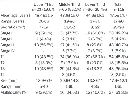
Table 1. Patients and tumour characteristics
Upper Third
n=23 (19.5%) n=65 (55.1%)Middle Third n=30 (25.4%)Lower Third n=118Total Mean age (years) 48.4±11.5 48.8±15.6 44.3±15.1 47.5±14.7 Range (years) 26-66 19-86 17-75 17-86 Sex ratio (m/f) 4/19 13/52 8/22 25/93 Stage I 9 (39.1%) 31 (47.7%) 18 (60.0%) 58 (49.2%) Stage II 1 (4.4%) 2 (3.1%) 2 (6.7%) 5 (4.2%) Stage III 13 (56.5%) 27 (41.5%) 8 (26.6%) 48 (40.7%) Stage IV - 5 (7.7%) 2 (6.7%) 7 (5.9%) T1 10 (43.5%) 24 (36.9%) 20 (66.7%) 54 (45.8%) T2 3 (13.0%) 9 (13.9%) 6 (20.0%) 18 (15.3%) T3 10 (43.5%) 29 (44.6%) 4 (13.3%) 43 (36.4%) T4 - 3 (4.6%) - 3 (2.5%) Size (mm) 13.9±7.9 20.6±14.3 13.8±7.1 17.6±12.1 Range (mm) 5-40 1-65 4-35 1-65 Multifocality (%) 9 (39.1%) 16 (24.6%) 12 (40.0%) 37 (31.3%)
and prophylactic), in ipsilater-al and contripsilater-alateripsilater-al side, were as follows: i) pre-operative FNAC nodal positivity or radio-logical findings suggestive for metastases; ii) intra-operative detection, either by visual inspection or touch, of sus-pect nodes; iii) multifocal and advanced tumours confirmed at the intra-operative frozen section examination of the thyroid gland; iv) in the case of multifocal neoplasm localised also in the isthmus, a supple-mentary prophylactic contra-lateral central compartment dissection was done. Recur- rent nerves and parathyroid glands were identified in all
Results
Overall, patients were 93 females aged 46.9±14.5 years (range 17-86) and 25 males aged 50.2±15.9 years (range 23-82), with 75/118 patients (63.6%) being over 45-years old at time of TT. All tumours were classified according to their localisation in the thyroid gland, and differentiated in upper, middle, or lower third. Isthmus lesions were counted as middle third. If the cancer was multifocal, we considered the largest neoplasm (Table 1). In the whole cohort, the localisation of the cancer was 23 (19.5%), 65 (55.1%), or 30 (25.4%) to the upper, middle, or lower third, respectively. The corresponding localisation in the 75 patients aged over 45 was: 18 (24.0%; 16 females and 2 males), 41 (54.7%; 33 females and 8 males), 16 (21.3%; 11 females and 5 males).
At the histopathological examination, the maxi-mum diameter of the malignant nodule aver-aged 17.6±12.1 mm, neoplasm multifocality was recorded in 37 patients, with involvement
(2.5%) died for distant recurrences, and 1 died for unrelated causes (severe multiple trauma in a road accident). In the whole cohort, local recurrences occurred in 7 patients (5.9%), of whom 6 required a surgical second-look and 1 a supplementary radioiodine therapy.
Nodal metastases were found in 66/118 patients in the IC, 8/14 in the CC, 47/118 in the IL and 4/10 in the CL group, respectively (Table 2).
A concordance between lymph node positivity for metastases in central and lateral compart-ment was found in 7 of 23 cases (30.4%) when the cancer was in the upper third of thyroid, in 26 of 65 cases (40%) when the cancer was in the middle third and in 6 of 30 cases (20%) when the cancer was in the lower third, but dif-ferences were not statistically significant. The LNMs in the IC group, either isolated or pooled with those of the IL group, were statistically related to age and tumour size (P=0.01), par-ticularly in patients over 45 years (Table 2). In these lymph nodes groups, tumour size and of the upper, middle or lower thyroid third in 9 (24.3% of 37; 7 females, 2 males), 16 (43.2%; 11 females, 5 males) and 12 cases (32.4%; 10 females, 2 males), respectively (Table 1) Hashimoto’s thyroid-itis was histologically present in 16 patients (13.5%). Cervical drainage, which was bilateral in 10 patients, was generally removed in the first post-operative day, except for 3 patients, in whom it was delayed on the next day for slight bleeding.
No mortality was observed. Early compli-cations were transient hypocalcaemia in 19 (16.1%), transient recurrent laryngeal nerve injuries in 7 (5.9%) and cervical hae-matomata in 2 (1.7%). Only 2 patients (1.7%) developed permanent hypoparathy-roidism, in which they required permanent substitutive treatment with oral calcium and cholecalciferol. Discharge occurred on the first, second or third postoperative day in 71, 42 or 5 patients, respectively. The mean follow-up time was 75.9±9.2 months (range 61-98), equivalent to 6.3±0.7 years. The 5-year overall survival was 96.6% (114/118), because 3 patients Table 2. Correlation among the number of LNMs, with
tumour size, age and gender of patients
IC
N=66 N=8CC N=47IL N=4CL N=113IC+IL Tumour size rho 0.29 0.05 0.07 0.77 0.21
P 0.01* 0.90 0.65 0.22 0.02* Patients Age rho -0.31 0.17 -0.10 0.25 -0.22
Total p 0.01* 0.67 0.49 0.74 0.01* Gender rho -0.04 -0.54 -0.02 0.57 -0.06 P 0.73 0.16 0.83 0.42 0.51 Tumour size rho 0.38 0.63 -0.13 n.v. 0.18 P 0.05 0.36 0.62 0.23 Patients Age rho <45 -0.19 0.10 0.11 n.v. -0.09 P 0.33 0.89 0.68 0.54 Gender rho -0.05 0.94 -0.16 n.v -0.11 P 0.39 0.05 0.55 0.47 Tumour size rho 0.38 -0.33 0.08 0.86 0.27 P 0.01* 0.66 0.63 0.33 0.02* Patients Age rho >45 0.004 0.73 -0.08 0.90 0.04
P 0.97 0.26 0.65 1.00 0.73 Gender rho -0.19 0.27 -0.04 0.50 -0.15 P 0.13 0.72 0.81 0.60 0.21
*statistically significant (P value<0.05). rho: Spearman’s correla-tion coefficient; n.v.: not value; LNM: lymph node metastases; IC group: ipsilateral central; CC group: contralateral central; IL group: ipsilateral lateral; CL group: contralateral lateral.
age may be considered as sensible dependent predictor variables. Instead, the other CC and CL groups were unrelated to tumour size, age and gender (Table 2). The number of lymph nodes to be removed to reach an optimal ade-quacy of treatment was assessed considering the average number of lymph node harvested ± SD values (e.g. considering A (average) ± B (SD), we reckoned the lymph node dissection insuf-ficient if <B, good if comprised between A and B, and optimal if > than A plus B values) (Table 3).
Discussion
PTC spreads to cervical nodal metastases in 30 to 80% of patients [4]. Patients with nodal involvement have an increased recurrence of PTC and a decreased survival rate [11, 12]. Accordingly, the appropriate surgical treatment of overt and occult nodal metastases improves overall survival [13], and indeed cervical node dissection represents the recommended treat-ment for the advanced thyroid malignant dis-eases [8]. Controversies remain on the correct management to achieve an adequate neck dis-section and on the cervical lymph node levels that require excision [4, 6]. In accord to the international guidelines for the management of differentiated thyroid cancer, the initial surgical treatment for PTC is TT with synchronous cen-tral node compartment dissection [8].Such dis-section is therapeutic if lymph nodes are clini-cally involved or prophylactic if thyroid cancer is advanced, i.e. stage T3 or T4 [8]. However, lit-erature reports a large variability of results about the total number of lymph nodes removed
imisation of the number of lymph nodes removed is unnecessary and unrelated to the recurrences [6, 22]. The lateral compartment dissection increases the complications rate for a raised surgical invasiveness [13, 21]. Some authors report a concordance between tumours localisation and nodal compartment involve-ment, particularly between superior lobe and lateral compartment [23, 24]. Others under-lined the usefulness of the sentinel node biop-sy to better assess the lateral neck compart-ment status for the purpose of detecting the occult lesions [25]. Recently, new surgical tech-niques for haemostasis and use of nerve-spar-ing devices have been introduced to reduce complications arising from monopolar diather-my thermal injury [26, 27].
On considering our low rate recurrences occurred during the follow-up period, and on the basis of our experience, we suggest that: i) the number of lymph nodes harvested to con-sider a cervical dissection as optimal may be accounted more than 8 and 11 in central and lateral compartments, and 6 and 10 in contra-lateral sides, respectively; ii) when tumour is localised either only or even in the isthmus, a supplementary prophylactic contralateral cen-tral compartment dissection is recommended, due to the increased spreading trend toward the locoregional lymph nodes.
Furthermore, we confirm that central lymph nodes compartment must be always removed, lateral lymph nodes compartment may be either removed in case of ipsilateral central nodes positivity or cleared despite of the pres-Table 3. Adequacy judgement of cervical lymph node
dis-section in PTC patients
Cervical Node Dissection Adequacy Judgement Lymph node metastases/
Total lymph nodes harvested
Total lymph nodes: mean ± SD Insufficient Good Optimal IC 182/655 (27.8%) 5.5±2.9 <3 3-8 >8 CC 22/60 (36.7%) 4.3±2.2 <2 2-6 >6 IL 114/966 (11.8%) 8.2±3.7 <5 5-11 >11 CL 5/87 (5.7%) 8.7±1.8 <6 6-10 >10
PTC: Papillary Thyroid Carcinoma.
and the nodal levels included in the dis-sections [4, 6, 9, 11].Due to occult lymph node metastases a routine pro-phylactic central node compartment dissection can be carried out in all PTC patients, even if in absence of pre-operative imaging suggestive for metastases or suspect nodes during the intra-operative research [14-18]. The prophylactic lateral node dissec-tion is not recommended in absence of cytological diagnosis of tumour [8], despite more analysis showed a con-cordance of the involvement in central and lateral compartment [19-21]. However, a few studies report that
max-ence of uninvolved central nodes, at least in more advanced diseases, and contralateral lymph nodes compartments should be inspect-ed and removinspect-ed only on the evidence of gross node metastases.
In our retrospective analysis, we worked up data from the above said 118 cases hypothe-sized and suggested which minimum number of lymph nodes better applies to achieve a quantitatively adequate cervical dissection. However, further studies with an increasingly larger number of patients are necessary to bet-ter strengthen and confirm this hypothesis as a future method of evaluation.
Acknowledgements
For the statistical analysis of data, we wish to thank Giuseppe Vita, Ph.D., University of Me- ssina.
Disclosure of conflict of interest None.
Address correspondence to: Dr. Marco Cicciù, De- partment of Odontostomatology, School of Dentistry University of Messina, Policlinico G. Martino, Via Consolare Valeria 98100, Italy. Tel: +39090221- 6920; E-mail: [email protected]
References
[1] Sobin LH, Gospodarowicz MK, Wittekind C. TNM classification of malignant tumours. Inter-national Union against Cancer. 7th edition. Chichester, West Sussex, UK: Hoboken, NJ, Wiley-Blackwell; 2010.
[2] Ceresini G, Corcione L, Michiara M, Sgargi P, Teresi G, Gilli A, Usberti E, Silini E, Ceda GP. Thyroid cancer incidence by histological type and related variants in a mildly iodine-deficient area of Northern Italy, 1998 to 2009. Cancer 2012; 118: 5473-80.
[3] Boyle P, Ferlay J. Cancer incidence and mortal-ity in Europe, 2004. Ann Oncol 2005; 16: 481-8.
[4] Machens A, Hinze R, Thomusch O, Dralle H. Pattern of nodal metastasis for primary and reoperative thyroid cancer. World J Surg 2002; 26: 22-8.
[5] Kupferman ME, Patterson M, Mandel SJ, LiVol-si V, Weber RS. Patterns of lateral neck metas-tasis in papillary thyroid carcinoma. Arch Oto-laryngol Head Neck Surg 2004; 130: 857-60. [6] Albuja-Cruz MB, Thorson CM, Allan BJ, Lew JI,
Rodgers SE. Number of lymph nodes removed
during modified radical neck dissection for papillary thyroid cancer does not influence lat-eral neck recurrence. Surgery 2012; 152: 1177-83.
[7] Shaha AR. TNM classification of thyroid carci-noma. World J Surg 2007; 31: 879-87. [8] Cooper DS, Doherty GM, Haugen BR, Kloos RT,
Lee SL, Mandel SJ, Mazzaferri EL, McIver B, Pacini F, Schlumberger M, Sherman SI, Stew-ard DL, Tuttle RM. Revised American Thyroid Association management guidelines for pa-tients with thyroid nodules and differentiated thyroid cancer. Thyroid 2009; 19: 1167-214. [9] Vergez S, Sarini J, Percodani J, Serrano E,
Caron P. Lymph node management in clinically node-negative patients with papillary thyroid carcinoma. Eur J Surg Oncol 2010; 36: 777-82.
[10] Bozec A, Dassonville O, Chamorey E, Poisson-net G, Sudaka A, Peyrottes I, Ettore F, Haude-bourg J, Bussière F, Benisvy D, Marcy PY, Sa-doul JL, Hofman P, Lassale S, Vallicioni J, Demard F, Santini J. Clinical impact of cervical lymph node involvement and central neck dis-section in patients with papillary thyroid carci-noma: a retrospective analysis of 368 cases. Eur Arch Otorhinolaryngol 2011; 268: 1205-12.
[11] Scheumann GF, Gimm O, Wegener G, Hunde-shagen H, Dralle H. Prognostic significance and surgical management of locoregional lymph node metastases in papillary thyroid cancer. World J Surg 1994; 18: 559-67. [12] Leboulleux S, Rubino C, Baudin E, Caillou B,
Hartl DM, Bidart JM, Travagli JP, Schlumberger M. Prognostic factors for persistent or recur-rent disease of papillary thyroid carcinoma with neck lymph node metastases and/or tu-mor extension beyond the thyroid capsule at initial diagnosis. J Clin Endocrinol Metab 2005; 90: 5723-9.
[13] Hughes DT, Doherty GM. Central neck dissec-tion for papillary thyroid cancer. Cancer Control 2011; 18: 83-8.
[14] Palestini N, Borasi A, Cestino L, Freddi M, Odasso C, Robecchi A. Is central neck dissec-tion a safe procedure in the treatment of papil-lary thyroid cancer? Our experience. Langen-becks Arch Surg 2008; 393: 693-8.
[15] Teixeira G, Teixeira T, Gubert F, Chikota H, Tu-fano R. The incidence of central neck micro-metastatic disease in patients with papillary thyroid cancer staged preoperatively and intra-operatively as N0. Surgery 2011; 150: 1161-7. [16] Iyer NG, Shaha AR. Central compartment dis-section for well differentiated thyroid cancer … and the band plays on. Curr Opin Otolaryngol Head Neck Surg 2011; 19: 106-12.
[17] Mulla M, Schulte KM. Central cervical lymph node metastases in papillary thyroid cancer: a
systematic review of imaging-guided and pro-phylactic removal of the central compartment. Clin Endocrinol (Oxf) 2012; 76: 131-6.
[18] Mamelle E, Borget I, Leboulleux S, Mirghani H, Suárez C, Pellitteri PK, Shaha AR, Hamoir M, Robbins KT, Khafif A, Rodrigo JP, Silver CE, Rinaldo A, Ferlito A, Hartl DM. Impact of pro-phylactic central neck dissection on oncologic outcomes of papillary thyroid carcinoma: a re-view. Eur Arch Otorhinolaryngol 2015; 272: 1577-8. doi: 10.1007/s00405-014-3104-5. [19] Bhattacharyya N. Surgical treatment of
cervi-cal nodal metastases in patients with papillary thyroid carcinoma. Arch Otolaryngol Head Neck Surg 2003; 129: 1101-4.
[20] Machens A, Hauptmann S, Dralle H. Lymph node dissection in the lateral neck for comple-tion in central node-positive papillary thyroid cancer. Surgery 2009; 145: 176-81.
[21] Keum HS, Ji YB, Kim JM, Jeong JH, Choi WH, Ahn YH, Tae K. Optimal surgical extent of lat-eral and central neck dissection for papillary thyroid carcinoma located in one lobe with clinical lateral lymph node metastasis. World J Surg Oncol 2012; 10: 221. doi: 10.1186/1477-7819-10-221.
[22] Caron NR, Tan YY, Ogilvie JB, Triponez F, Reiff ES, Kebebew E, Duh QY, Clark OH. Selective modified radical neck dissection for papillary thyroid cancer-is level I, II and V dissection al-ways necessary? World J Surg 2006; 30: 833-40.
[23] Hunt JP, Buchmann LO, Wang L, Abraham D. An analysis of factors predicting lateral cervi-cal nodal metastases in papillary carcinoma of the thyroid. Arch Otolaryngol Head Neck Surg 2011; 137: 1141-5.
[24] Fritze D, Doherty GM. Surgical management of cervical lymph nodes in differentiated thyroid cancer. Otolaryngol Clin North Am 2010; 43: 285-300.
[25] Lee SK, Kim SH, Hur SM, Choe JH, Kim JH, Kim JS. The efficacy of lateral neck sentinel lymph node biopsy in papillary thyroid carcinoma. World J Surg 2011; 35: 2675-82.
[26] Rahbari R, Mathur A, Kitano M, Guerrero M, Shen WT, Duh QY, Clark OH, Kebebew E. Pro-spective randomized trial of ligasure versus harmonic hemostasis technique in thyroidec-tomy. Ann Surg Oncol 2011; 18: 1023-7. [27] Glover AR, Norlén O, Gundara JS, Morris M,
Sidhu SB. Use of the Nerve Integrity Monitor during Thyroid Surgery Aids Identification of the External Branch of the Superior Laryngeal Nerve. Ann Surg Oncol 2015; 22: 1768-73.