R E S E A R C H A R T I C L E
Open Access
Ultrasound in the evaluation of enthesitis: status
and perspectives
Frédérique Gandjbakhch
1, Lene Terslev
2, Fredrick Joshua
3, Richard J Wakefield
4, Esperanza Naredo
5and
Maria Antonietta D
’Agostino
6*, for OMERACT Ultrasound Task Force
Abstract
Introduction: An increasing number of studies have applied ultrasound to the evaluation of entheses in
spondyloarthritis patients. However, no clear agreement exists on the definition of enthesitis, on the number and
choice of entheses to examine and on ultrasound technique, which may all affect the results of the examination.
The objectives of this study were to first determine the level of homogeneity in the ultrasound definitions for the
principal lesions of enthesitis in the published literature and second, to evaluate the metric properties of
ultrasound for detecting enthesitis according to the OMERACT filter.
Methods: Search was performed in PUBMED and EMBASE. Both grey-scale and Doppler definitions of enthesitis,
including describing features of enthesitis, were collected and metrological qualities of studies were assessed.
Results: After selection, 48 articles were analyzed. The definition of ultrasound enthesitis and elementary features
varied among authors. Grey-scale enthesitis was characterized by increasing thickness (94% of studies),
hypoechogenicity (83%), enthesophytes (69%), erosions (67%), calcifications (52%), associated bursitis (46%) and
cortical irregularities (29%). Only 46% of studies reported the use of Doppler. High discrepancies were observed on
frequency, type of probe and Doppler mode used. Face and content validity were the most frequently evaluated
criteria (43%) followed by reliability (29%) and responsiveness (19%).
Conclusions: Ultrasound has evidence to support face, content validity and reliability for the evaluation of
enthesitis, though there is a lack of well-reported methodology in most of the studies. Consensus on elementary
lesions and standardization of exam is needed to determine the ultrasound definition of enthesitis in grey-scale
and in Doppler for future applications.
Keywords: Systematic literature review, scoring system, ultrasound, power Doppler, enthesitis, enthesopathy,
spon-dyloarthritis, ankylosing spondylitis, OMERACT filter
Introduction
Enthesitis, that is, the inflammation of insertions of
ten-dons, ligaments and capsules into the bone, is the
char-acteristic sign of ankylosing spondylitis and related
pathologies, which are commonly regrouped as
spondy-loarthritis (SPA). The functioning enthesis dissipates
stress over a wide area, including the insertion,
immedi-ately adjacent tendon and adjacent bone. The soft tissue
components of an enthesis have traditionally been
evaluated by clinical examination based on the presence
of tenderness and/or swelling while X-rays have been
used to assess associated bony changes. The accuracy of
these methods, however, is uncertain, which is why new
imaging techniques such as ultrasound and magnetic
resonance imaging (MRI) have been sought. The role of
MRI for assessing the spectrum of pathology in SPA has
recently been reported [1,2]. This technique has been
most commonly used to assess axial disease. The MRI
pattern of SPA enthesitis has been described as a diffuse
bone edema adjacent to enthesis, associated with
sur-rounding soft tissue edema [3]. However, MRI lacks
sen-sitivity and specificity for peripheral enthesitis [4]. This
can be explained because changes in the fibrous part of
* Correspondence: [email protected]
6Rheumatology Department, Université Paris Ouest-Versailles-Saint Quentin
en Yvelines, Hôpital Ambroise Paré, APHP, UPRES EA 2506, 9 avenue Charles De Gaulle 92100 Boulogne-Billancourt, France
Full list of author information is available at the end of the article
© 2011 Gandjbakhch et al.; licensee BioMed Central Ltd. This is an open access article distributed under the terms of the Creative Commons Attribution License (http://creativecommons.org/licenses/by/2.0), which permits unrestricted use, distribution, and reproduction in any medium, provided the original work is properly cited.
the enthesis, where fibroblasts are tightly cross-linked
with little scope for accumulation of water, cannot easily
be detected with MRI [4,5]. Additionally, MRI cannot
easily assess multiple sites or be used to assess the
con-tralateral joints.
Most of the available data on the potential application
of ultrasound for rheumatology is currently about the
assessment of its role in rheumatoid arthritis with
lim-ited data or studies in other rheumatic diseases, among
which SPA is themost frequently studied [6-53]. For
routine use in daily practice and clinical trials, the
assessment of ultrasound performance in terms of
metric qualities is recommended [54]. Though several
studies have highlighted the value of ultrasound in
assessing inflammation of enthesis in SPA, there is no
clear agreement on which structures to examine. Even
though a clear distinction between the meaning of the
word enthesitis and enthesopathy exists in the
rheuma-tologic literature, no clear definition of an enthesitis
lesion has been reported in the ultrasound literature.
Thus, technical and anatomical issues, combined with a
lack of standardization, may have hampered the
devel-opment and validation of the ultrasound technique
applied to clinical practice, or to multicenter studies, in
SPA. Consensus definitions for ultrasound-related
pathologies were published by the OMERACT
(Out-come Measure in Rheumatology in Clinical Trials)
ultra-sound group in 2005, including enthesopathy [52].
However, no data are available about the
implementa-tion of this definiimplementa-tion in clinical and research practice.
The objective of this study was to first determine the
level of homogeneity in the ultrasound definitions for
the principal lesions of enthesitis in the published
litera-ture, and second, to evaluate the metric properties of
ultrasound for the detection of enthesitis according to
the OMERACT filter through a systematic literature
review. We focused our review on the anatomical
defini-tion of enthesitis, that is, attachment of ligaments or
tendons or capsules on bones, which does not imply
body tendon nor surrounding tissue, such as bursae.
Methods
Search strategy and study selection
The search for original articles concerning humans,
published in the English language between January 1985
and May 2010, and referring to peripheral enthesitis and
ultrasonography was carried out in PUBMED and
EMBASE databases. Reviews or abstracts from scientific
congresses were not included.
In order to obtain the largest number of references,
the search was performed in two steps in PUBMED
with different key words:
- Search 1 was carried out using the following key
words « ankylosing spondylitis OR spondylarthropathies
OR reactive arthritis OR psoriatic arthritis OR enthesis
OR enthesopathy OR rheumatic diseases OR definition
» AND « ultrasonography OR ultrasound OR
sonogra-phy OR Doppler ».
- Search 2 was performed including the key words
«entheses OR enthesis OR enthesitis OR enthesopathy
». For both searches key words referred to Mesh Terms
or, if not available, to key words present in the title/
abstract.
In EMBASE the search was performed with the key
words « ankylosing spondylitis OR spondylarthropathy
OR reactive arthritis OR Psoriatic arthritis OR Enthesis
OR Enthesitis OR Enthesopathy OR Definition » AND «
Ultrasonography OR Ultrasound OR Sonography OR
Doppler ».
Only references with available abstracts were assessed.
Titles, abstracts and full reports of articles identified
were systematically screened by one author (FG) with
regard to inclusion and exclusion criteria. The final
search was verified by a second author (FJ). Articles
concerning cadavers were not included in the final
selection if they concerned healthy subjects.
Articles which did not meet inclusion criteria were
excluded at any step of the study selection.
Data extraction
All data were extracted from the selected articles using a
standardized spreadsheet previously developed and
vali-dated for systematic reviews [55,56] . All selected
arti-cles were rated in order to determine ultrasound
definitions of enthesitis or its characteristics and to
eval-uate the quality of the studies according to the
OMER-ACT filter [54]. A standardized tool for assessing the
quality of the analyzed studies was developed and
assessed in a binary mode (yes/no) based on a set of six
predefined criteria: 1) Was the recruitment of patients
well-defined in the methods section? 2) Was the
defini-tion of ultrasound enthesitis clearly defined as well as
the definition of each elementary component? 3) Was
there a description of ultrasound scanning technique? 4)
Was there a description of attempted blinding of
obser-vers? 5) Was there a description of enthesitis scoring,
and which source was this scoring based on? 6) Was the
choice of comparator adequately explained and results
completely given? Quality was reported on a scale of 0
to 6, with higher results indicating higher quality.
Particular attention was also given to the definition,
quantification and site of detection of Doppler signals,
(that is, vascularization detected at enthesis, in the body
of the tendon, at cortical bony insertion, in the bursa).
Evaluation methods
Face and content validity, construct validity, criterion
validity and discriminant validity (that is, reliability and
responsiveness) were independently evaluated in every
paper, including whether the methods for assessing it
and their measurement were available or not. Face and
content validities, essentially subjective, were analyzed
according to the conclusions of authors. Criterion
valid-ity was considered achieved when ultrasound results
were concurrently or predictively compared with a true
“gold standard”.
Construct validity was achieved when ultrasound
eva-luation of enthesitis was demonstrated to be consistent
with theoretic concepts (that is, that ultrasound measure
of enthesitis is related to other measures of enthesitis).
The evaluation of reliability was divided into two
parts: the acquisition phase and reading of images
phase. For both we assessed the intra- and
inter-obser-ver evaluation. Responsiveness was evaluated by the
ability of the tool to demonstrate change, usually in
response to an intervention.
Statistical analysis
Descriptive statistics were used to report data.
Frequen-cies and percentages were used for categorical variables.
Results
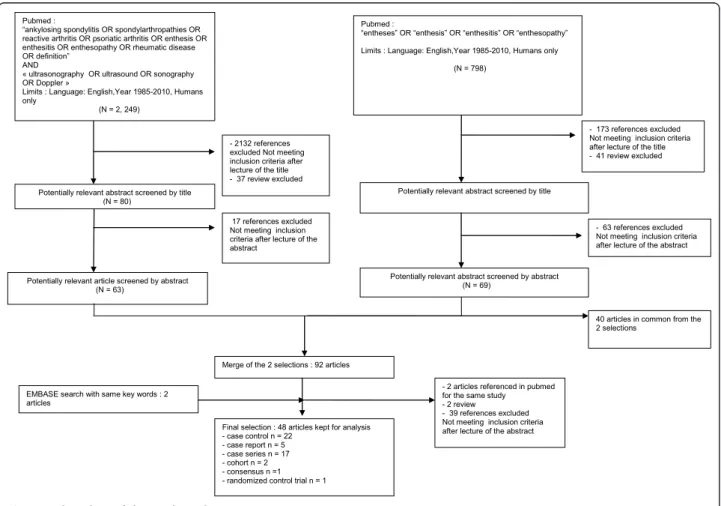
Figure 1 illustrates the flow chart of the selection of
the articles. Of the 3,852 references obtained from
databases, 237 abstracts were selected after reading
titles, 94 articles were selected after reading abstracts
and, finally, 48 articles were analyzed to determine the
ultrasonographic enthesitis definition and
characteris-tics. These articles included 22 case-control studies, 5
case-report studies, 17 case-series studies, 2 cohorts, 1
expert consensus and 1 randomized control trial
(Table 1). Most of them (n = 37) focused on
inflam-matory pathologies: spondylarthropathy or ankylosing
spondylitis (n = 24), spondylartropathy or other
inflammatory rheumatism (n = 3), and psoriatic
arthri-tis (n = 10). Only six studies focused on degenerative
involvement of enthesis. Two studies did not report
the patients
’ diagnoses.
Entheses of lower limbs were the most common
stu-died, especially Achilles tendon (80% of articles)
fol-lowed by the entheses of upper limbs. No consensus
concerning either the location or the number of enthesis
to be examined was observed.
(N = 132)
Pubmed :
“ankylosing spondylitis OR spondylarthropathies OR reactive arthritis OR psoriatic arthritis OR enthesis OR enthesitis OR enthesopathy OR rheumatic disease OR definition”
AND
« ultrasonography OR ultrasound OR sonography OR Doppler »
Limits : Language: English,Year 1985-2010, Humans only
- 2132 references excluded Not meeting inclusion criteria after lecture of the title - 37 review excluded Potentially relevant abstract screened by title
Potentially relevant article screened by abstract
17 references excluded Not meeting inclusion criteria after lecture of the abstract
Pubmed :
“entheses” OR “enthesis” OR “enthesitis” OR “enthesopathy” Limits : Language: English,Year 1985-2010, Humans only
- 173 references excluded Not meeting inclusion criteria after lecture of the title - 41 review excluded Potentially relevant abstract screened by title
- 63 references excluded Not meeting inclusion criteria after lecture of the abstract Potentially relevant abstract screened by abstract
Merge of the 2 selections : 92 articles
40 articles in common from the 2 selections
- 2 articles referenced in pubmed for the same study - 2 review
- 39 references excluded Not meeting inclusion criteria after lecture of the abstract Final selection : 48 articles kept for analysis
- - - - - - EMBASE search with same key words : 2
articles (N = 2, 249) (N = 798) (N = 80) (N = 63) (N = 69) case control n = 22 case report n = 5 case series n = 17 cohort n = 2 consensus n =1 randomized control trial n = 1
Table 1 Characteristics of the studies
Year Authors Type ofArticle
Sample Size
Population of interest Entheses sites of interest Face validity 1987 Maffulli [42] case series 47 Athletes A N 1989 Olivieri [47] case report
1 SPA A, PF, ischial tuberosity, trochanter N
1994 Lehtinen [40]
case control
39 SPA (ReA, PsA, AS) pelvic adductor origin, trochanter, ischial tuberosity, PT, A, PF
N
1995 Lehtinen [39]
cohort 23 SPA ischial tuberosity, trochanter, PT, A, PF N
1998 Olivieri [46] case series
14 SPA A (hanging free over the edge of the table) N
1999 Gibbon [32]
case control
370 clinically idiopathic plantar fasciitis, SPA, RA, Achilles tendon disease, ankle
instability, healthy subjects
A, PF (90°) N
2000 Balint [17] case report
1 PsA A (hanging free) N
2000 Galluzzo [29] case series 31 PsA A, PF N 2001 Cosentino [19]
RCT 60 patients with talalgia PF N
2002 Balint [16] case series
35 SPA (AS, PsA, ReA) GUESS score: A, PF (90°),PTPI, PTDI, Q (30°) N
2002 D’Agostino [21] case report 2 SPA A, PF N 2002 Falsetti [10] case control
450 SPA ,RA, OA, painful shoulders, healthy subjects
Deltoid tendon insertion N
2002 Falsetti [28] case control
178 PMR, SPA(PsA, AIBD, uSpA), RA wrist, elbow, shoulder, hip, knee, ankle, heel N
2002 Frediani [27]
case control
160 PsA, RA, healthy subjects Q (30°) N
2003 D’Agostino [22]
case control
228 SPA, MBP, RA A, PF, tibialis anterior tendon, CET, FCT, PT, Q, gluteus medius and minimus tendons
N
2003 De Simone [24]
case control
109 psoriatic with arthritis or spondylitis, healthy subjects
A N
2003 Falsetti [11] case control
598 EOA , NOA , RA, PsA, healthy subjects A, PF N
2003 Kamel [34] case series
32 SPA A, PF Y
2004 Falsetti [9] case control
157 CCA, OA, healthy subjects A, PF N
2004 Kamel [35] case series
16 SPA PT N
2005 Genc [30] case control
62 RA , AS, healthy subjects * N
2005 Ozçakar [48]
case control
50 Psoriasis, healthy subjects A (Neutral flexion) N
2005 Ozgocmen [49] case report 1 AS A N 2005 Wakefield [52] consensus - NA NA N 2006 Borman [18] case series
44 SPA (AS, PsA, ReA, uSpA) A, PF N
2006 Fournie [26]
case control
41 PsA, RA flexor tendons of the hand N
2006 Kiris [37] case series 30 AS MASES ** N 2006 Tse [51] case report 1 AS A, PF, Q, PT N
Ultrasound parameters and setting
The description of ultrasound examination was reported
in 35 (73%) studies and recommendations on the
posi-tion of the examined enthesis, especially for lower limbs,
were available in most of the studies. Authors
predomi-nantly used 90° flexion of the feet during examination of
Achilles tendon and Plantar Fascia, 30° to 60° flexion of
the knee during examination of the patella ligament and
the quadriceps tendon. In more recent studies, a neutral
position of the feet was used to perform Achilles tendon
entheses examination.
Definition and description of enthesitis in grey-scale and
Doppler modes
In grey-scale a 7.5 MHz or 7.5 to 10 MHz linear probe
frequency were used in 15/48 studies while a frequency
Table 1 Characteristics of the studies (Continued)
2007 Alcalde [14] casecontrol
54 AS, healthy subjects A(neutral flexion), PF (neutral flexion),PTPI(60°), PTDI (60°), Q (60°)
N
2007 Genc [31] cohort 54 RA, AS GUESS * N
2007 Kerimoglu [36] case series 49 Hemodialysis GUESS * N 2007 Scarpa [50] case control
47 PsA ,“sine psoriasis"patients All entheses with increased tracer uptake in scintigraphy
N
2007 Wiell [53] case control
20 PsA, RA, healthy subjects flexor/extensor tendons of the hands Y
2008 de Miguel [23]
case control
54 SPA(AS, ReA, uSpA, PsA, juvenile SPA), healthy subjects
MASEI score: A (90°), PF (90°), PTPI (70°), PTDI (70°), Q, brachial triceps tendon (90°)
N
2008 Filippou [12]
case series
7 Ochronosis flexor/extensor tendon of the hand, CET, CFT, distal brachial triceps tendon, gluteus medius and minimus
tendons, Q, PT, iliotibial band, A, PF, anterior and posterior tibialis tendon, peroneal tendon, toe extensor
tendon N 2008 Gisondi [33] case control
60 Psoriasis, healthy subjects GUESS * N
2008 Hatemi [7] case control
160 Behçet, AS, RA, healthy control GUESS * Y
2008 Klauser [38] case control
33 SPA (AS, PsA, uSpA, ReA, AIBD), RA, patients with non rheumatic disease
MASES ** N 2008 Mc Gonagle [44] case control
47 SPA(AS, PsA, ReA), healthy subjects A (90°) N
2009 D’Agostino [20]
case series
5 SPA(AS, uSpA, PsA, AIBD) Q, PTPI , CET, A, PF N
2009 Filippucci [13]
case series
28 SPA A (hanging in neutral position) N
2009 Matsos [43] case series 62 NA NA N 2009 Munoz-Fernandez [45] case control
79 SPA, anterior uveitis, healthy subjects MASEI *** N
2009 Filippucci [25] case series NA PsA A N 2009 Iagnocco [8] case series
93 SPA A(hanging in neutral position) N
2010 Gutierrez [6] case series 30 PsA U: A, Apo, PT N 2010 Li [41] case control
70 SPA(AS, ReA, PsA,AIBD, uSpA), healthy subjects
A (90°) N
2010 Aydin [15] Case series
43 SPA A (hanging in neutral position) N
A, Achilles tendon; AIBD, arthritis associated with inflammatory bowel disease; AS , ankylosing spondylitis; ASIS, anterior superior iliac spines; CET, commun extensor tendon insertion on lateral epicondyle; CFT, common flexor tendon insertion on medial epicondyle; EOA, erosive osteoarthritis; MBP, mechanical low back pain; NA, not available; NOA, nodular osteoarthritis; OA, osteoarthritis; PF, Plantar Fascia; PMR, polymyalgia rheumatica; PsA, psoriatic arthritis; PSIS, posterior superior iliac spines; PT, patellar tendon; PTDI, patellar tendon distal insertion; PTPI, patellar tendon proximal insertion; Q, quadriceps; RA , rheumatoid arthritis; RCT, randomized control trial; ReA, reactive arthritis; SPA, spondylarthropathy; uSpA, undifferenciated spondyloarthritis
* GUESS score: A; PF (90°),PTPI, PTDI, Q (30°), ** MASES: 1st and 7th
costosternal joints, PSIS, ASIS, iliac crests, A, 5th
lumbar spinous process, *** MASEI score: A (90°), PF (90°), PTPI (70°), PTDI (70°), Q, brachial triceps tendon (90°)
>10 MHz was used in 23 studies. Information
concern-ing probe characteristics was lackconcern-ing in four studies.
Table 2 shows definitions or description of ultrasound
enthesitis and ultrasound elementary components used
for defining enthesitis (for further details see also Table
S1 in Additional file 1). Table S2 in Additional file 2
shows ultrasound parameters and equipment used in
the different studies. In grey-scale, enthesitis was
charac-terized by the presence of increasing thickness in 45
(94%) studies, hypoechogenicity of the enthesis in 40
(83%) studies, enthesophyte in 33 (69%) studies, erosion
in 32 (67%) studies, calcification in 25 (52%) studies,
associated with bursitis in 22 (46%) studies or cortical
irregularities in 14 (29%) studies. Only 16 (33%) studies
described the ultrasound technique of thickness
mea-surement, which was prevalently measured at the point
of maximal thickness on the bony insertion (for further
details see also Table S3 in Additional file 3).
Only 22 out of 48 (46%) studies described the use of
Power Doppler to assess enthesitis (Table 3); all of them
were published after 2003. Most of the studies took into
account the presence of signal Doppler in different
loca-tions: tendon, enthesis and bursa. The exact site of
mea-surement of a Doppler signal was described in 12
studies. There were discrepancies regarding the
techni-cal recommendations of the use of Doppler with a huge
difference of the pulse repetition frequency (PRF) in the
studies ranging from 400 Hz to 1,000 Hz.
Scoring system of enthesitis (grey-scale and Doppler)
Table 4 shows the different ultrasound scoring systems
used for evaluating enthesitis. Ultrasound scoring of
enthesitis was performed in 20 studies. All of the
pro-posed scoring systems were primarily based on grey
scale changes, measuring the thickness of tendon
insertion, the presence of erosions, bursitis and
enthe-sophytes. Proposed grading was semi-quantitative in
most of them. Only nine studies reported scoring
sys-tems of Power Doppler activity of the enthesis, which
were generally semi-quantitative [7,8,13,15,20,22,
23,37,45], but also quantitative with a proposed cut-off
for differentiating between SPA and controls. Five
scoring systems were developed at the enthesis level
(and mostly concerned Achilles enthesis evaluation),
and 15 were developed at the patient level (that is, the
scoring system gave information regarding different
enthesis sites and allowed the evaluation of global
patient inflammatory activity or enthesis structural
damage). Two of them, the GUESS (Glasgow
Ultra-sound Enthesitis Scoring System) score, proposed by
Balint
et al. in 2002 [16] and the SEI (Spanish
Enthesi-tis Index) score, by Alcade
et al. [14], take into
account grey-scale elementary components alone. Both
of them are scoring systems developed at the enthesis
level and at patient level, and the GUESS was the
scor-ing method most frequently used (7/20).
Published scoring systems were used both for
diagnos-tic purposes [22,23,53], and for sensitivity to change
[15,19,31]. Performance of those scores varied according
to the purpose.
Evaluation of studies according to the OMERACT filter
Table 5 summarizes the characteristics of the 48
selected articles according to the OMERACT filter.
Truth
The face, content, criterion and construct validity of
ultrasound findings of the enthesis has been tested in
only 21 articles (44%). Comparators were clinical
exami-nation in 13 studies, MRI in 5 studies, X-ray in 5 studies
and histology in 1 study. In three studies, two
compara-tors were used, clinical and X-ray or MRI.
Ultrasound examination was performed blindly from
other data in 29 articles (62%).
Discrimination
Reliability
Detailed results of the reliability of the technique, which
were evaluated in 14 (29%) studies are only reported in
the additional online file (Table S4 in Additional file 4).
Among them, eight studies correctly reported the
meth-odology used. Reliability was most frequently tested on
static images reading and only two evaluated the
acqui-sition. Only four studies included information on both
inter-examiner and intra-examiner reliability. In general,
reading reliability was good but acquisition reliability
had some deficiencies.
Responsiveness
Responsiveness was evaluated in nine studies. Of them,
only four included power Doppler evaluation of the
enthesis [15,17,21,49] and three used a scoring system
[15,19,31]. Ultrasound evaluation of enthesitis was found
to be sensitive to change in six studies, whereas three
studies did not demonstrate responsiveness, but the
eva-luation concerned the Grey-scale aspect alone, while in
the studies also including Power Doppler the sensitivity
to change was greater. Only three articles reported
responsiveness regardless of statistical analyses, while six
articles were descriptive of changes but did not quantify
it.
Feasibility
None of the analyzed papers reported information about
feasibility of examining entheses using ultrasound.
Discussion
The present review has demonstrated that ultrasound is
considered a valuable tool for assessing enthesitis. Since
1985, when the first description was made by Lehtinen
and colleagues, an increasing interest for using this
Table 2 Ultrasound definition and description of enthesitis or of its elementary components
Lear AuthorsGrey-scale Doppler Definition or description of Enthesitis ♣ Elementary components
Echogenicity Thickness Calcific Deposits
Enthesophytes Tear Erosions Cortical Irregularities Bursitis 1987 Maffulli [42] Y NA Y Y Y Y NA NA NA NA NA 1989 Olivieri [47] Y NA NA NA Y NA NA NA NA NA NA 1994 Lehtinen [40] Y NA Y U Y Y Y NA NA U Y 1995 Lehtinen [39] Y NA Y U Y Y Y NA NA U NA 1998 Olivieri [46] Y NA NA Y Y NA NA NA NA NA NA 1999 Gibbon [32] Y NA Y Y Y Y Y NA Y NA NA 2000 Balint [17] Y Y NA NA Y NA NA NA NA NA Y 2000 Galluzzo [29] Y NA Y Y Y Y NA NA NA NA NA 2001 Cosentino [19] Y NA Y Y Y NA Y NA NA NA NA 2002 Balint [16] Y NA Y Y Y NA Y NA Y Y Y 2002 D’Agostino [21] Y Y NA Y Y Y NA NA Y NA NA 2002 Falsetti [10] Y NA Y Y Y NA Y NA Y Y NA 2002 Falsetti [28] Y NA Y Y Y NA Y NA Y NA NA 2002 Frediani [27] Y NA Y Y Y NA Y NA Y Y NA 2003 D’Agostino [22] Y Y NA Y Y Y U U Y Y NA 2003 De Simone [24] Y NA NA Y Y Y NA Y NA NA Y 2003 Falsetti [11] Y NA Y Y Y Y Y NA Y NA Y 2003 Kamel [34] Y NA Y Y Y Y Y Y NA NA NA 2004 Falsetti [9] Y Y Y Y Y NA Y NA Y NA NA 2004 Kamel [35] Y NA Y Y Y Y NA NA NA NA Y 2005 Genc [30] Y NA Y Y Y NA Y NA Y NA Y 2005 Ozçakar [48] Y NA NA U Y U U U U U Y 2005 Ozgocmen [49] U Y NA NA U NA NA NA NA NA NA 2005 Wakefield [52] Y Y Y Y Y Y Y NA Y Y NA 2006 Borman [18] Y NA Y Y Y NA Y NA Y NA Y 2006 Fournie [26] Y NA Y NA NA NA Y NA NA NA NA 2006 Kiris [37] Y Y NA Y Y Y Y U Y Y Y 2006 Tse [51] Y NA NA NA U U U U U U U 2007 Alcalde [14] Y NA Y Y Y Y NA Y Y NA Y 2007 Genc [31] Y NA Y Y Y NA Y NA Y NA Y 2007 Kerimoglu [36] Y NA Y Y Y Y Y NA Y NA Y 2007 Scarpa [50] Y Y Y Y Y Y Y NA Y Y NA 2007 Wiell [53] Y Y Y Y Y Y Y NA Y Y NA 2008 de Miguel [23] Y Y NA Y Y Y Y NA Y NA Y
Table 2 Ultrasound definition and description of enthesitis or of its elementary components (Continued)
2008 Filippou [12] Y Y Y Y Y Y NA NA NA NA NA 2008 Gisondi [33] Y NA Y Y Y NA Y NA Y NA Y 2008 Hatemi [7] Y Y Y Y Y NA Y NA Y Y Y 2008 Klauser [38] Y Y Y Y Y NA Y NA Y NA NA 2008 McGonagle [44] Y NA Y Y Y NA Y NA Y NA NA 2009 D’Agostino [20] Y Y Y Y Y Y Y NA Y NA NA 2009 Filippucci [13] Y Y Y Y Y Y Y NA Y Y Y 2009 Matsos [43] Y Y Y Y Y Y Y NA Y Y NA 2009 Munoz-Fernandez [45] Y Y NA Y Y Y Y NA Y NA Y 2009 Filippucci [25] Y Y Y Y Y N Y NA Y NA Y 2009 Iagnocco [8] Y Y Y Y Y Y Y NA Y Y Y 2010 Gutierrez [6] Y Y Y Y Y N Y NA Y NA Y 2010 Li [41] Y Y Y Y Y N N NA Y Y NA 2010 Aydin [15] Y Y Y Y Y Y Y NA Y Y YNA, not available, Y, yes
♣: Definition: enthesitis definition reported by the authors, description: enthesitis description or description of enthesitis elementary components, without clear definition reported by the authors
Table 3 Description of enthesitis in Doppler- mode
Year Authors Doppler parameters Description of site of vascularization
2000 Balint [17] PRF1000 Hz NA
2002 D’Agostino [21] PRF 750 Hz, power Doppler gain 50 periosteal bone and enthesis
2003 D’Agostino [22] PRF 750 Hz, power Doppler gain 50-53dB cortical bone insertion, body of the tendon, bursa, junction tendon/enthesis
2004 Falsetti [9] PRF 750-1000 Hz, highest gain level without background noise and low filter
tendon + bursa
2005 Ozgocmen [49] PRF 0.3-1.5kHz, dynamic range 55dB low wall filter periosteum and achilles tendon insertion
2005 Wakefield [52] NA NA
2006 Kiris [37] PRF 0.5-1 KHz , dynamic range 50-55dB - low wall filter tendon + enthesis : no precision concerning the exact location of vascularization
2007 Scarpa [50] NA NA
2007 Wiell [53] PRF 500 Hz NA
2008 De Miguel [23] PRF 400Hz, gain 20dB, low wall filter enthesis, tendon, bursitis
2008 Filippou [12] NA NA
2008 Hatemi [7] PRF 750 Hz NA
2008 Klauser [38] 8.3 MHz, PRF 500 Hz, low wall filter NA
2009 D’Agostino [20] 10 MHz, PRF 500 Hz, gain 113 dB enthesis insertion into the cortical bone 2009 Filippucci [13] PRF 750 Hz, colour-mode frequency of 9.1 MHz , low wall
filters
enthesis, tendon, bursitis
2009 Matsos [43] NA U
2009 Munoz-Fernandez [45]
NA enthesis, tendon, bursitis
2009 Filippucci [25] NA U
2009 Iagnocco [8] PRF 900Hz , Doppler frequency 9.1 MHz, low wall filters enthesis, tendon, bursitis 2010 Gutierrez [6] PRF750 Hz , Doppler frequency between 7.5 -14.3 MHz. U
2010 Li [41] 10 MHz for colour-mode scanning with a focus at 5 mm. peri-sesamoidal and periosteal areas 2010 Aydin [15] PRF 750 Hz, colour-mode frequency of 9.1 MHz, low wall
filters
enthesis, tendon, bursitis
Table 4 Description of enthesitis scoring system
Year Authors Enthesis studiedGrey-scale
Doppler mode
Scoring system Reliability Sensitivity to change 2001 Cosentino
[19]
PF Y N grade l: thickening of enthesis (<2 mm thicker than the controlateral asymptomatic side), heterogeneous hypoechogenicity of enthesis
and enthesophytosis.
grade 2: thickening of enthesis (>2 mm thicker than the controlateral asymptomatic side), heterogeneous hypoechogenicity of enthesis,
and enthesophytosis.
grade 3: grade 2 with peritendinous oedema.
NA U
2002 Balint [16] GUESS:
A, PF (90°),PTPI, PTDI, Q (30°)
Y N GUESS score (0 to 36): Each item scores one point. total possible score on both lower limb
is 36
superior pole of the patella- quadriceps tendon enthesis: quadriceps tendon thickness >=6.1mm, suprapatellar bursitis, superior pole of patella erosion, superior pole of patella
enthesophyte
inferior pole of the patella-proximal patellar ligament enthesis: patellar ligament thickness
> = 4 mm, inferior pole of patella erosion, inferior pole of patella enthesophyte tibial tuberosity-distal patellar ligament enthesis: patellar ligament thickness > = 4
mm, infrapatellar bursitis, tibial tuberosity erosion, tibial tuberosity enthesophyte superior pole of the calcaneus-achilles tendon
enthesis: Achilles tendon thickness >=5.29 mm, retrocalcaneal bursitis, posterior pole of calcaneus erosion, posterior pole of calcaneus
enthesophyte
inferior pole of the calcaneus -plantar aponeurosis enthesis: Plantar aponeurosis thickness >=4.4 mm, inferior pole of calcaneus
erosion, inferior pole of calcaneus enthesophyte.
U NA
2002 Falsetti [28] wrist, elbow, shoulder, hip, knee, ankle, calcaneum
Y N each item scored according to a semi quantitative score: 1: mild,2: moderate,3:
considerable
items scored: synovitis, tenosynovitis, enthesitis
U NA
2003 D’Agostino [22]
A, PF, tibialis anterior tendon, CET, CFT, PT, Q, trochanter
Y Y stage 1: Vascularization at the cortical junction without abnormal findings in Grey-scale stage 2a: Vascularization associated with swelling and/or decreased echogenicity at the
cortical junction in Grey-scale stage 3a: Same as stage 2a, plus erosions of cortical bone and/or calcification of enthesis,
and optional surrounding bursitis stage 2b: Abnormal findings in B mode as in
stage 2a, but without vascularization stage 3b: Abnormal findings in B mode as in
stage 3a, but without vascularization
Y NA
2003 Falsetti [11] A, PF, retrocalcaneal bursae, subcalcaneal fat pad, cortical bone of posterior and inferior aspects of
calcaneum
Y N Each inflammatory lesion was graded according to a semi-quantitative scale: grade
1: mild, grade 2: moderate, grade 3: considerable
NA NA
2006 Kiris [37] MASES * N Y 0 = absence, 1 = mild, 2 = moderate, 3 = severe
Y NA
2007 Alcalde [14] SEI:
A, PF (neutral flexion°),PTPI, PTDI, Q (60°)
Y N SEI = the total sum of SEI-A and SEI-C. the maximum SEI scoring is 76 points SEI-A (0 to 36): each variable is scored as 0
(absence) or 1 (presence): thickening of tendon/aponeurosis, hypoechogenicity of
tendon/aponeurosis, peritendinous/ periaponeurotic oedema, bursitis (where
applicable)
SEI-C (0 to 40): each variable is scored as 0 (absence) or 1 (presence): tendon tear, loss of
thickness, tendon calcification, bone erosion.
Table 4 Description of enthesitis scoring system (Continued)
2008 De Miguel[23]
MASEI:
A (90°), PF (90°), PTPI and PTDI (70°), distal Q tendon, distal brachial triceps tendon (90°)
Y Y MASEI score (0 to 136 on both sides): Calcifications were scored on a
semi-quantitative score of 0 to 3 Doppler and erosions were scored as 0 or 3
points
Scores for tendon structure, tendon thickness and bursa were either 0 or 1. Calcifications were examined at the area of
the enthesis insertion, and scored as 0 if absent, or 1 if a small calcification or ossification with an irregularity of enthesis cortical bone profile was seen. Calcifications
were given a score of 2 if there was clear presence of enthesophytes or if medium sized
calcifications or ossification were observed. Lastly, they were classified as a 3 if large calcifications or ossifications were present. To
simplify things, ossifications and enthesophytes at the enthesis were also
included as calcifications.
Y NA
2008 Hatemi [7] GUESS ** Y Y GUESS score* + Doppler: one point for each enthese with vascularization. Cumulative score
for Doppler (max = 10)
U NA
2009 D’Agostino [20]
Q, PTPI, CET, A, PF Y Y Grey-scale: hypoechogenicity/thickness: 0 to 1, calcification/enthesophyte: 0 to 1, erosion: 0 to
1
Doppler : (0 to 3): 0: no signal, 1: minimal (1 spot), 2: moderate (2 spot), 3: severe (> = 3 spots)or Doppler scored as 0 to 1
(absent-present)
Y NA
2008 Mc Gonagle
[44]
A (90°) Y N spur (0 to 3):0 absence, 1: minimal, 2: moderate, 3: large
NA NA
2009 Filippucci [13]
A Y Y soft tissue inflammation (seven items): tendon hypoechogenicity, Entheseal hypoechogenicity, Bursal effusion, PDS signal at tendon level, PDS signal at entheseal level, PDS signal at bursal
level
tissue damage (five items): Intratendineous calcifications, Entheseal calcifications, Enthesophytes, Bone erosions, Bone irregularities* (not used to calculate total
score)
(1) a total score for soft tissue inflammation, which resulted from the sum of the scores assigned to the 7 US findings indicative of soft
tissue inflammation, ranging from 0 to 7 with presence/absence data and from 0 to 14 with
semiquantitative scores; (2) a total score for tissue damage, which resulted from the sum of the scores assigned
to the 4 US findings indicative of tissue damage, ranging from 0 to 4 with presence/
absence data and from 0 to 8 with semiquantitative scores.
Y NA
2009 Iagnocco [8]
A (neutral position) Y Y All lesions scored on both a dichotomous scale (present/absent) and a 4-point semiquantitative scale (0 = absent, 1 = mild, 2
= moderate, 3 = severe)
enthesopathy: tendon hypoechogenicity at the level of bony attachment, tendon thickening at the at the level of bony attachment, intra-tendinous calcifications, enthesophytes, bony erosions, bony cortex irregularities, presence of
Doppler signal at the level of bony attachment, presence of intratendinous
Doppler signal
bursitis: enlargement of deep calcaneal bursa, enlargement of superficial calcaneal bursa tendon lesion: both partial and full-thickness
tendon lesions
technique in the evaluation of SpA enthesitis has been
observed, especially within the last 10 years. This is
probably due to the tremendous technological
progres-sion of ultrasound equipment. However, standardization
of enthesitis assessment by ultrasound would facilitate
the dissemination of this technique in daily practice, and
also allow adequately trained sonographers to participate
in multicenter research studies. A wide variability was
observed among studies in the definition of ultrasound
enthesitis, associated with a broad heterogeneity of
defi-nitions of its elementary components, and the absence
of a consensus on technical parameters and methods of
examination probably led to the observed heterogeneity
in metric properties of the studies according to the
OMERACT filter. No consensus concerning either the
location or the number of enthesitis to be examined was
observed.
Those discrepancies can be explained by the inclusion
of studies from 1985 until the present, assuming that
ultrasound equipment has improved considerably since
that time, and the differences in the quality of
equip-ment may have hampered the detection of those lesions.
However, the quality and the attention in the
descrip-tion of enthesitis features have improved in the studies
published after 2005, which may be explained by the
publication from our group on the preliminary
OMER-ACT definition of enthesopathy [52]. Indeed, previous
studies have shown that grey-scale elementary lesions
may be observed in both mechanical and inflammatory
enthesopathy [11,30]. Yet, in order to help diagnosis, a
more specific feature is the detection of inflammatory
signs, especially the vascularization.
Since the first observation on the utility of power
Doppler for visualizing vascularization of the enthesis as
a sign of inflammation made in 2003 [22], an increasing
number of studies have included Doppler evaluation.
Some authors have well demonstrated the presence of
vascularization of the enthesis/bone junction in SPA
patients [13,20,23,37]. Even if Doppler use seems to be
important, a wide heterogeneity in its use was recorded.
Most of the studies referred to the presence of Doppler
signal in different locations: tendon, enthesis, bursa. The
lack of consensus with regards to the site of
examina-tion of abnormal vascularizaexamina-tion may contribute to
explaining discrepancies among studies. Some authors
may call
“inflammatory enthesitis” what would be called
“tendonitis” by others. Moreover, this review has shown
a large difference in the Doppler parameters used
among studies. Doppler sensitivity to inflammatory flow
(low-velocity flow) depends partly on the settings and
partly on the type of equipment.
The differences found in the articles may, therefore, be
explained by the lack of consensus on the optimal
Dop-pler settings for enthesitis. Since no information
con-cerning inter-equipment reliability for enthesitis
evaluation is available, the different types of ultrasound
equipment used may also explain part of the
discrepan-cies observed. Indeed, Doppler sensitivity could have
been affected by the type of equipment used; better
sen-sitivity may have been reported with new generation
equipment with the highest quality of Doppler
parameters.
Only 73% of the studies clearly described acquisition
technique. For example, the method for measuring
enthesis thickness, which appears as one of the most
important features recorded by authors for
characteriz-ing enthesitis of the Achilles tendon, was only described
in 31% of the studies despite the fact that the necessity
of measuring the thickness for defining the presence of
enthesitis was reported by 94% of the authors.
Measure-ment methods and site of measureMeasure-ment varied
consis-tently and none of the proposed methods have been
extensively tested and validated yet.
The quantification of enthesitis by ultrasound was
pre-dominantly performed by using semi-quantitative
scor-ing methods. However, some differences were observed
in the evaluation of involvement as all of the proposed
scoring systems combined both evaluation of
inflamma-tory activity, mostly by taking into account echogenicity
and increased thickness and structural damage, mostly
enthesophytes and erosions. As these are all grey-scale
changes, this could explain the discrepancy observed in
the sensitivity to change. In recent years, there has been
more focus on enthesitis vascularization, probably the
most interesting and specific feature to differentiate
inflammatory enthesitis from mechanical enthesitis [22].
Consequently, enthesitis scoring systems taking Doppler
signal into account have been proposed. These scoring
systems, taking more into account the inflammatory
activity may better present sensitivity to change. Hatemi
et al. proposed to add a semi-quantitative scoring
con-cerning vascularization to the GUESS score [7].
The proposal of a scoring system validated at the
patient level, taking into account inflammatory activity
and structural damage is one of the challenges for future
studies regardless of ultrasound enthesitis. This
Y, yes; N, no; U, unclear; NA, not available
A, Achilles tendon; ASIS, anterior superior iliac spines; CET, commun extensor tendon insertion on lateral epicondyle; CFT, common flexor tendon insertion on medial epicondyle; PF, Plantar Fascia; PSIS, posterior superior iliac spines; PT, patellar tendon; PTDI, patellar tendon distal insertion; PTPI, patellar tendon proximal insertion; Q, quadriceps
* MASES: 1st and 7th
costosternal joints, PSIS, ASIS, iliac crests, A, 5th
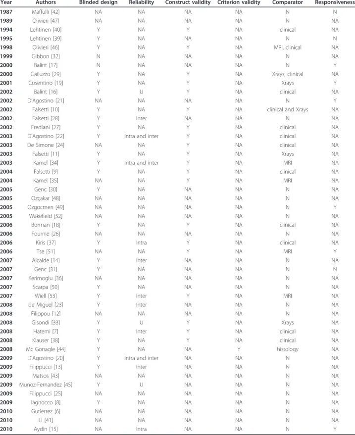
Table 5 Summary of reporting according to the OMERACT filter
Year Authors Blinded design Reliability Construct validity Criterion validity Comparator Responsiveness
1987 Maffulli [42] NA NA NA NA N N
1989 Olivieri [47] NA NA NA NA N NA
1994 Lehtinen [40] Y NA Y NA clinical NA
1995 Lehtinen [39] Y NA NA NA N N
1998 Olivieri [46] Y NA Y NA MRI, clinical NA
1999 Gibbon [32] N NA NA NA N NA
2000 Balint [17] N NA NA NA N Y
2000 Galluzzo [29] Y NA Y NA Xrays, clinical NA
2001 Cosentino [19] Y NA Y NA Xrays Y
2002 Balint [16] Y U Y NA clinical NA
2002 D’Agostino [21] NA NA NA NA N Y
2002 Falsetti [10] Y NA Y NA clinical and Xrays NA
2002 Falsetti [28] Y Inter NA NA N NA
2002 Frediani [27] Y NA Y NA clinical NA
2003 D’Agostino [22] Y Intra and inter Y NA clinical NA
2003 De Simone [24] NA NA Y NA clinical NA
2003 Falsetti [11] Y NA Y NA Xrays NA
2003 Kamel [34] Y Intra and inter Y NA MRI NA
2004 Falsetti [9] Y NA Y NA clinical NA 2004 Kamel [35] NA NA Y NA MRI NA 2005 Genc [30] Y NA NA NA N NA 2005 Ozçakar [48] NA NA NA NA N NA 2005 Ozgocmen [49] NA NA NA NA N Y 2005 Wakefield [52] NA NA NA NA N NA 2006 Borman [18] Y NA Y NA clinical NA 2006 Fournie [26] NA NA NA NA N NA
2006 Kiris [37] Y Intra Y NA clinical NA
2006 Tse [51] NA NA Y NA MRI Y
2007 Alcalde [14] Y Inter NA NA N NA
2007 Genc [31] Y NA NA NA N N
2007 Kerimoglu [36] NA NA NA NA N NA
2007 Scarpa [50] Y NA NA NA N NA
2007 Wiell [53] Y Inter Y NA MRI NA
2008 de Miguel [23] Y Inter NA NA N NA
2008 Filippou [12] NA NA NA NA N NA
2008 Gisondi [33] Y U Y NA Xrays NA
2008 Hatemi [7] Y Inter Y NA clinical NA
2008 Klauser [38] Y NA Y NA clinical NA
2008 Mc Gonagle [44] Y NA NA Y histology NA
2009 D’Agostino [20] Y Intra and inter NA NA N NA
2009 Filippucci [13] Y Inter NA NA N NA 2009 Matsos [43] NA NA NA NA N NA 2009 Munoz-Fernandez [45] Y U NA NA N NA 2009 Filippucci [25] NA NA NA NA N NA 2009 Iagnocco [8] Y NA NA NA N NA 2010 Gutierrez [6] NA NA NA NA N NA 2010 Li [41] NA NA NA NA N NA 2010 Aydin [15] NA Intra NA NA N Y
implicates to determine which enthesis are the most
relevant to include in the scoring system. Moreover,
dif-ferent scoring systems probably would have to be
pro-posed and validated for diagnostic purposes and for
monitoring treatment.
Are the analyzed studies correctly designed for
apply-ing one or all parameters of validity of the OMERACT
filter?
Concerning face validity, most of the authors agreed
on the ability of ultrasound to detect enthesitis and
related abnormalities. Thus, ultrasound measures of
enthesis involvement (both inflammation and structural
damage) must be considered to have face and content
validity according to the filter. Concerning construct
and criterion aspects, validity results are mitigated,
probably because of the lack of a good comparator (or
reference standard) for evaluating ultrasound enthesitis.
In fact, we cannot consider any other imaging
techni-ques, such as X-rays, MRI or clinical evaluation as a
true gold standard because they do not measure the
same phenomenon. X-rays can only detect structural
damage and do not give information concerning soft
tis-sue evaluation, and, therefore, do not give information
on inflammatory activity as ultrasounds do. Clinical
eva-luation underestimates enthesitis involvement due to the
difficulty to clearly appreciate the enthesis by physical
examination; and a conventional MRI, due to technical
limitations, is unable to visualize isolated enthesitis [57].
MRI findings, particularly the measures suggestive of
inflammatory activity, need further comparison with
ultrasound to evaluate the differences in the imaging
techniques and to determine which are the common
areas of involvement in order to help further
clarifica-tion of construct validity. The only real reference which
can correctly evaluate ultrasound capabilities is
histol-ogy, which cannot be currently used because of ethical
reasons.
Concerning the discrimination aspect of the filter,
published studies have demonstrated that ultrasound
can be a reliable and sensitive tool, even if some of the
aspects of reliability need to be improved. This applies
to the detection of grey-scale abnormalities which were
less reliable than the detection of a Doppler signal in
the two studies evaluating both the reading and
acquisi-tion phases.
Responsiveness was not always evaluated and
fre-quently only a merely description of changes was
reported. Among the nine studies in which sensitivity to
change was reported, responsiveness was not
demon-strated in three which used grey-scale evaluation alone,
while all the studies including Doppler evaluation
showed responsiveness. Doppler evaluation appeared to
be an important feature to take into account in order to
evaluate responsiveness to treatment and it should be
included in enthesis examination for this purpose.
Further evaluation of the responsiveness of enthesitis
evaluation should be performed on scoring systems with
evidence of statistical difference.
Conclusion
In conclusion, ultrasound enthesitis may be useful for
diagnosis or monitoring of SPA patients, but has still to
be validated. It appears as a valid (especially for face and
content validity) and reliable tool for enthesitis
evalua-tion. A consensus on enthesitis definition is required in
order to improve the quality of studies and to improve
the value of ultrasound in SPA management. This article
is part of the series Advances in the imaging of
rheu-matic diseases, edited by Mikkel Ostergaard. Other
arti-cles in this series can be found at
http://arthritis-research.com/series/imaging
Additional material
Additional file 1: Table A: Ultrasound definition and description of enthesitis or of its components. The table reports an exhaustive description or definition of ultrasound enthesitis reported in the original publications
Additional file 2: Table B: Characteristics of ultrasound parameters and equipments. The table reports a complete description of the ultrasound equipment and of all parameters (grey-scale and Doppler if present) used in the published studies.
Additional file 3: Table C: Technique of thickness measurement. The table reports the position of the joint for measuring the enthesis thickness.
Additional file 4: Table D: Intraobserver and interobserver reliability. The table reports the detailed reliability described into the studies.
Abbreviations
GUESS: Glasgow Ultrasound Enthesitis Scoring System; MRI: magnetic resonance imaging; OMERACT: Outcome Measure in Rheumatology in Clinical Trials; PRF: pulse repetition frequency; SEI: Spanish Enthesitis Index; SPA: spondyloarthritis.
Acknowledgements
OMERACT Ultrasound Task Force members: Philippe Aegerter, Sibel Aydin, Marina Backhaus, Peter V. Balint, David Bong, George A.W. Bruyn, Isabelle Chary-Valckenaere, Paz Collado, Eugenio De Miguel, Emilio Filippucci, Jane E. Freeston, Walter Grassi, Marwin Gutierrez, Annamaria Iagnocco, Sandrine Jousse-Joulin, David Kane, Helen I. Keen, Damien Loeuille, Ingrid Moller, Peter Mandl, Carlos Pineda, Wolfgang A. Schmidt, Marcin Szkudlarek. Hans-Rudolf Ziswiler.
Author details
1Rheumatology Department, Université Paris 6-Pierre et Marie Curie, Hôpital
La Pitié Salpetrière, APHP, 83 Boulevard de l’hôpital 75013 Paris, France.
2Rheumatology Department, Copenhagen University Hospital at Glostrup,
Nordre Ringvej 57 2600 Glostrup, Denmark.3Rheumatology Department,
Prince of Wales Hospital, Barker St Randwick NSW 2031 Australia.4Section of
Musculoskeletal Disease, LIMM, University of Leeds and NIHR Leeds Musculoskeletal Biomedical Research, Chapeltown Road Leeds LS7 4SA,UK.
5Rheumatology Department, Hospital Universitario Severo Ochoa; Doctor
Alvarez Sierra 4, 4° A, 28033 Madrid, Spain.6Rheumatology Department, Université Paris Ouest-Versailles-Saint Quentin en Yvelines, Hôpital Ambroise
Paré, APHP, UPRES EA 2506, 9 avenue Charles De Gaulle 92100 Boulogne-Billancourt, France.
Authors’ contributions
FG performed the literature search, analyzed the data and drafted the manuscript. LT participated in the analysis and interpretation of the literature search and contributed to the manuscript preparation. FJ participated in the literature search and the analysis and interpretation of data. EN and RJW took part in the analysis of data and the manuscript preparation. MADA designed the study, participated in the analysis of data and the preparation of the manuscript. All authors read and approved the final manuscript.
Competing interests
The authors declare that they have no competing interests.
Received: 8 February 2011 Revised: 6 July 2011
Accepted: 17 November 2011 Published: 17 November 2011
References
1. Rudwaleit M, Landewe R, van der Heijde D, Listing J, Brandt J, Braun J, Burgos-Vargas R, Collantes-Estevez E, Davis J, Dijkmans B, Dougados M, Emery P, van der Horst-Bruinsma IE, Inman R, Khan MA, Leirisalo-Repo M, van der Linden S, Maksymowych WP, Mielants H, Olivieri I, Sturrock R, de Vlam K, Sieper J: The development of Assessment of SpondyloArthritis international Society classification criteria for axial spondyloarthritis (part I): classification of paper patients by expert opinion including
uncertainty appraisal. Ann Rheum Dis 2009, 68:770-776, Erratum in: Ann Rheum Dis. 2011 Aug;70(8):1519.
2. Rudwaleit M, van der Heijde D, Landewe R, Listing J, Akkoc N, Brandt J, Braun J, Chou CT, Collantes-Estevez E, Dougados M, Huang F, Gu J, Khan MA, Kirazli Y, Maksymowych WP, Mielants H, Sørensen IJ, Ozgocmen S, Roussou E, Valle-Oñate R, Weber U, Wei J, Sieper J: The development of Assessment of SpondyloArthritis international Society classification criteria for axial spondyloarthritis (part II): validation and final selection. Ann Rheum Dis 2009, 68:777-783.
3. Marzo-Ortega H, McGonagle D, O’Connor P, Emery P: Efficacy of etanercept in the treatment of the entheseal pathology in resistant spondylarthropathy: a clinical and magnetic resonance imaging study. Arthritis Rheum 2001, 44:2112-2117.
4. McGonagle D, Marzo-Ortega H, O’Connor P, Gibbon W, Pease C, Reece R, Emery P: The role of biomechanical factors and HLA-B27 in magnetic resonance imaging-determined bone changes in plantar fascia enthesopathy. Arthritis Rheum 2002, 46:489-493.
5. Benjamin M, McGonagle D: The anatomical basis for disease localisation in seronegative spondyloarthropathy at entheses and related sites. J Anat 2001, 199:503-526.
6. Gutierrez M, Filippucci E, De Angelis R, Filosa G, Kane D, Grassi W: A sonographic spectrum of psoriatic arthritis:“the five targets”. Clin Rheumatol 2010, 29:133-142.
7. Hatemi G, Fresko I, Tascilar K, Yazici H: Increased enthesopathy among Behcet’s syndrome patients with acne and arthritis: an ultrasonography study. Arthritis Rheum 2008, 58:1539-1545.
8. Iagnocco A, Riente L, Delle Sedie A, Filippucci E, Salaffi F, Meenagh G, Scire CA, Grassi W, Montecucco C, Bombardieri S, Valesini G: Ultrasound imaging for the rheumatologist. XXII. Achilles tendon involvement in spondyloarthritis. A multi-centre study using high frequency volumetric probe. Clin Exp Rheumatol 2009, 27:547-551.
9. Falsetti P, Frediani B, Acciai C, Baldi F, Filippou G, Prada EP, Sabadini L, Marcolongo R: Ultrasonographic study of Achilles tendon and plantar fascia in chondrocalcinosis. J Rheumatol 2004, 31:2242-2250. 10. Falsetti P, Frediani B, Filippou G, Acciai C, Baldi F, Storri L, Bisogno S,
Marcolongo R: Enthesitis of proximal insertion of the deltoid in the course of seronegative spondyloarthritis. An atypical enthesitis that can mime impingement syndrome. Scand J Rheumatol 2002, 31:158-162. 11. Falsetti P, Frediani B, Fioravanti A, Acciai C, Baldi F, Filippou G,
Marcolongo R: Sonographic study of calcaneal entheses in erosive osteoarthritis, nodal osteoarthritis, rheumatoid arthritis and psoriatic arthritis. Scand J Rheumatol 2003, 32:229-234.
12. Filippou G, Frediani B, Selvi E, Bertoldi I, Galeazzi M: Tendon involvement in patients with ochronosis: an ultrasonographic study. Ann Rheum Dis 2008, 67:1785-1786.
13. Filippucci E, Aydin SZ, Karadag O, Salaffi F, Gutierrez M, Direskeneli H, Grassi W: Reliability of high-resolution ultrasonography in the assessment of Achilles tendon enthesopathy in seronegative spondyloarthropathies. Ann Rheum Dis 2009, 68:1850-1855.
14. Alcalde M, Acebes JC, Cruz M, Gonzalez-Hombrado L, Herrero-Beaumont G, Sanchez-Pernaute O: A sonographic enthesitic index of lower limbs is a valuable tool in the assessment of ankylosing spondylitis. Ann Rheum Dis 2007, 66:1015-1019.
15. Aydin SZ, Karadag O, Filippucci E, Atagunduz P, Akdogan A, Kalyoncu U, Grassi W, Direskeneli H: Monitoring Achilles enthesitis in ankylosing spondylitis during TNF-alpha antagonist therapy: an ultrasound study. Rheumatology (Oxford, England) 49:578-582.
16. Balint PV, Kane D, Wilson H, McInnes IB, Sturrock RD: Ultrasonography of entheseal insertions in the lower limb in spondyloarthropathy. Ann Rheum Dis 2002, 61:905-910.
17. Balint PV, Sturrock RD: Inflamed retrocalcaneal bursa and Achilles tendonitis in psoriatic arthritis demonstrated by ultrasonography. Ann Rheum Dis 2000, 59:931-933.
18. Borman P, Koparal S, Babaoglu S, Bodur H: Ultrasound detection of entheseal insertions in the foot of patients with spondyloarthropathy. Clin Rheumatol 2006, 25:373-377.
19. Cosentino R, Falsetti P, Manca S, De Stefano R, Frati E, Frediani B, Baldi F, Selvi E, Marcolongo R: Efficacy of extracorporeal shock wave treatment in calcaneal enthesophytosis. Ann Rheum Dis 2001, 60:1064-1067.
20. D’Agostino MA, Aegerter P, Jousse-Joulin S, Chary-Valckenaere I, Lecoq B, Gaudin P, Brault I, Schmitz J, Dehaut FX, Le Parc JM, Breban M, Landais P: How to evaluate and improve the reliability of power Doppler ultrasonography for assessing enthesitis in spondylarthritis. Arthritis Rheum 2009, 61:61-69.
21. D’Agostino MA, Breban M, Said-Nahal R, Dougados M: Refractory inflammatory heel pain in spondylarthropathy: a significant response to infliximab documented by ultrasound. Arthritis Rheum 2002, 46:840-841, author reply 841-843.
22. D’Agostino MA, Said-Nahal R, Hacquard-Bouder C, Brasseur JL, Dougados M, Breban M: Assessment of peripheral enthesitis in the
spondylarthropathies by ultrasonography combined with power Doppler: a cross-sectional study. Arthritis Rheum 2003, 48:523-533. 23. de Miguel E, Cobo T, Munoz-Fernandez S, Naredo E, Uson J, Acebes JC,
Andreu JL, Martin-Mola E: Validity of enthesis ultrasound assessment in spondyloarthropathy. Ann Rheum Dis 2009, 68:169-174.
24. De Simone C, Guerriero C, Giampetruzzi AR, Costantini M, Di Gregorio F, Amerio P: Achilles tendinitis in psoriasis: clinical and sonographic findings. J Am Acad Dermatol 2003, 49:217-222.
25. Filippucci E, De Angelis R, Salaffi F, Grassi W: Ultrasound, skin, and joints in psoriatic arthritis. J Rheumatol Suppl 2009, 83:35-38.
26. Fournie B, Margarit-Coll N, Champetier de Ribes TL, Zabraniecki L, Jouan A, Vincent V, Chiavassa H, Sans N, Railhac JJ: Extrasynovial ultrasound abnormalities in the psoriatic finger. Prospective comparative power-doppler study versus rheumatoid arthritis. Joint Bone Spine 2006, 73:527-531.
27. Frediani B, Falsetti P, Storri L, Allegri A, Bisogno S, Baldi F, Marcolongo R: Ultrasound and clinical evaluation of quadricipital tendon enthesitis in patients with psoriatic arthritis and rheumatoid arthritis. Clin Rheumatol 2002, 21:203-206.
28. Frediani B, Falsetti P, Storri L, Bisogno S, Baldi F, Campanella V, Acciai C, Filippou G, Chellini F, Cosentino R, Marcolongo R: Evidence for synovitis in active polymyalgia rheumatica: sonographic study in a large series of patients. J Rheumatol 2002, 29:123-130, Erratum in: J Rheumatol 2002 Mar;29(3):644.
29. Galluzzo E, Lischi DM, Taglione E, Lombardini F, Pasero G, Perri G, Riente L: Sonographic analysis of the ankle in patients with psoriatic arthritis. Scand J Rheumatol 2000, 29:52-55.
30. Genc H, Cakit BD, Tuncbilek I, Erdem HR: Ultrasonographic evaluation of tendons and enthesal sites in rheumatoid arthritis: comparison with ankylosing spondylitis and healthy subjects. Clin Rheumatol 2005, 24:272-277.
31. Genc H, Duyur Cakit B, Nacir B, Saracoglu M, Kacar M, Erdem HR: The effects of sulfasalazine treatment on enthesal abnormalities of inflammatory rheumatic diseases. Clin Rheumatol 2007, 26:1104-1110. 32. Gibbon WW, Long G: Ultrasound of the plantar aponeurosis (fascia).
33. Gisondi P, Tinazzi I, El-Dalati G, Gallo M, Biasi D, Barbara LM, Girolomoni G: Lower limb enthesopathy in patients with psoriasis without clinical signs of arthropathy: a hospital-based case-control study. Ann Rheum Dis 2008, 67:26-30.
34. Kamel M, Eid H, Mansour R: Ultrasound detection of heel enthesitis: a comparison with magnetic resonance imaging. J Rheumatol 2003, 30:774-778.
35. Kamel M, Eid H, Mansour R: Ultrasound detection of knee patellar enthesitis: a comparison with magnetic resonance imaging. Ann Rheum Dis 2004, 63:213-214.
36. Kerimoglu U, Hayran M, Ergen FB, Kirkpantur A, Turgan C: Sonographic evaluation of entheseal sites of the lower extremity in patients undergoing hemodialysis. J Clin Ultrasound 2007, 35:417-423. 37. Kiris A, Kaya A, Ozgocmen S, Kocakoc E: Assessment of enthesitis in
ankylosing spondylitis by power Doppler ultrasonography. Skeletal Radiol 2006, 35:522-528.
38. Klauser AS, Wipfler E, Dejaco C, Moriggl B, Duftner C, Schirmer M: Diagnostic values of history and clinical examination to predict ultrasound signs of chronic and acute enthesitis. Clin Exp Rheumatol 2008, 26:548-553.
39. Lehtinen A, Leirisalo-Repo M, Taavitsainen M: Persistence of enthesopathic changes in patients with spondylarthropathy during a 6-month follow-up. Clin Exp Rheumatol 1995, 13:733-736.
40. Lehtinen A, Taavitsainen M, Leirisalo-Repo M: Sonographic analysis of enthesopathy in the lower extremities of patients with
spondylarthropathy. Clin Exp Rheumatol 1994, 12:143-148.
41. Li CA, Kim HO, Lee SY, Lee SI: Assessment of Achilles enthesitis in the spondyloarthropathies by colour Doppler energy ultrasound in the context of the‘enthesis organ’. Scand J Rheumatol 2010, 39:141-147. 42. Maffulli N, Regine R, Angelillo M, Capasso G, Filice S: Ultrasound diagnosis
of Achilles tendon pathology in runners. Br J Sports Med 1987, 21:158-162. 43. Matsos M, Harish S, Zia P, Ho Y, Chow A, Ioannidis G, Khalidi N: Ultrasound
of the hands and feet for rheumatological disorders: influence on clinical diagnostic confidence and patient management. Skeletal Radiol 2009, 38:1049-1054.
44. McGonagle D, Wakefield RJ, Tan AL, D’Agostino MA, Toumi H, Hayashi K, Emery P, Benjamin M: Distinct topography of erosion and new bone formation in achilles tendon enthesitis: Implications for understanding the link between inflammation and bone formation in spondylarthritis. Arthritis Rheum 2008, 58:2694-2699.
45. Munoz-Fernandez S, de Miguel E, Cobo-Ibanez T, Madero R, Ferreira A, Hidalgo MV, Schlincker A, Martin-Mola E: Enthesis inflammation in recurrent acute anterior uveitis without spondylarthritis. Arthritis Rheum 2009, 60:1985-1990.
46. Olivieri I, Barozzi L, Padula A, De Matteis M, Pierro A, Cantini F, Salvarani C, Pavlica P: Retrocalcaneal bursitis in spondyloarthropathy: assessment by ultrasonography and magnetic resonance imaging. J Rheumatol 1998, 25:1352-1357.
47. Olivieri I, Gemignani G, Braccini G, Romagnoli C, Pasero G: Isolated HLA-B27 associated peripheral enthesitis. J Rheumatol 1989, 16:1519-1521. 48. Ozcakar L, Cetin A, Inanici F, Kaymak B, Gurer CK, Kolemen F:
Ultrasonographical evaluation of the Achilles’ tendon in psoriasis patients. Int J Dermatol 2005, 44:930-932.
49. Ozgocmen S, Kiris A, Ardicoglu O, Kocakoc E, Kaya A: Glucocorticoid iontophoresis for Achilles tendon enthesitis in ankylosing spondylitis: significant response documented by power Doppler ultrasound. Rheumatol Int 2005, 25:158-160.
50. Scarpa R, Cuocolo A, Peluso R, Atteno M, Gisonni P, Iervolino S, Di Minno MN, Nicolai E, Salvatore M, del Puente A: Early psoriatic arthritis: the clinical spectrum. J Rheumatol 2008, 35:137-141.
51. Tse SM, Laxer RM, Babyn PS, Doria AS: Radiologic Improvement of juvenile idiopathic arthritis-enthesitis-related arthritis following anti-tumor necrosis factor-alpha blockade with etanercept. J Rheumatol 2006, 33:1186-1188.
52. Wakefield RJ, Balint PV, Szkudlarek M, Filippucci E, Backhaus M, D’Agostino MA, Sanchez EN, Iagnocco A, Schmidt WA, Bruyn GA, Kane D, O’Connor PJ, Manger B, Joshua F, Koski J, Grassi W, Lassere MN, Swen N, Kainberger F, Klauser A, Ostergaard M, Brown AK, Machold KP, Conaghan PG, OMERACT 7 Special Interest Group: Musculoskeletal ultrasound including definitions for ultrasonographic pathology. J Rheumatol 2005, 32:2485-2487.
53. Wiell C, Szkudlarek M, Hasselquist M, Moller JM, Vestergaard A, Norregaard J, Terslev L, Ostergaard M: Ultrasonography, magnetic resonance imaging, radiography, and clinical assessment of inflammatory and destructive changes in fingers and toes of patients with psoriatic arthritis. Arthritis Res Ther 2007, 9:R119, Erratum in: Arthritis Res Ther. 2008;10(1):402.
54. Boers M, Brooks P, Strand CV, Tugwell P: The OMERACT filter for Outcome Measures in Rheumatology. J Rheumatol 1998, 25:198-199.
55. Joshua F, Edmonds J, Lassere M: Power Doppler ultrasound in musculoskeletal disease: a systematic review. Semin Arthritis Rheum 2006, 36:99-108.
56. Joshua F, Lassere M, Bruyn GA, Szkudlarek M, Naredo E, Schmidt WA, Balint P, Filippucci E, Backhaus M, Iagnocco A, Scheel AK, Kane D, Grassi W, Conaghan PG, Wakefield RJ, D’Agostino MA: Summary findings of a systematic review of the ultrasound assessment of synovitis. J Rheumatol 2007, 34:839-847.
57. Eshed I, Bollow M, McGonagle DG, Tan AL, Althoff CE, Asbach P, Hermann KG: MRI of enthesitis of the appendicular skeleton in spondyloarthritis. Ann Rheum Dis 2007, 66:1553-1559.
doi:10.1186/ar3516
Cite this article as: Gandjbakhch et al.: Ultrasound in the evaluation of enthesitis: status and perspectives. Arthritis Research & Therapy 2011 13: R188.
Submit your next manuscript to BioMed Central
and take full advantage of:
• Convenient online submission
• Thorough peer review
• No space constraints or color figure charges
• Immediate publication on acceptance
• Inclusion in PubMed, CAS, Scopus and Google Scholar
• Research which is freely available for redistribution
Submit your manuscript at www.biomedcentral.com/submit

![Table 2 Ultrasound definition and description of enthesitis or of its elementary components (Continued) 2008 Filippou [12] Y Y Y Y Y Y NA NA NA NA NA 2008 Gisondi [33] Y NA Y Y Y NA Y NA Y NA Y 2008 Hatemi [7] Y Y Y Y Y NA Y NA Y Y Y 2008 Klauser [38] Y Y](https://thumb-eu.123doks.com/thumbv2/123dokorg/8331154.132503/8.892.84.813.150.530/ultrasound-definition-description-enthesitis-elementary-components-continued-filippou.webp)