Università degli Studi di Pisa
Corso di Dottorato di Ricerca in Fisiopatologia e
Clinica dell’Apparato Cardiovascolare e
Respiratorio
Presidente Prof. A. Mussi
Tesi di Diploma
MULTISLICE
THOMOGRAPHY
EVALUATION
IN
CORONARY
ARTERY
DISEASE
RELATORE:Ch.mo Prof.A.BALBARINI CORRELATORE: Ch.mo Prof. PROF.S.NOVO
CANDIDATO: DR. SALVATORE EVOLA
______________________________________________________________________ ANNO ACCADEMICO 2008–2009
INDEX………. pg 1
Abbreviations………...pg 2 Introduction………..pg 3 History and Fundamentals of MSCT – CA……….. pg 4 Clinical Indication………... pg 8 Plaque Visualization………...pg 11 Inclusion Criteria………...pg 12 Exclusion Criteria………...pg 12 Our Own Experience………..pg 13 · Materials and Methods section……….. pg 14 · Inclusion Criteria……….pg 14· Exclusion Criteria………pg 15
· Patient preparation………..pg 15
· Scanning and image reconstruction protocol……….pg 16 · Quantitative Coronary Angiography………...pg 17
· Image assessment by MSCT………...pg 18 · Statistical Analysis………...pg 21 · Results………. ……….pg Myocardial Bridges……….pg 26 Discussion……….pg 28 Limitation……….pg 29 Conclusions………..pg 30 Clinical Cases ……….pg 31 · Clinical Case I………...………....pg 31 · Clinical Case II……….. ………..pg 31
· Clinical Case III……….... ...pg 32
ABBREVIATIONS:
ACS = Acute Coronary Syndrome AD = Aortic Dissection
CA = Coronary Angiography
CABG = Coronary Artery Bypass Graft CAD = Coronary Artery Disease
CS = Calcium Score DA = Descending Anterior ECG = Electrocardiogram FN = False Negative
FP = False Positive
fpVCT = Flat Panel Volume Computed Tomography MPR = Multi Planar Reconstruction
MIP = Maximum Intensity Projection CPR = Curved Planar Reconstruction
MSCT-CA = Multi-Slice Computed Tomography – Coronary Angiography NPV = Negative Predictive Value
PCI = Percutaneous Coronary Interventions PDA = Posterior Descending Artery
PE = Pulmonary Embolism PPV = Positive Predictive Value
SPECT = Single Photon Emission Computed Tomography TN = True Negative
TP = True Positive VR = Volume Rendering
Introduction
Conventional Coronary Angiography (CCA) is the diagnostic standard for identification and evaluation of coronary stenosis and coronary artery bypass graft (CABG) patency1,2. Limits of this technique (invasivity, undeniable costs, risk of mortality and
morbidity) and the large, worldwide, procedure number, whose only one third followed by interventional procedures, because of high percentage of uninjured coronary arteries, suggest the usefulness of a new non-invasive way to visualize the coronaric tree in patients with actual indication to CCA and Percutaneous Coronary Interventions (PCI)3-6.
Course, dimension and movement are essentially the most complex issues of non invasive coronary imaging. Several studies for evaluating coronaries movement, both their systolic speeds and different "rest" time in diastole, have been carried out 7-9
. To replace conventional CCA, an ideal non-invasive diagnostic imaging technique, because of the mentioned limitations, would need high spatial and temporal resolutions 7,10,11
. The introduction of the first non-invasive system of cardiovascular imaging with an ECG-gated acquisition mode, the electron beam computerized tomography (EBCT), dates back to 1984 12-16. In the course of the last decade ongoing, technological research to
improve computerized tomography equipments, produced scanner that, from the initial 2 layers, evolved up to arrive at present state-of-the-art: 64 row scanners 7 and more. The
first generations of multi-slice computerized tomography (MSCT) used, with 4 layers per "gantry" rotation and a rotation time of about 0.5 s, even if supplied appreciable images, did not allow complete evaluation of the coronaric tree especially in distal segments; images were adulterated from movement artefacts, that prevented from a correct coronaries evaluation 17-18. On the contrary, the following 16 row scanner
generation allowed to assess all coronary segments, halving the length of scanning, from 40 to 20 sec, up to reach less than 12 s by using 40-64 slices equipments. In literature few studies are published about MSCT machine gifted of double source and double
system of detectors. The real advantage of such MSCT scanner is the increase in time resolution, which abolishes, as pointed out by the Authors, the heart rate threshold of 70 b/ min, as necessary inclusion criterion to perform the examination. Trough to the detriment of a higher radiation dose administered to patient, these computerized tomography techniques found their greater field of application in the “triage” of chest pain 19-33 and seemed very useful in “triple rule-out” at the Emergency Department
(Runza). This allowed to reduce both cardiac and respiratory movement artefacts and led to consider this technology a good diagnostic method in selected groups of patients, with some important advantages too: invasivity, good patient tolerability, non-hospitalization. 34-40
.
The aim of this study is to evaluate MDCT diagnostic performance in identification and quantification of coronary and bypass stenosis, as compared with CCA.
History and fundamentals of MSCT technology
Since introduction, in 1972, of the first generation computerized tomography 41 , for whose discovery, in 1979 Goedfrey Hounssfiel and Allan Cormarck gained Nobel prize, a lot of tecnological innovations came out. In past decade, diagnostic evalutation by computerized tomography was an exclusive prerogative of electron beam computerized tomography (EBCT). This equipment was utilized to evaluate myocardial perfusion and cardiac function, and only in a second moment its use was finalized to coronary visualization42. EBCT requires 30-40 cranium-caudal transverse sections to evaluate coronaric tree in its full extension. Tomographic slices have a fixed thickness of 3 mm and require an acquisition time of 50-100ms.The heart is visualized during a single episode of 20-30 seconds apnoea. There are some important limitations of performing EBCT: it has an elevated temporal resolution but a low space resolution. Moreover
retrospective electrocardiographic sincronization, limiting the possibility of rebuilding a predefinite42,43 cardiac phase. In the first nineties, the introduction of spiral CT gave a precious contribution to” not invasive” tridimensional evaluation of big vessels. While in conventional CT gantry executes single rotations at constant intervals along the longitudinal axis (which causes the production of not adjacent scansions, and so a discontinuity in the study of anatomic structures), in spiral CT gantry can perform different rotations around patient without solution of continuity. Moreover, the use of a X-ray tube with a high dispersion of heat allows to obtain a great number of consecutive rotations . Technological evolution also allowed the passage from spiral CT with single detector to spiral multi-detector CT. The first allowed to obtain the acquisition of a single slice for rotation radiogen tube, with space and temporal resolutions that resulted insufficient to examine the heart during a single apnoea44,45. Since 1998, multi-detector or multi-slice CT was introduced, that is the most advanced expression of spiral method. It is characterized by small detectors that are overlapped in adjacent slices, in order to obtain more scansions at every gantry rotation. The first multi-detector CT allowed the simultaneous acquisition of 4 slice during a gantry single rotation in a range between 0.5-0.8 seconds46 . Later 8-16 channels multi-detector CT was introduced, with a minimal gantry rotation of 0.375 seconds. This method allowed an improvement in space resolution, assuring a better signal - to - noise ratio, that is, a fundamental element in image quality, in particular when small structures 43,45 must be visualizated.
Moreover, its temporal resolution is significantly lower than electrons beam CT (200-250 ms versus 50-100 ms), though its faster gantry rotation time (≤ 0,4 sec.) and retrospective ECG acquisition assure a good diagnostic accuracy.
The examination protocol includes an acquisition in basal conditions for the quantitative and qualitative evaluation of calcifications (vascular, valvular, parietal and pericardiac). A second administration is implemented after an administration of 120 ml
(4 ml/sec flux) of contrast medium, preceded by the execution of a bolo test of 40 ml of contrast medium in 4 ml/sec flux for the accurate and personalized calculation of the scanning delay.
The most important diagnostic information can be registered after a detailed postprocessing which includes Volume Rendering (VR), tridimensional reconstructions, that has a role in understanding anatomy, Multiplanar Reformat (MPR) and Maximum Intensity Projection (MIP), that are bidimensional reconstruction are necessaries in diagnosis, Curved Planar Reconstruction (CPR) that allows bidimensional visualization and Advantage Vessel Analysis (AVA) protocol, Navigator, Short and Long Axis Reformation and Smart-core, performed on dedicated work-station.
Recentely the latest generation of multidetector CT was also introduced, which allows to acquire 64 slices for each rotation simultaneously. The latest generation of MSCT scanners allow the acquisition of 64 sub-millimetric slices for each rotation and enable to reach on a routinary basis, combining an isotripic spatial resolution (0,4 mm3 ) and a rotational velocity of 0,33 s , an optimal image quality and the visualization, for instance, of small calibre vassels of the intracranial, pulmonary, mesenteric, renal and periferic district, as well as of the entire coronary tree.
Moreover, in this last area they redefined the MSTC analysis methodology of coronary plaques, allowing to evaluate the internal part of stents47.
The insertion of multi-slice systems made possible to acquire a larger body region in the same period of time, with a further improvement in image clarity.
In add to this there is the further advantage represented by a drastically reduction in times of examination performance: an important improvement, considering that for thoracic and cardiac examination patient has to hold inspiratory apnoea to garantee that image quality is not impaired by thoracic movements.
The improvement in spatial resolution concerns several aspects of the non-invasive coronary imaging:
· Increases the capability to visualize small calibre vassels (e.g. distal coronary ramifications) 48;
· Increases the capability to quantify the calcium inside the coronaries;
· Allows to decrease the stent “blooming” effect and therefore it visualizes the intracoronary volume inside the stents;
· Allows to better define the presence of coronary plaques and better quantify their caracteristics (e.g. volume, attenuation etc.)
The improvement in the temporal resolution concerns several other aspects of non-invasive coronary imaging:
· Increases the capability to capture the images in a specific time of the cardiac cycle;
· Allows to find additional reconstructing windows inside the caridac cycle;
· Increases the system performace when the left ventricular function needs to be evaluated;
· Decreases the scanning time.
Recent introduction of the dual-source MDCT has further improved temporal resolution for cardiac imaging. Work in progress on the potential of the dual-energy CT, the 256-slice MDCT, and the flat-panel Volume CT (fp VCT), suggest the possibility of new and interesting cardiac applications49 because of the reduction of limitations due to cardiac
frequency e/o cardiac rhythm Dual source technology improving temporal ( Dual source) and spatial resolution (fpVCT). Flat-panel volume CT scanners can be thought of as conventional multidetector CT scanners in which detector rows have been replaced by an area detector. The flat-panel detector has wide z-axis coverage that enables
imaging of entire organs in one axial acquisition. The high-volume coverage and continuous rotation of the detector may enable depiction of dynamic processes such as
coronary blood flow. The contrast resolution of flat-panel volume CT is slightly inferior to that of multidetector CT and a higher radiation dose is needed to achieve a
comparable signal-to-noise ratio50.
We attend highligths of these new technologies but their radiation dose it appears still high and represent an ingiustify biologic cost.
clinical Indications
For a correct patient selection proposed for MSCT coronary angiography, it would be necessary to refer to some guidelines, which nowadays have still not been realized . Real indication to use of MSCT meet controversial opinions of several Authors, but nowadays exist experts consensus statement 51, that trace the right way to use this
technology.
More studied and debated clinical applications with many data in literature concern: · Patients with atypical thoracic pain and low/intermediate risk of ischemic cardiopathy 51 . These patients, in experience acquired so far, should undergo any way an ergometric test first, as it has been stated in 1997 by the ACC-AHA guidelines for the exercise ECG test. In borderline patients, or with aspecific symtoms, with several cardiovascular risk factors (smoking, ipertension, dyslipidemia, diabetes mellitus, obesity, familiarity) and doubtful or not performable ergometric test, MSCT represents a valid and effective diagnostic alternative 52. On the contrary, patients with medium/high cardiovascular risk, who are very likely to have a more or less severe coronary pathology, should undergo coronary arteriography, after which a coronary angioplasty might be performed.
· MSCT patients follow- up with previous implant of aortic coronaric by-pass is actually unappropriated By - pass great diameter allows the evaluation of patency or occlusion with high diagnostic reliability. Sensibility in this event is included between 86-100%, while for specificity values swing between 95-100% 53-58 . It needs to underline that metallic clips very near to by-pass can impede an exact evaluation. By-pass distal anastomosis results often difficult to visualize and the presence of very calcific coronaries prevents from an adequate evaluation.
· About coronaric stent follow-up there are no sufficient evidences except in case in which stents are not positioned in the principal branches and / or great caliber. In fact, literature data indicate that 16 slices CTMS scanners are able to identify stents occlusion or patency that have been previously implanted 59-61 . Moreover proximal or distal stenosis can be found. Using 16 slices CTMS for stents patency evaluation, sensibility and specificity have been respectively reported equal to 78 and 100% 60 . The principal limitations of coronaric stent evaluation by CTMS are due to coronaric movement and “blooming” artefacts, linked to stents material and small diameter 61-63 . The use of “compatible CT” stents, that are constituted of materials without artefacts, could open a new fundamental chapter in CTMS follow-up of angioplastic coronary intervention. Absorbable stents tested in the ABSORBE study that do not leave trace of them are an example.
· Coronaric abnormalities evaluation. The anatomic course of abnormal vessel can be obtained by 3D Volume Rendering rebuildings that evidence also the relationship with other cardiac structure, aorta and pulmonary artery. The coronary anomalies can be a cause of sudden death in the young, such as when a coronary artery courses between aorta and pulmonary artery. This anomaly can be easily diagnosed by using AC-CTMS
65-67
.
· Morfologic and structural study of the heart and in particular of Aortic and Pulmonary Artery disease are specifi indications.
An interesting and debated application is the “ triple rule – out”: the exclusion of three life-threatening pathologies ( Acute Coronary Sindrome - ACS -; Pulmonary Embolism - PE -; Aortic Dissection – AD ) with one test. Acute myocardial infarction (AMI), PE, and AD are diseases associated with acute chest pain and may lead to severe morbidity and mortality. These diseases may not be trivial to diagnose in the settings of emergency room. ECG-gated multi-detector computed tomography (MDCT), already established for the assessment of pulmonary embolism and aortic dissection, provides reliable information regarding the triage of patients with acute coronary syndrome in the emergency room. MDCT recently appeared to be logistically feasible and a promising comprehensive method for the evaluation of cardiac and non-cardiac chest pain in emergency department patients. The possibility to scan the entire thorax visualizing the thoracic aorta, the pulmonary arteries, and the coronary arteries, could provide a new approach to the triage of acute chest pain. 68
Quantification of coronaric calcium and characterization of atherosclerotic plaque are two interesting fields in which the role of MSCT has been widely studied in literature.
Some studies proved correlation between quantity of calcium into coronaries and risk of a successive ischemic event, but there is not yet a general consent about the importance of calcium score as a further risk factor, besides the ones falling within the Framingham score for ischemic risk assessment in asymptomatic patients. Calcium Score ( CS) evaluation is feasible and indicate not only plaque place, but also plaque extension.69 When CS is > 400 there is high probability of coronary stenosis.70-71 CS
seems to have a major role in primary prevention.
CCA is a projective, bidimensional technique that, despite high spatial and temporal resolution, is a luminografic technique with conseguent stenosis underestimation. MSCT offers major informations on vessel wall and on its alterations. The Atherosclerotic plaque characterization by MSCT could be particularly important in order to make an accurate diagnosis of ischemic cardiopathy.72
Actually, plaque ulceration with subsequent thrombosis represents a dangerous and potentially lethal event.
Rupture risk seems to depend on composition of the same plaque: the most part of ulcerations takes place in adipose plaques, that are formed by a high percent of lipidic components and are covered by a thin fibrotic cap.
Small ulcerations may be asymptomatic at all, while larger lesions are a potential cause of unstable angina, myocardial infarction and sudden death. 72
Nowadays, intravascular ultrasonography (IVUS) represents the gold standard in plaque characterization. Therefore, by MSCT it is possible to distinguish among soft, fibrotic or calcified plaques 73, but an effective correlation between atherosclerotic plaque structure
and ischemic patient prognosis has still not been proved. MSCT substantially underestimate plaque volume as compared with IVUS.74 Further improvements in
image quality to achieve reliable assessment, especially of noncalcified plaque
throughout the coronary tree were evaluated in a post-mortem coronary fpVCT analysis that provided an accurate and reproducible method for the quantitative assessment of both luminal stenosis and atherosclerotic plaque size. 75
Inclusion criteria
Usually, inclusion criteria to make a scan include:
- Heart rate < 65 bpm (spontaneous or induced by administration of beta – blockers)
- Sinus rhythm
- Capability of holding the breath for a period compatible with scanning Times. 48, 51
These criteria are necessary to avoid movement artefacts.
As regards heart rate, the problem is the residual movement of coronaric movement in every phase of the cardiac cycle.
Bradycardia allows to stretch diastolic interval and this causes a prolongation of telediastole, during which the heart and the coronary arteries are almost lacking in movement.
Capability of holding the breath is necessary to avoid movement artefacts linked to respiratory movement. Actually, it is evident that patient respiration during scanning considerably reduces quantity and quality of acquired information.
Exclusion criteria
Patients with heart rate ≥ 75 bpm, hyperkinetic arrhithmias such as atrial fibrillation, known intolerance to iodinate contrast medium, renal insufficiency (serum creatinine >120 mmol/L), pregnancy, respiratory insufficiency, severe heart failure and all unstable clinical conditions on the whole, are excluded from angiographic study by MSCT.
Several data published in literature show that a high heart rate reduces scan performance and success in terms of diagnostic quality76-77.
50 – 200 mg of metoprolol are administered by mouth to all patients who show heart rate > 70 bpm, 45 – 60 min before scanning.
When patient reports to have had a mild or moderate allergic response to iodinate contrast medium, scan is performed after desensibilization by antihistaminic and cortisonic drugs.
In case of renal insufficiency, administration of contrast medium can be better tolerated if an isosmolar drug is chosen 78.
Respiratory insufficiency and an unstable clinical state can avoid an adequate apnea during scanning.
Patients affected by severe heart failure usually are not able to keep supine position and they usually show high heart rate which, as already said, represents a contraindication to performing an AC – MSCT.
OUR OWN EXPERIENCE
Our study is a performance test carried out to assess the specific capability of MSCT, as compared with coronaric angiography (gold standard) to find out significant stenosis in symptomatic patients or in the presence of aorto – coronaric grafts or stents in subjects who already have undergone revascularization interventions.
· Materials and methods section
Since May 2005 up to September 2007 all patients who underwent both AC – MSCT and traditional coronarography were evaluated.
So the enrolled population included 119 patients with atypical thoracic pain or unstable angina. This population was divided into two groups: the first was formed by 83 patients (63 men and 20 women; mean age: 62.7 ± 8.3 years), who were admitted to evaluate their coronaric disease; the second constituted of 36 patients (30 men, 6 women; mean age: 58 ±12 years), who previously had undergone revascularization by aorto – coronaric by – pass, and in which grafts had to be evaluated. From the first group 33 patients were excluded because of the inability to maintain a correct inspiratory apnea for at least 12 seconds (n = 4, 4.8%), average heart rate > 70 bpm, with proved failure of β – blocker administration (n = 3, 3,6%), with creatinine serum levels > 2 mg/dl (n = 5,6%) and known intolerance to iodinate contrast medium (n = 4, 4.8%); subsequently, patient population suitable for the study resulted equal to 50 patients (39 men, 11 women; mean age 60.9 ± 9.2 years). Patients, so selected, after signing an informed consent underwent coronaric angiography by MSCT (MSCT - CA) and then by traditional coronarography. Stent evaluation was performed assessing a subgroup taken from the previous two (18 stents).
· Inclusion criteria
Criteria listed below were considered essential to include patients in the study: · Indication for undergo an elective diagnostic coronarography
· Detection of sinus rhythm by EKG
· Capability of holding the breath for a period compatible with scanning times (13 – 14 seconds)
· Exclusion criteria
The presence of criteria reported below was considered enough to exclude studied subjects:
· Patients in which an effective control of heart rate did not result possible. · Hyperkinetic arrhithmias (for example, atrial fibrillation, extrasystolia) · Known intolerance to iodinate contrast medium
· Renal insufficiency (serum creatinine >120 mmol/L); · Pregnancy
· Unstable clinical conditions · Acute coronaric sindrome
· Patient preparation
100 mg of tartrate metoprolol were administered by mouth to all patients who showed not adequate heart rate, 45-60 min before scanning. Moreover, if considered opportune, a dose of tranquilizer was also administered (1 mg of lorazepam). We never prepare patients wih nitrates during this study.
· Scanning and image reconstruction protocol
Angiographic scan was performed by a CT 40 detector spiral scanner (Philips Brillance 40) till February 2006 and by a 64 detector scanner (Philips Brillance 64) until September 2007.
At the beginning a scanning without contrast medium was carried out, in order to choice interest volume and to visualize eventual coronaric calcifications (calcium score). By using a dedicated software, exact dose of ionizing radiations to which patients had been exposed during the examination was calculated: average value was different according to patient sex if examination was performed by the 40 detector equipment (14 mSv in women and 11,5 mSv in men) while it was equal to 20 mSv by the Brillance 60 scanner.
Then 100 ml of iodinate contrast medium were administered at a speed of 5 m/s, by an automatic injector linked to 18 – gauge – cannula, which was previously put into the right antecubital vein. To optimize coronaric opaqueness, the bolus – tracking approach was chosen, in order to have a synchronization effect between contrast medium injection in coronaric arteries and scanning beginning.
Data from angiographic scan were elaborated after a single 12 – 13 sec. inspiration. Retrospective reconstructions, based on EKG signal, were made in order to get a high – quality image which was free from movement artefacts as far as possible.
Raw data after every MSCT were reconstructed at different R – R interval percents (from 25% to 70%), so that each coronaric vessel could be visualized in its best phase. Diastolic phase of cardiac cycle was chosen in all patients (70% of the R – R interval).
Reconstruction algorithm collected data coming from a single heart beat, in the course of half radiogenic tube rotation.
· Quantitative coronaric angiography (QCA)
Traditional coronaric angiography was performed within 7 days from MSCT examination.
Two in – blind observers analyzed every coronaric segment, dividing coronaries into 16 segments according to the American College criteria; aorto – coronaric grafts were rather divided as follows:
· if venous grafts, into proximal anastomosis, body and distal anastomosis · if arterial grafts, into body and distal anastomosis
By – pass sequential segments were considered as further single grafts, whose body and distal anastomosis were taken into account.
All segments, without gauge limits, were included in the comparison between MSCT and conventional angiography.
An automatic algorithm to detect vessel border and subsequently to assess coronaries and grafts diameter was applied.
Stenosis were evaluated according to reduction percent as compared with reference diameter, which in turn was calculated in two orthogonal planes, using the average value of the two samples as final value.
Identified segments were classified according to two parameters: as “not significantly sick”, that is, with lumen irregularity or diameter reduction < 50%, or as “significantly sick”, if lumen stenosis was ≥50%.
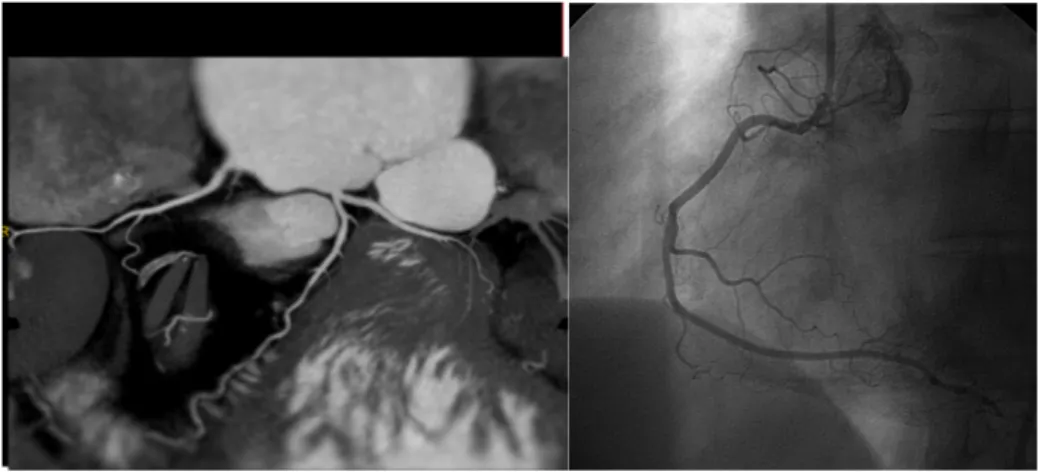
Fig.1 LIMA to anterior descending artery: conventional angiography and its corresponding angio- CT.
· Image assessment by MSCT
After acquiring them, images were sent to a dedicated work station. Single axial images were elaborated again both by a Multi – Planar – Reformat (MPR) two – dimensional reconstruction, which is able to give representations arranged on different planes from that of acquisition, and by three – dimensional Volumetric rendering (VR), which can visualize the heart and the anatomic structures near to it as three – dimensional objects, even adjustable in the space to own liking. In this phase of the study two blinded observers evaluated all patients enrolled in the experimentation. Images at the highest intensity were used to identify lesions in explored segments, while multiplan images were useful to classify lesions as more or less significant.
With regard to image quality, it was assessed per segment and classified as follows: · good, if vessel lumen was perfectly visualized
· low, in cases in which vessel lumen resulted scarcely visualized because of the presence of artefacts.
Therefore, stenosis were defined as significant just when they obstructed lumen ≥50%. Some cases of beginning disagreement among observatories were evaluated in agreement afterwards.
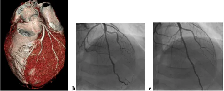
FIG.2. MSTC: Volumetric Rendering (VR) image and multi planar reconstruction of stented saphenous vein graft to seq. obtuse marginal branch – posterior artery.
Fig 3. Volume Rendering and MPR globe of uninjured coronaries.
Fig 4. Coronary angio-TC and traditional coronarography.
· Statistical Analysis
Data regarding diagnostic accuracy of angiography by MSCT in finding out significant atherosclerotic lesions or significant restenosis, evaluated by using coronaric angiography as reference technique, are reported below in terms of sensitivity, specificity, positive predictive value (PPV) and negative predictive value (NPV), with confidence intervals of 95% (tab. 1). CCA vs MSCT-CA comparison has been conduct per segment, per vessel and then per patient. As regards CABG, comparison between conventional coronaric angiography and MSCT was performed assessing every graft, both including body and anastomosis. Intra and inter – observatory variability in identifying significant stenosis was assessed (lumen reduction ≥ 50%) and evaluated by Kappa statstic. For statistical analysis we used a dedicated software ( Statistica 5.0, StatSoft Italia, Vigonza – PD )
· Results
By a dedicated software has been calculated radiation dose: average value presented sex based difference and resulted 14 mSv for female gender and 11,5 mSv for male with 40 slices technology; average value of 20 mSv with 64 slices.
Average heart rate of the sample was equal to 59,6 ±7,4 bpm; intra - and inter – observatory variability resulted to be 0,75. Second AHA classification 79, we considered 17 coronary segments, so potentially we 800 segments were considered. 177 segmnts don’t were visualized during CCA because of Intermedius branch absence or non dominant arteries ( n=130 seg), proximal arterial occlusion with lack of distal branch visualization ( n= 47 seg.). Only 0,8% of total segments ( n=5) non-evaluable because of movement artifacts. Scanning was succesfull in all patient. Telediastolic phase utilized
in 70% (35/50) of patients, the latter in telesistolic phase because of movement artifacts. An overall number of 618 coronaric segments were evaluated.
92 grafts were evaluated, of which 36 were arterial (26 left and 10 right internal mammary arteries) and 56 were venous; analyzed anastomosis were 102, o which 38 were proximal and 64 distal; explored bodies were 78; 8 venous grafts not evaluated because of the presence of artefacts caused by metal clips.
For the same reason distal anastomosis of an arterial graft was not evaluated, 8 saphenous grafts, 6 left internal mammary arteries and 2 right internal mammary arteries resulted obstructed.
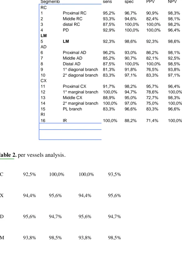
Results concerning diagnostic accuracy for coronaries are reported in table 1, 2 and 3 in tables 4 and 5 are listed data concerning grafts and stents, respectively.
Tab 1. Performance test per segments.
Segmento sens spec PPV NPV
RC 1 Proximal RC 95,2% 96,7% 90,9% 98,3% 2 Middle RC 93,3% 94,6% 82,4% 98,1% 3 distal RC 87,5% 100,0% 100,0% 98,2% 4 PD 92,9% 100,0% 100,0% 96,4% LM 5 LM 92,3% 98,6% 92,3% 98,6% AD 6 Proximal AD 96,2% 93,0% 86,2% 98,1% 7 Middle AD 85,2% 90,7% 82,1% 92,5% 8 Distal AD 87,5% 100,0% 100,0% 98,5% 9 1° diagonal branch 81,3% 91,8% 76,5% 93,8% 10 2° diagonal branch 83,3% 97,1% 83,3% 97,1% CX 11 Proximal CX 91,7% 98,2% 95,7% 96,4% 12 1° marginal branch 100,0% 94,7% 78,6% 100,0% 13 Middle CX 88,9% 95,0% 72,7% 98,3% 14 2° marginal branch 100,0% 97,0% 75,0% 100,0% 15 PL branch 83,3% 96,6% 83,3% 96,6% RI 16 IR 100,0% 88,2% 71,4% 100,0%
Table 2. per vessels analysis.
RC 92,5% 100,0% 100,0% 93,5%
CX 94,4% 95,6% 94,4% 95,6%
AD 95,6% 94,7% 95,6% 94,7%
Tab 3. Per patient diagnostic accuracy.
_______________________________________________________________________
n. pts Sens. Spec. VPP VPN DA LR+ LR - Odds pre-test (%, n) (%, n) (%, n) (%, n) (%)
___________________________________________________________________________________ 50 100 (36/36) 100 (14/14) 100 (36/36) 100 (14/14) 100 ∞ 0.00 1.00 ___________________________________________________________________________________ LR = likelihood ratio; NPV = negative predictive value; PPV = positive predictive value
Tab 4. Graft Performance test
Tab 5.Diagnostic Performance for stents
SENSITIVITY SPECIFICITY VPP VPN
STENT 100% 93,3% 80% 100%
Myocardial bridges
The presence of myocardial bridges represents another interesting aspect of coronaric pathology, of which MSCT shows more clearly anatomic features (fig. 6), resulting very useful for clinical management of this condition and for assessing of its
Bodies 100% 94% 80% 100% 95% Proximal Anastomosis 100% 100% 100% 100% 100% Distal Anastomosis 75% 96,5% 75% 96% 94% CABC in toto 94% 92% 88% 96% 93% Arterial CABG 100% 92% 86% 100% 95% Venous CABG 90% 93% 90% 93% 92% OCCLUSION 100% 100% 100% 100% 100%
actual incidence in population. Actually, this last is about 0,5 – 12% according to coronaric angiography and 5 – 86% according to necroscopic case studies 80.
In this study population we observed (86 patients who underwent AC – MSCT and coronarography) 17 bridges/courses (19,8%) were highlighted by CT and only 7 (8,2%) by coronarography.
In all cases the abnormality involved anterior descendent artery, and all bridges were single, 5 of them were symptomatic (as isolated coronaric abnormality) with a deep course.
After their identification, it is possible to choice specific therapeutic options, suggesting “ad hoc” medical treatment.
a b c
Fig 6. a) VR shows MB in II tract of DA; b)CCA DA systolic squeezing; c) CCA diastolic phase,
normal caliber; d)MPR, intramiocardial tract.
Discussion
Multi-slice computed tomography allows reliable evaluation of the coronary arteries in a non-invasive manner. In general, the high negative predictive value is considered to be the major strength of the technique and a normal MSCT virtually excludes significant CAD on conventional coronary angiography.
CAD is the leading cause of mortality and morbidity in occidental countries; such issues suggests that MSCT based coronary screening, is logic, feasible and careful, but it is today a concept widely refused because of high risk of radiation exposure. On the basis of current literature it is difficult understanding in wich class of patients may be helpful MSCT application. It’ s interesting evaluate high risk patients for CAD even if asym-ptomatic but up to day is ungiustified, while is more acceptable use in patient with dubious exercise test and aspecific symptoms or scheduled for valvular replacement.
Our experience shows good MSCT diagnostic performance when compared to CCA in evaluating CABG patency, in >50% coronary stenoses identification and much more in excluding coronary disease in symptomatic patients and, finally, in detecting other coronary aspects, such as myocardial bridge. Some functional index of perfusion, such as slow-flow fenomenon, that is index of CABG
degeneration, is not available with MSCT that seize only morphologic aspects. A
good exam execution requires careful patient selection and both farmacological and technical preparation, then patient education to maintain a correct breath- hold. Major diagnostic difficulties came from coronaric calcifications, that are responsible of modest percentage of false positives. High sensitivity and specificity, PPV and NPV in per segment analysis, that increase in per vessel analysis and raise up to 100% in a per patient evalua-tion, shows that MSCT is capable in identifying CAD patient that need CCA. Metallic parts such as post-toracotomic suture, metallic clips, valvular prostesis, electrocatheters, heavly limit images lecture. Consideration on stent evaluation are not feasible in our study because of low prevalence of restenosis in a small sample dimension. Another issue is high radiation dose exposure, that we consider an important limitation and is reason of technical research besides right clinical exam indication. Radiation dose exposure needs ulterior studies and probably may be solved by newer software and scanner introduction. Prospective ECG-gating( i.e.) using a 'step-and-shoot' axial scanning protocol has been shown to reduce radiation exposure effectively while maintaining diagnostic accuracy. 81
Limitations
The esteemed radiation dose CA-TCMD is more raised than conventional AC. The ex-posure to ionizing radiations can be reduced with perspective modulation. This tech-nique can reduces the exposure to ionizing radiations of the 50% in patients with low cardiac frequency, but is more sensitive to arrhythmias and limits the possibilities of datasets reconstructions during telediastolic phase. Exist, besides, automatic modulation dose systems (Dose-Ri- ght-ACS, Automatic Current Selector, e DOM, Dose Modula-tion, Philips Medical Systems) that may combine modulation of delivered current both in relation with angular attenuation coefficient of the examined region and through a modulation dose system in Z axis. Referring to initial “scout view”, the milliamperage is automatically modulated along acquisition volume on the basis of angular attenuation coefficient of the examined region and on the basis of attenuation values of tissues along Z axis 82-83. So an elevated Calcium Score (CS) (i.e. >400) reduces MDCT diagnostic accuracy 38,84. Severe calcifications, in a single coronaric segment, make impossibile lumen evaluation with stenosis overestimation. For this ragion in our study we evalua-ted incorrectly a Left Main stenosis ( >50%), in a patient with total Calcium Score 1331,8 using Agatston system, of whom 83,4% localized in Left Main. It is important to consider not only total calcium score ( i.e. > 400 sec. Agatston ) but the calcic distri-bution in coronaries. In this study, moreover, cardiac and respiratory movements arti-facts impaired evaluation capability in 29,4% of coronaric tree, in one patient. Patients collaboration is important in performing correct examination, so this technique up to day is also a patient-dependent exam. Better technology and newer scanner with better temporal resolution such as double source gifted with double detector systems can improve our diagnostic capabilities, diminishing the so called “patient-dependence”.
Multi-slice computed tomography (MSCT) is a rapidly developing technique and allows reliable evaluation of the coronary arteries and CABG in a non-invasive manner.
Despite limitations due to calcium, movement, metallic parts and high radiation dose, MSCT – CA showed a good diagnostic capability in detecting significant coronary artery stenosis in patient with suspected or known significant coronary artery disease. Moreover, in our experience, is a valuable tool for assessing coronary artery bypass graft patency in patients with clinical suspect of occlusion. It’ s a good, adjunctive, way to correctly select CCA patient in dubious case. Multiphasic diagnostic approach with new sistems of automatic modulation of radiation dose and new algorithms in images
reconstruction are fundamental to reduce the important “patient-dependence” threshold. Candidates to MDCT should remain, however, restricted and selected on the basis of patient’s balanced diagnostic iter to avoid unjustified risk of ionizing radiation.
Clinical cases
Case I
Female 64 y.o., ipertension, diabetes, referred for chest pain
Dubious exercise test, negative SPECT. MDCT-CA shows healty coronaries with long myocardial bridge of Descending Anterior artery. (Figure below)
Case I.
Image A. VR; Im. B. particular of
intramiocardial tract; Im. C and D, sisto-diastolic caliber variation due to
Case II
Male 58 y.o., hypercholesterolemia, diabetes, obesità,
referred to exertional stable angina. Dubious exercise test, negative SPECT. MDCT-CA shows significant (>50%) Left Main stenosis ( fig. below).
Case II. Significant Left Main disease
Case III
Male 73 y.o., referred for exertional angina; in 1993 two venous sequential bypass for multisegmental occlusive disease: 1, first obtuse marginal branch (1st OM ) – second
obtuse marginal branch (2nd OM) – posterior descending artery (PDA); 2, Descending Anterior (DA) - second obtuse marginal branch (2nd OM). MDCT-CA shows proximal occlusion of SV-DA-2nd OM ( thick arrows) and >50% stenosi in distal tract of SV – 1st OM – 2nd OM – PDA. (fig. below)
References.
1. Passariello R, De Santis M. Coronary artery disease. Update and prospects of radiologic imaging with CT and MR. Radiol Med (Torino) 2001; 101: 411-23.
2. Traversi E, Aldrovandi A, Barazzoni G, Bertoli G, Baldi M, Tramarin R. Non-invasive coronary angiography by multislice computed tomography:a new diagnostic method? Ital Heart J 2002; 3: 665-8. 3. Johnson LW, Lozner EC, Johnson S, et al. Coronary arteriography 1984-1987: a report of the Registry of the Society for Cardiac Angiography and Interventions. I: Results and complications. Cathet Cardiovasc Diagn 1989; 17: 5-10.
4. American Heart Association. Heart disease and stroke statistics - 2004 update. Dallas, TX: American Heart Association, 2004.
5. Budoff MJ, Achenbach S, Duerinckx A. Clinical utility of computed tomography and magnetic resonance techniques for noninvasive coronary angiography. J Am Coll Cardiol 2003; 42: 1867-78. 6. Shaw LJ, Tarkington L, Callister T, et al. The HCA National Disease Management Program for coronary disease detection and treatment in women. Am J Manag Care 2001; 7 (Spec No): SP25-SP30. 7. Cademartiri F, Malagutti P, Belgrano M, et al. Non-invasive coronary angiography with 64-slice computed tomography. Minerva Cardioangiol 2005; 53: 465-72.
8. Kopp AF, Schroeder S, Kuettner A, et al. Coronary arteries: retrospectively ECG-gated multi-detector row CT angiography with selective optimization of the image reconstruction window. Radiology 2001; 221: 683-8.
9. Hong C, Becker CR, Huber A, et al. ECG-gated reconstructed multi-detector row CT coronary angiography: effect of varying trigger delay on image quality. Radiology 2001; 220: 712-7.
10. Cademartiri F, Luccichenti G, Marano R, et al. Non-invasive angiography of the coronary arteries with multislice
computed tomography: state of the art and future prospects. Radiol Med (Torino) 2003; 106: 284-96. 11. Achenbach S. Detection of coronary stenoses by multidetector computed tomography: it’s all about resolution. J Am Coll Cardiol 2004; 43: 840-1.
12. Achenbach S, Hoffmann U, Ferencik M, Wicky S, Brady TJ. Tomographic coronary angiography by EBCT and MDCT. Prog Cardiovasc Dis 2003; 46: 185-95.
13. Lipton MJ, Higgins CB, Farmer D, Boyd DP. Cardiac imaging with a high-speed cine-CT scanner: preliminary results. Radiology 1984; 152: 579-82.
14. Marshall W, Hall E, Doost-Hoseini A, Alvarez R, Macovski A, Cassel D. An implementation of dual energy CT scanning. J Comput Assist Tomogr 1984; 8: 745-9.
15. Brateman L, Jacobs AM, Fitzgerald LT. Compton scatter axial tomography with X-rays: SCAT-CAT. Phys Med Biol 1984; 29: 1353-70.
16. Christ G. Exact treatment of the dual-energy method in CT using polyenergetic X-ray spectra. Phys Med Biol 1984; 29: 1501-10.
17. Nieman K, Oudkerk M, Rensig BJ, et al. Coronary angiography with multislice computed tomography. Lancet 2001; 357: 599-603.
18. Achenbach S, Ulzheimer S, Baum U, et al. Noninvasive coronary angiography by retrospectively ECG-gated multislice spiral CT. Circulation 2000; 102: 2823-8.
19. Kirsch J, Williamson EE, Araoz PA. Non-compaction visualization using ECG-gated dual-source CT. Int J Cardiol 2007; 118: e46-e47.
20. Dewey M, Hamm B. CT coronary angiography: examination technique, clinical results, and outlook on future developments. Rofo 2007; 179: 246-60.
21. Flohr TG, Schoepf UJ, Ohnesorge BM. Chasing the heart: new developments for cardiac CT. J Thorac Imaging 2007; 22: 4-16.
22. Nikolaou K, Saam T, Rist C, et al. Pre- and postsurgical diagnostics with dual-source computed tomography in cardiac surgery. Radiologe 2007; 47: 310-8.
23. Busch S, Nikolaou K, Johnson T, et al. Quantification of coronary artery stenoses: comparison of 64-slice and dual source CT angiography with cardiac catheterization. Radiologe 2007; 47: 295-300. 24. Reimann AJ, Rinck D, Birinci-Aydogan A, et al. Dualsource computed tomography: advances of improved temporal resolution in coronary plaque imaging. Invest Radiol 2007; 42: 196-203.
25. Rist C, Johnson TR, Becker A, et al. Dual-source cardiac CT imaging with improved temporal resolution: impact on image quality and analysis of left ventricular function. Radiologe 2007; 47: 287-94.
26. Johnson TR, Nikolaou K, Fink C, et al. Dual-source CT in chest pain diagnosis. Radiologe 2007; 47: 301-9.
27. Johnson TR, Krauss B, Sedlmair M, et al. Material differentiation by dual energy CT: initial experience. Eur Radiol 2007; 17: 1510-7.
28. Ropers D. Multislice computed tomography for detection of coronary artery disease. J Interv Cardiol 2006; 19: 574-82.
29. Scheffel H, Alkadhi H, Plass A, et al. Accuracy of dual- source CT coronary angiography: first experience in a high pre-test probability population without heart rate control. Eur Radiol 2006; 16: 2739-47.
30. Kalender WA. X-ray computed tomography. Phys Med Biol 2006; 51: R29-R43.
31. Johnson TR, Nikolaou K, Wintersperger BJ, et al. Dualsource CT cardiac imaging: initial experience. Eur Radiol 2006; 16: 1409-15.
32. Achenbach S, Ropers D, Kuettner A, et al. Contrast-enhanced coronary artery visualization by dual-source computed tomography - initial experience. Eur J Radiol 2006; 57: 331-5.
33. Flohr TG, McCollough CH, Bruder H, et al. First performance evaluation of a dual-source CT (DSCT) system. Eur Radiol 2006; 16: 256-68.
34. Pugliese F, Mollet NR, Runza G, et al. Diagnostic accuracy of non-invasive 64-slice CT coronary angiography in patients with stable angina pectoris. Eur Radiol 2006; 16: 575- 82.
35. Leschka S, Alkadhi H, Plass A, et al. Accuracy of MSCT coronary angiography with 64-slice technology: first experience. Eur Heart J 2005; 26: 1482-7.
36. Musto C, Simon P, Nicol E, et al. 64-Multislice computer tomography in consecutive patients with suspected or proven coronary artery disease: initial single center experience. Int J Cardiol 2007; 114: 90-7.
37. Nikolaou K, Knez A, Rist C, et al. Accuracy of 64-MDCT in the diagnosis of ischemic heart disease. AJR Am J Roentgenol 2006; 187: 111-7.
38. Mollet NR, Cademartiri F, van Mieghem CA, et al. Highresolution spiral computed tomography coronary angiography in patients referred for diagnostic conventional coronary angiography. Circulation 2005; 112: 2318-23.
39. Leber AW, Knez A, von Ziegler F, et al. Quantification of obstructive and nonobstructive coronary lesions by 64-slice computed tomography: a comparative study with quantitative coronary angiography and intravascular ultrasound. J Am Coll Cardiol 2005; 46: 147-54.
40. Raff GL, Gallagher MJ, O’Neill WW, Goldstein JA. Diagnostic accuracy of noninvasive coronary angiography using 64-slice spiral computed tomography. J Am Coll Cardiol 2005; 46: 552-7.
41.
Hounsfield GN, Computerized transverse axial scanning (tomography). 1. Description of system. Br J Radiol 46: 1016-1022, 1973.
42.
De Feyter P, Nieman K, Van Ooijten P, Oudkerk M, Imaging techniques: non-invasive coronary artery imaging with electron beam computed tomography and magnetic resonance imaging. Heart 2001; 84: 442-8.
43.
Ohnesorge BM, Becker CR, Flohr TG, Reiser MF, Multislice CT in cardiac imaging. Berlin: Springer–Verlag, 2002.
44.
Becker CR, Ohnesorge BM, Shoepf UJ, Reiser FM, Current development of cardiac imaging with multidetector row CT. Eur J Radiol 2000; 36: 97-103.
45.
Traversi E, Bertoli G, Barzzoni G, Baldi M, Tramarin M, Non-invasive coronary angiography with multislice computer tomography. Technology, methods, preliminary experience and prospects. Ital Heart J 2004; 5: 89-98.
46.
Kopp AF, Kuttner A, Heushmid M, Schroder S, Ohnesorge B, Claussen CD, Multidetector-row CT cardiac imaging with 4 and 16 slices for coronary CTA and imaging of atherosclerotic plaques. Eur Radiol 2002; 2 (Suppl): S17-S24.
47.
Cademartiri F, Runza G, Krestin GP, Introduction to coronary imaging with 64- slice Computer Tomography. Radiol Med 110: 16-41.
48.
Ropers D, Baum U, Pohle K et al, Detection of coronary artery stenoses with thin-slice multi-detector row spiral computed tomography and multiplanar reconstruction. Circulation 107: 664-666, 2003.
49. Kalra MK, Brady TJ. Current status and future directions in technical developments of cardiac computed tomography. J Cardiovasc Comput Tomogr. 2008 Mar-Apr;2(2):71-80
50. Rajiv Gupta, Arnold C. Cheung, Soenke H. Bartling, Jennifer Lisauskas, Michael Grasruck, Christianne Leidecker, Bernhard Schmidt, Thomas Flohr, Thomas J. Brady. Flat-Panel Volume CT: Fundamental Principles, Technology, and Applications. Radiographics November 2008 28:2009-2022;
51.ACCF/ACR/SCCT/SCMR/ASNC/NASCI/SCAI/SIR2006 Appropriateness Criteria for Cardiac Computed Tomography and Cardiac Magnetic Resonance Imaging. JACC 2006, vol 48, n 7, 1475-1497.
52
. Giuseppe Runza, Marco Rizzo, Salvatore Evola, Valerio Alaimo, Giuseppina Novo, Egle Corrado,, Giovanna Evola, Giuseppina Palazzolo, Oreste Fabio Triolo,Francesca Gennaro, Enrico Hoffmann, Massimo Midiri, Salvatore Novo. Approccio diagnostico non invasivo con tomografia computerizzata multidetettore a 40 strati per lo studio della malattia aterosclerotica coronarica. G Ital Cardiol 2007; 8 (6): 000-000
53.
Ropers D, Ulzheimer S, Wenkel E et al, Investigation of aortocoronary artery bypass grafts by multislice spiral computed tomography with electrocardiographic-gated image reconstruction. Am J Cardiol 88: 792-795, 2001.
54.
Nieman K, Pattynama PM, Rensing BJ et al, Evaluation of patients after coronary artery bypass surgery: CT angiographic assessment of grafts and coronary arteries. Radiology 229: 749-756, 2003.
55.
Yoo KJ, Choi D, Choi BJ et al, The comparison of graft patency after coronary artery bypass grafting using coronary angiography and multislice computed tomography. Eur J Cardiothorac Surg 24: 86-91; discussion 91, 2003.
56.
Martuscelli E, Romagnoli A, D’Eliseo A et al, Evaluatin of venous and arteria conduit patency by 16-slice computed tomography. Circulation 110: 3234-3238, 2004.
57.
Schlosser T, Konorza T, Hunold P et al, Noninvasive visualisation of coronary artery bypass grafts using 16-detector row computed tomography. J Am Coll Cardiol 44: 1224-1229, 2004.
58.
Giuseppina Novo, Giuseppe Runza, Salvatore Evola, Oreste Fabio Triolo, Valerio Alaimo, Marco Rizzo, Antonella La Fata, Giuseppina Palazzolo, Fiorella Sutera, Giuseppe Andolina, Enrico Hoffmann, Salvatore Novo, Massimo Midiri. Utilità dell’angiografia coronarica eseguita mediante tomografia computerizzata multidetettore nel controllo della pervietà del bypass aortocoronarico. G Ital Cardiol 2007; 8 (12): 770-776.
59.
Cademartiri F, Mollet N, Nieman K et al, Images in cardiovascular medicine. Neointimal Hyperplasia in carotid stent detected with multislice computed tomography. Circulation 108: e 147, 2003.
60.
Schuijf JD, Bax JJ, Jukema JW et al, Feasibility of assessment of coronary stent patency using 16-slice computed tomography. Am J cardiol 94: 427-430, 2004.
61.
Mollet NR and Cademartiri F, Images in cardiovascular medicine, In-stent neointimal hyperplasia with 16-row multislice computed tomography coronary angiography. Circulation 110: e514, 2004.
62.
Maintz D, Seifarth H, Flohr T et al, Improved coronary stent visualisation and in-stent stenosis detection usin 16-slice computed tomography and dedicated image reconstruction technique. Invest Radiol 38: 790-795, 2003.
63.
Maintz D, Grude M, Fallenberg EM et al, Assessment of coronary arterial stents by multislice CT angiography. Acta Radiol 44: 597-603, 2003.
64.
Mollet NR, Hoye A, Lemos PA et al, Value of preprocedure multislice computed tomographic coronary angiography to predict the outcome of percutaneous recanalisation of chronic total occlusions. Am J Cardiol 95: 240-243, 2005.
65.
Cademartiri F, Nieman K, Raaymakers RH et al, Non-invasive demonstration of coronary artery anomaly performed using 16-slice multidetector spiral computed tomography. Ital Heart J 4: 56-59, 2003.
66.
Horisaki T, Yamashita T, Yokoyama H et al, Three-dimensional reconstruction of computed tomographic images of anomalous origin of the left main coronary artery from the pulmonary trunk in an adult. Am J Cardiol 92: 898-899, 2003.
67.
Lessick J, Kumar G, Beyar R et al, Anomalous origin of a posterior descending artery from the right pulmonary artery: report of a rare case diagnosed by multidetector computed tomography angiography. Circulation 109: e185-186, 2004.
68.G. Runza, L. La Grutta, V. Alaimo, S. Evola, F. Lo Re, T.V. Bartolotta, F. Cademartiri, M. Midiri. Comprehensive cardiovascular ECG-gated MDCT as a standard
diagnostic tool in patients with acute chest pain. European Journal of Radiology 64 (2007) 41–47. 69. Rumberger JA, Simons DB, Fitzpatrick LA et al. Coronary artery calcium area by electron beam computed tomography and coronary atherosclerotic area. A histopathologic correlative study. Circulation 1995, 92: 2157-2162.
70. Runberger JA, Brundage BH, Rader DJ, Kondos G. Electron beam computed tomography coronary calcium scanning: a review and guidelines for use in asymptomatic person. 1999 Mayo Clinic Proc. 74: 243-252.
71.
Becker CR, Coronary calcification scoring with CT: history, methodology, EBCT vs Helical. Applied Radiol 32: 30-32, 2003.
72. Achenbach S, Moselewski F, Ropers D, et al. Detection of Calcified and Non-calcified CoronaryAtherosclerotic Plaque by Contrast-Enhanced,Submillimeter Multidetector Spiral ComputedTomography. Circulation 2004; 109:14–7.
73. Stephan Achenbach, Fabian Moselewski, Dieter Ropers, Maros Ferencik, Udo Hoffmann, Briain MacNeill, Karsten Pohle, Ulrich Baum, Katharina Anders, Ik-kyung Jang, Werner G. Daniel, Thomas J. Brady. Detection of Calcified and Noncalcified CoronaryAtherosclerotic Plaque by
Contrast-Enhanced, Submillimeter Multidetector Spiral Computed Tomography A Segment-Based Comparison With Intravascular Ultrasound. Circulation 2004;109;14-17.
74.
Schroeder S, Kopp AF, Baumback A et al, Non invasive characterisation of coronary lesion morfology by multislice computed tomography: a promising new technology for risk stratification of patients with coronary artery disease, Heart 85: 576-578, 2001.
75. Friedrich D. Knollmann, Annika Wieltsch, Simone Peters, Anika Mahlke, Susanne Niederberger
and Tereza Kertesz. Flat Panel Volume Computed Tomography of the Coronary Arteries. Academic Radiology Volume 16, Issue 10, October 2009, Pages 1251-1262
76.
Ohnesorge B, Flohr T, Becker C et al, Cardiac imaging by means of electrocardiographically gated multisection spiral CT: initial experience. Radiology 217: 564-571, 2000.
77.
Kachelriess M, Kalender A, Electrocardiogram-correlated image reconstruction from subsecond spiral computed tomography scans of the heart. Med Phys 25: 2417-2431, 1998.
78.
Aspelin P, Aubry P, Fransson SG et al, Nephrotoxic effects in high risk patients undergoing angiography. N Engl J Med 348: 491-499, 2003.
79. Austen WG, Edwards JE, Frye RL, et al. A reporting system on patients evaluated for coronary artery disease. Report of the Ad Hoc Committee for Grading of Coronary Artery Disease, Council on Cardiovascular Surgery, American Heart Association. Circulation 1975; 51: 5-40.
82. Kalender WA, Wolf H, Suess C, Gies M, Greess H, Bautz WA. Dose reduction in CT by on-line tube current control: principles and validation on phantoms and cadavers. Eur Radiol 1999; 9: 323-8. 83. Kalender WA, Wolf H, Suess C. Dose reduction in CT by anatomically adapted tube current modulation. II: Phantom measurements. Med Phys 1999; 26: 2248-53.
84. Cademartiri F, Runza G, Mollet NR, et al. Impact of intravascular enhancement, heart rate, and calcium score on diagnostic accuracy in multislice computed tomography coronary angiography. Radiol Med (Torino) 2005; 110: 42-51.