Medical Oncology (2018) 35:157 https://doi.org/10.1007/s12032-018-1222-4
ORIGINAL PAPER
Autotaxin is a novel molecular identifier of type I endometrial cancer
Antonio Mazzocca1 · Luca Maria Schönauer2 · Rosalba De Nola2,3 · Antonio Lippolis2 · Teresa Marrano2 · Matteo Loverro2 · Carlo Sabbà1 · Edoardo Di Naro2
Received: 10 September 2018 / Accepted: 25 October 2018
© Springer Science+Business Media, LLC, part of Springer Nature 2018 Abstract
Endometrial cancer is the most common cancer of the female genital tract in Western Countries, with an incidence of 150.000 new cases/year. Despite high incidence, little is known about the molecular pathogenesis of this tumor. Phospholipids including lysophosphatidic acid (LPA) are involved in proliferation and dissemination of cancer. LPA is a potent bioactive phospholipid synthesized by autotaxin (ATX) through its lysophospholipase D activity. Recent evidence suggests that the ATX/LPA signaling axis plays a role in endometrial cancer. We carried out a prospective study involving two groups of patients classified in accordance to hysteroscopic-guided biopsy. Patients with histological diagnosis of endometrial cancer were enrolled into group one, whereas control patients with pelvic organ prolapse were assigned group two. Both groups underwent hysterectomy, with either open or laparoscopic surgery. After uterine extraction, a second endometrial biopsy was performed to collect tissues. Real-Time PCR was performed to evaluate ATX gene expression in collected tissues. Sta-tistical analysis including unpaired two-way or one-way Student’s t test and ANOVA was performed. We found ATX gene expression significantly higher in neoplastic endometrium compared with normal tissue (P value = 0.0002). In particular, the expression of ATX was significantly elevated in type I endometrial cancer (i.e., endometrioid histotype) compared to type II, in premenopausal women and in patients affected either by obesity (BMI > 30) or diabetes. We propose ATX as a novel potential biomarker particularly implicated in the pathobiology of type I endometrial cancer. Also, we propose ATX as a useful theranostic target in endometrial cancer.
Keywords Autotaxin (ATX) · Endometrial cancer · Lysophosphatidic acid (LPA) · Biological marker · Obesity Abbreviations
ATX Autotaxin
LPA Lysophosphatidic acid RT Reverse transcription PCR Polymerase chain reaction BMI Body mass index
PTEN Phosphatase and tensin homolog PIK3CA Phosphatidylinositol 3-kinase KRAS Kirsten rat sarcoma virus
p53 Protein 53
HER-2/neu Human epidermal growth factor receptor 2/ proto-oncogene Neu
p16 Protein 16 PLD Phospholipase D RNA Ribonucleic acid DNA Deoxyribonucleic acid
GAPDH Glyceraldehyde 3-phosphate dehydrogenase gene
SEM Standard error of the mean SMB Somatomedin B-like
ENPP 2 Ectonucleotide pyrophosphatase/phosphodi-esterase type 2
PLA2 Phospholipase A2
LPARs Lysophosphatidic acid receptors
* Antonio Mazzocca [email protected]
1 Interdisciplinary Department of Medicine, University of Bari
School of Medicine, Piazza G. Cesare, 11, 70124 Bari, Italy
2 Interdisciplinary Department of Medicine, Gynaecology
and Obstetrics Clinic, University of Bari School of Medicine, Piazza G. Cesare, 11, 70124 Bari, Italy
3 Department of Tissues and Organs Transplantation
and Cellular Therapies, D.E.O.T, School of Medicine, University of Bari, Piazza G. Cesare, 11, 70124 Bari, Italy
Medical Oncology (2018) 35:157
157 Page 2 of 8
Introduction
Endometrial cancer is the most common neoplasm of the female genital tract in Western Countries, with a global incidence of 287,000 and a mortality rate of 74,000 per year in 2008 [1]. The endometrial cancer originates from the endometrial lining and glandular epithelium and occurs mainly at a mean age of 55–60 years. Nevertheless, epi-demiological data have shown an increasing incidence of the disease both in under-50-year-old women (1.5%) and in over-50s (2.6%) from 2006 to 2010, whereas the endo-metrial cancer mortality rate is increased of 1.5% among under 50s women and remained stable in over-50-year-old women [2]. This increasing incidence has been associated with an epidemic of obesity and absence of physical inac-tivity [3]. Two types of endometrial carcinoma have long been identified and they display differences in the field of pathology, biology, and clinics. In particular, type I is an estrogen-driven tumor affecting usually pre- and peri-menopausal woman, characterized by endometrioid histo-logical features, deriving from pre-malignant hyperplasia, with good prognosis (5-year survival > 90%) and it har-bors mutations in PTEN, PIK3CA, KRAS, b-catenin, and microsatellite instability. However, the type I incidence is facing a recent increase probably due to the pandemic obe-sity, which is a known risk factor for endometrial cancer. By contrast, type II represents only the 10% of all endo-metrial cancers and it is composed of non-endometrioid histotypes, namely in particular: papillary serous carci-nomas, sarcomas, and clear cell carcinomas. The type II arises from an atrophic endometrium background and it occurs in an older onset age (about 10 years later than type I). Type II also has a worse prognosis (all stage have an overall survival of 35%) and a trend of reduced survival in the last years, harboring genetic mutations in p53, HER-2/ neu, p16, and E-cadherin [4]. Moreover, all together endo-metrial cancer stages are characterized by a 5-year overall survival of about 80%, even without any valid screening method [3]. In Europe, one out 9000 women undergoes death for endometrial cancer, so it seems unlikely that this trend could decrease in the near future, probably due to the slight influence of early detection and kind of treatment on endometrial cancer mortality [3]. The main therapy for endometrial cancer is surgery followed, in selected cases, by radiotherapy to treat the microscopic disease of pel-vic lymph nodes, the central pelpel-vic region, and the upper vagina [3].
There is a growing interest among physicians in the standard radiotherapy, whose role has been re-evaluated in many studies (PORTEC-1, Post Operative Radiation Ther-apy in Endometrial Carcinoma Trial 1; GOG9, Gynaeco-logic OncoGynaeco-logic Group 9; ASTEC/EN.5, Adjuvant external
beam radiotherapy in the treatment of endometrial cancer randomized trials). In advanced stages, adjuvant chem-otherapy seems to be more effective than radichem-otherapy. Conversely, chemotherapy is associated with more side effects [5]. These data have encouraged the scientific com-munity to focus on the physiopathology of endometrial cancer in order to find new therapeutic approaches. More recently, researchers are investigating the role of lipid metabolism in the pathogenesis of several solid cancers, including endometrial and ovarian cancer [6]. For exam-ple, dyslipidemia is frequently associated with conditions that promote endometrial cancer. Therefore, we focused on the role of phospholipids in this type of cancer [7]. Autotaxin (ATX), also known as ectonucleotide pyroph-osphatase/phosphodiesterase family member 2 (ENPP 2), is a transmembrane phospholipase D (PLD) that catalyzes the hydrolysis of the distal phosphoester bond between monoacylglycerol phosphates and the polar head group of lysophospholipids, determining the biosynthesis of LPA. When the biosynthesis and the signaling pathway of LPA become altered, many features of cancer pathobiol-ogy, oncogenesis, and cancer progression can be affected [8]. In the present study, we investigated the ATX gene expression in the endometrium of normal and neoplastic perimenopausal women to clarify the role and the clinical significance of the ATX/LPA signaling pathway in this disease.
Materials and methods
Patients
This is a prospective study involving 55 patients, subdi-vided into two groups (normal and neoplastic endome-trium) who came to our attention during a 6-month period starting from January to July 2015, treated with total hysterectomy, for uterine prolapse without any endome-trial pathology o previous endomeendome-trial cancer diagnosis. Patients were divided into two groups based on histo-pathology: (i) the first group included 26 patients with diagnosis of endometrial cancer; (ii) the second group of 29 patients with a normal endometrium. Ultrasound and MRI evaluation were performed as pre-operative assess-ment in case of endometrial cancer. Endometrial cancer was treated with type A hysterectomy, bilateral salpingo-oophorectomy, and pelvic/lomboaortic lymphadenectomy in medium or high-risk patients [9]. Patients with prolapse underwent simple hysterectomy with or without adjunctive surgical procedure to restore normal pelvic support. Exclu-sion criteria encompass previous or coexistent tumors and concomitant suspicious adnexal masses. We collected data about clinical history, previous pregnancies, age, and BMI.
Medical Oncology (2018) 35:157 Page 3 of 8 157
Each patient underwent clinical and laboratory investiga-tions including trans-vaginal ultrasound examination; hys-teroscopy with endometrial biopsy; Pap test, and serum oncological markers. Informed consent was given by all patients enrolled in the study. Endometrial biopsy was performed for each uterine extraction and the collected sample tissues were stored at − 80 °C. Endometrial can-cers were classified into different stages and histological subtype (Table 1) according to the International Federation of Gynecology and Obstetrics Consensus of 2009 [10]. Patients with endometrial cancer were subjected to a clini-cal follow-up every 3–6 months for at least 30 months. The ATX expression levels were evaluated in both the groups: patients with endometrial cancer and women with normal endometrium. The level of ATX expression in endometrial cancer samples was then correlated with histological type, stage of the disease, BMI, menopause onset age, type II diabetes, and hypertension.
Sample collection and RNA extraction
Frozen tissues were homogenized in TriReagent®
(Molecu-lar Research Center) and RNA was precipitated with isopro-panol, applied to RNeasy spin columns (Qiagen), eluted, and treated with RNase-free DNase for 30 min at 37 C, followed by heat inactivation at 75 °C and storage at − 80 °C.
Quantitative real-time PCR
Total RNA was isolated using the RNeasy Mini Kit (Qia-gen) and underwent DNAse treatment (TURBO DNA-free™ Ambion®). Reverse Transcription (RT) was then carried out
with random primers using 1 µg of total RNA from each sample. Quantitative RT-PCR was performed using pre-designed primers and SYBR Green® master mix (Biorad).
Housekeeping Glyceraldehyde 3-phosphate dehydrogenase gene (GAPDH) was used as endogenous control. ATX/PLD Primers were obtained from Integrated DNA Technologies®
(Leuven, Belgium): Forward: 5′-TGA CGG CCC TCT CTC TGT GT-3′. Reverse: 5′-ATT GCA GCT CTC CTC GTT GTC-3′.
Statistical analyses
For correlations, Pearson’s r coefficients were calculated. Data are represented as the mean ± SEM. Significant dif-ferences between groups were determined by unpaired two-way or one-two-way Student’s t test or ANOVA. P < 0.05 was considered statistically significant. The study was approved by local Independent Ethics Committee of the Polyclinic Consortium University Hospital, University of Bari. The patients gave prior written informed consent to the use of their tissues for clinical research.
Results
To evaluate the involvement of ATX in endometrial can-cer, we carried out analysis in endometrial tissues by using quantitative PCR. We found that ATX expression levels were significantly higher in endometrial cancer compared with control samples (P value = 0.0002) as shown in Fig. 1a. Surprisingly, more elevated values of ATX were observed in patients with endometrioid histotype compared with spe-cial histotypes (P value = 0,038) (Fig. 1b). Furthermore, we found a higher rate of ATX expression in premenopausal population, compared to post-menopausal women with endometrial cancer (P value of 0.0437) (Fig. 1c). Meta-bolic diseases are often associated to endometrial cancer. To assess the role of ATX in endometrial cancer patients with these metabolic conditions, we performed quantita-tive PCR in RNA extracted from endometrial biopsies. The
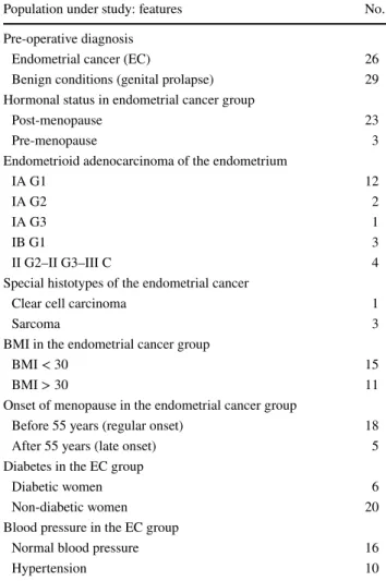
Table 1 Clinico-pathological characteristics of patients with endome-trial cancer enrolled in the study
Population under study: features No.
Pre-operative diagnosis
Endometrial cancer (EC) 26
Benign conditions (genital prolapse) 29
Hormonal status in endometrial cancer group
Post-menopause 23
Pre-menopause 3
Endometrioid adenocarcinoma of the endometrium
IA G1 12
IA G2 2
IA G3 1
IB G1 3
II G2–II G3–III C 4
Special histotypes of the endometrial cancer
Clear cell carcinoma 1
Sarcoma 3
BMI in the endometrial cancer group
BMI < 30 15
BMI > 30 11
Onset of menopause in the endometrial cancer group
Before 55 years (regular onset) 18
After 55 years (late onset) 5
Diabetes in the EC group
Diabetic women 6
Non-diabetic women 20
Blood pressure in the EC group
Normal blood pressure 16
Hypertension 10
Medical Oncology (2018) 35:157
157 Page 4 of 8
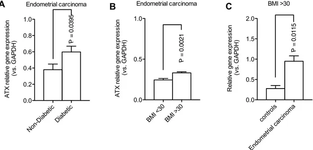
comparative analysis showed elevated ATX expression in diabetic (P value = 0.0395) compared with non-diabetic patients (Fig. 2a). In addition, we found a highly signifi-cant increased in ATX expression (P value = 0.0021) when we compare obese versus non-obese patients with endo-metrial cancer (Fig. 2b). A further analysis revealed that ATX expression levels were significantly higher in obese
women (BMI ≥ 30) with endometrial cancer compared with obese women with normal endometrium (P value = 0.0115) (Fig. 2c). Next, we evaluated the correlation of ATX expres-sion with cancer stage. The expresexpres-sion levels of ATX did not show any significant difference across various stages of endometrial cancer. However, although non-significant, a trend showing increased ATX expression levels was
A
B
C
Endometrial carcinoma Endometrioi d Special Histotype s 0.0 0.5 1.0 1.5 2.0 2.5 3.0 P = 0.0380 AT X re la tive gene ex pr essi on (vs. GA PD H ) Endometrial carcinoma Pre-menopausePostmenopaus e 0.0 0.5 1.0 1.5 2.0 P = 0.0437 AT X re la tive gene ex pr essi on (vs. GA PD H ) ATX expression Controls Endometrial carcinoma -0.5 0.0 0.5 1.0 1.5 P = 0.0002 No rma liz ed ex pr es si onFig. 1 ATX expression levels evaluated by a real-time PCR analysis in patients with endometrial cancer. a ATX expression levels were significantly higher in endometrial cancer tissues compared with con-trols (P value = 0.0002). b Increase of ATX expression in
endometri-oid histotype compared to special histotypes (P value = 0.038). c ATX expression levels were higher in women who had received the diag-nosis of endometrial cancer before menopause compared with post-menopause patients diagnosed with endometrial cancer
Endometrial carcinoma Non-Diabetic Diabetic 0.0 0.2 0.4 0.6 0.8 1.0 P = 0.0395 AT X re la tive gene ex pr essi on (vs. G APD H ) BMI >30 controls Endometrial carcinoma 0.0 0.5 1.0 1.5 2.0 P = 0.0115 Re la tive gene ex pr essi on (vs. G APD H ) Endometrial carcinoma BMI <3 0 BMI >3 0 0.0 0.5 1.0 P = 0.0021 AT X re la tive gene ex pr essi on (vs. G APD H )
A
B
C
Fig. 2 ATX expression in metabolic conditions associated with endo-metrial cancer. a Significant increase of ATX expression in diabetic patients with endometrial cancer (P value = 0.0395). b Obese patients with endometrial cancer show a significant higher expression of
endometrial ATX (P value = 0.0115). c Significant higher expression of endometrial ATX was observed in obese patients compared to non-obese with endometrial carcinoma (P value = 0.0021)
Medical Oncology (2018) 35:157 Page 5 of 8 157
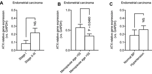
observed in advanced (II–III) compared to early stages (I) (Fig. 3a). Lastly, we found a significant ATX expression (P = 0.0460) in patients with endometrial cancer with nor-mal menopausal onset (before 55 years) compared to those with late menopausal onset (post 55 years) (Fig. 3b). Lack of statistical significance was found in ATX results, when we compared patients with endometrial cancer with or without hypertension (Fig. 3c).
Discussion
In the present study, our main goal was to investigate the role of ATX expression in normal and neoplastic endome-trium. We found a significant difference between these two groups with elevated ATX expression in endometrial cancer. We have also shown that ATX is significantly increased in patients with endometrial cancer associated to hormonal sta-tus and metabolic conditions including obesity and diabetes. ATX, originally discovered as an autocrine factor promoting chemotaxis in melanoma cells, is a 100 kDa multi-domain glycoprotein, consisting of two N-terminal somatomedin B-like (SMB) domains, a central catalytic phosphodiesterase domain and a C-terminal nuclease-like domain, which acts as an ecto-lysophospholipase D. ATX is encoded by the ENPP2 gene, which occupies a 116 kilo-base-long DNA segment of human chromosome 8 [11–14]. The product of the ATX catalysis is LPA, a small glycer-ophospholipid (1-acyl-2-hydroxy-sn-glycero-3-phosphate)
generated from the cleavage of the choline/serine head group from lysophosphatidylcholine or from lysophos-phatidylserine [15]. Although LPA can be produced by different enzymes (i.e., PLA2, phospholipase A2), the main source of all circulating bioactive LPA is due to the enzymatic activity of ATX [8]. LPA acts on different LPA-specific G protein–coupled receptors eliciting a great vari-ety of cellular responses [16, 13].
LPA and ATX have been also studied in the pathogen-esis of cancer, since they are involved in various processes essential for cancer progression, such as migration, inva-sion, tumor cell survival, and angiogenesis [17–19]. The ATX/LPA signaling axis promotes the proliferation and the dissemination of ovarian cancer and evidence from the present study and other studies indicate that it could be involved in the pathobiology of endometrial cancer [20]. In the gynecological field, LPA is thought to be a tumor-promoting factor, inducing, for example, the proliferation and the mitotic activity of leiomyomas [21, 22]. Indeed, ATX influences the functions and the proliferation of the muscular uterine components because of the synthesis of LPA in the smooth muscle cells [22]. Interestingly, ATX could interact with the cellular microenvironment induc-ing many biological effects. ATX also binds to integrins through the SMB-like domains and modulates the mito-genic activity of cancer in mouse models [8]. Seo et al. revealed that ATX is highly secreted by ovarian cancer stem cells and that either drug inhibition or ATX gene sup-pression reduces the LPA biosynthesis and consequently
Endometrial carcinoma Stage I Stage II-III 0.0 0.1 0.2 0.3 0.4 0.5 NS AT X re la tive gene ex pr essi on (vs. G APD H ) Endometrial carcinoma Menopause age <5 5 Menopause age >5 5 0.0 0.5 1.0 P = 0.046 0 AT X re la tive gene ex pr essi on (vs. G APD H ) Endometrial carcinoma Normal BP Hypertensio n 0.0 0.1 0.2 0.3 0.4 0.5 NS AT X re la tive gene ex pr essi on (vs. G APD H )
A
B
C
Fig. 3 ATX expression evaluated by real-time PCR analysis in dif-ferent subgroups of endometrial cancer. a Although non-significant, an increased trend in ATX expression was observed in advanced stages (II–III) compared with early stages (I) of endometrial cancer. b Significant (P = 0.0460) higher ATX expression in endometrial
cancer diagnosed in women with normal menopausal onset (before 55 years), compared to endometrial cancer diagnosed in women with late menopausal onset (post 55 years). c Absence of ATX significant endometrial expression in women with or without hypertension
Author's personal copy
Medical Oncology (2018) 35:157
157 Page 6 of 8
the ability of self-renewing in ovarian cancer stem cells and chemoresistance [23].
In the present study, we show that ATX RNA is more expressed in endometrial cancer compared with controls (pelvic organ prolapses). A previous study conducted by Wasniewski demonstrated a positive correlation (P < 0.005) of endometrial ATX/LPA signaling axis with endometrial cancers [20]. Wasniewski et al. carried out an interesting study based on the comparison of mRNA and protein lev-els of LPARs (lysophosphatidic acid receptors), ATX and PLA2, between type I endometrial cancers and normal endometrium samples (control), using qRT-PCR and west-ern blotting. The first group was made of 37 post-menopau-sal women who underwent total abdominal hysterectomy because of endometrial cancers, whereas the second group (controls) was composed of 10 premenopausal women who underwent hysterectomy for leiomyomas. Regarding the various endometrial histotypes, we found that the expression of ATX gene is significantly elevated in the endometrioid endometrial cancer when compared to “special” histotypes. This clear difference reflects the pathogenic and the clini-cal divergence between the two histotypes, related to dis-tinct metabolic and endocrine milieu. Endometrial cancers have long been classified into estrogen-dependent type I and estrogen-independent type II. The Type II is less common but clinically more aggressive than Type I, affecting older women, usually without any metabolic derangement or pre-existing pre-cancerous lesions (hyperplasia with atypia), harboring p53 mutations, but with a wild PTEN pheno-type [3, 24, 25]. Our data corroborate the aforementioned observations, in fact in patients with endometrial cancer, we found a remarkable elevated ATX gene expression in pre-menopause women still exposed to estrogenic milieu compared with those with a post-menopausal status, thus confirming the negative effect of estrogens in a dysmeta-bolic condition. This suggests a long developmental time for endometrioid endometrial cancer, whose roots mainly lie in the estrogen exposure during premenopausal period among genetically and metabolically predisposed women. In premenopausal women, overweight determines insulin resistance, excessive ovarian androgen biosynthesis, ano-vulation, and chronic progesterone deficiency [3]. Unlike Wasniewki et al. that enrolled only post-menopausal cases of endometrial cancer, we had the possibility to evaluate endo-metrial cancer in both pre-menopause and post-menopause status. Therefore, we correlated the hormonal influence in the pathogenesis of endometrial cancer and the higher ATX expression found in premenopausal patients. Moreover, we confirmed the correlation between type I endometrial cancer and ATX expression.
In our study, we also evaluated the role of metabolic dis-eases in endometrial cancer, in particular type II diabetes and obesity. We demonstrated a significantly higher expression
of ATX in diabetic patients. This is in contrast with findings by Ref. [20]. The absence of correlation between diabetes and ATX expression in the study by Wasniewski may be due to the cohort that was mainly composed of over-60-year-old obese women with other conditions related to aging and dysmetabolism. Our observations are instead in line with the well-known correlation between ATX, insulin resistance, and metabolic syndrome [26]. Among women with diabe-tes, LPA downregulates blood glucose levels by triggering translocation of Glucose transporter type 4 to the plasma membrane of myocytes and adipocytes. This determines an increased dose-dependent glucose uptake in muscles and adipose tissues [27]. In dysmetabolic mice, this may apparently suggest a possible advantage. However, it can eventually cause deleterious effect by inhibiting glucose-induced insulin secretion. Indeed, a chronic pharmacological inhibition with Ki16425 of LPA receptors improved glucose homeostasis in obese/pre-diabetic mice [19]. Nevertheless, in the present study, we do not show laboratory information supporting the potential correlation with insulin resistance in such patients. Due to the known correlation between high values of BMI and endometrial endometrioid cancer [10,
26], we decided to study the expression of the endometrial ATX gene expression in obese women, by comparing con-trols (healthy endometrium) with patients with endometrial cancer. Remarkably, ATX gene expression was higher in obese endometrial cancer group compared with controls similarly to Wasniewski et al. [20], who demonstrated an up-regulation of all the investigated LPARs (1–4) and of the LPA biosynthetic enzymes (ATX, PLA2) in endome-trial cancer tissues. Next, we evaluated the entire group of endometrial cancer divided into two subgroups, endometrial cancer patients with BMI < 30 versus endometrial cancer patients with BMI ≥ 30, and we confirmed the statistically significant positive correlation between obesity and ATX gene expression. Undoubtedly, obesity induces a hyper-estrogenic status with a continuous peripheral oestrone production in the adipose tissue associated with an imbal-anced secretion of pro-inflammatory cytokines that have a supposed triggering role in the pathogenesis of endometrial cancer [26]. Interestingly, our study also shows a signifi-cantly higher value of ATX expression in endometrial cancer diagnosed in women with normal menopausal onset (before 55 years), compared to endometrial cancer diagnosed in women with late menopausal onset (post 55 years). This could be due to the longer exposure of endometrium to dys-regulated metabolic and imbalanced endocrine status cor-related to the late onset of menopause in the former group, known as a promoting factor of type I endometrial cancer [28, 29, 26]. All these factors confirm that endometrioid endometrial cancer is an estrogen correlated disease with a long latent phase characterized by dysmetabolic condi-tions of hyperinsulinemia (related to metabolic syndrome),
Medical Oncology (2018) 35:157 Page 7 of 8 157
type II diabetes and obesity. This dysmetabolic conditions is also characterized by a prolonged estrogen production within the adipose tissue among perimenopausal period in genetically predisposed subjects. Furthermore, obesity promotes an imbalanced level of insulin-like growth factor and its binding proteins. This condition can be enhanced mainly by ovarian oestradiol in pre-menopause or mostly by a peripheral transformation of oestron within the adipose tissue in post-menopause. In addition, this vicious cycle can be sustained by an insufficient biosynthesis of progesterone. Notably, the transcriptional activity of estrogen-receptors can be induced by insulin-like growth factor 1 even with-out sustained levels of oestradiol (i.e., in post-menopause), supporting the evidence of a cross-talk between these dif-ferent pathways [3]. Finally, we suppose that ATX expres-sion could act as a metabolic link between estrogen milieu and insulin resistance, mainly in obese women acting as a promoting factor in endometrial carcinogenesis. Despite its apparent low aggressiveness, endometrial cancer is often related to a considerable and persistent mortality. For this reason, researchers are trying to elucidate the causes that determine mortality of this form of gynecological cancer. Therefore, new insights into pathophysiology of endometrial cancer are urgently needed to identify new targets for more effective therapeutic interventions than those available today. These considerations led us to study the role of ATX expres-sion in endometrial cancer. Notably, we found an increased expression of ATX gene in endometrial cancer, particularly in patients with endometrioid histotypes, BMI ≥ 30, type II diabetes, pre-menopause , and regular menopause. Given the high 5-year survival rate (approximately about 95%) [3] of early-stage endometrial cancer, we hypothesize that ATX expression, evaluated on endometrial biopsies or serum, as already demonstrated for LPA in ovarian cancer [17] may become an early marker for endometrial cancer detection. Similarly, a targeted therapy directed against ATX could be postulated as an effective tool in the treatment of endome-trioid carcinoma, especially in the early stages.
Acknowledgements The work was supported by AIRC (Italian Asso-ciation for Cancer Research) Investigator Grant (IG) 2015 Id.17758 (to A. Mazzocca).
Author contributions AM conceived and designed the study, planned and performed experiments, analyzed data, and wrote the manuscript. RDN, TM, analyzed data, reviewed the literature, and wrote the manu-script. AL collected all the endometrial samples and organized data. ML and CS helped with discussions and comments. LMS and EDN performed ultrasound, clinical evaluation, hysteroscopic biopsies, and the surgeries.
Compliance with ethical standards
Conflict of interest The authors declare no conflicts of interest.
References
1. Ferlay J, Shin HR, Bray F, Forman D, Mathers C, Parkin DM. Estimates of worldwide burden of cancer in 2008: GLOBOCAN 2008. Int J Cancer. 2010;127(12):2893–917.
2. Raspagliesi F, Gadducci A, Franchi M. Requisiti minimi per il trattamento delle neoplasie ginecologiche; 2007.
3. Amant F, Moerman P, Neven P, Timmerman D, Van Lim-bergen E, Vergote I. Endometrial cancer. The Lancet. 2005;366(9484):491–505.
4. Evans T, Sany O, Pearmain P, Ganesan R, Blann A, Sundar S. Differential trends in the rising incidence of endometrial cancer by type: data from a UK population-based registry from 1994 to 2006. Br J Cancer. 2011;104(9):1505.
5. Galaal K, Al MM, Bryant A, Lopes AD, Lawrie TA. Adjuvant chemotherapy for advanced endometrial cancer. Cochrane Data-base Syst Rev. 2014;5:CD010681.
6. Sonoda K. Molecular biology of gynecological cancer. Oncol Lett. 2016;11(1):16–22.
7. Fujita T, Miyamoto S, Onoyama I, Sonoda K, Mekada E, Nakano H. Expression of lysophosphatidic acid receptors and vascular endothelial growth factor mediating lysophosphatidic acid in the development of human ovarian cancer. Cancer Lett. 2003;192(2):161–9.
8. Federico L, Jeong KJ, Vellano CP, Mills GB. Thematic review series: phospholipases: central role in lipid signaling and disease: autotaxin, a lysophospholipase D with pleomorphic effects in oncogenesis and cancer progression. J Lipid Res. 2016;57(1):25.
9. Querleu D, Morrow CP. Classification of radical hysterectomy. Lancet Oncol. 2008;9(3):297–303.
10. Colombo N, Creutzberg C, Amant F, Bosse T, González-Martín A, Ledermann J et al. ESMO-ESGO-ESTRO consensus confer-ence on endometrial cancer: diagnosis, treatment and follow-up. Ann Oncol. 2015;27(1):16–41.
11. Aoki J. Mechanisms of lysophosphatidic acid production. In Seminars in cell and developmental biology, vol. 15, no. 5. Cambridge: Academic Press; 2004, pp. 477–89.
12. Bächner D, Ahrens M, Betat N, Schröder D, Gross G. Develop-mental expression analysis of murine autotaxin (ATX). Mech Dev. 1999;84(1–2):121–5.
13. Hausmann J, Christodoulou E, Kasiem M, De Marco V, Van Meeteren LA, Moolenaar WH, et al. Mammalian cell expres-sion, purification, crystallization and microcrystal data collec-tion of autotaxin/ENPP2, a secreted mammalian glycoprotein. Acta Crystallogr Sect F. 2010;66(9):1130–5.
14. Okudaira S, Yukiura H, Aoki J. Biological roles of lysophos-phatidic acid signaling through its production by autotaxin. Biochimie. 2010;92(6):698–706.
15. Gotoh M, Fujiwara Y, Yue J, Liu J, Lee S, Fells J, et al. Control-ling cancer through the autotaxin–lysophosphatidic acid recep-tor axis; 2012.
16. Aoki J, Inoue A, Okudaira S. Two pathways for lysophosphatidic acid production. Biochim Biophy Acta. 2008;1781(9):513–8. 17. Choi JW, Herr DR, Noguchi K, Yung YC, Lee CW, Mutoh T
et al. LPA receptors: subtypes and biological actions. Annu Rev Pharmacol Toxicol. 2010;50:157–86.
18. Li TT, Alemayehu M, Aziziyeh AI, Pape C, Pampillo M, Pos-tovit LM, et al. β-Arrestin/Ral signaling regulates lysophospha-tidic acid–mediated migration and invasion of human breast tumor cells. Mol Cancer Res. 2009;7(7):1064–77.
19. Rancoule C, Attane C, Gres S, Fournel A, Dusaulcy R, Bertrand C, et al. Lysophosphatidic acid impairs glucose homeostasis and inhibits insulin secretion in high-fat diet obese mice. Diabeto-logia. 2013;56(6):1394–402.
Medical Oncology (2018) 35:157
157 Page 8 of 8
20. Wasniewski T, Woclawek-Potocka I, Boruszewska D, Kowalczyk-Zieba I, Sinderewicz E, Grycmacher K. The significance of the altered expression of lysophosphatidic acid receptors, autotaxin and phospholipase A2 as the potential biomarkers in type 1 endo-metrial cancer biology. Oncol Rep. 2015;34(5):2760–7. 21. Billon-Denis E, Tanfin Z, Robin P. Role of lysophosphatidic acid
in the regulation of uterine leiomyoma cell proliferation by phos-pholipase D and autotaxin. J Lipid Res. 2008;49(2):295–307. 22. Tokumura A, Majima E, Kariya Y, Tominaga K, Kogure K,
Yas-uda K, Fukuzawa K. Identification of human plasma lysophos-pholipase D, a lysophosphatidic acid-producing enzyme, as autotaxin, a multifunctional phosphodiesterase. J Biol Chem. 2002;277(42):39436–42.
23. Seo EJ, Kwon YW, Jang IH, Kim DK, Lee SI, Choi EJ, et al. Auto-taxin regulates maintenance of ovarian cancer stem cells through lysophosphatidic acid-mediated autocrine mechanism. Stem Cells. 2016;34(3):551–64.
24. Binder PS, Mutch DG. Update on prognostic markers for endo-metrial cancer. Women’s Health. 2014;10(3):277–88.
25. Di Saia PJ, Creasman WT, Mannel RS, McMeekin DS, Mutch DG. Clinical gynecologic oncology e-book. Amsterdam: Elsevier Health Sciences; 2017
26. Setiawan VW, Yang HP, Pike MC, McCann SE, Yu H, Xiang YB, et al. Type I and II endometrial cancers: have they different risk factors? J Clin Oncol. 2013;31(20):2607.
27. Yea K, Kim J, Lim S, Park HS, Park KS, Suh PG, Ryu SH. Lysophosphatidic acid regulates blood glucose by stimu-lating myotube and adipocyte glucose uptake. J Mol Med. 2008;86(2):211–20.
28. D’Amore S, Palasciano G, Moschetta A. Peroxisome proliferator-activated receptors (PPARS), metabolic sindrom and atheroscle-rosis. G Ital Arterioscler. 2013;4(2):8–22.
29. Reeves VL, Trybula JS, Wills RC, Goodpaster BH, Dubé JJ, Kie-nesberger PC, Kershaw EE. Serum autotaxin/ENPP 2 correlates with insulin resistance in older humans with obesity. Obesity. 2015;23(12):2371–6.