Editors and authors
Advisory Board ChairsProfessor Valerie J Lund (Royal National Throat, Nose & Ear Hospital, University College Ear Institute London, United Kingdom) [General Secretary, ERS]
Professor Heinz Stammberger (Dept of ORL, H&NS, Medical University Graz, Austria)
Professor Piero Nicolai (Dept of ORL – University of Brescia, Italy) Professor Paolo Castelnuovo (Dept of ORL University of Insubria, Varese, Italy)
Advisory Board Participants
Tim Beale (Royal National Throat, Nose and Ear Hospital, London, United Kingdom)
Alfred Beham (Institute of Pathology, Medical University Graz, Austria) Manuel Bernal-Sprekelsen (Hospital Clinic Servico ORL, Barcelona, Spain)
Hannes Braun (Dept of ORL, H&NS, Medical University Graz, Austria) Paolo Cappabianca (Dept of Neurosurgery, Federico II University, Naples, Italy)
Ricardo Carrau (John Wayne Cancer Institute at St. John´s Health Center, Santa Monica, CA, USA)
Luigi Cavallo (Division of Neurosurgery & Maxillo-Facial Surgery, Frederico II University, Naples, Italy)
Georg Clarici (Dept. of Neurosurgery, Medical University Graz, Austria) Wolfgang Draf (International Neuroscience Institute, Hannover, Germany)
Felice Esposito (Division of Neurosurgery & Maxillo-Facial Surgery, Universita degli Studi di Napoli Frederico II, Naples, Italy) Juan Fernandez-Miranda (Dept. of Neurosurgery, University of Pittsburgh, PA, USA)
Wytske Fokkens (Dept. of ORL, AMC, Amsterdam, The Netherlands) Paul Gardner (Minimally Invasive EndoNeurosurgical Center, UPMC, University of Pittsburgh, PA, USA)
Verena Gellner (Dept.of Neurosurgery, Medical University Graz, Austria)
Henrik Hellquist (Dept of Histopathology, Norfolk and Norwich University Hospital, United Kingdom)
Philippe Herman (Service ORL – Hopital Lariboisiere, Paris, France) Werner Hosemann (Dept. of ORL, H&NS, University of Greifswald,
Germany)
David Howard (Dept of ENT & HNS, Charing Cross Hospital & Imperial College London, United Kingdom) Nick Jones (Dept of ORL & HNS, Queens Medical Centre, Nottingham, United Kingdom)
Mark Jorissen (ENT Dept UZ Leuven, Leuven, Belgium) Amin Kassam (John Wayne Cancer Institute at St John´s Health
Center, Santa Monica, CA, USA)
Daniel Kelly (John Wayne Cancer Institute at St John´s Health Center, Santa Monica, CA, USA)
Senta Kurschel-Lackner (Dept of Neurosurgery, Medical University Graz, Austria)
Samuel Leong (Department of Paediatric Otorhinolaryngology, Alder Hey Children's NHS Foundation Trust, Liverpool, United Kingdom) Nancy McLaughlin (Neuroscience Institute & Brain Tumor Center, John Wayne Cancer Institute at St John’s Health Center, Santa Monica, CA, USA)
Roberto Maroldi (Dept of Radiology, University of Brescia, Italy) Amir Minovi (Universitats HNO Klinik, St Elisabeth Hospital, Bochum, Germany)
Michael Mokry (Dept.of Neurosurgery, Medical University Graz, Austria)
Metin Önerci (Hacettepe University, Ankara, Turkey) Yew Kwang Ong (Minimally Invasive EndoNeurosurgical Center, UPMC, University of Pittsburgh, PA, USA) Daniel Prevedello (Dept. of Neurosurgery, Ohio State University, Columbus, OH, USA)
Hesham Saleh (Dept ORL & HNS, Charing Cross Hospital/Imperial College, London, United Kingdom)
Dharambir S Sethi (Singapore General Hospital, Singapore) Daniel Simmen (The Hirslanden Clinic, ORL-Zentrum, Zürich, Switzerland)
Carl Snyderman (Minimally Invasive EndoNeurosurgical Center, UPMC, University of Pittsburgh, PA, USA) Arturo Solares (Medical College of Georgia, Augusta, GA, USA) Margaret Spittle (Dept of Oncology, University College London, United Kingdom)
Aldo Stamm (Sao Paulo ENT-Center, Federal University of Sao Paulo, Brazil)
Peter Tomazic (Dept of ORL, H&NS, Medical University Graz, Austria) Matteo Trimarchi (Dept of ORL, IRCCS San Raffaele Hospital/Vita Salute University, Milano, Italy)
Frank Unger (Dept. of Neurosurgery, Medical University Graz, Austria) Peter-John Wormald (Dept of Surgery-Otolaryngology Head and Neck
Surgery, University of Adelaide, South Australia) Adam Zanation (Dept. of ORL, University of North Carolina, Chapel
Hill, NC, USA )
This group includes a wide expertise including otorhinolaryngology, head and neck surgery, neurosurgery, medical oncology, histopathology and radiology.
R H I N O L O G Y
Official Journal of the International and European Rhinologic SocietiesAcknowledgments:
The chairs of this Advisory Board would like to express their gratitude for the great help in preparing this document: Claire Zwerina, Secretary and Assistant to Professor Stammberger, Medical University Graz.
Sabine Tuerl, AIMS International Services, Vienna.
Alex Stagg, Senior Librarian, Ear Institute, University College London.
Supported by an Educational Grant from the Medical University of Graz, Austria
Printers:Quantes, Rijswijk, the Netherlands
Contents
Summary 2
1 Introduction 3
1-1 Aims and Objectives 3
1-2 Methodology 3
1-3 Search strategy 3
2 Surgical anatomy 4
3 Incidence & epidemiology 5
4 Diagnosis 11
4-1 Clinical 11
4-2 Imaging 11
4-3 Histopathology 15
5 Endonasal endoscopic surgical approaches 20
6 Benign sinonasal tumours 25
6-1 Epithelial 25 6-1-1 Inverted papilloma 25 6-2 Bony 29 6-2-1 Fibrous dysplasia 29 6-2-2 Ossifying fibroma 29 6-2-3 Osteoma 30 6-3 Vascular 31 6-3-1 Juvenile angiofibroma 31
7 Malignant sinonasal tumours 38
7-1 Epithelial 38
7-1-1 Squamous cell carcinoma 38
7-2 Non-epithelial 42
7-2-1 Adenocarcinoma 43
7-2-2 Salivary gland-type carcinoma 44
7-3 Neuroectodermal 46
7-3-1 Olfactory neuroblastoma/
Esthesioneuroblastoma 46
7-3-2 Malignant melanoma 51
7-4 Bony & cartilaginous 54
7-4-1 Chondrosarcoma 54 7-4-2 Osteosarcoma 56 8 Pituitary tumours 59 8-1 Introduction 59 8-2 Treatment 59 8-2-1 Surgical approaches 59
9 Cranial tumours involving the skull base 64
9-1 Benign soft tissue 64
9-1-1 Meningioma 64
9-2 Cranial nerve tumours 69
9-3 Craniopharyngioma 71
9-4 Chordoma 76
10 Paediatric skull base tumour surgery 81
11 Outcome measures, prognostic factors and staging 83
12 Reconstruction 86
12-1 Endonasal skull base reconstruction 86 12-2 Management of CSF leak and skull base repair 89
12-3 Antibiotic use 100
13 Adjunctive non-surgical treatments 101
14 Management Algorithms 105
15 Research needs and future priorities 108
16 Sinonasal and skull base tumour database 109
REFERENCES 111
of the Nose, Paranasal Sinuses and Skull Base
NB. The authors are aware that certain rare histologies have been omitted. As this is an evidence based review, there is insufficient informa-tion in the literature thus far on all individual types of tumour managed endoscopically to warrant a separate secinforma-tion in every case.
Valerie Lund*, Heinz Stammberger, Piero Nicolai, Paolo Castelnuovo on behalf of the European Rhinologic Society Advisory Board on Endoscopic Techniques in the Management of Nose, Paranasal Sinus and Skull Base Tumours.
Tumours affecting the nose, paranasal sinuses and adjacent skull base are fortunately rare. However, they pose significant problems of management due their late presentation and juxta-position to important anatomical structures such eye and brain. The increasing application of endonasal endoscopic techniques to their excision offers potentially similar scales of resection but with reduced morbidity. The present document is intended to be a state-of-the art review for any specialist with an interest in this area
• to update their knowledge of neoplasia affecting the nose, paranasal sinuses and adjacent skull base;
• to provide an evidence-based review of the diagnostic methods;
• to provide an evidence-based review of endoscopic tech-niques in the context of other available treatments; • to propose algorithms for the management of the disease; • to propose guidance for outcome measurements for
research and encourage prospective collection of data. The importance of a multidisciplinary approach, adherence to oncologic principles with intent to cure and need for long-term follow-up is emphasized.
Key words: sinonasal, nasal cavity, pituitary, benign, malig-nant, tumours, endonasal, endoscopic, nose, paranasal sinuses, skull base
* corresponding author: Valerie Lund, Royal National Throat, Nose and Ear Hospital, 330 Grays Inn Rd, London WC1X 8DA, United Kingdom. E-mail: [email protected]
1-1 Aims and Objectives
As part of an initiative by the European Rhinologic Society, a group of internationally recognised experts from many disci-plines have been invited to contribute to an Advisory Board, which has considered the present knowledge and published evidence concerning endoscopic techniques in the manage-ment of tumours, both benign and malignant, affecting the nose, paranasal sinuses and adjacent skull base. The aims have been to consider endoscopic techniques in the context of exist-ing techniques, to highlight areas where further high quality evidence is required and to consider ways in which this may be achieved. In undertaking this evidence based review, the authors have concentrated on those tumours subjected to endoscopic resection, which have most frequently been report-ed in the literature and as a consequence certain less common histologies have been omitted. Nonetheless similar principles may be applied in the endoscopic management of these rarer tumours.
1-2 Methodology
A similar methodology has been used as for the European Position Paper on Rhinosinusitis and Nasal Polyps (EP3OS) (1). However, the participants recognise that when considering a surgical technique, the ability to provide placebo-controlled randomised trials, ie levels I & II is compromised by ethical considerations. Furthermore, the rarity of these tumours makes the acquisition of large cohorts difficult. Nonetheless, the group are cognisant of the necessity to critically examine what has been published in the light of evidence based recom-mendations and recognise that most series can only be consid-ered as providing evidence at level III & IV and recommenda-tions at levels C & D.
1-3 Search strategy
A Medline review of the literature was performed to identify relevant contributions from 1966 onwards. More detail is given in the specific sections.
1. Introduction
Table 1-1. Category of evidence (2).
Ia Evidence from meta-analysis of randomised controlled trials Ib Evidence from at least one randomised controlled trial
IIa Evidence from at least one controlled study without randomisation IIb Evidence from at least one other type of quasi-experimental study
III Evidence from non-experimental descriptive studies, such as comparative studies, correlation studies, and case-control studies IV Evidence from expert committee reports or opinions or clinical experience of respected authorities, or both
Table 1-2. Strength of recommendation.
A Directly based on category I evidence
B Directly based on category II evidence or extrapolated recommendation from category I evidence C Directly based on category III evidence or extrapolated recommendation from category I or II evidence D Directly based on category IV evidence or extrapolated recommendation from category I, II or III evidence
It goes without saying, that there is no “new” anatomy in the sinus and skull base region since the advent of endoscopic approaches – no new anatomic terminology, no new topo-graphical relations. What has changed however, is the way one looks at anatomical structures with the endoscope(3-93). Unlike traditional or microscope-based open approaches where the surgical field usually narrows from a wide external exposure to the structures in question, endoscopes allow for an “inverted funnel” approach with the nostrils being the narrowest seg-ment of access. From here, the surgical field widens depending on the specifications of the endoscopes used. Classical anatom-ical dissection techniques either follow structures like vessels, nerves, muscles or are displayed in different CT- or MRI-sec-tions. The latter technique results in excellent comparability of anatomical slices and images in various planes, enabling the surgeon to identify and follow structures both on consecutives scans as well as anatomical dissection planes. Endoscopic endonasal surgery initially relied on coronal scans in CT, as this was the plane in which anatomical structures would pre-sent to surgeons on their way through the sinuses and along the anterior skull base. Nontheless the challenge remained for surgeons to fuse the serial sections into a three-dimensional, spatial model of reality in their minds.
The transnasal endoscopic techniques take advantage of an optical possibility not available to the unaided eye, nor a microscope: to view “around the corner”. Rigid endoscopes with deflecting lenses require an initially linear straight corri-dor for the anatomical approach; at the end of this straight
cor-ridor however, they allow a view onto structures well beyond the right angle, which today can be reached with special instru-ments also passed through the same corridors.
Anatomical descriptions must therefore take into consideration these “new” ways and possibilities of visualisation. Anatomical structures for instance in a lateral recess of the sphenoid sinus look different when viewed from medially, i.e. the sphenoid sinus proper, via a 30° or 45° lens, compared to a view from “anteriorly” with 0° lenses or the microscope. This applies even more to the structures in the sella and parasellar region, the clivus and intracranial compartments such as the third and the lateral ventricles, the optic chiasm or vascular structures like the circle of Willis.
This supplement therefore does not aim to provide a detailed description of well known topographical features, but to give some examples of surgical endoscopic approaches and their relating anatomical structures. This is best seen in Section 5: “Endonasal Endoscopic Surgical Approaches”, but in many of the other chapters as well. Detailed anatomical descriptions and relevant studies are listed in the references of this section. The feasibility of endoscopic approaches to a significant degree depends on critical anatomical structures in the corridors and their immediate topographical vicinity. The risk and complica-tion potential is not so much related to the size of a lesion but rather to the endoscopic corridor(s) of approach.
3-1 Benign tumours
Benign tumours of the sinonasal tract are a histologically diverse group of neoplasms, which can be divided into several groups: fibro-osseous (osteoma, chondroma, ossifying fibroma, and fibrous dysplasia), neural-related (schwannoma, neurofi-broma, and meningioma), hamartomatous (respiratory epithe-lial adenomatoid hamartoma), odontogenic (ameloblastoma, and calcifying epithelial tumour of Pindborg), vascular (hae-mangioma, haemangiopericytoma, juvenile nasopharyngeal angiofibroma, and pyogenic granuloma), and inverted papillo-ma(94).
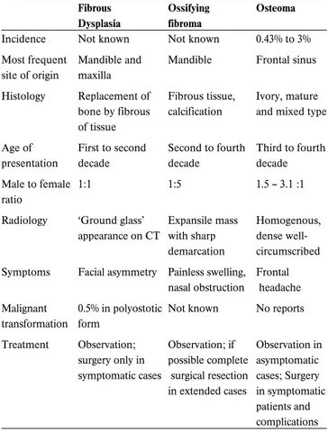
Osteoma is the most common benign sinonasal tumour, and it has been reported to be seen on 1% of routine sinus radi-ographs, most commonly localized to the frontal sinus (95). The overall incidence of paranasal sinus osteomas in patients with coronal sinonasal CT scans due to sinonasal symptoms was 3%
(96)
. Osteomas usually presents between the second and sixth decades, with a predilection of fifth and sixth decades (96,97). Male to female ratio is 1.3-2:1. The frontal sinus was the most frequently involved site (57%)(of these lesions 37% were in the immediate vicinity of the frontonasal duct and 21% above and lateral to the ostium), followed by the maxillary, ethmoid and the sphenoid sinuses. Maxillary sinuses are affected in about 20% of cases (96,98). Involvement of the sphenoid sinus region is extremely rare (99,100). Osteomas can occur in conjunction with Gardner’s syndrome, which is an autosomal-dominant condi-tion consisting of osteomas (usually multiple), soft tissue tumours (such as epidermal inclusion cysts or subcutaneous fibrous tumours), and polyposis of the colon. This triad of symptoms should prompt gastroenterology referral if this is suspected, as malignant degeneration of these colonic polyps will occur in 40% of patients (101,102).
Fibro-osseous lesions are a heterogeneous group and the com-monest fibro-osseous lesions of the sinonasal tract are ossify-ing fibroma and fibrous dysplasia. Other lesions include giant cell granuloma, fibromyxmoma, osteoblastoma. Both fibrous dysplasia and ossifying fibroma are more common in females than in males (103,104). Usually fibrous dysplasia presents in the first two decades (105). There are two forms: polyostotic (15%– 30%), involving more than one bone, and monostotic (70%– 85%), involving only one bone (106). McCune-Albright syn-drome (the polyostotic form fibrous dysplasia, precocious puberty, cafe´-au-lait spots) is the most rare and preferentially involves young girls (107). Twenty-five percent of monostotic cases arise in the facial skeleton (108). The maxilla and mandible are the most common sites in the head and neck, although it has been reported throughout the maxillofacial skeleton,
including the sphenoid intersinus septum (106). Asymptomatic fibrous dysplasia is often an incidental finding on X-ray obtained for other reasons (trauma or evaluation of hearing loss). The sphenoid bone and central skull base are frequently involved in such cases. Growth is variable, and usually slows after puberty but this is not invariable (109). Fibrous dysplasia has a low rate of malignant transformation (106,108,110). Transformation occurs in 0.5% of polyostotic forms and in 4% of lesions in patients with McCune-Albright syndrome (106,111). Ossifying fibroma is usually diagnosed in the third to fourth decade and mainly affects bones. It is more commonly found in the mandible (75%) or maxilla (10—20%) and lesions in the nasoethmoidal region are rare (103). A more aggressive variant called ‘juvenile ossifying fibroma’ occurs in the young. Juvenile ossifying fibromata was defined by Reed and Hagy
(112)
as ‘a localised actively growing destructive lesion occurring predominantly in children. It predominantly affects male sub-jects.
Less than 5% of aneurysmal bone cysts occur in the craniofa-cial bones (103). The mandible is involved in 66% of the cases while maxilla only in 33% of cases. Aneurysmal bone cysts in the orbitoethmoid complex is very rare (113,114). Aneurysmal bone cyst is slightly more frequent in females and develops in about 90% of patients during the first two decades of life
(115,116)
. Giant cell tumours of the craniofacial bones are rare. The most common sites involved are the sphenoid and eth-moid bones. Osteoblastoma is a benign bone tumour. Clinical presentation can be similar to other fibro-osseous lesions
(103,117)
.
Inverted papilloma (IP) accounts for 0.5 percent to 4 percent
(118,119)
of the surgically removed nasal tumours with an inci-dence ranging from 0.6 to 1.5 cases per 100,000 inhabitants per year (120,121). The age at onset ranged from 15 to 96 years old, with the highest incidence was seen in the 5th and 6th decades of life (122). The male to female ratio was reported as 2 to 5:1
(123,124)
. There were no significant racial differences (125). The duration of symptoms varied from 5 months to 20 years with mean duration of 3.9 years (122). The frequency of inverted papilloma in apparently normal bilateral polyps varies between 0.00% and 0.92%. The incidence of inverted papilloma in unre-markable recurrent cases of nasal polyps is rare. This rate is similar to the one observed in patients undergoing first surgery. Age, gender, and number of recurrences did not influ-ence the frequency of this diagnosis (126). Since multiple sites within the nasal cavities and paranasal sinuses were involved, it is not always easy to determine the location of the origin of inverted papillomas. However, the ethmoid region, the lateral wall of nasal fossa and the maxillary sinus are the most
quent sites of origin of inverted papilloma. The frontal sinus is exceedingly rare (the ethmoid region in 48%, the maxillary sinus in 28%, the sphenoid sinus in 7.5%, the frontal sinus in 2.5%, the inferior turbinate in 2.5%, and the septum in 2.5%
(125)
. Inverted papilloma is generally unilateral, bilateral involve-ment of the sinonasal tract is very rare, reported in less than 1% (124) to 9% (127) of patients. The numerous publications on inverted papilloma differ widely in their reported incidence of associated carcinoma, which ranges from 0 to 53 per cent
(128,129)
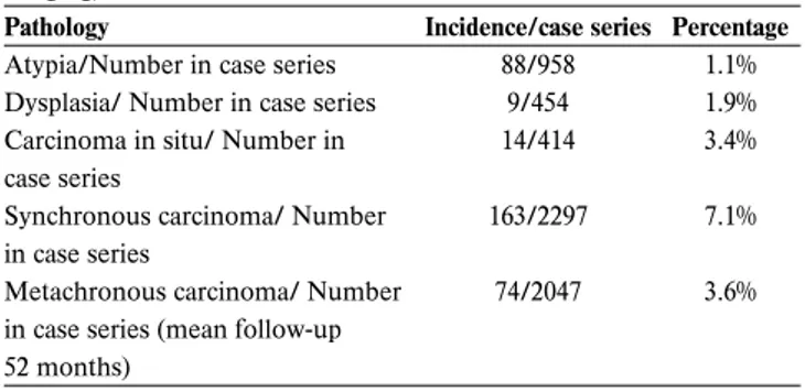
. The true population base for these figures is uncertain, given that many series were reported from tertiary centers, where recurrent and problematic cases are likely to be over-represented (130). Mirza et al. (130) found that the rates of syn-chronous and metasyn-chronous carcinomatous transformation of inverted papilloma are 7.1 and 3.6 per cent, respectively and 11 per cent malignant transformation in recurrent inverted papil-lomas after carefully reviewing the literature. The mean time taken to develop a metachronous carcinoma was 52 months (range six to 180 months). Some reports suggest that the devel-opment of squamous cell carcinoma in IP is heralded by a reduced cellular apoptosis, which is triggered by human papil-loma virus infection (131).
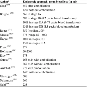
Juvenile angiofibroma (JA) is a rare benign vascular tumour that accounts for 0.5% of all head and neck tumours and its general incidence is approximately 1:150,000 (132,133). In a national retrospective cohort study to estimate the incidence rate of JA in the Danish population, an overall incidence rate of 0.4 cases per million inhabitants per year was found. The median age at diagnosis was 15 years (range 10-24 years), the incidence rate of the population at risk reached 3.7 cases per million(134). Carrillo et al. (135) reported that the median age at diagnosis was 18.5 years (range 18-35 years) and all of their patients were males. Only male adolescents were identified. Altogether only 30 female patients have been reported in the literature. It is questionable whether all the previously present-ed female cases in the literature are in fact JA (134). In many of the papers, the female cases cannot be further analyzed and could not be decided whether the tumours were angiomyofi-broma or variants of angiofiangiomyofi-broma. However, a larger series of 299 JA cases from 6 studies included no female patients (136-141). In spite of the reports of hormonal disorders in patients with JA and the presence of androgen and/or estrogen receptors in tumour tissues, apparently no alterations of hormonal serum levels were observed, and the hormonal influence on JA is still controversial (142,143). The findings of the partial or complete losses of the Y chromosome and gains of the androgen recep-tor gene due to gains of chromosome X in the recent genetic analysis of JA indicates an androgen related pathophysiological process in JA(143).
Schwannomas can occur in all parts of the body but are rela-tively more frequent in the head and neck region (25–45% of all cases) (144). Approximately 4% of these lesions of the head
and neck involve the nasal and paranasal cavities (145). In the paranasal sinuses they are reported mainly in the nasoethmoid compartment, less frequently in maxillary sinus, septum, sphe-noid and frontal sinuses (146). Most cases occur between the second and fifth decade of life; there is no specific association with sex or race (147). There is, by contrast, a very low risk of malignancy for schwannomas; however, in literature there are reports of malignant degeneration in long standing benign schwannomas (148). The risk of malignant transformation rises to 10%-15% in Von Recklinghausen’s disease (149).
Lobular capillary hemangioma (pyogenic granuloma) mainly affects the female population, with a peak incidence in the third decade (range 11-65 years) (150,151). Sinonasal localisation ranges from 7% to 29%, the anterior portion of the nasal sep-tum and the turbinates being the most frequently involved areas (152,153). Nasal trauma and hormonal imbalances have been postulated as possible etiologic factors (154).
Pleomorphic adenoma is the third most common benign tumour of the sinonasal tract after osteoma and inverted papil-loma (155). It usually affects patients in the fifth decade of life, with a slight female predominance. The nasal septum is the most frequently affected site followed by maxillary sinus
(156,157)
. There are many other benign tumours such as leiomy-oma, paraganglileiomy-oma, hemangileiomy-oma, myoepithelileiomy-oma, oncocy-toma, etc. Since they occur sporadically in the sinonasal area, the information in the literature to draw conclusions is scarce. However, they share the same epidemiologic profile with other benign tumours. They will not be mentioned separately.
3.2 Malignant Tumours
Sinonasal neoplasms are uncommon neoplasms, they account only for 1% of all malignancies (158,159) and for 3% of all upper respiratory tract malignancies and accounting for only 3% to 5% of all head and neck malignancies (160,161). Annual incidence is 0.5-1 new cases /100,000 inhabitants in Italy (162), whereas rela-tively high rates for sinonasal malignancies (SNM) were found in Asian and African populations, the highest age-adjusted rates, between 2.5 and 2.6 per 100,000 per annum, occurring in Japanese males (163). Ayoutunde reports sinonasal malignancies (SNM) account for 1.57% of all rhinologic diseases in his series. Sinonasal malignancies are more common in males. The male to female ratio is reported between 1.2-2.7/1 (164,165). In the antrum the male:female ratio is 2:1, and in the ethmoid sinus the male:female ratio is 1.4:1. Overall 75% of all malignant tumours occur in persons older than 50 years (166).
The most common sinonasal malignancies are the primary epithelial tumours followed by the non epithelial malignant tumours. In the group of epithelial SNMs the squamous cell carcinoma dominated, and in the nonepithelial SNMs, the most common group was malignant lymphoma. The prevalence of
the different malignant tumours in the literature is extremely variable. The incidence of epithelial SNMs ranges between 52.9% to 91% (166,167). In a series of 115 patients, Svane-Knudsen (168) reported that 64% had well-differentiated squamous cell carcinomas (SCC), adenocarcinomas, and adenocystic carcino-mas. Non-Hodgkin’s lymphomas (NHL) and undifferentiated carcinomas represented 9% and 2.6%, respectively. Zbaren et al.
(169)
, in a German series of 216 cases, found 56% to be epider-moid carcinomas and 14% to be adenocarcinomas. Similarly, Haraguchi et al. (170), in 60 Japanese patients, found a predomi-nance of well-differentiated SCCs (25%) followed by melanomas and NHLs (23%) and a small number of undifferen-tiated carcinomas (5%). On the other hand, in areas where there is a high incidence of nasal-paranasal neoplasms, the histopathological spectrum is different from the one described in low-risk areas. Undifferentiated carcinomas in Chinese high-risk areas (ie, Hong Kong) constitute more than 80% of all nasal-paranasal malignancies (171). Incidence, site and histologi-cal type can vary in different geographihistologi-cal areas which may be due to occupational, social and genetic factors (172). For all nose and sinus tumours, the nose is the primary site in 25% and the sinuses 75%, and of all sinus neoplasms 60% to 80% originate from maxillary sinus (166). However, it is not easy to determine the exact site of origin with large tumours. As a result, the tumour distributions in the literature are variable (166).
Squamous cell carcinoma is the most common tumour of the sinonasal malignancies. Approximately 60% to 73% of squa-mous cell carcinomas originate in the maxillary sinus, 20% to 30% in the nasal cavity, 10% to 15% in the ethmoid sinus, and 1% in the sphenoid and frontal sinuses (155,165,166). Among the carcinomas of the nose and paranasal sinuses, sinonasal gland carcinomas represent the second most frequent type of malig-nant epithelial tumour and the paranasal sinuses are the most common site of minor salivary gland involvement (173-176). Adenoid cystic carcinoma (ACC) accounts for fewer than 1% of all head and neck malignancies and 10% of all salivary gland neoplasms (176,177). Lupinetti et al.(178) reported that most patients were Caucasians (72.4%), non-smokers (48.4%), and non-drinkers (74.4%) in their ACC series. Sinonasal ACC accounts for 10% to 25% of all head and neck ACC (176). The maxillary sinus (47%) and the nasal cavity (30%) were the most common primary tumour sites. ACC has a propensity for per-ineural spread and bony invasion, which can lead to significant skull base involvement and intracranial extension (178). Adenocarcinoma is the third most common mucosal epithelial malignancy found in this area, after squamous cell carcinoma and adenoid cystic carcinoma (179), and represents approximate-ly 8% to 15% of all sinonasal cancers (180,181). The incidence is less than 1 case per 100,000 inhabitants per year (182), occurring predominately among men with a mean age of presentation of 60 to 65 years (183). However, in the northern part of Spain, the incidence is 0.19 cases/100,000 inhabitants per year (184). The
median age of onset lies between 50 and 60 years (183)and in wood dust related tumours even earlier (185). Men develop ade-nocarcinoma four times more frequently than women, reflect-ing the occupational hazard implicated(186). It is located most frequently (85%) in the ethmoid sinus and the upper part of the nasal cavity. One study using endoscopic endonasal surgery showed that woodworkers' adenocarcinomas constantly origi-nated in the olfactory cleft, appearing as polyp-like neoplasms with well-defined bodies(187). Nasal adenocarcinoma on sinus CT scans showing a unilateral expanding opacity of the olfac-tory cavity should raise the suspicion of nasal adenocarcinoma
(34)
. It only exceptionally arises in other sites of the nasal cavity (maxillary sinus in 10%) and these cases are usually not related to wood dust exposure(188).
The association between wood dust exposure and adenocarci-noma of the sinuses is also well established. It is estimated that woodworkers have 500 times elevated risk compared to the male population and up to 900 times compared to the popula-tion in general(185). It has been shown that the true risk factor is the actual exposure to wood dust particles, and not the pos-sible exposure to chemical products used in the industry, such as polish, varnish or protectors(185). Hard wood types as ebony, oak and beech confer the highest risk of developing sinonasal adenocarcinomas(189), increased further by inhalation of formaldehyde or substances normally used in this type of industry(190). The strong relation of adenocarcinoma to expo-sure to wood dust makes it a disease almost exclusive to car-penters and furniture makers. Therefore, in many countries (Australia, Germany, Great Britain, Belgium, France, etc.), it is considered an industrial disease (189,191-194). The furniture mak-ers who are likely to be exposed to the fine wood dusts of threshold >5mg/m3/day are at greater risk (167,195,196). Many findings indicate a dose-response relationship (190,197,198), with a higher incidence of tumours occurring among workers exposed for longer periods. Recent studies have shown that even short periods of exposure (< 5 years) can lead to an increased risk of carcinoma. In general, the normally long latency period is esti-mated at 40 years(199), although it can range between 20 and 70 years(195). Despite this clear etiology, it is still unknown by what molecular mechanism sinonasal adenocarcinomas devel-op. Because wood dust does not have mutagenic properties, it is hypothesized that prolonged exposure to and irritation by wood dust particles stimulate cellular turn-over by inflammato-ry pathways(186,200).
Sinonasal mucoepidermoid carcinoma accounts for 0.6% of all salivary tumours and 4.8% of all mucoepidermoid carcinomas. The most common site is maxillary antrum followed by nasal cavity, nasopharynx and ethmoid sinuses in order of decreas-ing frequency(163,201). Thorium dioxide, when used as an imag-ing agent, may cause antral squamous and mucoepidermoid carcinomas (166). Although no other definitive risk factors for mucoepidermoid carcinoma have been identified, minor
trau-ma and chronic irritation have been implicated in the etiology of sinonasal tract cancers in general (202). Acinic cell carcinomas account for approximately 1% of salivary gland tumours and 10% to 15% of all sialocarcinomas (203-206). Acinic cell carcino-mas are found predominantly in the parotid gland and are somewhat unusual in other locations (203-206). In the sinonasal tract, acinic cell carcinomas are extremely rare (207-210). To date, only 11 cases of sinonasal acinic cell carcinoma have been described in the English-language literature (211). Acinic cell carcinomas are most common in the fifth and sixth decades of life but are seen in patients of all ages, including children
(203-206)
. In the sinonasal tract, the ages in reported cases range from 42 to 76 years (median, 59 years). There is a discrepancy in the literature about whether acinic cell carcinomas occur more often in men or women in general; however, there is no sex related difference in the occurrence of sinonasal acinic cell carcinomas (203,205).
Relative risk rates for sinonasal epithelial malignancies have been determined for several chemical agents (chromates, nick-el compounds, isopropylic alcohol, and mustard gas) and for several occupations (i.e. nuclear refinery work, leather work in boot and shoe manufacturing, chrome pigment work, metal-worker, textile metal-worker, construction metal-worker, bakers, flour milling and farmer) even in the apparent absence of causal agents (212). Reported increased risks to different industries were reported as follows: the metal industry (relative risk rang-ing from 3.1 to 5.9), the textile industry (rangrang-ing from 2.9 to 17.0), the mining and construction industry (ranging from 2.3 to 5.3), and the agricultural industry (ranging from 1.9 to 3.3)
(212)
. Among women, exposure to textile dust was associated with an elevated risk of squamous cell carcinoma and adeno-carcinoma. For squamous cell carcinomas, the risk increased with the duration and the level of exposure. The risks associat-ed with the different types of textile fibers (cotton, wool, and synthetic fibers) were similar and the results did not permit to incriminate a particular type of textile (213). Thorium dioxide, when used as an imaging agent, causes antral squamous and mucoepidermoid carcinomas(166).
Smoking is associated with an elevated risk of nasal cancer, especially squamous cell carcinoma of the maxillary antrum
(165,166,214)
. However, some reports did not find any increased risk for sinonasal tract malignancies associated with tobacco and alcohol(215). Since no persuasive evidence was found that smoking is an etiologic factor in sinonasal carcinoma, further research is needed.
Since the early 1980s, when evidence was provided on the pos-sible involvement of human papilloma virus (HPV) in the aeti-ology of both benign respiratory papillomas and squamous cell carcinomas, a substantial number of studies have explored this issue and its deoxyribonucleic acid has been found in both the inverted papilloma and the cells of neighbouring, apparently
normal mucosa(216). The current evidence linking HPV to at least a proportion of benign sinonasal papillomas is convincing. Syrjaenen(217)based on the analysis of over 1,000 such lesions, reported that HPV-6 and HPV-11 is present in one third (33.3%) of inverted papillomas and this detection rate is higher than most other reported extragenital papillomas, except those of the larynx and bronchus. Tang et al.(218)detected human papilloma virus in up to 86% of inverted papilloma cases. The 2005 International Agency for Research on Cancer evaluation on the carcinogenicity of HPV in humans(219)concluded that there is sufficient evidence for the carcinogenicity of HPV in the oral cavity and oropharynx, limited evidence in the larynx, and inadequate evidence in sinonasal cavities(220). However, some previous reports have suggested a possible implication of HPV in the development of several carcinomas of the sinonasal region(217,221,222). In 1993, Kashima et al.(223)found human papillomavirus (HPV) positive in 4% of squamous cell carcinomas. Alos et al.(220) detected HPV DNA in tumour tis-sue of 20% of sinonasal squamous cell carcinoma patients. The tumours affected predominantly men by a proportion of approximately 3:1; no significant differences in sex and age were found between HPV-positive and-negative groups. There were no significant differences in tumour stage at presentation. Despite the similar clinical characteristics and staging at pre-sentation, patients with HPV-positive tumours had a signifi-cantly better prognosis than those with HPV-negative neo-plasms(220). Syrjaenen(217) showed that 21.7% of sinonasal carci-nomas analysed were positive for HPV. Low risk HPV types 6 and 11 are usually confined to benign lesions, whereas the reverse is true for the oncogenic HPV types 16 and 18; and the presence of squamo–columnar junctions and squamous cell metaplasia in the sinonasal system(217). The discrepancies reported by several studies might result in part from technical reasons, but it is also possible that sinonasal lesions have a het-erogeneous aetiology (HPV related and non-related) and/or that some novel (yet unidentified) HPV types exist in these lesions, which are detected by some studies but not by others. Primary sinonasal tract mucosal malignant melanomas are rare, accounting for between 0.3% and 2% of all malignant melanomas and about 4% of head and neck melanomas (224-228). The head and neck represents the most common site of mucosal malignant melanoma with a suggested incidence of about 0.018/105 to 0.051/105 per year(225,229,230). Sinonasal tract mucosal malignant melanomas represents up to 4% of all sinonasal tract neoplasms(226,229,231). In the National Cancer Data Base report by the American College of Surgeons Commission on Cancer and the American Cancer Society of more than 84,000 melanomas seen from 1985 to 1994(224), only 1.3% were melanomas that arose from mucosal surfaces, of which 55% were of the head and neck. Sinonasal tract mucosal malignant melanomas were found to be equally common in men and women. A higher proportion of melanoma was iden-tified in black patients (10.4%)(232). In general, the mean age
for sinonasal tract mucosal malignant melanomas (64.3 years) is later in life than cutaneous malignant melanomas. Sinonasal tract mucosal malignant melanomas is a more lethal disease in patients older than 60 years, a finding similar to cutaneous melanoma(232). Tumours originating in the sinuses are less common than those arising in the nasal cavity, but sinus tumours may grow asymptomatically until late in the disease course(231,233). A third of patients had neck mestastases, which often preceded distant metastasis, and distant metastasis always were rapidly fatal(234).
The incidence of olfactory neuroblastoma was found 0.4 cases/million inhabitants per year(235). The incidence of olfac-tory neuroblastoma is difficult to establish, but the tumour is not as rare as is commonly reported and probably represents more than 5% of all nasal malignant tumours (235-237). Olfactory neuroblastoma occurs over a wide age range (3–90 y), with a bimodal peak in the second and sixth decades of life (238-240). Occasional cases have also been reported in children younger than 10 years(168,237). Olfactory neuroblastoma affects male and female patients with similar frequency and can be found in all age groups(237,241). No known cause exists for this tumour (242), although diethylnitrosamine injections can induce tumours in hamsters at the site of the olfactory epithelium(243). No heredi-tary patterns have been described for this neoplasm, and there is no apparent racial predilection(238).
Extrapulmonary neuroendocrine carcinomas (SNEC) only account for 4% of all SNECs(244), and very few cases of SNECs of head and neck have been reported previously(245). Less than 250 cases of head and neck SNEC have been published so far, including 48 cases of SNEC in the nasal and paranasal cavities
(246,247)
. The majority of patients with SNEC of the head and neck are male. Although there seems to be an association with cigarette smoking, the association is not as strong as that with pulmonary SNEC(248,249). Although the neoplasm has been described at any age between 16 and 77 years, the prevalent distribution is in the fifth and sixth decade(250,251). No particular risk factors for this tumour appears to have been identified
(247,252,253)
.
Although tumours of this type occur in a variety of organs and sites, small cell undifferentiated carcinoma (ScCC) of the sinonasal tract is a rare malignancy, with the reported series all having fewer than 10 patients(248). In the M.D. Anderson series of neuroendocrine tumours, there were only 7 cases of small cell carcinoma(254). The paucity of well-documented cases pre-cludes generalization about the clinical features. Within this limitation, the mean age at presentation is approximately 50 years (range 26-77 years), and there is no sex predilection. Anatomic sites include the nasal cavity, ethmoid sinuses, and maxillary sinus(255).
Sinonasal undifferentiated carcinoma (SNUC) is a rare tumour, with fewer than 100 reported cases in the world litera-ture (256).There is a male predominance (2–3:1). The age range is broad, usually ranging from the third to ninth decades; the median age at presentation is in the sixth decade(257-259). There are no known etiologic agents. SNUCs are typically negative for Epstein–Barr virus (EBV)(258-260). Some cases have been reported to develop following radiation therapy for nasopha-ryngeal carcinoma(258).
The annual incidence of the Ewing’s Sarcoma Family of Tumours (ESFT) in the United States is 2.1 cases per million children, and they account for approximately 2% of all cancers in children and young adults(261). ESFT is more common in male than in female patients and has a greater incidence in white and Hispanic children than in black or Asian children
(262,263)
. ESFT is not felt to be inherited and is not associated with any cancer syndromes. In 95% of cases, at (11;22)(q24;q12) translocation is detected(264). Sinonasal tract involvement is extremely rare, with about 50 cases reported in the English literature. Most of them were observed in the max-illary sinus, whereas less than 10 cases each involved the eth-moid and nasal fossa [also known as PNET- primitive neuroec-todermal tumour].
Hemangiopericytomas are unusual vascular tumours, which accounts for only 1% of all vascular neoplasms and for 3%-5% of sarcomas (179). They rarely occur in the paranasal sinuses and nasal cavity. The rate of head and neck involvement ranges between 15% and 25%, with sinonasal tract localisation present in 5% of patients(179). Ethmoid, nasal cavity and sphenoid sinus are the preferential sites of origin(265). Although the tumour affects all ages, they occur most commonly in adults in the sixth and seventh decades of life(266,267). No gender predomi-nance is reported(267,268). Trauma, steroid therapy, and altered hormone secretions are proposed as predisposing factors
(269,270)
.
Sarcomas of the head and neck are rare tumours, accounting for 4–10% of all sarcomas(271-274) and fewer than 1% of all malig-nancies of the head and neck region(275,276). Sarcomas of the sinonasal tract make up for about 15% of sinonasal tumours
(277)
. Oral and maxillofacial sarcomas present at any age from 5 months to 77 years (mean 42) and male to female ratio of 3:1 with predilection for the mandible.1,2. The mean age and male: female ratio in Africa is lower than in Western series (278). Osteosarcoma is a rare bone tumour that occurs primarily in long bones. Overall incidence is 1:100,000 inhabitants per year, and 6-13% of cases occur in the head and neck region (279). Osteosarcoma had peak prevalence in the third decade with equal gender distribution. The occurrence in the paediatric age is very rare (280). In the maxillofacial region, it tends to occur a decade later than in long bones. Osteosarcoma accounts for 0.5%-1% of all sinonasal tract tumours (281). In children with
osteosarcoma, about 3% carry a germ line mutation in p53, with the majority of these having a family history suggesting Li-Fraumeni syndrome(282). The incidence of osteosarcoma has been increasing by about 1.4% per year for the past 25 years
(283)
. The etiology of osteosarcomas remains unknown. Bone abnormalities and diseases as Paget’s disease of bone, fibrous dysplasia, myositis ossificans, other hereditary pathologies like retinoblastoma, Li-Fraumeni syndrome, and previous chemotherapy and irradiation for other malignancies have been suggested as specific risk factors(284,285).
Rhabdomyosarcoma is the most frequent soft tissue sarcoma in the paediatric age, accounting for up to 75% of all child sar-comas and 6% of all paediatric cancers. The embryonal subtype is the most common. Mesenchymal rhabdomyosarcoma is rare and the head and neck region is the most commonly involved site (37%)(286). Occurrence of head and neck rhabdomyosarco-ma in adults is rare. Only 10% of all soft tissue tumours and 1% of all neoplasms in the sinonasal tract are rhabdomyosarcomas
(287)
. Sinonasal tract localisation is present in about 8% of all adult age rhabdomyosarcomas(288).
Chondrosarcoma make up only 10–20% of malignant primary bone tumours, with 5–10% located in the head and neck. Maxillary sinus is the most frequently involved site (289,290). In the skull base, chondrosarcomas typically occur at the petrocli-val synchondrosis. The lesion is commonly diagnosed in the sixth decade. The paediatric population is rarely affected. There is no gender predominance (288). However, in some reports it is more frequently seen in males (278,291). The etiology of chondrosarcomas remains unknown. Meanwhile, associated conditions include multiple hereditary exostosis, Ollier’s dis-ease, Maffucci’s syndrome, previous intravenous thorium diox-ide contrast use, Paget’s disease of bone, chondromyxoid fibro-ma, and previous irradiation (292).
Leiomyosarcoma is rare in the oro-facial region (272) and accounts for approximately 7% of all soft tissue sarcomas. It comprised the fourth most common sarcoma, mainly in the maxilla, and with a 5:1 male:female ratio(278). Sinonasal tract localisation is rare with about 40 cases reported in the litera-ture(293).
Fibrosarcoma with mandibular predominance and equal sex distribution was the fifth most common sarcoma. Liposarcoma, fibromyxosarcoma, neurofibrosarcoma, ameloblastic sarcoma and synovial sarcoma are rare(272,278). Sinonasal lymphomas, either primary or secondary are mostly non-Hodgkin lymphomas and are the second most common malignant tumours following carcinomas occurring in the sinonasal tract. Non-Hodgkin lymphomas are classified into B and T-NK subtypes according to lymphocytic phenotype
(155,294)
. There is a difference in incidence, epidemiology, and
cell type between Western and Asian countries. In Western countries, lymphomas are infrequent and sinonasal tract involvement varies between 0.2%-2% of all non-Hodgkin lym-phomas(295). They constitute 5.8 to 8% of the extranodal lym-phomas arising in the head and neck area(294,296). B-cell lym-phomas are predominant and tend to affect paranasal sinuses in the elderly(155,294). In Asian and South American countries, the incidence of non-Hodgkin lymphomas of the nasal region is much higher than in the United States and they account for 2.6 to 6.7% of all lymphomas and are the second most frequent group of extranodal lymphomas after gastrointestinal lym-phomas. T or NK cell lymphomas are predominant and the nasal cavity is mainly involved in younger people(297,298). Epstein-Barr virus is considered important in the etiopathogen-esis of lymphomas, especially for specific lymphomas such as Burkitt lymphoma and nasal NK-T lymphoma. In Asian coun-tries, the prevalence of EBV-positive T-cell lymphomas is simi-lar to the prevalence of EBV virus infection and differ from the findings of the more common EBV-negative B-cell nasal lym-phomas in the United States. These findings suggest that EBV plays a role in the development of nasal T-cell lymphomas and that the incidence of EBV infection may explain the reported “East-West” difference in the incidence of nasal T-cell lym-phomas(155,294,297).
Most of the malignant tumours of the sinonasal regions are pri-mary in origin. Metastasis of malignant tumours to the sinonasal area occurs infrequently and usually presents at the late stage of primary disease. More than 50% of sinonasal metastases take origin from a renal carcinoma(299). Other most common primary sources in decreasing order after the kidney, are lung (12%), uro-genital ridge (12%), breast (9%), and gastrointestinal tract (GI tract) (6%) (300). The most common metastatic sites are the max-illary sinus (50%), followed by the ethmoid sinus (18%) and nasal cavity (15%) (299-302). However, some reports from East Asia are very different when compared with European and North American reports. Different incidences of malignant neoplasms in the primary site may explain the result of different incidences of sinonasal metastatic tumour(303). Although the mean age of patients with sinonasal metastases varies in different primary origins, the highest incidence is in the sixth decade in men and the seventh decade in women (304).
It is more appropriate to consider chordomas as low-grade malignancies with the potential for metastasis. Chordomas are slow-growing, locally aggressive tumours. Approximately 25% of chordomas arise in the clivus and constitute about 0.15% of all primary intracranial neoplasms (305). The ages of the patients ranged from 4 to 76 years, with a predominance for the third and fourth decades in intracranial localizations(306). The male-to-female ratio is reported 2:1 by Weber et al.(307) and 2:3 by Stippler et al.(306). No associations with irradiation or any other environmental factors has been observed. A small percentage of cases have a familial pattern of inheritance(308).
4-1 Clinical
Tumours affecting the sinonasal region and skull base usually present late as their presenting symptoms are often banal and therefore, overlooked by patients and their clinicians, particu-larly in primary care where these conditions are rarely encoun-tered. The recent onset of unilateral nasal symptoms, which do not improve with a short course of medical therapy should prompt referral for specialist assessment. Orbital and neurolog-ical symptoms should generally be referred ab initio.
4-2 Imaging
Imaging studies play a key role in pretreatment assessment and preoperative planning of benign and malignant lesions involv-ing the sinonasal tract, nasopharynx, and adjacent skull base. Computed tomography (CT) and magnetic resonance imaging (MRI) are used in a complementary fashion when characteriz-ing tumours in this region to accurately assess their
loco-regional extent including any bone and neurovascular exten-sion or nodal involvement. They are also of paramount impor-tance in intraoperative guidance and post-operative surveil-lance. These anatomic areas are very complex and understand-ing the radiological features typical of the different pathologies requires a thorough knowledge of anatomy. For the sake of clarity, the present review will separately analyze pre-treatment assessment, intra-operative evaluation, and post-operative sur-veillance.
4-2-1 Pre-treatment evaluation
The aims of imaging are to distinguish tumour from inflamma-tory reactions and secretions, elucidate the nature of the tumour (benign vs. malignant), and map its extent (309). These goals are best achieved by MR. However, CT is the first inves-tigation commonly obtained in a patient with symptoms sug-gesting a disease involving the sinonasal tract and/or the adja-cent skull base. Apart from patients with fibro-osseous lesions, where CT can properly define the nature of the lesion and its
4. Diagnosis
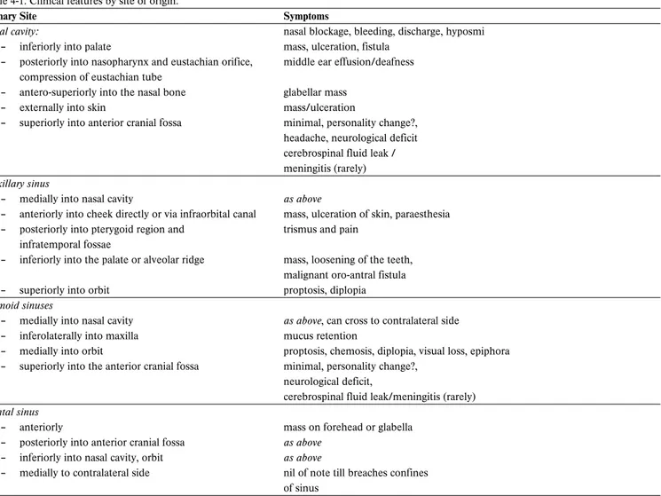
Table 4-1. Clinical features by site of origin.
Primary Site Symptoms
Nasal cavity: nasal blockage, bleeding, discharge, hyposmi
– inferiorly into palate mass, ulceration, fistula
– posteriorly into nasopharynx and eustachian orifice, middle ear effusion/deafness compression of eustachian tube
– antero-superiorly into the nasal bone glabellar mass
– externally into skin mass/ulceration
– superiorly into anterior cranial fossa minimal, personality change?, headache, neurological deficit cerebrospinal fluid leak / meningitis (rarely) Maxillary sinus
– medially into nasal cavity as above
– anteriorly into cheek directly or via infraorbital canal mass, ulceration of skin, paraesthesia – posteriorly into pterygoid region and trismus and pain
infratemporal fossae
– inferiorly into the palate or alveolar ridge mass, loosening of the teeth, malignant oro-antral fistula
– superiorly into orbit proptosis, diplopia
Ethmoid sinuses
– medially into nasal cavity as above, can cross to contralateral side
– inferolaterally into maxilla mucus retention
– medially into orbit proptosis, chemosis, diplopia, visual loss, epiphora – superiorly into the anterior cranial fossa minimal, personality change?,
neurological deficit,
cerebrospinal fluid leak/meningitis (rarely) Frontal sinus
– anteriorly mass on forehead or glabella
– posteriorly into anterior cranial fossa as above – inferiorly into nasal cavity, orbit as above
– medially to contralateral side nil of note till breaches confines of sinus
borders, the other lesions require an MR with gadolinium enhancement. This should include high-resolution (3 mm) T1-weighted and T2-T1-weighted images of the sino-nasal tract, orbit, skull base, and the adjacent intracranial compartment, acquired in the axial, coronal, and sagittal planes. Fat-saturated T1-weighted techniques are usually included to identify the pres-ence of disease beyond the paranasal sinuses (i.e., perineural spread or intracranial extension) (310). Additional sequences, to be used in selected conditions, include the following: 1) MR cisternography with heavily T2-weighted (3DFT-CISS, DRIVE) thin sections (0.6 mm or less), a technique, which has been advocated(311) to determine the relationships of tumour with cisternal cranial nerve segments; 2) high resolution GE sequences with sub-millimetric isotropic slices (FIESTA; VIBE) to image the intraforaminal segment of cranial nerves
(312)
; 3) FLAIR (fluid-attenuated inversion recovery), which helps in differentiating CSF from the cystic/fluid content of tumours or secondary mucoceles abutting the skull base; 4) MR angiography, which enables a detailed visualisation of the entire course (or segments) of internal carotid artery (ICA). Sinonasal tract and anterior skull base
Benign tumours of the sinonasal tract and anterior skull base are rare. Osteomas are the most frequent but many are diag-nosed by chance and do not require surgery. Papillomas (inverted, oncocytic, fungiform) are the most common indica-tion for surgery. CT commonly shows a space-occupying lesion with low/moderate bone resorption involving the eth-moid-nasal complex and/or the maxillary sinus. While the pos-sibility of characterizing the lesion based on CT imaging is quite low, the endoscopic appearance may be highly sugges-tive. MR has the well known advantage over CT to differenti-ate inflammatory changes from the lesion and to identify the so-called “cerebriform” or “columnar” pattern, which is evident on T2 and contrast-enhanced T1 sequences and reflects the histological architecture of the lesion. The loss of the cerebri-form pattern may be a sign of a concomitant carcinoma, which is usually associated with a more extensive bone erosion. The finding on CT of focal hyperostosis or osteitic changes (313,314) as indicators of the area of origin of the lesion, can also be appreciated on MR (315).
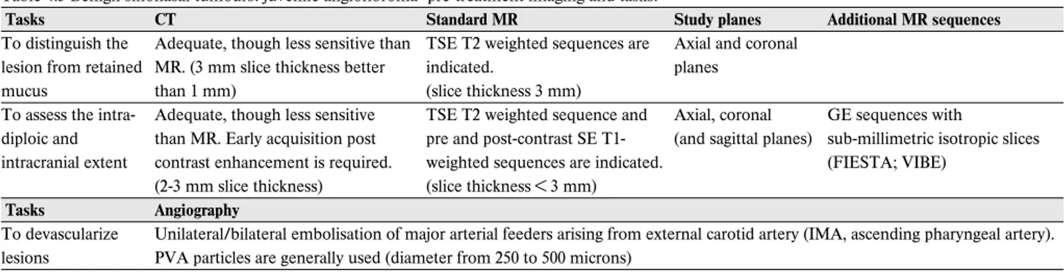
Juvenile angiofibroma (JA) originates from the pterygo-pala-tine fossa but at an early stage tends to involve the adjacent sino-nasal cavities. Submucosal growth and invasion of the cancellous bone of the basisphenoid are distinct features that are easily identified by enhanced CT or MR (316). “Finger-like projections” with sharp and lobulated margins are the hallmark of JA growth in the soft tissues and along canals and foramina of the skull base. Intradiploic spread of the lesion can be better differentiated from normal medullary content on MR by com-bining a plain T1 with a post-contrast T1 without or with fat-saturation (312). Furthermore, the presence of several signal voids within the lesion on both T1 and T2 sequences,
indicat-ing major intra-lesional vessels, corroborates a diagnosis of JA. In intermediate/large-size JA pre-operative assessment includes angiography, which provides a detailed map of the lesion’s vascular supply, showing connections with the external and occasionally internal carotid artery and the vertebral arter-ies and branches of the contralateral carotid system. This examination is commonly performed concomitantly to emboli-sation, which has the intent of decreasing intraoperative bleed-ing. While the currently available methods of embolisation provide excellent devascularization of feeders coming from the internal maxillary artery and its branches as well as from the ascending pharyngeal artery, control of major vascularization from the internal carotid artery is demanding. Devascularization by direct tumour puncture and embolisation is associated with an unacceptable risk of major neurologic complications (317)and has been therefore abandoned. In case of encasement of the internal artery, which is indeed a very rare event, the balloon occlusion test and sacrifice of the ICA or, as a less invasive procedure, stenting of the intratemporal carotid artery may be considered.
Olfactory groove meningioma typically presents as an extra-axial, durally based lesion involving the anterior skull base. Characteristic MRI features are isointensity to slight hypointensity on T1-weighted images; the appearance on T2-weighted images is variable. Homogenous enhancement is invariably obtained after contrast agent administration, even when the lesion is densely calcified. Specific information required for planning surgery includes defining the relation-ship of the lesion with neurovascular structures, especially the anterior cerebral arteries and their branches, and the presence of subpial invasion, suggested by an increased signal on flair MR sequences (318).
The key issues in imaging patients with a malignant tumour of the sinonasal tract are mapping of anterior skull base and orbit involvement, and assessment of perineural spread. All these goals are better achieved by MR than with CT. One crucial point when assessing anterior skull base involvement is analy-sis of the signal intensity at the interface between the ethmoid and brain: the cribriform plate with its double periosteal layer; the dura mater and the subarachnoid space. On enhanced T1 or fat saturated T1 (VIBE), the three layers compose a “sand-wich” of different signals. When a neoplasm abuts against the cribriform plate without interrupting the hypointense signal, the lesion should be considered extracranial. Effacement of the hypointense signal lower layer by tumour implies bone-perios-teum penetration. In this case, if an uninterrupted thickened and enhancing dura is visible, the neoplasm may be defined as intracranial-extradural. Conversely, focal or more extensive replacement of enhanced thickened dura by tumour signal indicates intracranial-intradural extension. Brain invasion is suggested by the presence of edema (312).
Displacement and distortion of orbital walls are frequently observed in both ethmoid and maxillary cancers. The presence of these findings does not necessarily imply that the patient requires a clearance of the orbit, which is a mutilating opera-tion indicated only when tumour breaches through the perior-bita. Though definitive assessment of the integrity of this structure is obtained in most cases intraoperatively, informa-tion provided by MR may be relevant for surgical planning and informed consent discussion (312). When a thin and regular hypointensity is still visible on T2 images between tumour and orbital fat, the periorbita should be considered intact (319). Perineural spread is typically observed in adenoid cystic carci-noma and more rarely in squamous cell carcicarci-noma, lymphoma, and melanoma. MR can correctly predict the presence of per-ineural spread with 95% sensitivity but can only map the entire extent of spread in around 60% of cases (320). Apart from nerve enhancement and nerve enlargement, which are signs more predictive of perineural spread (312), there are other suggestive features: enlargement or, less frequently, destruction of skull base foramina, obliteration of fat planes around a nerve or within a foramen, replacement of the normal CSF signal with-in Meckel’s cave, and convexity of the lateral cavernous swith-inus wall. The use of high-spatial resolution post-contrast fat satura-tion VIBE permits a detailed evaluasatura-tion of skull base foramina without artifacts with special reference to the discrimination between the nerve and surrounding vascular plexus (312). Although all malignancies of the sinonasal tract share similar imaging features, there are some specific findings that can help in characterizing the lesion. For instance, olfactory neuroblas-toma, which more commonly has the epicenter in the upper nasal cavity and/or adjacent ethmoid cells, may present a mar-ginal cyst in the intracranial component or hyperostosis of adjacent bones. MR features of chondrosarcoma reflect the presence of chondroid avascular matrix surrounded by a more vascularized peripheral growing tissue. On T2 sequences, the chondroid matrix is hyperintense because of high water con-tent, while ossified or cartilaginous areas appear hypointense. Gadolinium administration results in enhancement of the vas-cularized peripheral rim, which contrasts with the non-enhanc-ing chondroid matrix. Other than perineural spread, there are two other patterns of spread that can suggest a diagnosis of adenoid cystic carcinoma: subperiosteal bone invasion and extent into fat spaces (155).
Planum, tuberculum, and sella
A series of different lesions can be observed in these areas: meningiomas, craniopharyngiomas, and pituitary adenomas. Understanding the relationship of the lesion with the pituitary stalk, optic chiasm, ICA, cavernous sinus, middle and anterior cerebral arteries is crucial for establishing the feasibility of an endoscopic approach. This information is best obtained by MR.
The most typical MR finding for craniopharyngioma is a het-erogeneous suprasellar-sellar signal mass containing a cystic component, well defined, with internal uniform signal, hyper-intense on both T1 and T2 sequences. However, a variety of different MR patterns may be present, including solid, calci-fied, CSF-like, hematin-like, and protein-like signals. A solid component is also invariably present, often partially calcified
(321)
.
Regardless of their size, pituitary adenomas share some com-mon MR findings. T1 sequences are the most appropriate to identify these lesions, which are generally hypointense and to discriminate them from the adjacent normal parenchyma, which, unless completely compressed, is more hypointense. On T2 sequences, hyperintensity is more commonly observed in macroadenomas (321). According to Iuchi et al. (322), high T2 signal suggests a soft tumour and, therefore, better predicts respectability.
Clivus
Chordomas and chondrosarcomas are the most frequently encountered clival lesion. The MR appearance of chordoma varies in relation to the histologic pattern of the lesion. In a rather high percentage of cases, MR shows heterogeneous hyperintensity on T2, with possible dark areas reflecting the presence of mucoid or old hemorrhagic areas, respectively. Soft tissue components show iso to hypointensity on T1, and variable degrees of contrast enhancement. Sagittal plain T1 sequences are particularly useful, as the hypointense tumour tissue replacing the hyperintensity of the clival bone marrow can be clearly seen. Some of the typical features of chondrosar-coma have been previously reported. It is noteworthy that cal-cifications largely vary, ranging from small and scattered to large, dense, and diffuse.
Petrous apex
Lesions in this area include chordomas, chondrosarcomas, and extensions of petroclival meningiomas and nasopharyngeal carcinomas. Among benign lesions the most frequently encountered is cholesterol granuloma, which appears hyperin-tense in both T1 and T2 sequences and does not enhance after contrast medium administration. A peripheral hypointense rim on both T2 and T1-weighted images, due to expanded cortical bone and hemosiderin deposits, can be observed (323). Assessment of the relationship with ICA is critical to select the proper corridor of endoscopic access.
4-2-2 Intraoperative evaluation
The development of image-guided systems (optical and elec-tromagnetic) has enabled sinus/skull base surgeons to monitor the position of surgical instruments, based on a pre-operative CT scan, and to navigate the skull base and the sino-nasal cavi-ties with more precision. Recent updates include the integra-tion of CT angiography and CT and MR fusion techniques into
image-guided systems (324). However, these systems do not pro-vide a real-time update of intraoperative findings. This limita-tion may lead the surgeon to underestimate the risk of injuring vital structures, as in the case of tumour lesions of the sphenoid massively eroding bone and laterally displacing internal carotid arteries (ICA) or optic nerves (ON). In such instances, removal of the lesion starts along the midline and invariably results in medialization of the ICA and ON, which is obviously not recorded by the system and may be misleading for the surgeon. These problems have been overcome with the advent of intra-operative imaging. Imaging data are acquired during surgical procedures and subsequently uploaded in the navigation sys-tem. The ideal requirements for intraoperative imaging include portability, rapid image acquisition, compatibility with commer-cially available image-guided surgery systems, and patient safe-ty. Although both CT and MR may be used for intraoperative imaging, CT is more commonly accepted (325).
Cone beam CT (CBCT), which has been extensively used in the dental and oral surgery fields for in-office examination, is gaining popularity even in otorhinolaryngology for both office
and intraoperative diagnosis. In comparison to standard CT, CBCT permits the structure of interest to be imaged within a single rotation with a subsequent decrease in time and radia-tion exposure. One limitaradia-tion of CBCT is some loss of quality in soft tissue imaging, which can hinder the differentiation between fluid in the dissected cavities and polyps, inflammed mucosa, or retained secretions(325). Endoscopic findings may obviously help the surgeon to overcome this limitation. Apart from the cost of a high-field MR, which is the major drawback in comparison to CT, interventional MRI still requires dedicated operating rooms and instrumentation com-patible with the magnetic field generated by the coils. Introduction of portable MR will undoubtedly contribute to its more common use.
4-2-3 Postoperative surveillance
Postoperative surveillance, mainly based on MR studies, is aimed at detecting residual/ recurrent lesions and possible complications (i.e., mucocoele). Understanding the radiologic features of the healing process in a large surgical cavity created
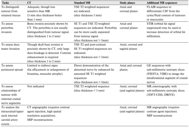
Table 4.2. Malignant sinonasal tumours. Pre-treatment imaging.
Tasks CT Standard MR Study planes Additional MR sequences
To distinguish Adequate, though less TSE T2 weighted sequences Axial and FLAIR sequence to tumour from sensitive than MR. are indicated. coronal planes differentiate CSF from the retained mucus (3 mm slice thickness better (slice thickness 3 mm) cystic/fluid content of tumours
than 1 mm) or mucoceles
To assess Bone erosion precisely shown by SE T1 and TSE T2-weighted Axial and STIR (orbital fat signal periorbita CT. The periorbita is not usually sequences are indicated. Periorbita coronal planes suppressed) may be used to invasion distinguished from tumour signal. can be more easily separated increase detection of orbital fat
(slice thickness 1 to 2 mm) from tumour signal. infiltration.
(slice thickness not > 3mm)
To assess dura Though skull base erosion is TSE T2 and post-contrast Axial, coronal and mater invasion precisely shown by CT, only large SE T1-weighted sequences are sagittal planes
dura breakage is detected. Contrast indicated.
enhancement is required. (slice thickness not > 3mm) (slice thickness 1 to 2 mm)
To assess Limited to indirect signs Direct demonstration of the Axial and coronal GE sequences with perineural spread (fat effacement or enlargement of abnormal nerve by enhanced fat planes sub-millimetric isotropic slices
foramina, muscular atrophy) saturated SE T1 weighted (FIESTA; VIBE) to image the
sequences intraforaminal segment of cranial
(slice thickness not > 3mm) nerves
To assess Not indicated TSE T2 weigheted sequence Axial, coronal MR cisternography with relationships of (slice thickness < 3 mm) (and sagittal planes) sub-millimetric isotropic slices
tumour with (3DFT-CISS; DRIVE)
cisternal cranial nerve segments
To analyse the CT angiography (requires contrast Axial, coronal MR angiography (requires intracranial/upper agent injection, high spatial (and sagittal planes) contrast agent injection).
neck internal resolution acquisition). MIP reconstructions
carotid artery MIP reconstructions course
SE: Spin Echo sequence; TSE: Turbo Spin Echo sequence; STIR: Short Tau Inversion Recovery sequence; GE: Gradient Echo sequence; FIESTA: Fast Imaging Emploting Steady State sequence; VIBE: Volume Interpolated Breath-Hold Examination sequence; 3DFT-CISS: three-dimensional constructive interference in a steady state sequence; DRIVE: driven equilibrium radio frequency reset pulse sequence; MIP: Maximum Intensity Projection