DOTTORATO DI RICERCA IN
Scienze Chimiche
Ciclo XXV
Settore Concorsuale di afferenza:
03/B1
Settore Scientifico disciplinare:
CHIM/03
DEVELOPMENT OF NEWLY CONCEIVED BIOMIMETIC
NANO-STRUCTURED BIOMATERIALS AS SCAFFOLDS
FOR BONE AND OSTEOCHONDRAL REGENERATION
Presentata da: Dott.ssa D’Alessandro Teresa
Coordinatore Dottorato
Relatore
Prof.ssa Adriana Bigi
Prof. Norberto Roveri
Correlatore
Dott.ssa Anna Tampieri
Contents
Aim of research
PART I: GENERAL TOPICS
1 Introduction1.1 Biomaterials……….. 1.2 Biomaterials for bone substitution………
1.2.1 Biomimetic materials……….. 1.3 Bone tissue………
1.3.1 Biology of bone tissue ……… 1.3.2 Morphology and structure of bone tissue ……… 1.3.3 Articular bone region………... 1.3.4 Vertebral bone region.……….. 1.3.5 Degeneration of bone tissue………. 1.3.6 Biomineralization process……… 1.3.7 Biological bone mineral phase………
References
2 Biomaterials for regenerative medicine: treatment of musculoskeletal diseases
2.1 Rigeneration of bone and osteo-cartilaginous tissue: problems and
solutions……… 2.2 The most important musculoskeletal diseases: Osteoporosis (OP) and
Osteoarthritis (OA)………... 2.3 Current clinical treatments for OA and OP………...
2.3.1 Main problems in the treatment of OA……… 2.3.2 Main problems in the tratment of OP……….. 2.3.3 Newly conceived biomaterials as scaffolds for the treatment of
OA and OP……….. References
Ι
1 1 1 3 4 4 5 7 9 11 13 15 16 19 20 23 24 25 26 27 313 Magnetism in biomedical applications: a new concept for tissue regeneration
3.1 Biomedical applications.………... 3.1.1 Magnetic bioseparation………. 3.1.2 Drug delivery……… 3.1.3 MRI contrast agents………. 3.1.4 Hyperthermia for the treatment of cancer……… 3.1.5 Magnetic therapy for the treatment of bone disease……… 3.2 Magnetic materials………
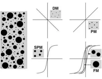
3.2.1 The influence of temperature……… 3.2.2 Factors affecting magnetic properties………... 3.2.3 Intelligent magnetic nanobiomaterials for Tissue Engineering …...
References
4 Biomimetic materials
4.1 Calcium phosphate in biological systems………. 4.2 Tricalcium phosphate (TCP)………. 4.3 Bioactivity of calcium phosphate……….. 4.4 Apatites………. 4.4.1 Hydroxyapatite: structure and chemistry……….. 4.4.2 Biological Apatites………... 4.4.3 Effect of Carbonate……….. 4.4.4 Effect of Magnesium……… 4.4.5 Effect of Strontium………... 4.5 Non-stoichiometric and substituted HA……… 4.6 Natural polymer……… 4.6.1 Collagen……… 4.6.2 Alginate……… 4.6.3 Gelatin……….. References 35 35 35 36 37 38 39 41 43 44 45 47 51 51 52 53 54 54 56 57 58 58 60 63 64 66 68 70
PART II: EXPERIMENTAL
5 Analytical Tecnique5.1 X-Ray Diffraction (XRD)………. 5.1.1 Quantitative phase analysis: Reference Intensity Ratio (RIR)
method………. 5.1.2 Rietveld Method………... 5.2 Fourier-Transform Infrared Spectroscopy (FTIR)……… 5.3 Inductively Coupled Plasma Atomic Emission Spectroscopy (ICP-AES)... 5.4 Scanning Electron Microscopy (SEM)………. 5.5 Thermogravimetric and Thermoanalytical analyses (TG-DTA)………….. 5.6 Powder analysis……… 5.6.1 Specific Surface Area………... 5.6.2 Granulometric Analysis……… 5.7 Mechanical properties……….. 5.8 Magnetic measurements………
5.6.1 Magnetic Susceptibility………
6 Magnetic Fe2+/Fe3+ doped Hydroxyapatite (FeHA)
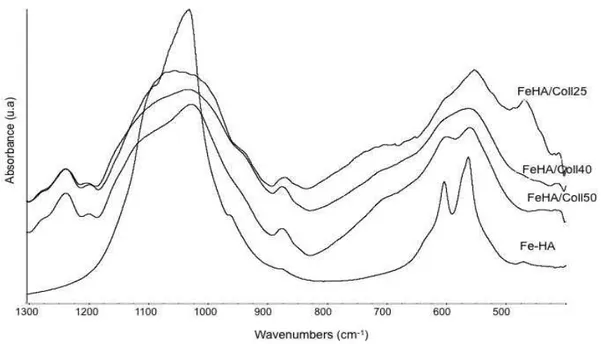
6.1 Synthesis of FeHA powder………... 6.2 Chemical, structural and magnetic characterization of FeHA powder……. 6.2.1 Phase analysis by XRD………. 6.2.2 Chemical analysis by ICP………. 6.2.3 Magnetic and microstructural investigation………. 6.2.4 Hyperthermia effect of FeHA powder………. 6.3 Development of FeHA granulate………. 6.4 Biocompatibility tests……….. 6.4.1 In vitro evaluation of biocompatibility and cell morphology……. 6.4.2 Effects of the in vitro applications of a static magnetic field…….. 6.4.3 In vivo pilot experimental and histological analysis……… 6.5 Conclusion………
References
7 Development of magnetic biohybrid FeHA/Coll scaffolds
7.1 Synthesis process of HA/Coll composite………. 7.2 Synthesis process of FeHA/Coll composites………
75 75 80 81 81 83 84 88 89 89 91 92 94 94 97 98 100 102 105 107 111 112 113 113 115 117 119 121 123 123 124
7.3 Chemico-physical and magnetic characterization of FeHA/Coll
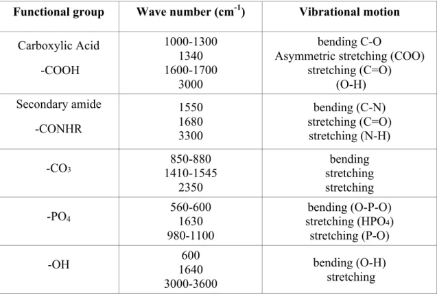
composites……… 7.3.1 X-ray diffraction analysis (XRD)………. 7.3.2 Thermogravimetric analysis (TG-DTG)……….. 7.3.3 Infrared spectroscopy analysis (FTIR)………. 7.3.4 Morphological analysis by scanning electron microscopy (SEM)
and by transmission electron microscope (TEM)……… 7.3.5 Chemical analysis by ICP………. 7.3.6 Magnetic properties of FeHA/Coll composites……… 7.4 In vitro evaluation of FeHA/Coll scaffolds……….. 7.5 Development of a three-layer magnetic scaffold……….. 7.6 Conclusion………....
References
8 Bioactive calcium phosphate bone cement: basic concepts
8.1 Features of injectable bone cements………. 8.2 Hardening mechanism………... 8.3 Cement microsctucture and porosity………. 8.4 Bioactivity and resorption of calcium phosphate cement……….
References
9 Development of biomimetic calcium phosphate bone cement
9.1 Synthesis of metastable CaP phase……… 9.1.1 Synthesis of α-TCP………. 9.1.2 Synthesis of Srα-TCP……….. 9.2 Development of biomimetic hydroxyapatite based bone cement………….
9.2.1 Hydrolysis of α-TCP powder: synthesis of HA cement…………... 9.2.2 Hydrolysis of Srα-TCP powder: synthesis of SrHA cement……… 9.2.3 Intoduction of carbonate ions during Srα-TCP hydrolysis:
synthesis of SrCHA………. 9.3 Introduction of polymeric phase into calcium phosphate cement………… 9.3.1 Calcium phosphate/gelatine bone cement……… 9.3.2 Calcium phosphate/Sodium alginate bone cement……….. 9.4 Porosity measurements………. 9.5 Mechanical properties………... 125 125 126 128 131 133 134 138 140 142 143 145 145 147 147 148 150 153 154 154 154 159 159 163 169 171 173 177 181 182
9.5.1 Compression strength………... 9.5.2 Young’s Modulus………. 9.6 Route to enhance the mechanical properties of CaP bone cement………... 9.6.1 Incorporation of TiO2 as bio-active reinforcing phase………. 9.6.2 Sodium alginate cross-linking……….. 9.7 Conclusion………
References
Conclusion and Future Perspective
183 184 185 186 187 192 194 197
Aim of research
The present research activity is focused on the development of a new class of biomaterials and device for application in regenerative medicine, particularly in the repair/regeneration of hard tissue such as bone and osteochondral regions.
The new biomaterials were designed to provide new solutions towards the local treatment of hard tissues affected by degenerative diseases such as Osteoarthritis (OA) and Osteoporosis (OP) or serious traumas. These pathologies affect an ever increasing number of people worldwide with huge socio-economic impact. Although the incidence of such diseases has historically been linked to ageing, more recently the number of relatively young people affected by these diseases has increased, due to new life-styles (e.g. intense sport activity, obesity, alcohol consumption). In the most serious cases, lesions involving osteocartilaginous regions or vertebral bodies can lead to severe complications affecting the neuro-muscular system and can significantly impair the patient’s quality of life and longevity. The current therapies mainly aim at blocking the progress of the diseases and/or to provide a physical sustains, in case of vertebral weakening. Such approach, however, has several limitations and drawbacks, and does not provide tissue regeneration and the recovery of the original functions.
During the last decade the scientific research has increasingly pushed towards the development of tissue-mimicking constructs addressed to tissue regeneration. In particular, it is widely accepted that the regeneration of hard connective tissues requires the implantation of porous bio-resorbable scaffolds exhibiting chemical-physical and morphological mimicry with the target tissues so to provide environmental cues for cell adhesion, proliferation and specific differentiation.
With regard to bone, biomimetic apatites, i.e. nanostructured calcium phosphates mimicking the composition and the nanostructure of the mineral bone, are considered the golden biomaterials for regenerative bone scaffolds. In this respect, the Research Group on Biomaterials at ISTEC-CNR has 20 years of experience in the synthesis and characterization of nano-apatites with different crystal order and ionic substitutions, suitable to activate specific biologic functions relevant for bone tissue regeneration. With regard to complex osteochondral regions, ISTEC-CNR developed in 2001 a biologically-inspired mineralization processes mimicking the cascade of events yielding
new bone formation, that enabled the synthesis of biomimetic hybrid hydroxyapatite/collagen composites mimicking multifunctional osteocartilaginous regions. By this process, biomimetic, ion-substituted nanoapatites can be heterogeneously nucleated on collagen fibrils, the assembling and organization of which is mediated by several physical, chemical, morphological and structural control mechanisms, activated by pH and temperature control. These scaffolds demonstrated very high regenerative potential and were subsequently patented, scaled up and brought to the market as bone and osteochondral scaffolds.
With respect to this previous knowledge, the present work intended to develop new improved scaffolds for bone and osteochondral tissue regeneration, aimed to achieve faster regeneration and improved texturing of the newly formed tissues. Indeed scientific research is increasingly being devoted to develop new approaches for stimulating cell activity, including the design of specific 3D morphologies and the development of smart, stimuli-responsive, drug delivery systems. A particularly intriguing approach is based on the use of magnetism: recent findings show that the local application of weak magnetic fields may stimulate cells to produce new bone tissue in a more ordered manner.
With this in mind the present research thesis was specifically focused on the synthesis and physico-chemical-morphological characterization of:
i) a new iron-substituted apatite phase (FeHA) with intrinsic superparamagnetic properties;
ii) new biomimetic hybrid scaffolds with superparamagnetic properties for bone and osteochondral regeneration, obtained by biologically-inspired mineralization processes; iii) new self-setting injectable pastes based on calcium phosphates functioning as apatitic bone cements for regenerative vertebroplasty.
More specifically, in the present work a new apatite phase with superparamagnetic properties was developed, by performing ionic substitutions in the apatite lattice, particularly of bi- and tri-valent iron ions replacing calcium, achieved by fine control of the synthesis conditions. The new apatite phase resulted of great potential, due to the possibility of using biocompatible, bioactive and bio-resorbable superparamagnetic phases for a number of applications, spanning from regenerative medicine to theranostics and imaging, thus overcoming the current use of magnetite nanoparticles, which raise several concerns for their long-term cyototoxicity.
The development of this new phase was exploited in the generation of biologically-inspired mineralization process enabling the heterogeneous nucleation of superparamagnetic nano-apatite on collagen fibrils. By reproducing the conditions of formation of new bone tissue, thus obtaining highly biomimetic bone, osteochondral scaffolds with intrinsic superparamagnetic properties were synthesized. This approach may open the way to the generation of new scaffolds with multiple functionality, i.e. the possibility to activate the scaffold by remote magnetic control and provide local signals to enhance the regenerative processes. Additionally, the remote magnetic activation can enable the recall and controlled delivery of osteogenic and angiogenic growth factors linked to magnetic nanoparticles so to achieve scaffolds functioning as a flexible “workstation” able to regulate and guide tissue regeneration on the basis of the personal needs of the patient.
In the view of specific treatments for patients affected by osteoporosis or traumas involving vertebrae weakening or fracture, the present work was also dedicated to the development of new self-setting injectable pastes based on strontium-substituted calcium phosphates, able to harden in vivo and transform into strontium-substituted hydroxyapatite. The addition of strontium may provide an anti-osteoporotic effect, aiding to restore the physiologic bone turnover. The ceramic-based paste was also added with bio-polymers, able to be progressively resorbed thus creating additional porosity in the cement body that favour cell colonization and osseointegration. This approach may enable innovative therapies against the weakening of vertebral bodies due to osteoporosis or serious traumas. In this respect, the current clinical approach is mainly the percutaneous injection of acrylic bone cements, which can harden in few minutes and allow the early dismission of the patient after surgery. However, due to several drawbacks related to the exothermy of the polymerization reaction, the bioinertness of acrylic phases and the excessive stiffness of the cement in relationship with the surrounding bone, this solution is not suitable for relatively young patients (<60-65 years) and/or severe patients, and reliable regenerative solutions for vertebroplasty are still missing. The set up and development of regenerative bone cements were made difficult so far by the high complexity of the processes required to establish pastes characterized, at the same time, by high bioactivity, osteogenic and osteoconductive character, the absence of any adverse reactions, in association with good injectability, low setting times (in the order of
The new bio-devices developed in the present work may enable new tissue regeneration-based treatments and minimally-invasive surgical approach that have the potential to overcome the limitations and drawbacks associated with the current therapies. In these clinical situations, the development of biomimetic, bioresorbable scaffolds with regenerative ability is regarded by clinicians as breakthrough solutions in the cure of these debilitating pathologies. In fact, autologous bone grafts are not always readily available in sufficient amounts, and its use poses concerns on the patient health and pain, bone allografts are inferior to autologous bone, are expensive, and have known risks of bacterial contamination, viral transmission and immunogenicity. Moreover the use of the new superparamagnetic iron-substituted apatite nanoparticles and hybrid scaffolds may pave the way to the establishment of newly conceived therapies for tissue regeneration, as well as novel applications in theranostics and imaging characterized by virtual absence of any drawbacks related to the cytotoxicity of nanoparticles.
PART I:
CHAPTER 1
INTRODUCTION
1.1 BiomaterialsIn the last few years the biomedical research area is going towards materials science aiming applications of materials to health care, the so-called biomaterials. They can be defined as implantable materials that must be in contact with living tissues with the final aim of achieving a correct biological interaction between the material and the host1. In the first Consensus Conference of the European Society for Biomaterials (ESB) in 1976, a biomaterial was defined as “a nonviable material used in a medical device, intended to interact with biological systems”; however the ESB’s current definition is a “material intended to interface with biological systems to evaluate, treat, augment or replace any tissue, organ or function of the body”. This subtle change in definition is indicative of how the field of biomaterials has evolved. Biomaterials have moved from merely interacting with the body to influencing biological processes toward the goal of tissue regeneration. However, a more recent definition has been published: A biomaterial is a substance that has been engineered to take a form which, alone or as part of a complex system, is used to direct, by control of interactions with components of living systems, the course of any therapeutic or diagnostic procedure, in human or veterinary medicine2.
1.2 Biomaterials for bone substitution
Biomaterials for bone substitution are employed mainly as fillers, or as structural substitutes or as coating for screws or similar device. The ultimate aim of biomaterials for bone substitution is to restore the structural integrity of the damaged bone.
The substitution of bone intends mainly to restore the structural functionality of the missing part, but it is not able to completely restore all its properties; the very complex composition and morphology of the bone tissue are responsible of its unique ability to
continuously adapt to the ever changing mechanical solicitations, so to be simultaneously light, tough and elastic and able to locally regenerate after traumas of limited extension. Thus, with the increasing of the expectance of life and well-being, the development of bone substitutes able to restore the whole functionality of the original tissue was of increasing importance in the last decades; beyond the commercial aspects, the problem has a highly social relevance as the patients can regain a more active and satisfactory life and sooner than in the past.
An ideal bone substitute should be biomimetic, i.e. able to perfectly mimic in vivo the behaviour of the natural bone. For this reason, not only biocompatibility is required, but features of bioactivity, osteoinductivity, osteoconductivity and bio-resorbability are also strongly needed.
Biocompatibility is the ability of a material to perform with an appropriate host response
in a specific application, without having any toxic or injurious effects on biological systems. The scope of this definition is very wide and many subgroups of applications can be found to make more narrow definitions of biocompatibility. Anyway, for a long-term implantable medical device, like a bone substitute, the biocompatibility refers to the ability of the device to perform its intended function, with the desired degree of incorporation in the host, without eliciting any undesirable local or systemic effects in that host.
Bioactivity is the ability of the implant to bond to bone tissue. The process of bone
bonding is the result of multiple, parallel and sequential reactions at the material-tissue interface. These interactions are related to either physicochemical phenomena that occur in the presence or absence of cells, or are related to reactions affected by cellular activity.
Osteoinductivity is the ability of a material to induce cell differentiation oriented to the
synthesis of a bone matrix, able to mineralize in bone tissue.
Osteoconductivity is a passive property of the implant consisting in the ability of making
easy the formation and diffusion of the new bone both through its chemical (presence of ions and substances able to enhance the cell activity for osteogenesis, the process of new bone development) and morphological (hierarchically organized porous structure able to host the growing bone tissue and the vascular system) features.
Bioresorbability is the ability of the implant to be dissolved by the in vivo processes of
1.2.1 Biomimetic materials
Biomimetics is a new, very important, field of science that studies how Nature designs, processes and assembles/disassembles molecular building blocks to fabricate high performance mineral-polymer composites (e.g. mollusc shells, bone, tooth) and/or soft materials (e.g. skin, cartilage, tendons) and then applies these designs and processes to engineer new molecules and materials.
In nature, living organisms synthesize mineralized tissues and this process of biomineralization is under strict biological control. It involves the interactions of several biological macromolecules among themselves and with the mineral components. Generally, natures design principles are based on a “Bottom-Up” strategy. Such processes lead to the formation of hierarchically structured organic-inorganic composites with mechanical properties optimized for a given function. A common theme in mineralized tissues is the intimate interaction between the organic and inorganic phases and this leads to the unique properties seen in biological materials. Therefore, understanding natures design principles and ultimately mimicking the process may provide new approaches to synthesize biomaterials with unique properties for various applications.
In fact, in the last decades the activity of material scientists was more and more directed to the development of biomimetic scaffolds, able to drive and address cell activity towards proper differentiation and the repair of diseased human tissues3. In case of bone, this requires the synthesis of three-dimensional constructs able to exchange chemical signals promoting osteogenesis and to progressively be resorbed during the formation and remodelling of new bone. Besides, particularly for the regeneration of extensive portions of bone, a morphological and mechanical biomimesis is also required, to allow cell colonization and formation of a proper vascularization tree. The healing of load-bearing bones also requires scaffolds with a hierarchically organized morphology, to provide improved biomechanical behaviour and allow a proper mechano-transduction of the mechanical stimuli down to the cell level4.
1.3 Bone tissue
Bone is a bioceramic composite that has long held the attention of the materials engineer who seeks to duplicate its enviable mechanical properties, in which both high strength and fracture toughness can be achieved due to the unique architecture of this organic-inorganic composite.
1.3.1 Biology of bone tissue
Bone is a specialised connective tissue which forms the basis of the skeleton and, as such, its functions are numerous and complex. One of these functions, although not necessarily the most important, is to protect the internal organs from damage which could result from the physical trauma of everyday life. In combination with the associated musculature, the bones of the skeleton also provide a means of physical support, locomotion and related movement. One further role is that of a reservoir for a multitude of inorganic ions.
Three distinctly different cell types can be found within bone (Fig.1): the matrix-producing osteoblast, the tissue-resorbing osteoclast, and the osteocyte, which accounts for 90% of all cells in the adult skeleton5.
Osteoblasts (from the Greek words for "bone” and "germ" or embryonic) are the cells
within bone that lay down the extracellular matrix and regulate its mineralization. Morphologically, these cells are cuboidal in shape and located at the bone surface together with their precursors, where they form a tight layer of cells. Osteoblasts are located on the surface of osteoid seams and also manufacture hormones, such as prostaglandins, to act on the bone itself. They robustly produce alkaline phosphatase, an enzyme that has a role in the mineralisation of bone, as well as many matrix proteins. Osteoblasts are the immature bone cells. When osteoblasts are trapped in the bone matrix, which they themselves produced, they become star-shaped cells named osteocytes, the most abundant cell found in bone.
Osteocytes are osteoblasts that remain in bone matrix until they degrade according to
natural cycle of life. They are set both in cortical and trabecular bone localizing in hollows or bone gaps, among one lamella and the other. They have a flat and long shape with cytoplasmatic prolongations. Their function is to guarantee the survival of the surrounding tissue taking nourishing substances from the medullar hollow through cytoplasmatic channels (canaliculi)6. Osteocytes are not active producers or destroyers of
bone matrix, but are able to feel mechanical solicitations and, if necessary, control the other bone-cells activity such as deposition and reabsorption processes of mineral phase.
Osteoclasts (from the Greek words for "bone" and "broken") are multinucleated cells,
like macrophages, derived from the hematopoietic lineage. They degrade the mineralized bone tissue giving rise to isolated micro-ambients where they release acid products dissolving organic and inorganic bone matrixes7. Their activity can be controlled, both from chemical signals (hormones, vitamins, low levels of calcium) and mechanical stresses. Those signals stimulate the bone remodelling: the demolition of some lamellae and rebuilding of others differently oriented to better compensate the applied load8. In brief, osteoclasts digest while osteoblasts reconstruct again bone tissue9.
Fig. 1. Bone cells
Osteoid is comprised of type I collagen (~94%) and noncollagenous proteins. The hardness and rigidity of bone is due to the presence of mineral salt in the osteoid matrix, which is a crystalline complex of calcium and phosphate (hydroxyapatite). Calcified bone contains about 25% organic matrix (2-5% of which are cells), 5% water and 70% inorganic mineral (hydroxyapatite).
1.3.2 Morphology and structure of bone tissue
There are three different types of bone tissue, compact (cortical bone), trabecular (cancellous bone), and subchondral (Fig. 2). The compact one is the harder, outer tissue
of bones, the trabecular one is the sponge-like tissue inside bones, and the subchondral tissue is the smooth one at the apical and basal parts of bones, which is covered with cartilage (made up in turn of chondrocytes, collagen fibers and proteoglycans). Although macroscopically and microscopically different, cortical bone and cancellous bone are identical in their chemical composition.
Cortical bone, synonymous with compact bone, is one of the two types of osseous tissue
that form bone. Cortical bone facilitates bone's main functions: to support the whole body, protect organs, provide levers for movement, and store and release chemical elements, mainly calcium. As its name implies, cortical bone forms the cortex or outer shell, of most bones. Cortical bone contributes about 80% of the weight of a human skeleton. The primary anatomical and functional unit of cortical bone is the osteon. Each osteon consists of concentric layers, or lamellae, of compact bone tissue that surround a central canal, the Haversian canal. The Haversian canal contains the bone's nerve and blood supplies. The boundary of an osteon is the cement line.
Fig. 2. Schematic drawing of the bone structure
Cancellous bone, synonymous with trabecular bone or spongy bone, is the other type
of osseous tissue that forms bones. Compared to compact bone, it has a higher surface area but is less dense, softer, weaker, and less stiff. It typically occurs at the ends of long
bones, proximal to joints and within the interior of vertebrae. Cancellous bone is highly vascular and frequently contains red bone marrow where hematopoiesis, the production of blood cells, occurs. The primary anatomical and functional unit of cancellous bone is the trabecula, that is a small tissue element usually composed of dense collagenouse tissue.
Subchondral bone region is the zone of epiphyseal bone just beneath the articular
cartilage, and includes the subchondral plate and the underlying trabecular and subarticular bone. The subchondral plate comprises the deepest area of the articular cartilage, which is the calcified cartilage, and a thin cortical bone layer. The calcified cartilage is separated from the overlying hyaline cartilage by a line of demarcation called the tidemark.
Nevertheless, subchondral bone is recognised as a key factor in normal joint protection. In fact, subchondral bone has been shown to exert important shock-absorbing and supportive functions in normal joints. Subchondral bone can attenuate about 30% of the joint load, providing a mechanical base for joint cartilage10. Moreover, subchondral bone supplies nutrients to cartilage and facilitates the removal of metabolic waste products11,12.
1.3.3 Articular bone region
Cartilage is a flexible connective tissue found in many areas in the bodies of humans and
other animals, including the joints between bones, the rib cage, the ear, the nose, the bronchial tubes and the intervertebral discs.
The cartilaginous tissue, together with the bone tissue, belongs to the support skeletal tissues or connective tissues that have high mechanical properties. The cartilaginous tissue has high specific mechanical-elastic properties that allow to constantly reducing the friction induced by the loads on the articular surface during the normal movement activities of the human body. Moreover, the cartilaginous tissue is able to dissipate the peaks of mechanical stress on the sub-chondral bone. The connective tissue surrounding the cell cartilaginous component, namely the chondrocytes, (2% of the total volume of the cartilage) is dipped in an extracellular matrix/lattice consisting of collagen, proteogly- cans and glycoproteins. For example, 2-40% of the connective tissue includes: 50-60% of collagen, 25-35% of proteoglycans and 10-15% of glycoproteins. The collagen molecules are uniformly distributed throughout the tissue and are responsible of its shape and mechanical strength. The proteoglycans and the glycoproteins bind to the collagen by
trapping the water within the matrix.
Cartilage is classified in three types that differ in the biochemical composition and structure of their extracellular matrix (ECM), the resulting mechanical properties and therefore their occurrence in the human body: elastic cartilage, hyaline cartilage and fibrocartilage.
Elastic cartilage is characterized by chondrons with only a few cells, a small concentration of proteoglycans (PGs), but much elastin. Elastin is interwoven into the collagen mesh and the more this protein is present, the more Xexibility is provided to the tissue. Elastic cartilage is surrounded by perichondrium and appositional growth is guaranteed through cell differentiation of the connective tissue. This tissue type is less vulnerable to degenerative changes. Elastic cartilage is suitable to resist bending and so it can be found in the epiglottic cartilage, the smaller laryngeal cartilage, the external ear and auditory tube, or the small bronchi13.
A second type of cartilage is the fibrocartilage, providing other physical characteristics. Like the elastic type it contains a small concentration of PGs, but by contrast far less elastin. The meniscus is a fibrocartilaginous tissue composed primarily of an interlacing network of collagen fibers with a longitudinal, circumferential orientation which gives it unique functional properties. The ECM of fibrous cartilage consists of approximately 60-70% collagen, 8-13% non-collagenous proteins, and 1% PGs in dry weight. The water content in native meniscal tissue is around 70-75%14.
The third and most widespread cartilage in the human body is the hyaline type. As the name already implies, it appears as a white and slightly bluish tissue with a macroscopically smooth surface. Its resistance to compression or tensile forces is due to the net-like organized structure of the collagen type II fibers combined with a high concentration of PGs. Hyaline cartilage can be found in the nose, the trachea, bronchi, and most joints in synarthroidal as well as in diarthroidal joint15. In diarthroidal joints hyaline cartilage covers the contact zones of two interlocked bones and is called articular
cartilage (Fig. 3).
Articular cartilage is a unique type of connective tissue. Although appearing homogeneous, articular cartilage has a highly ordered structure. It is organized on two levels: the structure and composition of articular cartilage varies according to its distance from the surface and also in relation to the distance from the cells. Typically, articular
cartilage is divided into four zones: uperficial, middle (or transitional), deep (or radial), and the zone of calcified cartilage. Chondrocytes from the different zones differ in size, shape, and metabolic activity. The superficial zone is the thinnest, and forms the gliding surface of the joint. It is composed of thin collagen fibrils aligned parallel to the joint surface, with elongated, inactive chondrocytes directly subjacent. The middle zone is thicker than the superficial zone, with more spherical cells and with larger collagen fibrils that are not oriented in a parallel fashion. In the deep zone, the cells are spheroidal, arranged in a columnar orientation. The collagen fibers here are oriented in a parallel fashion, similar to the cells, vertical to the joint surface. In the zone of calcified cartilage, collagen fibrils insert into the calcified cartilage, providing both a mechanical transition from the cartilage to bone, as well as fixation between the two tissues16.
Fig. 3. Articular cartilage
1.3.4 Vertebral bone region
The intervertebral discs lie between the vertebral bodies, linking them together (Fig. 4). They are the main joints of the spinal column and occupy one-third of its height. Their major role is mechanical, as they constantly transmit loads arising from body weight and muscle activity through the spinal column. They provide flexibility to this, allowing bending, flexion and torsion.
They are approximately 7-10 mm thick and 4 cm in diameter (anterior - posterior plane) in the lumbar region of the spine17. The intervertebral discs are complex structures that consist of a thick outer ring of fibrous cartilage termed the annulus fibrosus, which surrounds a more gelatinous core known as the nucleus pulposus; the nucleus pulposus is sandwiched inferiorly and superiorly by cartilage end-plates. The central nucleus pulposus
contains collagen fibres, which are organised randomly, and elastin fibres (sometimes up to 150µm in length), which are arranged radially18; these fibres are embedded in a highly hydrated aggrecan-containing gel.
The annulus is made up of a series of 15-25 concentric rings, or lamellae, with the collagen fibres lying parallel within each lamella. The fibres are orientated at approximately 60° to the vertical axis, alternating to the left and right of it in adjacent lamellae. Elastin fibres lie between the lamellae, possibly helping the disc to return to its original arrangement following bending, whether it be flexion or extension. They may also bind the lamellae together as elastin fibres pass radially from one lamella to the next. The cells of the annulus, particularly in the outer region, tend to be fibroblast-like, elongated, thin and aligned parallel to the collagen fibres. Toward the inner annulus the cells can be more oval. Cells of the disc, both in the annulus and nucleus, can have several long, thin cytoplasmic projections, which may be more than 30µm long. Such features are not seen in cells of articular cartilage19. Their function in disc is unknown but it has been suggested that they may act as sensors and communicators of mechanical strain within the tissue.
Fig. 4. A schematic view of a spinal segment and the intervertebral disc. The figure shows the organization
of the disc with the nucleus pulposus (NP) surrounded by the lamellae of the annulus fibrosus (AF) and separated from the vertebral bodies (VB) by the cartilaginous end-plate (CEP). The figure also shows the relationship between the intervertebral disc and the spinal cord (SC), the nerve root (NR), and the apophyseal joints (AJ).
The third morphologically distinct region is the cartilage end-plate, a thin horizontal layer, usually less than 1mm thick, of hyaline cartilage. This interfaces the disc and the vertebral body. The collagen fibres within it run horizontal and parallel to the vertebral bodies, with the fibres continuing into the disc. The healthy adult disc has few (if any) blood vessels, but it has some nerves, mainly restricted to the outer lamellae, some of which terminate in proprioceptors. The cartilaginous end-plate, like other hyaline cartilages, is normally totally avascular and aneural in the healthy adult. Blood vessels present in the longitudinal ligaments adjacent to the disc and in young cartilage end-plates (less than about 12 months old) are branches of the spinal artery. Nerves in the disc have been demonstrated, often accompanying these vessels, but they can also occur independently, being branches of the sinuvertebral nerve or derived from the ventral rami or grey rami communicantes. Some of the nerves in discs also have glial support cells, or Schwann cells, alongside them20.
1.3.5 Degeneration of bone tissue
Deseases of articular bone region
articular cartilage which, in a non negligible percentage, can evolve in chronic arthrosis disease. The cartilaginous tissue subjected to a damage, in particular to a deep damage, has a poor self-reparative ability considering the poorly vascularized anatomic region and the limited replication ability of the chondrocyte cells. The result of the self-reparative process of the cartilage is often a formation of fibrous tissue which certainly can not be compared, in terms of mechanical performances and physiological features, to a normal hyaline cartilage. Accordingly, the alteration of the viscoelastic and mechanical balance can lead to considerable pathological consequences with a following compromission of the articular function and with a pain occurrence, although the cartilaginous degeneration process in most of cases is an asymptomatic phenomenon.
The situation evolves in a more complex way in case of lesions involving the sub-chondral bone structure, when the complete demolition of the cartilaginous layer directly exposes the bone to the mechanical stress of the joint.
In conclusion, between the pathological conditions which can induce nearly always irreversible degenerative processes there may be listed, by mere way of example: the bad alignments (varus and valgoid conditions), the avascular necrosis, the osteoarthritis and the rheumatoid arthritis. The known surgical techniques currently utilized for the repair of the cartilaginous tissue are of course applied as a function of the dimensions and the depth of the defect.
Deseases of vertebral bone region
One of the major problems facing the elderly spine is the occurrence of vertebral fractures, which are associated with height loss, back pain, disability and even increased mortality rates. As these vertebral fractures are strongly correlated to low bone density, they are commonly attributed to osteoporosis.
Disc degeneration begins with the nucleus becoming dehydrated and more fibrous, followed by a loss of distinction between the nucleus and annulus, disorganized fibers in the annulus and, finally, a decreased disc height. With disc pressure measurements it was found that healthy discs show an area of constant pressure under the nucleus and the inner annulus. For degenerated discs the area of constant pressure was much smaller and the pressure within it was lower. Such a change in pressure in the disc and on the endplates would lead to bone adaptation in the adjacent vertebrae. Indeed, disc degeneration has
been found to be correlated to a changed bone density distribution within the vertebra21.
1.3.6 Bio-mineralization processes: the basis of bone formation
The formation of bone tissue takes place through different events22 that comprise the synthesis and modification of specific polypeptide chains inside osteoblast cells, which are then extruded into the extracellular space. Here, the collagen microfibrils, with a mean diameter of 1.5 nm, start to assemble into fibres. During the self-assembly of collagen fibres, a quarter-staggered arrangement of parallel molecules of tropocollagen (a triple helical-shaped molecule, precursor of collagen) is established, leaving a regular array of 40 nm gaps within each periodic unit. These areas are known as hole zones (40 nm in length and 5 nm in width) and as reported23 are the preferential sites for the nucleation of the mineral phase (Fig. 5).
The nucleation of mineral nanocrystals takes place in specific loci corresponding to non-collagenous acidic macromolecules; many of which are highly charged from carboxylate groups that can bind Ca2+ ions. Following the chemical binding of Ca2+, the supersaturation of physiological fluids in phosphate ions and other minor species (e.g. Na+ and K+) provokes the precipitation and nucleation of the mineral phase as nano-sized, plate-shaped particles, which are initially confined to the hole zones, and progressively extend along the collagen fibrils. The organic template transfers information to the mineral phase at the molecular level: (i) the chemical interaction of hydroxyapatite (HA, Ca5(PO4)3OH) with collagen prevents the crystallization of the mineral phase, which is almost amorphous but is characterized by an apatite-like lattice; (ii) the size and shape of the nuclei are constrained by the activation of structural and morphological control mechanisms during the bio-mineralization process; (iii) the growth of nuclei is limited to very thin lamellae, which are spatially confined and have a specific crystal orientation; and (iv) lamellae organize through different hierarchical levels up to the macroscopic bone.
Fig. 5. Schematic illustration of nucleation of bone mineral crystals during the self-assembling of collagen
fibres.
The crystal state of the substrate induces the nucleation and growth of the apatite nanocrystals along a specific crystallographic direction (Fig. 6). Crystal growth is limited to very few unit cells of the apatite lattice, with values ranging from 30-50 nm long, 15-30
nm wide, and 2-10 nm thick24. The apatite platelets develop along the long axis of collagen, so that the apatite crystals grow preferentially along the c axis of the hexagonal apatite lattice.
Fig. 6. Schematic illustration: (a) an arrangement of mineralized collagen fibrils aligned both with respect
to crystal layers and fibril axes; (b) arrangement of mineralized collagen fibrils with only the fibril axes aligned.
1.3.7 Biological bone mineral phase
Bone mineral has the structure of a calcium-deficient apatite, with a Ca:P ratio <1.67, which is the theoretical value for pure HA25. Bone is a living tissue that is continuously undergoing remodelling and repair, therefore, the small size and/or non-stoichiometry of the crystals presumably endows the mineral phase with the solubility needed for resorption of the bone by osteoclasts (bone-resorbing cells). In fact, synthetic stoichiometric HA, although bioactive (stimulatory for bone formation), is rather slow to resorb because of the low solubility of HA under physiological conditions26. Besides the very small crystallite size, the low crystallinity of bone mineral (in relation to synthetic HA) is also caused by incorporation of ions into its lattice, such as CO32–, Na+, Mg2+, SiO44– or HPO42–, which are present in the physiological environment and extremely important for the biochemistry of bone remodelling (see Chapter 4)27.
References
1. Vallet-Regí M. Chem. Eng. J. 2008, 137, 1 2. Williams DF. Biomaterials 2009, 30, 5897.
3. Yannas, I.V., 2001. Tissue and Organ Regeneration in Adults. Springer, New York.
4. Sprio S, Ruffini A, Valentini F, D’Alessandro T, Sandri M, Panseri S, Tampieri A. J. Biotech. 2010, 156, 347.
5. Sommerfeldt DW, Rubin CT. Eur. Spine. J. 2001, 10, S86. 6. Burger EH, Klein-Nulend J. FASEB J. 1999, 13, S101.
7. Vaananen HK, Zhao H, Mulari M, Halleen JM. J.Cell.Sci. 2000, 113, 377. 8. Frost M. Anat. Rec. 1990, 226, 403.
9. RJ Moore “BONE” © Woods and Ellis 2000.
10. Imhof H, Sulzbacher I, Grampp S, Czerny C, Youssefzadeh S, Kainberger F. Invest. Radiol. 2000, 35, 581.
11. Santos Castaneda, Jorge A. Roman-Blas, Raquel Largo, Gabriel Herrero-Beaumont. Biochemical Pharmacology 2012, 83, 315.
12. The Role of Subchondral Bone in Joint Disease, Christopher E. Kawcak; C. Wayne McIlwraith, Richard D. Park and S. P. James, AAEP PROCEEDINGS, Vol. 47, 2001
13. Montes GS. Cell Biol. Int. 2006, 20, 15.
14. Flugge LA, Miller-Deist LA, Petillo PA. Chem. Biol. 1999, 6, R157.
15. Buckwalter JA (1997) Cartilage. In: Dulbecco R (ed) Encyclopedia of human biology, vol 2. pp 431–445.
16. Newman AP. Am. J. Sport. Med. 1998, 26, 309. 17. Twomey LT, Taylor JR. Clin. Orthop. 1987, 224, 97.
18. Yu J, Winlove CP, Roberts S, Urban JP. J. Anat. 2002, 201, 465.
19. Errington RJ, Puustjarvi K, White IR, Roberts S, Urban JP. J. Anat. 1998, 192, 369.
20. Johnson WE, Evans H, Menage J, Eisenstein SM, El Haj A, Roberts S. Spine
2001, 26, 2550.
21. Homminga J, Aquarius R, Bulsink VE, Jansen CTJ, Verdonschot N. Medical Engineering & Physics. 2012, 34, 453.
22. Olszta MJ et al. Mater. Sci. Eng. 2007, 58, 77. 23. Landis WJ et al. J. Struct. Biol. 1993, 110, 39.
24. Lowenstam HA and Weiner S. (1989) On Bio-mineralization, Oxford University Press.
25. LeGeros RZ (1991) Calcium Phosphatein Oral Biology and Medicine, Karger. 26. Dorozhkin SV and Epple M. Angew. Chem. Int. 2002, 41, 3130.
CHAPTER 2
BIOMATERIALS FOR REGENERATIVE
MEDICINE: TREATMENT OF
MUSCOLOSKELETAL DISEASES
Regenerative medicine is a critical frontier in biomedical and clinical research. It is the process of creating living, functional tissues to repair or replace tissue or organ function lost due to age, disease, damage, or congenital defects. This field holds the promise of regenerating damaged tissues and organs in the body by stimulating previously irreparable organs to heal themselves1.
Regenerative medicine applies the principles of engineering and life sciences to enhance the comprehension of the fundamental biological mechanisms underlying the structure function relationships in physiologic and pathologic tissues and to accomplish alternative strategies for developing in vitro biological substitutes which are able to restore, maintain, or improve tissue, and organ function2.
Musculoskeletal tissue damage is an increasing problem with a growing aging population. Regenerative medicine aims to repair and replace lost or damaged tissues by initiating the natural regeneration process. Most approaches currently pursued or contemplated within the framework of regenerative medicine, including cell-based therapies and living tissue engineering, rely greatly on the ability to synthesize or otherwise generate novel biomaterials, to fabricate or assemble biomaterials into appropriate two-dimensional (2D) and three-dimensional (3D) forms, and to tailor physical, chemical, structural and biological properties to achieve desired clinical efficacy.
In a regeneration strategy, biomaterials promote new tissue formation by providing adequate space (porosity) and appropriate surface to foster and direct cellular attachment, migration, proliferation, desired differentiation of specific cell phenotypes throughout the
scaffold where new tissue formation is needed3,4.
Consequently, the 3D architecture plays a pivotal role. It refers to the way in which a bulk material is distributed in space from the macro, micro to nano scales (corresponding to tissue, cellular and molecular scales in a specific tissue, respectively)5. Such hierarchical porous architectures not only define the mechanical properties of the scaffold, but also the initial void space that is available for regenerating cells to form new tissues (including new blood vessels) as well as the pathways for mass transport via diffusion and/or convection. While interconnected macroporosity of a biomaterial is important to provide sufficient space for cellular activity6-9interactions between cells and biomaterials occur at the interface, i.e. the entire internal pore walls of a 3D scaffold. The surface morphology or topography directly and significantly affects cell-scaffold interactions and ultimately tissue formation and function10.
2.1 Rigeneration of bone and osteo-cartilaginous tissue: problems and solutions
Regeneration of bone tissue
The bone loss can occur in various situations such as trauma, tumors, atrophy and in different areas of bone tissue such as long, flat or short bones presenting a peculiar organization. The regeneration of bone tissue is a complex process that is known to be regulated by multiple factors and which occurs spontaneously under normal conditions. Nevertheless, the regeneration of bone does not occur in a complete way and this often leads to the formation of a fibrous tissue. In fact, the bone regeneration requires the formation of new bone tissue and blood vessels that mediate the transport of osteogenic precursors, of secretory molecules that act as activators for osteoblasts, transporters of nutrients and oxygen.
There are various therapeutic strategies for promoting the regeneration of bone tissue; some are based on the use of autologous bone tissue other on the use of biomaterials alone or associated with osteo-progenitor cells and/or growth factors.
Autografts have primarily been the material of choice to replace lost bone, although the use of autografts comes with some disadvantages such as the limited quantity available and its use also requires additional surgical procedures, and therefore, longer surgical time and possible complications of the wound of the donor site such as bleeding, pain and
infection among others. Allografts and xenografts have the potential of transferring pathogens. To avoid the biological risk, these materials are subjected to exhaustive procedures which have dramatic effects primarily on their osteogenic and osteoinductive properties and these procedures can also reduce their structural integrity leading to graft fracture11.
An alternative is allogeneic bone grafting, obtained from human cadavers or living donors, which bypasses the problems associated with harvesting and quantity of graft material. Allogeneic bone is available in many preparations, including demineralised bone matrix (DBM), morcellised and cancellous chips, corticocancellous and cortical grafts, and osteochondral and whole-bone segments, depending on the recipient site requirements. Their biological properties vary, but overall, they possess reduced osteoinductive properties and no cellular component, because donor grafts are devitalised via irradiation or freeze-drying processing. There are issues of immunogenicity and rejection reactions, possibility of infection transmission, and cost.
Bone-graft substitutes have also been developed as alternatives to autologous or allogeneic bone grafts. They consist of scaffolds made of synthetic or natural biomaterials that promote the migration, proliferation and differentiation of bone cells for bone regeneration. A wide range of biomaterials and synthetic bone substitutes are currently used as scaffolds, including collagen, hydroxyapatite (HA), β-tricalcium phosphate (β-TCP) and calcium-phosphate cements, and glass ceramics, and the research into this field is ongoing12.
The use of biomaterials as scaffolds for bone regeneration requires that scaffold is able to exchange chemical and biochemical signals with the surrounding physiological environment, in order to address the activity of progenitor cells towards selective differentiation. The proper cell behaviour also depends on physical signals imparted by the scaffold morphology and structure, which determine the cell conductivity and colonization of the inner part of the scaffold. Likewise, the establishment of an efficient vascular network is crucial for the process of tissue regeneration; in fact, biologic structures are organized on a basis of minimum energy expenditure, so the vascular system in living tissues is organized to exchange the fluids necessary for the organ activity with the highest efficiency.
reproducing the structure and the hierarchically organized morphology of long bones is still a challenge, due to limitations in the current manufacturing technologies. In this view, the new trends in materials science research are looking at the peerless characteristics and properties of natural structures as new sources of inspiration to obtain innovative and smart biomorphic devices13.
Regeneration of osteo-cartilagineouse tissue
Currently available treatments depend upon the size of the osteo-chondral defect and the condition of the overlying cartilage. Using reparative surgery, cartilage treatments include arthroscopic debridement, abrasion arthroplasty, and microfracture. These procedures stimulate the body to heal the injury, mainly resulting in the formation of fibrocartilage. Fibrocartilage is a scar tissue presenting diminished resilience, reduced stiffness, and poor wear characteristics when compared to hyaline cartilage. Thus, fibrocartilage is unlikely to withstand physiological loading and cannot guarantee to function successfully in long term. Nevertheless, other options are available with restorative surgery, namely, autografts recurring to mosai-coplasty procedures, allografts14,15 and biologic replaement using cultured autologous chondrocytes. The biggest challenge with autografts is to achieve a final round shape that mimics the surface of the articular joints. Allograft procedure is similar to autografts and mostly used after other surgeries have failed. It is not recommended for patients with OA, and the limited supply of donor tissue is a major problem of this practice. Autologous chondrocyte transplantation/implantation16 has also been described to help restoring the structural make-up of the articular cartilage. The intermediate and long-term functional and clinical results are promising, especially regarding the durability of the repair in human patients follow-up17.
Despite the availability of procedures, all current treatment options inflict further tissue destruction before any therapeutic effect can be achieved.
The implantation of cells in the afflicted area could be a direct approach in osteo-chondral strategies, but the request for a support material to promote regeneration, especially in large sized defects, is to be critically considered. This idea is inspired in nature itself as, in the body, the majority of cells subsist in a 3D world, anchored onto a network of extracellular matrix (ECM), which scaffolding design proposes to recreate.
demanding environment to which cells are exposed to. Besides the tissue structural support and stimulation, either chemically or mechanically, the optimal scaffold should assist tissue functionality promoting the easy diffusion of nutrients, growth factors and cellular waste products. Additionally, the ideal scaffold should be biocompatible and its biodegradability adjustable to the time required for tissue regeneration18.
2.2 The most important musculoskeletal diseases: osteoporosis (OP) and osteoarthritis (OA)
Osteoarthritis (OA) and osteoporosis (OP) are among the most disabling degenerative diseases that affect millions of people each year, resulting in a strong negative socio-economic impact, particularly related to the progressive aging of the population's increased of life. In severe cases, these conditions may lead to severe complications affecting the neuromuscular system and may significantly impair quality of life of patients and their longevity.
Osteoporosis is the most prevalent skeletal disorder characterised by decreased bone
mass and bone mineral density. Osteoporosis is characterized by a reduction of bone in the skeleton that results in skeletal fragility and an increased risk of fracture after minimal trauma. The three major osteoporotic fractures occur in the forearm, vertebral body, and hip, although fractures of the humerus, tibia, pelvis, and ribs are also common19.
Osteoporotic fractures are a major cause of morbidity, mortality, and health and social service expenditures in both sexes. More than half of all patients with hip fracture have pain at the fracture site 6 months after surgery, and only one third regain their former mobility. Osteoporotic patients with vertebral crush fractures complain not only of back pain, loss of height, and kyphosis, but also have substantially less energy, poorer sleep, more emotional problems, immobility, and social isolation20.
Overall mortality is increased by about 17% after both femoral and vertebral fractures, but in the case of vertebral fracture, this may be due to coexisting conditions rather than to fracture itself 21.
In OP the bone micro-architecture is distrupted, and the amount and variety of non-collagenous proteins in bone is altered. Furthermore, fractures are significantly exacerbated and healing is often impaired in osteoporotic patients. Post-menopausal OP is the most common and significant form of OP in which oestrogen deficiency gives rise to
a high turnover rate in bone metabolism. Accelerated bone resorption by osteoclasts has been established as a principal mechanism in OP, although the imbalance in bone remodelling as occurs in OP can be in part due also to inappropriate apoptosis of osteoblasts/osteocytes22-23.
Osteoarthritis is a chronic disease in which articular cartilage undergoes degeneration
with age or after focal injury24. OA is characterised by the progressive deterioration and sclerosis of articular cartilage and sub-chondral bone and changes in the synovial membrane as a result of mechanical and biological processes that modify cartilage homeostasis 25,26.
Two major retrogressive changes occur during the onset and progression of osteoarthritis. The first is calcification, which decreases the diffusion of nutrients and oxygen to the chondrocytes. Chondrocytes of the calcified matrix die, and the matrix is resorbed. The second change is termed cartilage fibrillation, and involves a splitting of the matrix of the articular surface along the direction of orientation of the collagen fibers to expose these fibers, giving the surface a fuzzy appearance. This occurs at first in patches, which then enlarge. As the condition progresses, there is variable loss of cartilage by apoptosis and autophagy and exposure of underlying bone, accompanied by increasing pain27,28.
2.3 Current clinical treatments for OA and OP
Although OA and OP have completely different etiology and course, their degenerative effects lead to severe and almost similar symptoms. Currently (i) pharmacological treatments and ultimately (ii) tissue replacement and augmentation by implants are the main clinical interventions. However, both drug administration and implants suffer from significant limitations and they are not able to restore completely the patient’s mobility and quality of life.
(i) The currently available pharmacological treatments are mainly designed to
reduce inflammation and pain, to increase calcium uptake and to modify the equilibrium among bone formation and resorption, favouring the bone apposition on the trabecular surfaces. However, the efficiency of such drugs is limited, in some cases controversial and often they are poorly tolerated and elicit side-effects following systemic administration. Alternative non-surgical
interventions including injectable visco-supplements are also used for the treatment of early osteoarthritis, but patients with severe OA tend not to respond to such treatments.
(ii) Tissue replacement and augmentation by implants is adopted in cases
where pharmacologic treatment proves ineffective and OA and OP conditions cause persistent pain and disability. In particular, implants such as artificial knee prostheses and vertebroplasty and/or kyphoplasty are adopted when the tissue structure and functionality is irreversibly compromised. However, the prosthetic solution is generally reserved for older patients, due to the fact that the life span of prosthetic materials is limited to 10-15 years. For that reason the treatment of early arthritis in relatively young and active patients is even more problematic as it leads to revision surgery. In recent years, innovative bioengineering approaches have been proposed for the early treatment of arthritis. However, despite the emergence of encouraging laboratory data, the clinical application of tissue engineering products have not produced the expected clinical benefits.
2.3.1 Main problems in the treatment of OA
The main challenge in the treatment of OA by regenerative medicine approaches is represented by the need to regenerate two different and adjacent tissues: the cartilage and the underlying trabecular bone. These two tissues are closely connected within the osteo-chondral unit despite their distinct physical, chemical and biological characteristics and different healing potential. Thus, aided regeneration of the osteo-chondral compartment by biomaterials or tissue engineering constructs requires tailored technological solutions able to fulfil the different histological and physiological features of both the tissue types. Several therapeutic strategies used are employed to limit the pain associated with the disease including simple analgesics, non steroidal anti-inflammatory drugs (NSAIDS), intra-articular injection of glucocorticoids, hyaluronic acid preparations, and COX-2 specific inhibitors, rendered in combination with non-pharmacological therapy, such as patient education and physical/occupational therapy. However no medical intervention has been shown to halt disease progression or reverse joint damage in humans. Moreover,
many of these drugs are beset with serious side effects. Mesenchymal stem cells (MSCs) are now receiving enormous attention in numerous biomedical fields, including regenerative medicine, tissue protection, graft enhancement and immune-modulation. MSCs operate through a variety of effects or mechanisms on key cells of the innate and adaptive immune systems, mostly through manipulation of the cell cycle or inducing maturation arrest without apoptosis.
In recent decades a variety of biomaterials have been developed and launched on the market in order to face the increasing need for bone substitutes29-31.
Solutions ranging from autografts and allografts, synthetic polymers (e.g. PLA, PGA) and coral-derived materials up to the more advanced bioceramics or bioceramic-polymers composites have been proposed. These biomaterials are mainly used as bone fillers and in cartilage mosaicplasty, but they are not suitable for supporting the regeneration of complex anatomical regions the biochemical requirements of which have been affected by pathologies. Despite the improvements of the clinical outcomes delivered by these biomaterials, ideal tissue regeneration has not yet been achieved. Furthermore, source limitations and production costs reduce the clinical potential of these biomaterials.
2.3.2 Main problems in the treatment of OP
To date, drug therapy is indicated both for the prevention and for the treatment of osteoporosis. There are several classes of drugs, whose main action is to inhibit bone resorption by stabilizing or increasing the amount of bone mass. The most effective drugs for the recovery of bone mineral density and especially to reduce the risk of pathologic fractures are bisphosphonates (alendronate, risendronate, pamidronate, clodronate), strontium ranelate, raloxifene and parathyroid hormone peptides. All these interventions have been shown to reduce the risk of vertebral fracture when given with calcium and vitamin D supplements32. Bisphosphonates for example are widely used to ameliorate the effects of osteoporosis by inhibiting osteoclast functions. These drugs also act to inhibit apoptosis of both osteocytes and osteoblasts and to increase the bone forming potential of osteoblasts. Despite their potential, bisphosphonates appear to lead to excessive reduction in bone turnover thus causing increase of micro-damages33. Furthermore, experimental evidence supports the view that bone gained after anabolic treatment will be lost after the treatment is discontinued. Thus, it is vital to identify a treatment regimen that creates a
permanent reduction in fracture risk even after the therapy is discontinued. This might be achieved with a combination of biomaterials and drugs or with an entirely new approach. The surgical treatment of OP by vertebroplasty is performed to relieve pain related to pressure on the spinal nerves caused by the deformation and/or fracture of the vertebral bodies (VCF). Vertebroplasty and balloon kyphoplasty (injection of acrylic bone cement into a fractured vertebra) are widely used for this purpose, but they can only reduce the pain and do not provide tissue regeneration.
However, for OP and OA both the administration of the drug, both plants suffer from significant limitations and are not able to completely restore the patient's mobility and quality of life. In this clinical picture is strongly desired a tissue regeneration based on a minimally invasive surgical approach in order to allow early treatment and repeated tissue still bearing and with good biomechanical properties. The development of such treatments is considered by the clinical sector as a highly innovative solution in the treatment of these debilitating conditions, as it will overcome the limitations associated with current treatments.
2.3.3 Newly conceived biomaterials as scaffolds for the treatments of OA and OP
Although the systemic nature of OP and OA will always require a degree of pharmaceutical treatment, new biomimetic, minimally invasive biodegradable biomaterials and tissue engineering constructs, will provide a jump-start for bone regeneration and will enhance the chance of successful clinical outcome.
These new regenerative materials will eliminate the need for more radical surgical intervention and offer the accurate control of cell functions in the different histological compartments (i.e. bone and cartilage) through the simultaneous regulation of biochemical and chemical processes.
The repairing of the osteochondral defect, which often represents a pathological condition of a difficult solution for the surgeon, particularly when the subject is a young patient, results more complex. In these conditions, besides the restoration of the cartilaginous layer, it is important the reconstruction of the sub-chondral bone structure.
An emerging trend in the field of biomaterials research is the production of layered osteochondral scaffolds for the simultaneous treatment of compromised cartilage and subchondral bone in articular joint defects. These scaffolds seek to treat articular cartilage