i
DOTTORATO DI RICERCA IN
Scienze Veterinarie
Ciclo XXIX
Settore Concorsuale di afferenza: 07/H5
Settore Scientifico disciplinare: VET/10 – Clinica ostetrica e ginecologia veterinaria
TITOLO TESI
MODELLING LEIGH SYNDROME IN PIGS.
Presentata da: Dott.ssa Corinne QUADALTI
Coordinatore Dottorato
Relatore
Prof. Arcangelo GENTILE
Prof. Cesare GALLI
ii Table of contents ii Acknowledgments v Thesis abstract ix CHAPTER 1: INTRODUCTION 1 Introduction 1
1.1 Human neuropathology of interest: Leigh Syndrome 1
1.2 An insight into mitochondria and oxidative phosphorylation 6
1.3 Genetic basis of Leigh Syndrome: Surf1 (surfeit1) gene 11
1.4 Existing animal models for Leigh Syndrome 14
1.4.1 Saccharomyces cerevisiae and Surf1 analogue gene SHY1 (Barrientos et al.,
2002) 14
1.4.2 Drosophila melanogaster models 16
1.4.3 Early developmental pathology due to cytochrome c oxidase deficiency is revealed
by a new zebrafish model (Baden et al., 2007) 19
1.4.4 Mus musculus models 20
1.5 The swine as animal model for human diseases and biomedical applications 23
1.6 Critical step in the use of swine as animal model: porcine embryo in vitro
production (IVP) 26
1.7 Somatic Cell Nuclear Transfer (SCNT) 31
1.7.1 SCNT technique description 37
1.7.2 Recipient sows synchronization and transfer of reconstructed embryos 39 1.8 Genetic engineering: emerging technologies and new frontier for precise genome
iii
1.8.2 Most common site-specific nucleases (SSN) features and their applications 45
CHAPTER 2: AIMS OF THE STUDY
2 Aims of the study 50
CHAPTER 3: MAERIALS AND METHODS
3 Materials and methods 51
3.1 Animal experiments 51
3.2 Chemicals 51
3.3 Sequencing of swine Surf1 gene 51
3.4 Swine Surf1 gene disruption 53
3.4.1 Cell isolation and culture 53
3.4.2 Site-specific nucleases design and validation 53
3.4.3 Homologous recombination (HR) vector construction 56
3.4.4 Transfection of pig fibroblasts and screening 57
3.5 Somatic Cell Nuclear Transfer (SCNT) 60
3.5.1 Recipient sows synchronization, surgical Embryo Transfer (ET), post-implantation
development and farrowing 61
3.5.2 Univocal identification and genotyping of Surf1 piglets and specimens biobanking 62
3.6 Clinical phenotype assessment 64
3.7 Mitochondrial state and functioning 65
3.7.1 Spectrophotometric biochemical analysis on MRC enzymes activity 65
3.7.2 Mitochondria isolation 65
3.7.3 Western blot, Blue Native PAGE and In-Gel Activity 66
3.7.4 Mitochondrial morphology evaluation 66
iv
3.8.2 Histopathological and immune-histochemical analyses of brain specimens 67
3.9 Statistical analysis 68
CHAPTER 4: RESULTS
4 Results 69
4.1 Swine Surf1 gene complete sequence 69
4.2 Swine Surf1 gene disruption 70
4.3 Somatic Cell Nuclear Transfer 73
4.4 Surf1 KO piglets clinical phenotype 79
4.5 Mitochondrial state and functioning 84
4.6 Histological and histochemical findings in Surf1 KO tissues 89
BOX 1 97
BOX 2 101
CHAPTER 5: DISCUSSION
5 Discussion 102
CHAPTER 6: CONCLUSIONS AND FUTURE PERSPECTIVE
6 Conclusions and future perspective 111
CHAPTER 7: BIBLIOGRAPHY
v
L’attività di ricerca presentata in questa tesi è stata svolta presso AVANTEA srl (Cremona), presso la quale sono stata ospite per la maggior parte del dottorato, ed è stata finanziata dal progetto ERC del Prof. Massimo Zeviani e di AVANTEA, MitCare FP7 322424 (Mitochondrial Medicine: developing treatments of OXPHOS-defects in recombinant mammalian models - MitCare FP7 322424).
Il periodo all’estero, svolto presso i laboratori della Mitochondrial Biology Unit dell’MRC (Cambridge), sotto la supervisione del Prof. Massimo Zeviani, è stato finanziato in parte da una borsa Marco Polo (Gennaio – Aprile 2016) e in parte da una Short Term Scientific Mission STSM finanziata da COST Action BM1308, Biomedicine And Molecular Biosciences, Sharing Advances on Large Animal Models - SALAAM (Maggio – Giugno 2016). I miei più sentiti ringraziamenti vanno al Prof. Zeviani per avermi gentilmente accolta ed ospitata presso i suoi laboratori. Grazie anche al Dott. Carlo Viscomi per avermi supportata nell’organizzazione burocratica del mio periodo all’estero.
Un ringraziamento va anche alla Dr. Valeria Tiranti e al suo team (BESTA, Milano) e al Prof. Davide Zani (Università degli studi di Milano) per aver collaborato alla realizzazione del progetto.
Un grazie immenso va al mio tutor, Prof. Cesare Galli, e alla Dr. Giovanna Lazzari, per aver avuto fiducia in me nell’affidarmi questo progetto di ricerca e per l’immancabile sostegno nel corso del suo svolgimento.
Grazie ad Andrea, Irina e al Prof. Franco Lucchini, per avermi insegnato a muovermi nel mondo della ricerca con grande pazienza e passione, tante risposte sempre soddisfacenti e altrettante domande fondamentali alla riflessione.
vi
Grazie ad Elisa, Silvia, Gabriella, Paola e Gaia per l’instancabile lavoro di team, i sorrisi di conforto, le pause tè e le indispensabili torte del Lunedì.
E ora, i miei ringraziamenti si rivolgono ai colleghi oltre manica. Grazie a Dario, per aver fornito un fondamentale contributo al progetto disegnando il vettore di targeting, svolgendo le trasfezioni con CRISPR/Cas9 e contribuendo in modo essenziale alla caratterizzazione del modello, fornendo le relative immagini necessarie alla stesura di questa tesi. E grazie per avermi seguita con pazienza e sempre il buon umore nel mio periodo a Cambridge, mostrando tutto il buon cuore che sotto sotto anche un ottimo scienziato può conservare.
La caratterizzazione istologica e immunoistochimica di tessuti muscolari e intestinali è stata svolta dal Dr. Raffaele Cerutti presso l’MRC di Cambridge, che ha gentilmente fornito le immagini relative presenti in questa tesi. Grazie Raffaele, per avermi introdotto nell’ignoto mondo dell’istologia con attenzione e inquantificabile sapienza ed essere stato un esempio da seguire per l’insaziabile sete di conoscenza per la scienza a 360 gradi. Grazie poi per essermi stato vicino ed avermi supportata nei momenti di sconforto, rivelandoti un prezioso ed inaspettato amico.
Many thanks to Manu, Erika, Gabriele, Sara, Anil and to all the Mitochondrial Biology Unit of the MRC (MRC Mitochondrial Biology Unit, Wellcome Trust / MRC Building, Cambridge Biomedical Campus) for their kind support during my stay.
La caratterizzazione neurologica del modello presentato in questa tesi è stata svolta presso l’IZSTO dal gruppo della Prof. Casalone (Istituto Zooprofilattico Sperimentale del Piemonte, Liguria e Valle d'Aosta, Via Bologna 148, 10154 Torino). In particolare, ringrazio di cuore il Dr. Corona e la Dr.ssa Crociara per aver partecipato con passione agli interminabili
vii
Niente di tutto ciò sarebbe mai stato possibile senza il sostegno e l’affetto della mia famiglia, che sono qui a ringraziare con tutto il cuore per avermi cresciuta fin da piccola come una mini scienziata senza mai mettere in dubbio l’importanza delle mie passioni e appoggiando ogni mia decisione. E’ merito vostro, Mamma, Papà e Nonna, se oggi posso scrivere questa tesi e lo sarà quando Domani potrò continuare a seguire il mio lavoro e la mia passione.
Un ringraziamento speciale va a Luca, da sempre compagno di anima, confessore e spalla.
Grazie a Francesco, mio unico ed insostituibile amico cremonese, compagno di lunghe passeggiate riflessive e solitarie serate da fuorisede.
Grazie ad Anna, per le chiacchiere tra donne e i pomeriggi in piscina.
Grazie a Federico, per esserci stato un po’ sì, un po’ no e un po’ forse, contribuendo in maniera essenziale a rendermi forte ed indipendente, ma consapevole dei miei limiti.
viii
“Stick to a task, 'til it sticks to you. Beginners are many, finishers are few." Marjorie Pay Hinckley, Small and Simple Things
ix
The terms “neurodegenerative diseases” (NDs) denote a range of conditions affecting primarily the central nervous system (CNS) with a deterioration of specific neuronal populations, consequently determining the clinical phenotype typical of each disease (usually a mixture of motor and cognitive defects). Neurodegenerative diseases, most common are Parkinson’s and Alzheimer’s disease and Multiple Sclerosis, are an increasing phenomenon and affect a relevant number of people in different age ranges. Nevertheless, NDs consist of a heterogeneous group of differentially prevalent pathologies, including rare diseases as Huntington’s disease and Amyotrophic Lateral Sclerosis, and the vast majority of mitochondrial-related NDs (e.g. Leigh Syndrome). However, all NDs cause a reduction of life quality and expectancy in the patients, therefore representing a heavy burden for family members and a financial load on the healthcare system. Currently, efficient therapies are lacking for the vast majority of NDs.
A chance to deepen scientific and medical knowledge of NDs is to generate genetically modified animal models in order to study pathogenesis, prognosis, diagnosis, treatment and possibly prevention (Holm et al., 2016). In this thesis, we present the generation and the descriptive characterization of the first large animal model (swine) of Leigh Syndrome (LS), a well-characterized mitochondrial-derived ND. Leigh syndrome associated with cytochrome C oxidase (COX) deficiency is an early onset, fatal mitochondrial encephalopathy characterized by neurodevelopmental regression and brain stem and basal ganglia lesions, frequently caused by mutations in the Surf1 gene, a nuclear gene that encodes a mitochondrial protein involved in COX assembly.
The animal models of LS generated so far (Drosophila, Zebrafish, Mouse) fail to recapitulate simultaneously the human pathological and biochemical phenotype, which is the main goal of an animal model in order to unveil the mechanisms by which the pathology of interest occurs and therefore pave the way for the discovery of novel therapies. In the last decades, the swine has been increasingly used in biomedical research, mostly thanks to its high similarity to humans, in particular in anatomy, physiology and organogenesis. The project carried out in this thesis consisted in the generation of a putative swine Leigh Syndrome model. To this purpose, we used the most recent genetic engineering technologies in terms of site-specific nucleases, as TALENs and CRISPR/Cas9, associated to a homologous recombination-based (HR) vector, to target swine Surf1 gene and generate an exon-specific double-strand break in order to knock out gene activity and thus reproduce null mutations reported in most human patients. The first problem we
x
of the project, we sequenced the whole swine Surf1 gene using our cell line of interest as a template to customize our genetic tools and precisely edit swine genome. Knockout (KO) and heterozygous animals were finally generated through Somatic Cell Nuclear Transfer (SCNT), the most well established technique to turn a genome edited somatic cell into live animals in laboratory swine production.
A significant number of KO and heterozygous animals was generated and characterized, from both a clinical and a biochemical point of view, and compared with age-matched wild type individuals. Knockout and heterozygous animals were monitored from birth for the detection of clinical signs attributable to the specific pathology of interest and tissue and organ samples were collected post-mortem for the biochemical, molecular and morphological characterizations of our animal model. Surf1 KO piglets exhibit an early onset lethal clinical phenotype characterized by a high perinatal mortality, failure to thrive, muscle weakness and a highly reduced life span due to high susceptibility to general infections and respiratory impairment. On the other hand, Surf1 heterozygous animals were phenotypically normal and completely comparable to wild type individuals, as expected thus ruling out any off target effects of the nucleases. Histochemical analysis on Surf1 KO specimens revealed the presence of isolated COX deficiency in jejunum villi and a reduction of intramuscular fat in skeletal muscle fibers, suggesting an impairment of energy metabolism. Surprisingly, biochemical analysis revealed that, unlike LS patients, Surf1 KO pigs did not develop a severe muscular isolated COX deficit during their short lifespan. In conclusion, we successfully generated a Surf1 KO swine model, which needs to be further analysed as a promising tool to unveil the effective roles of Surf1 protein in metabolic pathways, in particular highlighting the perinatal period as a crucial moment. Thus, this study confirmed that the extremely heterogeneous range of findings and clinical outcomes already found in other Surf1 KO animal models described so far are also present in swine Surf1 model, which is concordant with the wide range of phenotypes recorded in LS patients.
1 1. Introduction
1.1 Human neuropathology of interest: Leigh Syndrome
Leigh syndrome (LS) (OMIM 256000) is a rare genetic neurometabolic disorder characterized by the progressive degeneration of the CNS. LS is considered the most common clinical paediatric manifestation of a defined mitochondrial disease, affecting 1 per 40,000 live births. The pathology took its name after Denis Leigh, who first described a novel neuropathology in an infant who died of the disease (Leigh, D., 1951). Leigh syndrome is also known as “subacute necrotizing encephalomyelopathy” because of the unique combination of neuropathological features observed in the majority of LS patients. Pathological lesions of classical LS (infantile necrotizing encephalopathy) are bilateral symmetrical lesions in the brainstem and basal ganglia associated with gliosis, capillary proliferation, and vacuolation of the neuropil though neurons are relatively preserved (Leigh, Denis, 1951). Clinical records of LS can be subdivided into three categories, i.e. infantile necrotizing encephalopathy (referred to as classical LS), X-linked infantile necrotizing encephalopathy and adult onset subacute necrotizing encephalopathy (https://rarediseases.org/rare-diseases/leigh-syndrome/). A brief description of these three categories is presented below.
I. Clinical onset of classical LS generally occurs between the ages of three months and 2 years after an initial period of normal development. Late onset has been observed both in adolescence or late childhood and in adulthood, but it is a very rare occurrence (<17%) (Lake et al., 2015). In most children, the first pathognomonic sign is the loss of previously acquired motor skills. In infants, loss of head control and poor sucking ability are often the first symptoms, which can be followed by reduced appetite with vomiting, irritability and possible seizure activity. Delays in reaching developmental milestones (e.g. head control, sitting, crawling, etc) may also occur. Besides, LS infants are often characterized by failure to thrive. If the onset of LS is in childhood, evident symptoms are often dysarthria and ataxia. Previously acquired intellectual skills may diminish and intellectual disability may also occur. Progressive deterioration of CNS associated with LS is characterized by generalized weakness, hypotonia, clumsiness, tremors, spasticity and/or the absence of tendon reflexes; finally, further neurological development is delayed. Abnormally high levels of lactic acid in blood and cerebrospinal fluid are often detected in LS patients, contributing to heart, lungs and/or kidney impairment and failure. Other frequently encountered symptoms of LS patients
2
are respiratory problems (e.g. apnea, dyspnea, hyperventilation, Cheyne-Stokes), dysphagia, visual problems (e.g. nystagmus, strabismus, ophthalmoplegia, optic atrophy, blindness) and heart problems (e.g. hypertrophic cardiomyopathy, asymmetric septal hypertrophy). Disease affecting the nerves outside of the central nervous system (peripheral neuropathy) may eventually occur, causing progressive weakness of the arms and legs. Other manifestations include microcephaly and hypertrichosis (Santoro et al., 2000; Rahman et al., 2001).
II. Mutations in X chromosome-encoded subunits of MRC enzymes (e.g. pyruvate dehydrogenase) have been reported to be at the basis of a maternal-inherited form of Leigh Syndrome (Matthews et al., 1993). The symptoms of the X-linked infantile form of LS are similar to those of classical Leigh syndrome.
III. The adult-onset form of LS is very rare and its onset is generally during adolescence or early adulthood, often associated with visual impairment (e.g. central scotoma, colour-blindness, bilateral optic atrophy). The CNS degeneration typically associated with LS progression develop slowly in this form of the disorder and late symptoms can be ataxia, spastic paresis clonic jerks, generalized tonic-clonic seizure, and/or varying degrees of dementia (https://rarediseases.org/rare-diseases/leigh-syndrome/).
The abnormalities associated with LS in the pre-natal life can be intrauterine growth retardation, cardiomegaly, oligohydramniosis, microcephaly, enlarged ventricles, intracranial (pseudo)cystis and white matter abnormalities, most of these being non-specific findings.
Generally, the vast majority of LS cases involve infants and the key feature of LS are acute neurological and sometimes systemic events called “decompensation”, usually occurring during an altered immunological state which taxes the body’s energy production (e.g. infections, vaccinations, etc) and associated with psychomotor retardation or regression (Gerards et al., 2016).
Diagnostic criteria for LS include the detection of symptoms typically associated with NDs as bilateral CNS lesions with characteristic neuroimaging (MRI) (Fig.1.1) and/or post-mortem neuropathological alterations and mitochondrial dysfunction. In the majority of cases, lactate levels are increased in blood and/or cerebrospinal fluid, although this is not a mandatory finding as at least 25% of cases present normal lactate levels. For this reason, recent diagnostic criteria exclude raised lactate levels as a prerequisite. The results of analysis on muscular biopsies of LS patients can be extremely heterogeneous, presenting lipid accumulation, COX-negative fibres, succinate-dehydrogenase deficiency and abnormal mitochondrial configurations, or, on the other hand, being completely normal. Ragged red fibres are rare in LS patients, whereas in the majority of cases abnormality in the mitochondrial respiratory chain (MRC) enzyme activity are detected, most
3
frequently isolated complex I or complex IV defects, in particular COX activity is usually less than 20% of that observed in normal fibroblasts, lymphocytes, or muscles biopsies (Gerards et al., 2016).
Fig.1.1 Magnetic resonance images (MRI) of a 6-months patient with LS showing bilateral signal intensity in coronal (A) and axial (B) sections of the putamen, nucleus accumbens, and mesencephalon. Figure taken from (Lake et al., 2015).
The variability in the pathological phenotype presentations reflects the extremely heterogeneous genetic background of the pathology. The main genetic cause of LS has been identified in Surf1 gene mutations, reported to be the cause of one third of LS cases (Darin et al., 2003; Sacconi et al., 2003). Nevertheless, even in Surf1-related LS cases, the clinical symptoms can be highly variable. Some LS patients were reported to present with an atypical course, without lactic acidosis or without the unique brain lesions (Rahman et al., 1996; Rahman et al., 2001). Moreover, some Surf1 mutated patients did not present typical LS, but developed different clinical signs e.g. demyelinating Charcot-Marie-Tooth disease (Echaniz-Laguna et al., 2013), villous atrophy, hypetrichosis without the typical brain lesions (Von Kleist-Retzow et al., 2001), or severe renal involvement (Tay et al., 2005). A case of leukodystrophy with systemic COX deficiency was also reported (Rahman S et al 2002). Diabetes, short stature and dysmorphic features have also been reported. However, gene mutations associated with LS onset have been detected in more than 60 mitochondrial and nuclear encoded genes, though still explaining only half of cases (Gerards et al., 2016). Leigh Syndrome can be defined as a distinct mitochondrial oxidative phosphorylation (OXPHOS) syndrome, meaning that the main cause of the
4
pathology is an impairment in the function of mitochondrial respiration pathway (Koopman et al., 2013). Mutations causing LS have been described in genes coding for subunits of each MRC complex as well as in related assembly factors (Fig.1.2).
Fig.1.2 Cellular pathways and related genes impaired in LS patients. Genes in which mutations have been identified in patients with LS are underlined. Nuclear encoded proteins are depicted in black, mitochondrial encoded subunits in red and MRC assembly factors in blue. Figure taken from (Gerards et al., 2016).
Briefly, mutations causing LS have been identified as follows:
Complex I: between 35% and 50% of patients with CI deficiency are diagnosed with LS and it is often a multisystem disease.
Complex II: CII deficiency is a rare cause of LS and only two mutations have been identified.
Complex III: LS is caused by mutations in two assembly factors.
Complex IV: next to CI, CIV mutations are the most common cause of LS, being the main cause of LS in the United Kingdom. Surprisingly, COXIII is the only structural subunit in which mutations have been related to LS; all other mutations affect the assembly factors or proteins involved in the translation of mtDNA-encoded subunits. The most frequently CIV mutated gene (∼75%) in LS is the assembly factor SURF1, presenting frequently truncating
5
mutations resulting in an absence of the functional protein with a consequent decreased formation of complex IV. One third of LS cases are accounted to be caused by mutations in Surf1 gene (Darin et al., 2003; Sacconi et al., 2003).
Complex V: ATP6 mutations have been described and linked to 5-10% of cases of LS; it is the most common form of maternally inherited LS.
Multiple complex deficiency: mostly caused by mutations in proteins involved in the mitochondrial translation.
The most frequent pathological finding, however, which remains common among LS patients, is the severe isolated COX deficiency.
6
1.2 An insight into mitochondria and oxidative phosphorylation
The term “mitochondrial disorders” usually refers to diseases that are caused by disturbances in the mitochondrial oxidative phosphorylation (OXPHOS) system, the final biochemical pathway assigned to produce ATP (Smeitink et al., 2001). Indeed, the most important task of mitochondria is to continuously replenish the cellular ATP storage. The energy required for the ATP synthesis comes from the stepwise oxidation of monosaccharides (glucose, fructose and galactose), fatty acids and amino acids coming from food intake. Monosaccharides oxidation starts in the cytosol with a process called glycolysis, where part of the chemical bond energy is transferred to the electron carrier nicotinamide adenine dinucleotide (NAD+), reducing it to form NADH, and a small part is used to directly produce ATP. The final product of glycolysis is pyruvate: in the case of glucose, cytosolic oxidation produces two molecules each of pyruvate, ATP and NADH. Pyruvate is also produced from processing of lactose (converted from glucose by mature red blood cells and muscle cells at work) and some amino acids. The oxidation of pyruvate occurs within the mitochondrial matrix, where a series of specific enzymes are located. Fatty acids oxidation is performed completely in the mitochondrial matrix in a stepwise process called β oxidation and the final product is either acetil-CoA (even-numbered fatty acids) or propionyl-acetil-CoA (odd-numbered fatty acids). Amino acids oxidation also occurs entirely in the mitochondrial matrix and they are finally processed to pyruvate, acetilCoA or an intermediate of the tricarboxylic acid cycle (TCA) depending on the amino acid type. The products of these partial oxidations (pyruvate, acetil-CoA, propionyl-CoA and intermediates) are further processed in the TCA by the enzymes and electron carriers of the electron transport chain (ETC), located in the inner mitochondrial matrix. This process, called oxidative phosphorylation (OXPHOS), converts the energy stored in the electron carriers in ATP (Fig.1.3).
7
Fig.1.3 Schematic representation of energy metabolism in a mammalian cell. The main energy source, ATP, is
generated by the glycolysis pathway (blue), the tricarboxylic acid (TCA) cycle and the OXPHOS system. Figure taken from (Koopman et al., 2013).
The relative proportion of cytosolic and mitochondrial ATP necessary for cellular processes depends on the cell type considered. Neurons, for instance, are high consumers of ATP and they do not have glycogen stores, so they depend totally on the efficient function of mitochondria. The OXPHOS system (Fig.1.4) is composed of five multiprotein enzyme complexes (I-V) and two electron carriers (coenzyme Q10 and cytochrome c). Nuclear DNA (nDNA) and mitochondrial DNA (mtDNA) encode for all OXPHOS complexes, except for CII, which is exclusively encoded by nDNA (Fig.1.4). Complex I (NADH:ubiquinone oxidoreductase) is the largest OXPHOS complex, recently proposed to consist of 44 subunits: 7 are encoded by the mtDNA and 37 subunits are encoded by nDNA. The highly conserved core subunits, needed for functioning, are the seven mtDNA-encoded subunits and seven of the nDNA-encoded subunits. There are at least 11 assembly factors involved in the biogenesis of holo-CI. CI oxidizes NADH to NAD+, transferring the electrons to the electron carrier coenzyme Q10 (CoQ10, ubiquinone). Complex II (succinate:ubiquinone oxidoreductase) works in both the OXPHOS system and in the TCA cycle; it oxidizes FADH2 to FAD, transferring electrons to CoQ10. The CII holoenzyme is heterotetrameric, consisting of four subunits all encoded by nDNA and its assembly is assisted by two assembly factors. Complex III (ubiquinol:cytochrome c oxidoreductase) is composed of 11 subunits, only one of which is encoded by mtDNA, and it is
8
assisted in the assembly by two identified assembly factors. Complex IV (cytochrome c oxidase) consists of 14 subunits, three encoded by mtDNA (COI, CO2, CO3) and the remaining encoded by nDNA; its assembly is assisted by at least 18 assembly factors. CIV transfers electrons to molecular oxygen to form water, and at the same time, the energy released by the electron transport is used to pump protons from the mitochondrial matrix, generating an electric charge and pH difference across the mitochondrial internal membrane. Finally, Complex V (F0F1-ATP-synthase) has the fundamental role to couple the energy from the controlled proton backflow to form ATP from ADP and inorganic phosphate. This last complex is composed by 19 subunits, two encoded by mtDNA and the remaining by nDNA, and four nDNA-encoded assembly factor are required in this process.
Fig.1.4 Nuclear and/or mitochondrial DNA coding origin and functional interaction of the mitochondrial OXPHOS complexes. The mitochondrial OXPHOS system is composed of five complexes (CI–CV) embedded in the
mitochondrial inner membrane (MIM). The genetic origin of complexes subunits can be the mitochondrial DNA (mtDNA, red) and/or the nuclear DNA (nDNA, blue); the same is true for assembly factors (green). Figure taken from (Koopman
et al., 2013).
Of the overall quantity of ATP produced, only a small percentage is used for the mitochondria own needs, whereas the great majority of ATP molecules is transferred outside the mitochondria via the adenine nucleotide translocator and used for a wide range of cellular functions (Smeitink et al., 2001). Different studies on bovine heart mitochondria and various rat tissues (heart, kidney, muscle, brain,
9
liver) suggest that the relative quantity of CoQ10 and cytochrome c (cyt-c) vary in a tissue-specific manner, indicating that the brain is more sensitive than liver and kidney, but less sensitive than skeletal muscle and heart tissue. On the other hand, CII, CIII and CIV do not show tissue-specificity. Moreover, experimental evidence of inter-complexes dependency suggest that OXPHOS complexes are not individually embedded in the mitochondrial matrix, but more likely, they are organized in supercomplexes, also known as “respirasomes”: for instance, CIII is required to maintain CI and deficiency of CIV reduces CI function. The last identified version of respirasomes suggest the involvement of CI, CIII and CIV in their formation; also CV forms higher oligomeric structures from dimeric units thought to be needed for maintaining of cristae structure (Koopman et al., 2013). The high level of complexity characterizing the OXPHOS system, composed of about 85 proteins encoded partially by the mtDNA and partially by the nDNA, explains the variety of clinical phenotypes associated with genetic defects in oxidative phosphorylation (Smeitink et al., 2001).
1.2.1 The OXPHOS system dual genetic control
The OXPHOS system dual genetic control involves both mitochondrial and nuclear genomes. The human mitochondrial genome was first described 30 years ago, and soon afterwards, some deviations from the universal genetic code were revealed. Moreover, a simplified genetic mechanism is used, so that for translation only 22 transfer RNA (tRNA) are required, instead of the 31 tRNA predicted by Crick’s wobble hypothesis. Mitochondrial genome is present in 103 – 104 copies in each cell, with two to ten copies per mitochondria. The sequence of mtDNA from unrelated individuals differs by about 0.3%; most individuals have a single mtDNA sequence (homoplasmy) inherited completely from the mother, though some cases of heteroplasmy have been reported. Another important characteristic of the mitochondrial genome is the threshold effect, whereby a critical number of mutated mtDNAs must be present for the OXPHOS system to present dysfunctions. The great majority of OXPHOS proteins are encoded by the nuclear genome (at least 70) and there are nuclear genes also to regulate mitochondrial gene expression. Mitochondria-involved nuclear genes are randomly distributed in the genome and most gene products are ubiquitous, though the predominance is in tissues with a high energy demand. The causes of mitochondrial disorders can be found both in nuclear genome and in mitochondrial DNA. Nuclear gene mutations that affect OXPHOS functions have been classified as follows:
Defects in genes coding for structural OXPHOS components: in the last two decades, different mutations affecting structural OXPHOS nuclear genes have been described; this
10
is helpful for diagnosis and to better understand the functional properties of the subunits involved. For instance, the first mutation to be discovered in structural OXPHOS-gene was described in two sisters with LS associated with isolated complex II deficiency and mapped in the flavo-protein gene.
Faulty intergenomic communication: mtDNA replication, maintenance and integrity rely on several nuclear-encoded factors. The impairment in mt-nDNA intercommunication can potentially cause a quality (e.g. mutations) and/or a quantity (e.g.depletion) drop in mtDNA molecules, therefore influencing MRC enzymes activity (Lamperti e Zeviani, 2009). There are two known diseases in which the primary cause seems to be defective interplay between mitochondrial and nuclear genomes. These diseases are mitochondrial neurogastrointestinal encephalomyopathy syndrome (MNGIE) and autosomal dominant progressive external ophthalmoplegia (adPEO).
OXPHOS assembly, homeostasis and import defects: no mutations have been mapped in the genes coding for the structural proteins of complex IV. The first mutations characterized in an assembly factor gene have been mutations in the Surf1 (surfeit1) gene in patients with COX-deficient Leigh syndrome. (Smeitink et al., 2001).
Mitochondrial DNA mutations affecting OXPHOS are mainly point mutations and are transmitted maternally. Different kind of mutations, such as deletions, are rarely transmitted from an affected mother to her child. To date, precise epidemiological data about the frequency of known mtDNA mutation is not available. However, mutations in tRNA genes account for ∼75% of mtDNA-related diseases. At first, mtDNA mutations were associated only with multi-system syndromes, but recently they have also been observed in patients with tissue-specific disorders and different mutations can be associated with the same clinical phenotype.
11
1.3 Genetic basis of Leigh Syndrome: Surf1 (surfeit1) gene
The severe, isolated defect of complex IV is the most common biochemical feature associated with Leigh Syndrome. In the 90’s, a disease locus has been identified in chromosome 9q34 and further analysis indicated Surf1 as a candidate gene. Surf1 is a nuclear encoded and its product is a protein localized in the MIM, believed to be involved in the assembly of the COX functional complex. The protein is part of the Surf1 family, which includes the homologue yeast protein SHY1 and rickettsial protein RP733. The gene is located in the Surfeit gene cluster, a locus of 60 Kb of DNA. The mouse Surfeit locus has been characterized as the tightest mammalian gene cluster so far described, and it presents some unusual features as overlapping genes and a bidirectional transcriptional promoter. There is no amino acid or DNA sequence homology among the genes of the cluster, the 5’ end of each gene is flanked by a CpG-rich island and all genes are ubiquitous expressed in all cell types tested. The genetic organization of the human Surfeit cluster is close to the mouse one, but differs in some details (Fig.1.5). For example, in mouse Surf2 and Surf4 genes overlap by 133 bp in their 3’ UTRs, whereas in human they are separated by 302 bp. Besides, the human Surf5 gene is characterized by the presence of an additional intron not present in the mouse homologue. So, Surfeit genes are six sequence-unrelated housekeeping genes, highly conserved in the amino acid sequence of Surfeit homologues from different vertebrates (Duhig et al., 1998).
Fig.1.5 Surfeit locus organization in mouse, human, and chicken. The DNA is indicated as a solid line with CpG
islands are indicated as black boxes and relative distances are indicated below. The directions of transcription are indicated with arrows (in the chicken the direction has not yet been determined. The mouse Surfeit locus is modified from Garson et al. (1995) and the chicken Surfeit Locus modified from Colombo et al. (1992). Figure taken from (Duhig et al., 1998).
12
A study by Tiranti et al. demonstrated that in a selected group of patients with defined LS and isolated COX deficiency, Surf1 gene was frequently mutated (Tiranti et al., 1999). The available information on LS pathogenesis were compatible with the hypothesis that the primary cause of cellular metabolism impairment is a critical depletion of ATP, which causes a state of stress in high-consuming cell populations (e.g. neurons). The first symptom of neuronal dysfunction seems to be gliosis, which, if prolonged, may contribute to lesion development and vacuolation of the neuropil. On the other hand, hyperlactacidemia, therefore the local pH changes, can be a cause of increased ROS production and vessel hypertrophy. Nonetheless, the overall neuronal state in LS pathology is relatively conserved, and further studies need to be done in order to understand if cell death is delayed or if a cellular population is more resistant to consequences of energy deprivation then others (Lake et al., 2015).
Fig.1.6 Schematic representation of cellular biochemical pathways impairments, consequences and pathogenic pathways leading to LS-characteristic lesions formation.
Mitochondrial activity is severely impaired in LS and this has a range of consequences on cellular biochemical pathways that ultimately lead to lesion formation. The depletion of ATP and the consequent increase in glycolysis could directly act on the reduction of neurotransmitter recycling and excitotoxicity, increasing lactate production and hyperlacticacidemia, and promoting excessive production of ROS, the main cause of oxidative damage. These metabolic changes could be the cause of the specific pathologic lesion formation (gliosis, neuronal death, vascular hypertrophy and proliferation, and, ultimately, vacuolation and lesion formation). Figure taken from (Lake et al., 2015).
13
In most cases, the mutations detected in Surf1 gene were loss-of-function mutation, in particular nonsense mutations, frameshift and splice mutations; in addition, a few silent polymorphisms were detected. No correlation between position, type of mutation and clinical phenotype was observed (Tiranti et al., 1999). In LS patient only two missense mutations in Surf1 gene have been described (Poyau et al., 2000). A case-report was published, reporting an in frame deletion mutation in Surf1 gene that is not specifically associated with LS (Von Kleist-Retzow et al., 2001). In particular, the patient, a female of 1 year of age, was admitted to hospital presenting feeding difficulties and vomiting associated with partial villous atrophy, generalized hypertrichosis, coarse face, strabismus and kyphosis. Moreover, a significant growth retardation was reported, and the patient was hypotonic and unable to walk. Surprisingly, the cerebral CT scan was normal, as were hepatic and cardiac functions. Biochemical analysis showed a markedly reduced COX activity in muscle homogenate and skin fibroblasts, whereas the activity of other complexes were in the normal range. Sequencing analysis revealed two heterozygous mutation in Surf1 gene: a transition G-to-A in position 553 of the cDNA, causing the change of an evolutionarily conserved glycine into a glutamic acid (G180E) in exon 6 and a transversion G-to-C in the intron 6 acceptor splice site (G603-1C). This transversion unmasked a cryptic acceptor in exon 7, resulting in an in frame 6 bp deletion at the start of exon 7 (Von Kleist-Retzow et al., 2001). The conclusion of this work indicate a tissue specific variability in the stability, and therefore in the function, of the Surf1 protein depending on the nature of the mutations. Truncating mutations have been described to disrupt the C-terminal region of the Surf1 protein, necessary for protein stability and function, therefore impairing protein function (Yao e Shoubridge, 1999). The in frame deletion G180E described in the work by Von Kleist-Retzow et al. is predicted to change a charge in a conserved amino acid, therefore probably influencing the membrane insertion of the protein or its interaction with other proteins.
In any case, the striking and conserved isolated COX deficiency detected in LS patients muscle biopsies is believed to be caused by the complete absence of Surf1 protein or its function, even if a residual 10%-20% activity of COX is still present, indicating a redundancy in the function of Surf1 protein.
14 1.4 Existing animal models for Leigh Syndrome
The increasing knowledge of NDs genetic basis has been fundamental in the last two decades to generate transgenic animal models, mostly rodents, in order to understand the main mechanisms of pathogenesis, disease progression and possible therapy. One crucial point is the characterization of new drugs in term of preclinical efficiency, pharmacology and toxicology to produce a significant slowdown or even stop of the pathology progression.
A successful animal model is able to recapitulate the human disease, both in the symptomatology and in the generation of pathological lesions. Unfortunately, such animal models are still lacking for the majority of NDs, LS among them (Holm et al., 2016). In particular, the most common animal model, the mice, tend to fail in recapitulating the neuronal loss and even in showing phenotypic alterations, probably due to the activation of compensatory mechanisms. The observed phenotype usually is different from the expected one both in models based on neurotoxins application and in models created by directly targeting the identified causative genes. This provides evidence that humans and mice have a significantly diverse vulnerability to the same triggers inducing neurodegeneration (Kreiner, 2015).
1.4.1 Saccharomyces cerevisiae and Surf1 analogue gene SHY1 (Barrientos et al., 2002) The yeast homologue of the human Surf1 gene is SHY1 gene, which codes for a mitochondrial protein involved in the full expression and functioning of cytochrome c oxidase (COX). The homology between the two genes has been proven by the similarity of the biochemical phenotype of mitochondria from shy1 mutants and Leigh syndrome patients, which suggests a common function of the two gene products, though their precise function is yet not known. A striking common feature between shy1 mutant yeasts and Surf1 deficient patients is that shy1 mutants produce 10-15% of fully assembled and functional COX, unlike other assembly-defective strains that show a complete absence of COX, and LS patients also have 10-30% of normal COX. A possibility is that both Surf1 and shy1 have a redundant function or that their activity simply increase the assembling efficiency of COX. In addition to the low COX content, shy1 mutants show elevated levels of cytochrome c and NADH cytochrome c reductase activity is two times higher than in wild type. To better understand the function of shy1 in yeast, Barrientos et al. investigated from a genetic and biochemical point of view some revertant shy1 mutants and showed that the recovery of the respiratory defect in shy1 revertants is directly related to an increased mitochondrial concentration of COX. Moreover, a yeast strain
15
defective for MSS51 and shy1 was analysed, and interestingly MSS51 partially supress the COX defect of shy1 mutants. MSS51 is proposed to be a Cox1 (COX subunit) translation factor and Barrientos et al. verified that the two proteins behave as distinct proteins, though do not form complexes even if both are located in the inner mitochondrial membrane. This result indicate that shy1 is directly implicated either in expression or in stability of Cox1 subunit. Further northern analysis excluded a participation of shy1 in the expression of Cox1, as normal levels of fully processed Cox1 mRNA were detected in shy1 mutants, so it is likely to be implicated in Cox1 stability. However, a possibility is that Cox1 expression could be indirectly affected if its translation is coupled to a shy1 protein-dependent downstream assembly event. This type of translational control is termed CES (Control by Epistasy of Synthesis). Besides, another possible explanation for the low expression of Cox1 in the mutant could be an increased protein turnover, coupling the conversion of Cox1 from a protease-labile state to a protected one to an event catalysed by shy1 protein. In Barrientos model, the 15-30% of newly-synthetized Cox1 protein is protease-protected and this fraction is increased in MSS51 revertants (Fig.1.7). Finally, this yeast Leigh syndrome model propose that shy1 protein/Surf1 protein can directly interact with Cox1 subunit or with an assembly intermediate in which Cox1 is one of the interacting partners.
Fig.1.7. Growth capacity of shy1 mutants and revertants. A) shy1 null mutant photographed after 6 and 8 days of culture at 30°C or 37°C. B) Growth curve of shy1 mutant and different strains of revertants inoculated in liquid media and incubated at 30°C. Doubling times for different strains are indicated. C) Serial dilutions of the different strains incubated at 30°C or 37°C for 2-3 days. Figure taken from (Barrientos et al., 2002).
16 1.4.2 Drosophila melanogaster models
The first D. melanogaster Surf1 knockdown (KD) model was described by (Zordan et al., 2006) and was based on the use of transgenic double-stranded RNA interference to obtain post-transcriptional silencing (PTS) of D. melanogaster Surf1 homolog (CG9943). Although Surf1 gene, and presumably its function, are highly conserved between species, interestingly in D. melanogaster the surfeit genes are not organized in cluster as in humans, but are dispersed throughout the genome. Two different models were generated to investigate the egg-to-adult effect of KD in both a systemic KD and a central nervous system-restricted (CNS) KD. The systemic KD model resulted in 100% egg-to-adult lethality, most of the individuals dying as larvae; the observation of 7-day-old larvae showed a sluggish aspect, related with impaired development and smaller dimensions compared with age-matched controls (Fig.1.8). Only a few larvae reached the pupae stage, but they did not progress any further. The systemic KD larvae showed a reduced locomotor activity, but no specific functional or structural defects were detected. Therefore, the altered motor pattern is likely to be caused by defective energy supply due to Surf1 silencing. The systemic silencing of Surf1 is also the most probable cause of the early developmental arrest at larvae-pupae stages, as early larvae stages can progress thanks to a maternal contribution, but KD individuals fail to proceed in successive energy-requiring metamorphosis stages. To bypass the larval lethality of systemic KD model, the expression of dsRNAi was restricted to the CNS. This CNS-restricted KD model showed no lethal effects on egg-to-adult viability and no signs of impairment, but surprisingly individuals showed a significant increase in lifespan compared to controls. Moreover, histological section from the head of CNS-restricted KD individuals showed a significantly reduced COX activity and a parallel strong increase in the SDH levels, indicating an increase in the mitochondrial mass as a compensatory reaction. Finally, a slight abnormality in the visual pattern was measured by the optomotor test paradigm, likely to be due to complex alterations in the eye and CNS involving several visual circuits. In conclusion, this study showed a concordance between human LS Surf1-/- phenotype and Surf1 KD D. melanogaster models because both predominantly affect the brain and, to a lesser extent, the skeletal muscle, and are associated with poor growth and failure to thrive.
17
A second study was made by Da-Re et al. (Da-Re et al., 2014) to explore the possibility to generate a new Surf1 KD model taking advantage of the GAL/UAS binary system to obtain post-transcriptional silencing of CG9943 (Drosophila homolog of Surf1) and further investigate the biochemical and morphological features of Surf1 KD fly model. In the previous model, two different Surf1 KD models were generated: one systemic model and one CNS-restricted KD model. In this second paper, a third model was developed as a muscle-restricted KD, under the control of how24b promoter, which is ubiquitously expressed in embryonic and larval somatic muscle during embryogenesis, late stages of somatic muscle development and during metamorphosis for muscle reorganization. Results confirmed the 100% egg-to-adult lethality in the systemic KD model, in particular at the larval stage. Larvae were smaller, but they possessed mouth hooks (3rd larval stage) indicating that the reduced size was not due to a developmental delay. Interestingly, from a
Fig.1.8 Images of Actin5C–GAL4 Surf1 KD individuals at different metamorphosis stages. 7 day-old KD larvae (B) appear drastically smaller than controls (A) and have undersized optic lobes (C is a control brain, D is a brain from Actin5C–GAL4-driven Surf1 KD). KD larvae have present mouth hooks (E) and anterior spiracles (F) (see red arrows), indicating the third instar. On the other hand, in Actin5C–GAL4 Surf1 KD79.1 larvae, cuticular structures are typical of the second instar (G and H); see red arrows. Some KD larvae reach the pupal stage and do not progress any further. Dissection of the dead pupae shows that individuals die at early imago stages. I and J are control pupae, respectively, with and without puparium; K and L are Actin5C– GAL4 Surf1 KD pupae. Figure taken from (Zordan et al., 2006).
18
biochemical point of view a marked decrease in activity of all respiratory complexes and complex V was highlighted (Fig.1.9), instead of the specific deficit in COX activity present in human Surf1 patients. The same generalized deficit in respiratory enzymes activity was observed in the muscle-restricted KO model. Moreover, individuals died during the early stages of pupae metamorphosis, failing to progress beyond pupal stage. In particular, fluorescence microscope images showed how adult muscle did not develop in this model, with the loss of a specific longitudinal fibre in the abdominal segment 4. However, the muscle-restricted KD model showed levels of mtDNA close to controls, assessing that decrease in respiratory enzymes activity is not due to mitochondrial loss. In the CNS-restricted model Da-Re et al. confirmed that there was no negative effects on egg-to-adult development, but interestingly they found that only COX activity was decreased, whereas other MRC complexes and complex V were not affected. These results open the possibility of a more general role of Surf1 in the organization of OXPHOS complexes, in particular at early developmental stages.
Fig.1.9 Effects on development and MRC biochemistry of ubiquitous Surf1 KD. A) Surf1 KD larvae (GFP positive) were significatively smaller in comparison with stage-matched controls (GFP negative). B) enzymatic activities of MRC complexes (I-IV) and F-ATP synthase (complex V) were measured in w1118 (white column), Actin5C-Gal4>+ (gray
column), and Actin5C-Gal4>UAS-GFP-Surf1-IR (black column) 3rd instar larvae. Complexes I-V activities were normalized to CS activity. Data plotted are mean S.D. (Student’s t test, *, p<0.005). Figure taken from (Da-Re et al., 2014).
19
1.4.3 Early developmental pathology due to cytochrome c oxidase deficiency is revealed by a new zebrafish model (Baden et al., 2007)
In this study, Baden et al. used antisense morpholino oligonucleotides (MO) to specifically block expression and sterically inhibit either translation or splicing of the gene of interest. Therefore, they titrated MO to show the range of phenotypes from wild type to complete loss of function of Surf1 gene (COX assembly factor) and CoxVa (COX structural component). In particular, by titrating the injections of MOs they obtained a reduction of COX activity to ∼50% of wild type levels, thus impairing normal development but without an immediate lethality. The target of both CoxVa and Surf1 produced the same pathological phenotype, characterized by a shortened rostral-caudal body axis, no or uninflated swim bladder, no or delayed gut development, oedematous and unabsorbed yolk sac at 5 dpf, microphtalmia, no or diminished jaw tissue and abnormal heart shape (Fig.1.10). Moreover, larvae showed severely impaired motility and a serious cardiovascular pathology characterized by significant pericardial edema, bradycardia, ventricular asystole, where edema and bradycardia were progressive, eventually leading to death at ∼7 dpf. Further investigations showed a significant increase in apoptosis, especially in the neural tube, with a severe impairment of development of secondary motor neurons, which are more similar to mammalian motor neurons. Based on these findings, Baden et al. conclude that COX deficiency during early development is particularly detrimental to the peripheral nervous system and to cardiac function, resulting in early lethality.
20
Fig.1.10 RNA resistant to Morpholino (MO) can rescue the COX-deficient phenotype in zebrafish. cox5aa-MO and surf1-MO were co-injected with in vitro transcribed target RNA resistant to the respective MO. A) range of COX-deficient phenotypes and relative severity. B) pie charts of phenotypes resulting from 10 ng of cox5aa-MO alone and 10 ng of cox5aa-MO with 4 ng of cox5aamRNA with silent mutations in the MO binding site. C) pie charts of phenotypes resulting from 20 ng of surf1-MO alone and 20 ng of surf1-MO with 0.5 ng of surf1 mRNA. Figure taken from (Baden et al., 2007).
1.4.4 Mus musculus models
In a first study by Agostino et al. (Agostino et al., 2003) a constitutive Surf1 KO mouse was generated through the use of a targeting vector carrying a substitution of murine Surf1 exons 5-7 with a neomycin-resistance (NeoR) cassette. The resulting murine KO model was characterized by high post-implantation embryonic lethality (∼90%) and early-onset mortality of post-natal individuals (∼30% died during the first month afterbirth). Interestingly, foetuses where morphologically normal a part from a significant reduction in size compared to littermates controls. Surf1 KO mice were characterized by a significant reduction in muscle strength and motor performance associated with a significant and isolated deficit of COX activity in skeletal muscle and liver and, to a lesser extent, in hearth and brain. Other findings were reduction of histochemical COX reaction and mitochondrial proliferation in skeletal muscle and a slight but significant increase in plasma lactate. In contrast to the human phenotype, this murine Surf1 KO model failed to recapitulate the typical progressive neurological pathological phenotype as no abnormalities in brain morphology were detected, with a consequent absence of overt neurological symptoms. A few years later, the same group of researchers presented a novel murine Surf1 KO model, were the targeting vector only introduced a loxP sequence in murine Surf1 exon 7 and no embryolethality was observed, indicating a possible negative influence
21
of the NeoR cassette used in the previous model on neighbouring genes (Dell'agnello et al., 2007). The characterization of this second model confirmed a significant smaller size of Surf1 KO animals compared to control littermates, but this difference progressively disappeared after weaning. No pathological phenotype was observed in KO animals in terms of neurological symptoms, reaction to stimuli, behaviour, cognitive ability and fertility. Only a mild decrease in motor skills resulted from the rotarod test. Histochemically, the specific reduction of COX activity was confirmed in all tissues analysed, where COX activity resulted 30%-50% of controls (Fig.1.11). In addition, blood lactate was slightly but significantly higher than controls. Both COX activity and blood lactate were altered to a lesser extent than human patients. Surprisingly, Surf1 KO mice showed an increased longevity compared to controls and further analysis showed a protection against Ca2+-related excitotoxic brain damage. The results concerning increased lifespan, protection against excitotoxic insults and increased blood lactate levels are confirmed in a study by Lin et al. (Lin et al., 2013), where in addition increased brain glucose metabolism, cerebral blood flow and enhanced memory are described. Finally, no difference was found in reactive oxygen species (ROS) generation, membrane potential and ATP production in Surf1 KO mice; markers of mitochondrial biogenesis (PGC-1α and VDAC) resulted increased in both skeletal muscle and heart, with the induction of mitochondrial stress pathways that can account for a protective effect on cellular homeostasis (Pulliam et al., 2014). In conclusion, this Surf1lox KO model was characterized by mild biochemical phenotype, associated with an apparently null clinical phenotype, suggesting that, in spite of the ubiquitous expression and high evolutionary conservation of Surf1 protein, its function in COX assembly is either ancillary or redundant, so it can be partly rescued by species-specific unknown compensatory mechanisms (Dell'agnello et al., 2007).
22
Fig.1.11 Histochemical and biochemical characterization of Surf1loxP KO mice. A) Serial quadriceps sections of from 5-month-old Surf1loxP KO
versus controls. Several fibres in the Surf1loxP KO
muscle are characterized by a reduction of COX and an increase in SDH reaction, compared to controls muscle. B) COX/CS activities of 3-month-old Surf1loxP KO mice (n = 10) compared to control littermates (n = 10) taken as 100%. *P < 0.01; ** P < 10-5. C) Western blot
analysis of 2D-BNE on isolated mitochondria from KO and control animals and from fibroblasts of an LS patient, using an anti-COXI antibody. S4 indicates mature, fully assembled COX; S1–S3 indicate early assembly intermediates. Figure taken from (Dell'agnello et al., 2007).
23
1.5 The swine as animal model for human diseases and biomedical applications
The failure of small animal models, in particular rodents, to mimic the main features of several human diseases is prompting the generation of large animal models. Over the last 20 years, swine have been increasingly used as general surgical models, for both training and research, for toxicologic testing of pharmaceuticals and as animal models in biomedical research programs. The pig had an increasingly high commercial demand, favouring the development of a vast species-specific knowledge and genetic information and thus allowing the breeding of “laboratory pigs”, the use of which can present a great advantage over primates and other non-rodents animal models, for ethical and economic reasons (Fig.1.12).
Fig.1.12 Pros and cons in the use of different animal species in research. Figure taken from (http://www.csnn.eu/en/?confirm_rules=1).
24
Both domestic and miniature swine breeds (commonly known as minipig) are commonly used, where the two differ mainly in the growth rate and size at sexual maturity, whereas anatomic and physiologic characteristics are conserved. As for minipig breeds, since the late 1940s they have been crossed in order to specifically select suitable animals for biomedical research purposes (Gutierrez et al., 2015). On the other hand, some breeds, like Yucatan pigs, are naturally occurring stocks. In any case, miniature breeds are in no way to be considered as transgenic or genetically modified animals. It is nonetheless fundamental to highlight that commercial breeds available today come from a strong selection among the centuries with the aim to increase the commercial-related features (large litters, muscle mass production, etc) of animals.
The pig share a number of anatomic and physiologic characteristics with humans that make it a better model compared with other animals commonly used: it has a long life span (12-15 years), it is relatively easy to breed and produce large litters, and with a gestation period of 114 days on average; puberty occurs at around 6-8 months in commercial breeds and 10-12 months in miniature swine breeds. From an anatomical point of view, the most cited swine systems known to be suitable models are the cardiovascular system (Fig.1.13), the urinary system, the integumentary system and the digestive system.
Fig.1.13 Examples of the successful use of pig in biomedical research. 1) Gross necropsy of pig with idiopathic thrombocytopenia syndrome and multiorgan hemorrhage. 2) Surgical model demonstrating implantation of a vascular access port. 3) Arterial graft being implanted in a carotid artery. 4) Coronary occlusion by ligation of the left anterior descending coronary artery to produce an acute infarct. Figure taken from (Swindle et al., 2012).
25
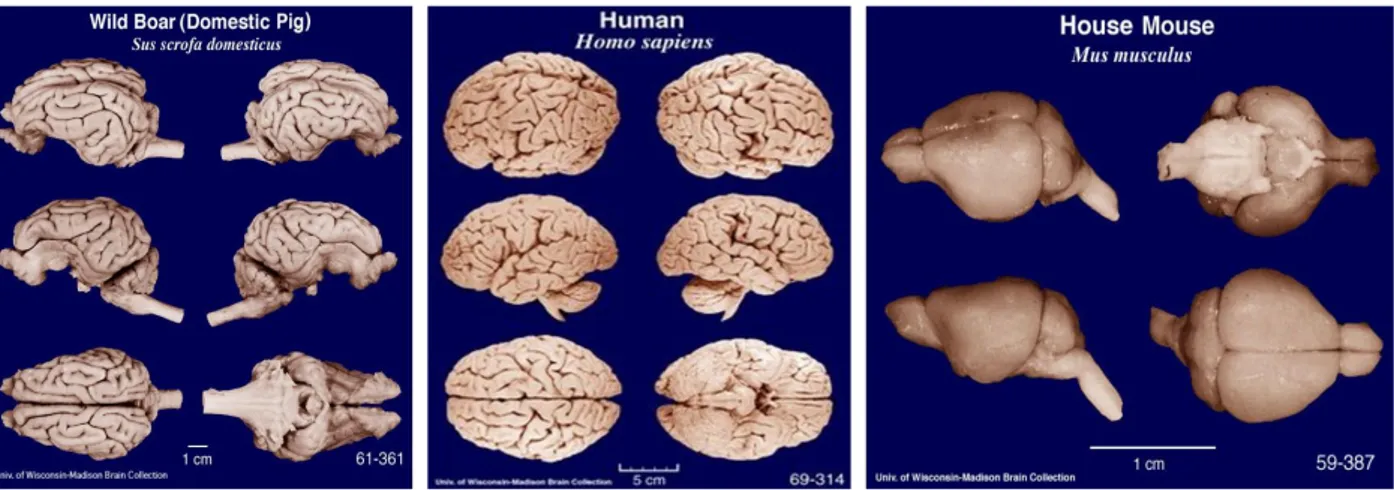
The swine central nervous system (CNS), and the brain in particular, have been rapidly evolving as a model system for humans given both the size and the anatomic characteristics: it is a relatively large gyrencephalic brain, white matter predominant with similar developmental peaks as human brain, which facilitates the study of specific pathological lesions (Fig.1.14). The internal carotid arteries provide most of the blood supply to the brain and blood flow characteristics are also similar, however pigs have a rete mirabile. In particular, the potential of pig models in paediatric neurological research was recognized fifty years ago due to the similarities in the extent of peak brain growth at the time of birth, the gross anatomy (gyral pattern and the distribution of grey and white matter) and the growth pattern of neonatal porcine brain to that of human infants. Moreover, similarities in the gross anatomy of pig brain to that of human brain has also been demonstrated for the hippocampus, a limbic structure, as well as for subcortical and diencephalic nuclei and brainstem structures.
Fig.1.14 Comparative brain photos: from left to right, Sus scrofa domesticus, Homo sapiens and Mus musculus. Figure taken from University of Wisconsin-Madison brain collection (http://neurosciencelibrary.org/).
The production of transgenic or genetically modified pigs was boosted almost a decade ago, when a Chinese-Danish pig genome-sequencing consortium has published the sequence of large portions of genomic DNA from five pig breeds and a porcine brain cDNA library is also available. Then, the introduction of Somatic Cell Nuclear Transfer (SCNT) paved a way of unexpected success in the genetic manipulation of this species, which demonstrated to be particularly difficult with the most used techniques (pronuclear injection, retroviral infection, ES cells).
26
1.6 Critical step in the use of swine as animal model: porcine embryo in vitro production (IVP)
In the 1970s, for the first time, gametes of livestock species started to be used for embryo biology-connected research and for the development of advanced reproductive technologies. In particular, the porcine IVP techniques were not working to an acceptable level until the early 1990s, when the need of large numbers of embryos for research purposes drove the continuous improvement of the system. The porcine in vitro production is almost completely based on the use of ovaries collected at local slaughterhouses, a source of large numbers of immature oocytes (Fig.1.15). The first report of live piglets born after IVP from abattoir-sourced oocytes was in 1986 (Cheng et al., 1986) and from that moment, a great effort from different laboratories in the world was taken to improve significantly the efficiency of the IVP procedure (Tab.1.1).
Unlike IVP procedure in other livestock species, the porcine IVP showed to have a very low efficiency right from the beginning, pointing the main problems as follows:
1. Male pronuclear formation: only a limited percentage of oocytes penetrated by the sperm were able to undergo sperm head decondensation and male pronuclear formation. It was clear that the maturation media was suboptimal and different studies demonstrated that the addition of cysteamine, a GSH precursor, could significantly increase the intra-oocyte GSH concentration and the relative percentage of oocytes able to undergo synchronous pronuclear formation (Grupen et al., 1995). Now it is well known that for oocytes of all species, including humans, cysteamine supplementation in the maturation media is essential to boost pronuclear formation.
2. Polyspermic fertilization: polyspermy is always been the major problem in porcine IVP practice. Different studies demonstrated how the zona pellucida (ZP) of ovulated oocytes was significantly thicker than the one of IVM oocytes, suggesting a possible failure to block polyspermic fertilization. In addition, the rate of polyspermic fertilization could depend on the sexual maturity of the donor, as it was lower in oocytes from sows than in IVM oocytes from gilts. This phenomenon is observed not only in swine, but also in adult ewes compared to pre-pubertal ewes. Other factors that could possibly interfere with the block of polyspermy are the ratio sperm:oocyte during fertilization and the exposure of oocytes to oviductal fluid, which contains specific glycoproteins and glycosaminoglycans that bind to the ZP specific protein forming complexes which can be further stabilized with heparin exposure, therefore increasing ZP resistance to sperm penetration (Coy et al., 2008). However, a big issue in porcine IVP is the ability of polyspermic embryos to develop up to the blastocyst stage, at the
27
same rate as those containing two normal pronuclei. This makes the blastocyst formation rate an inaccurate measure of viable embryo IVP efficiency. Nowadays, polyspermy in the porcine IVP procedure is still an unresolved issue.
3. Quality of oocytes selected for in vitro maturation: pigs are generally slaughtered at 6-7 months of age to meet the commercial demand for meat. Therefore, ovaries available at the slaughterhouse are often from pre-pubertal gilts. Studies showed that the quality of oocytes increases with the progression from first to third oestrous cycle of the donor gilt (Archibong et al., 1992). In porcine pre-pubertal ovaries, the antral follicle distribution is such that there are fewer large follicles (5-8 mm) and many more small (∼3 mm) compared to adult ovaries. Besides, porcine oocyte quality is also affected by season, being the developmental efficiency lower in summer (because of heat stress) than in winter.
28
Fig.1.15 Swine in vitro embryo production (IVP). Representative images of IVP swine embryos produced through in vitro maturation (IVM), in vitro fertilization (IVF) and in vitro culture (IVC) in our laboratory at AVANTEA. A) Swine
ovaries collected at a local slaughterhouse from pubertal gilts. B) Immature swine oocytes collected through aspiration. C) In vitro matured swine oocytes. D) Swine embryos at day 2 post-IVF. E) Swine morulae at day 5 post-IVF. F) Swine blastocyst at day 7 post-IVF. G) High magnification of a correctly in vitro fertilized swine zygote presenting female and male pronuclei (PNs, black arrows), two extruded polar bodies (PBs, red arrows) and the residual sperm tail (ST, blue arrow). H) High magnification of polyspermic swine zygote (IVF) presenting three visible pronuclei. Scale bars: 100 µm in B), C), D), E), F); 10 µm in G), H).
These problems have guided the advancement of a porcine IVP system. The in vitro maturation process takes 40 hours, a much longer duration than usual (i.e. 22-24 hours), and different protocols are now available for single-step culture rather than step-wise IVM procedures to suit the changing requirements of the oocytes during this long process as it was done in the past (Grupen, 2014). A long-held belief was that the majority of immature oocytes selected for IVM were still in the germinal
29
vesicle stage (GV), but nuclear staining revealed that almost half of the oocytes were already well into the germinal vesicle break down stage (GVBD). This fact also contributes to the known difference in timing between oocytes, for which the time taken to reach the metaphase II stage varies substantially among oocytes and this could enhance cytoplasmic deficiency (Funahashi et al., 1997; Grupen et al., 1997). The treatment of oocytes with dibutyryl cAMP (dbcAMP) during the first 20 hours of IVM reversibly inhibits meiotic progression, promoting synchrony of oocyte nuclear and cytoplasmic maturation. Anyway, some studies showed problems related with the temporary inhibition of nuclear maturation, such as detrimental effects on blastocyst rate. Moreover, the efficiency of the treatment with dbcAMP was found to be restricted to oocytes from small follicles (∼ 3 mm). Recently, a chemically defined maturation medium was develop that enables porcine oocytes to acquire full developmental competence. The key features of this medium are the presence of polyvinyl alcohol and a total of 22 amino acids (Yoshioka et al., 2008). At the end of the in vitro maturation process, oocytes are fertilized: many protocols are available, but none of them completely eliminate the problems. Once the fertilization has taken place, the zygote needs a suitable culture condition to reach successfully the blastocyst stage. In the pig female reproductive tract, embryos migrate from the oviduct to the uterine horns at the four-to-eight-cell stage about 2 days post-fertilization. A critical point for the embryo development was demonstrated to be the glucose concentration, as in the porcine oviductal fluid it was found to decrease markedly from the pre- to post-ovulatory period via unidentified systemic mechanism (Nichol et al., 1992; Nichol et al., 1998). Furthermore, the glucose utilization increases from one-cell to the blastocyst stage. For this reason, most commonly used protocols for porcine in vitro embryo culture are based on a two-step system in which media changes emulate the oviductal and uterine environment. Despite these advances, the porcine in vitro production is still considered suboptimal, as the in vitro blastocyst development rate is still poorer than the one obtained in vivo. (Lind et al., 2007; Swindle et al., 2012; Grupen, 2014; Holm et al., 2016).