SCUOLA DOTTORALE IN BIOLOGIA
Sezione di Biologia Applicata alla Salute dell’Uomo (BASU) – XXV Ciclo
EFFECTS OF FOOD CONTAMINANT MIXTURES ON
PUTATIVE TARGETS OF ENDOCRINE DISRUPTERS
EXPOSURE: AN INTEGRATED
TOXICOGENOMIC-BIOMARKERS APPROACH
EFFETTI DI MISCELE DI CONTAMINANTI
ALIMENTARI SU TARGET PUTATIVI DI
ESPOSIZIONE A INTERFERENTI ENDOCRINI: UN
APPROCCIO BIOMARKER-TOSSICOGENOMICO
PhD Student: Dr. Laura Stecca
Academic Year 2011/2012
Supervisor: Prof. Maria Marino Supervisor: Dr. Cinzia La Rocca Assistant supervisor: Dr. Timo Hamers
2
Index
SUMMARY ... 6
RIASSUNTO ... 9
BACKGROUND ... 12
Endocrine disruptors ... 12
PCBs, a class of Endocrine disruptors ... 13
PCB congeners classification ... 14
Liver as PCBs target organ ... 16
PCBs effects on liver cells ... 17
PCBs and nuclear receptors: ... 17
Estrogen receptors α and β ... 17
Aryl hydrocarbon receptor (AhR) ... 18
Androgen receptor ... 19
Thyroid receptor α (THRα) ... 20
Peroxisome proliferator receptor γ ... 21
Orphan receptors: Costitutive Androstane Receptor (CAR) and Pregnane X Receptor (PXR) ... 21
Retinoic acid receptor α (RARα) ... 23
PCBs and detoxification metabolism enzymes: CYPs and NRF2 ... 24
CYP1A1 ... 26
CYP1A2 ... 26
CYP3A4 ... 27
3
NF-E2-related factor 2 (Nrf2) ... 27
PCBs and oxidative stress biomarkers... 28
Reactive Oxygen Species (ROS) ... 29
GSH-GSSG Ratio ... 30
Superoxide dismutase (SOD) ... 30
Adipose tissue as PCBs target organ... 31
Peroxisome Proliferator-Activated Receptor γ (PPARγ) ... 33
Genes involved in adipogenesis upstream Pparγ induction ... 34
Hairy and enhancer of split-1 (HES1) ... 34
Protein delta homolog 1 (Dlk1) ... 35
CCAAT-enhancer-binding proteins (C/EBPs) ... 35
Nuclear receptor co-repressor 2 (Ncor2) ... 36
Genes involved in adipogenesis downstream Pparγ induction ... 37
Cyclin-dependent kinase inhibitor 1B (Cdkn1B) ... 37
Sonic hedgehog (Shh) ... 37
Tafazzin (Taz) ... 38
Vitamin D receptor (VDR) ... 38
AIMS ... 39
MATERIALS AND METHODS ... 42
Chemicals ... 42
PCB: standard solutions and mixtures composition ... 42
HuH6, HepG2 human hepatic cell lines: analysis of
biomarkers of effect at gene expression and protein level ... 43
Hepatic cell lines and culture conditions ... 43
4
Cytotoxicity assay ... 44
Total RNA extraction and cDNA synthesis ... 44
Gene expression evaluation by real-time PCR ... 44
Cytochrome P-450 monooxygenases activity assays ... 46
Superoxide Dismutase (SOD) Activity assay ... 47
Oxygen reactive species (ROS) detection assay ... 48
Reduced, oxidized, total glutathione (GSH) detection assay ... 49
Gene expression profiling of HepG2 cell line
... 50
Technical procedure ... 50
Data analysis ... 51
Cluster and functional annotation analysis ... 51
Obesogenic effect evaluation of PCB mixtures on murine
(3T3-L1) and human (SGBS) pre-adipocytes ... 52
3T3–L1 Mouse embryonic fibroblast and SGBS Human preadipocytes culture conditions and differentiation induction ... 52
Cell lines treatments ... 54
Adypocyte differentiation induction evaluation ... 54
3T3-L1 total RNA extraction and cDNA synthesis ... 55
3T3-L1 gene expression evaluation by real time RT-PCR ... 56
Statistical analysis ... 57
RESULTS ... 58
Citotoxicity assay results ... 58
PCB mixtures exposure effect on hepatic HuH6 and HepG2
cell lines ... 58
Nuclear receptors gene expression evaluation ... 58
5
Oxidative stress biomarkers analysis in HuH6 and HepG2 cell lines .... 60
Microarray profiling in HepG2 cells in response to PCB
mixtures treatments
... 63
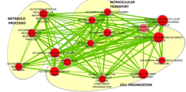
Ontology terms and Pathways affected PCB mixtures treatment ... 64
Gene Ontology enriched terms ... 64
KEGG pathways enrichments ... 67
PCB mixtures exposure effect on adipose tissue in vitro
models: 3T3-L1 mouse cell line and SGBS human cell line . 72
Evaluation of differentiation induced by PCB mixtures treatments ... 72Adipored assay results ... 72
Target genes expression modulation evaluated in 3T3-L1 cells through qPCR ... 74
DISCUSSION ... 75
CONCLUSIONS ... 82
BIBLIOGRAPHY ... 84
6
SUMMARY
The Endocrine Disruptors (EDs) are a class of chemicals that may interfere with the endocrine system that plays a crucial role in maintaining the physiological homeostasis of the human body as well as in regulating body growth, metabolism, reproduction and behavior (Faroon et al., 2001; Schell and Gallo, 2010). The EDs can exert their effects through a number of different mechanisms in particular interacting with nuclear (NRs) and orphan receptors, enzymatic pathways involved in steroid biosynthesis and detoxification metabolism (Diamanti-Kandarakis, 2009).
Polychlorinated biphenyls (PCBs) form a group of fat soluble and environmentally persistent EDs able to bioaccumulate in lipid fraction of animal tissue leading to biomagnification in the food chain. PCBs intake via food of animal origin represents the main route of human exposure (EFSA, 2010).
PCBs can be divided into two groups according to their biochemical and toxicological properties: Dioxin-like (DL) PCBs, mainly interacting with the Aryl Hydrocarbon Receptor (AhR) and Non Dioxin-like (NDL) PCBs whose mechanisms of action are not fully clarified. Actually, organisms are exposed to several congeners at the same time and, accordingly, the observed toxicological effects, such as on liver, thyroid, immune function, reproduction and behavior as well as carcinogenicity (EFSA, 2005), are exerted by mixtures. Therefore, new researches are required to study EDs mixtures, in particular on possible additive/synergistic effects at “real-life” dose levels.
In this study, 21 environmentally relevant PCB congeners were divided in three groups according to the proposed classification of Wolff et al. (1997) and Negri et al. (2003), based on structural and modes of action similarities: Mix1 (PCB 44, 49, 52, 101, 174, 177, 187, 201), potentially estrogenic NDL congeners, Mix2 (PCB 77, 81, 105, 114, 118, 126, 169) featuring DL-PCB and Mix3 (DL-PCB 99, 153, 180, 183, 196, 203), NDL-DL-PCBs highly persistent in the organism and phenobarbital-like inducers of Cytochrome P450 (CYP450).
The aim of the present project was to evaluate potential effects on liver and on adipose tissue as target organs of PCBs, elicited by the mixtures, at realistic human exposure levels, as derived from human exposure data through the evaluation of biomarkers response.
Two in vitro models have been selected as representative of liver tissue and corresponding to two different life stages: infant (HuH6) and pubertal (HepG2) cells.
7
To evaluate synergic effect, biomarkers of effect were used as indicators to measure PCB-induced variation in cellular or biochemical processes (NRs, NRF2, CYP450 enzymes and markers of oxidative stress). Moreover gene expression profiles of PCBs treated HepG2 cell line were evaluated by microarray analysis.Moreover, PCBs bioaccumulation and permanence in animal fat could affect preadipocyte programming and differentiation and adipocytes metabolism. Therefore, differentiation of mouse (3T3-L1) and human preadipocytes (SGBS cells) was evaluated to determine the effect of PCBs exposure. Moreover gene expression of biomarkers involved in adipocytes differentiation and metabolism were analyzed in 3T3-L1.
Previous reports classified Mix1 as potentially antiandrogenic congeners (Wolff et al., 1995), on the contrary, we observed in HepG2 cell line, that Mix1 significantly up-regulated only AR suggesting a potential androgenic effect. In HepG2 cells, we also observed an uncoupling in ROS-CYP1A1 regulation and in HuH6, GSH/GSSG ratio was significantly higher. As for Gene Ontology enrichment analysis, Mix1-modulated gene list was uniquely significant for terms related to Nuclear Transport and significantly affected only one pathway, the Intestinal Immune Network for IgA production, implying a stimulation of the immune response. Overall, these molecular processes may be considered specific markers of Mix1 effect in hepatic cells.
As regards effects on adipocytes, Mix1 increased SGBS differentiation at 3x concentration and in 3T3-L1 affected only C/EBPα involved in adipose tissue development.
Mix2, featuring DL-PCB congeners, exerted the up-regulation of AR and AhR in HepG2 cells. Moreover, Mix2 increased ROS levels as well as CYP1A1 mRNA expression both mediated by AhR (Kopf et al., 2010). However Mix2 significantly decreased the CYP1A1 enzyme activity, indicating a different effect possibly due to the low concentrations used. In HuH6, Mix2 induced a down regulation of CAR mRNA expression that may explain the reduction of CYP1A1 and CYP3A4 activity. Therefore, CAR gene expression and CYP1A1 and CYP3A4 activities may be assumed as target of DL-PCBs disruption on this hepatic cell line.
Microarray results evidenced Mix2 affected genes involved in RNA processing and splicing, protein localization and catabolism and macromolecules degradation. Noteworthy, main and characteristic effects of Mix2 relied a) on enrichment of genes related several cancer pathways as p53, activator of genes involved also in cell cycle arrest (Lanni and Jacks, 1998), apoptosis and in communication in adjacent cells, and b) on cell
8
cycle progression, inducing of G1 phase and inhibiting S phase. So we may hypothesize that a sum of contrasting mode of action exerted by PCB congeners in Mix2 occurred.As regards effects on adipocytes, Mix2 significantly induced SGBS differentiation only at 3x concentration, probably due to the mixture effect and/or the lower concentration levels used in this study.
Mix3 treatment affected the wider number of NRs expression (ERα, ERβ, AR, PXR, RARα, THRα) as well as NRF2, in particular, in HuH6. To our knowledge, it is the first study that showed AR mRNA expression due to PCB congeners included in Mix3, whereas the observed ERα and ERβ induction confirmed the estrogenic effect of PCB 99 and 153 (Warner et al, 2012).The decrease in the CYP3A4 enzyme activity in HuH6 cells was in agreement with NRF2 gene expression down-regulation (Itoh et al, 1997). Microarray analysis showed that Mix3 modulated the higher number of genes (5979 genes)with 1501 shared with Mix2-affected genes. Apart some GO enriched terms related to cell organization, Mix3 did not share other effects with Mix1, the other NDL mixture.
One of the most interesting evidence is the enrichment of genes involved in the Wnt signaling pathway by only Mix3, suggesting an unbalance toward pluripotency promotion (Takemarua and Moona, 2000).
Mix3 also affected some cancer pathways as well as the adherens junction pathway as Mix2 but modulating a different panel of involved genes. Moreover Mix3 exerted the higher magnitude of effect on pre-adipocytes differentiation in both murine and human cell lines. Gene expression evaluation analysis outlined that Mix3 down regulated Hes1, a DNA binding protein whose expression blocks adipogenesis (Ross et al., 2006). Overall results confirmed that the adopted grouping of PCBs in three mixtures served to highlight different modes of action as well as biomarkers responses, also among mixtures featuring NDL congeners. Moreover, the two hepatic cell lines and the two pre-adipocytes cell lines appear to be differently reactive to PCBs, indicating the need to use different in vitro models and a panel of biomarkers in order to characterize the EDs effects. These represented new results since there are no studies on PCB mixtures summarizing effects on oxidative stress, metabolism, nuclear receptors gene expression and adipocytes differentiation responses following treatment at human real exposure concentrations.
As prompted by EFSA (2005), it is necessary to provide evidences concerning comprehensive toxicological end-points of NDL effects. Therefore, these data may serve as a basis for developing relative toxicological factors for the NDL congeners risk assessment.
9
RIASSUNTO
Gli interferenti endocrini (IE) sono una classe di sostanze chimiche che possono interferire con il sistema ormonale. Il sistema endocrino svolge infatti un ruolo cruciale nel mantenimento della omeostasi fisiologica, nella regolazione della crescita, nel metabolismo, sviluppo e comportamento (Faroon et al., 2001). Gli IE possono esercitare i loro effetti attraverso una serie di meccanismi diversi come l’interazione con recettori nucleari (NR), con pathway enzimatiche coinvolte nella biosintesi degli steroidi, nel metabolismo e nella detossificazione (Diamanti- Kandarakis, 2009). I policlorobifenili (PCB) costituiscono un gruppo di IE liposolubili e persistenti nell'ambiente. Negli organismi, hanno la caratteristica di bioaccumulare nella frazione lipidica dei tessuti con conseguente biomagnificazione nella catena alimentare. L'esposizione umana avviene principalmente attraverso il consumo di alimenti di origine animale (EFSA, 2010). I PCB possono essere suddivisi in due gruppi in base alle loro proprietà biochimiche e tossicologiche: PCB diossina-simili (DL), che interagiscono principalmente con il recettore arilico (AhR) e PCB non diossina-simili (NDL), i cui meccanismi di azione non sono completamente chiariti. In realtà, gli organismi sono esposti contemporaneamente a diversi congeneri presenti nei cibi e gli effetti tossicologici osservati sono quindi imputabili a miscele di PCB che possono esercitare una varietà di effetti su fegato, tiroide, immunità, riproduzione cosi come il cancro (EFSA, 2005). Nuovi studi sono quindi necessari per evidenziare i possibili effetti additivi o sinergici di miscele di IE, in particolare a livelli di ‘dose reale’ per l’esposizione umana.
In questo studio, 21 congeneri di PCB di rilevanza ambientale sono stati divisi in tre gruppi sulla base di somiglianze strutturali e modalità di azione seguendo la classificazione proposta di Wolff et al. (1997) e Negri et al. (2003): Mix1 (PCB44, 49, 52, 101, 174, 177, 187, 201), che include congeneri NDL potenzialmente estrogenici, Mix2 (PCB77, 81, 105, 114, 118, 126, 169) che include DL-PCB e Mix3 (PCB99, 153, 180, 183, 196, 203) che contiene congeneri altamente persistenti nell'organismo umano e induttori dei citocromo P450 (CYP450).
Lo scopo del presente progetto è stato quindi quello di valutare i potenziali effetti provocati da miscele di congeneri di PCB di rilevanza ambientale sul fegato e sul tessuto adiposo, a livelli realistici di esposizione nell'uomo, in quanto derivati da dati umana misurati nella popolazione generale italiana. Sono stati selezionati due modelli in vitro rappresentativi del fegato, tessuto bersaglio dei PCB e principalmente e coinvolto nel metabolismo,
10
corrispondenti a due diverse fasi della vita: bambino (HuH6) e adolescente (HepG2). Per valutare l'effetto sinergico delle miscele di PCB, sono stati utilizzati biomarcatori di effetto (NR, NRF2, enzimi CYP450 e marcatori di stress ossidativo) associabili ad una possibile variazione nei processi cellulari o biochimici indotta dall’esposizione a PCB; inoltre, nelle HepG2, è stato valutato il profilo di espressione genica mediante analisi microarray. Ancora, lo studio esamina gli effetti dei PCB sul tessuto adiposo addominale poiché la permanenza dei PCB nella frazione grassa potrebbe influenzare il metabolismo degli adipociti e il programming e la differenziazione dei preadipociti. E’ stato quindi valutato il tasso di differenziamento in fibroblasti di topo (3T3-L1) e preadipociti umani (SGBS) e, nelle 3T3-L1 esposte alle tre miscele, è stata valutata l'espressione genica di biomarcatori coinvolti in questi processi.Precedenti studi classificano i congeneri della Mix1 come potenzialmente antiandrogeni (Wolff, 1995), al contrario, i nostri risultati evidenziano che Mix1 incrementa significativamente l’espressione di AR suggerendo un effetto androgenico. Nelle cellule HepG2, abbiamo anche osservato una regolazione della coppia di marcatori ROS-CYP1A1 e, in HuH6, il rapporto GSH/GSSG era significativamente più alto. Per quanto riguarda lo studio del profilo genico, si è evidenziato che l’elenco di geni modulati da Mix1 è significativo per termini relativi al trasporto nucleare e risulta significativamente influenzato solo il pathway del Network immunitario intestinale per la produzione di IgA che implica una stimolazione della risposta immunitaria. Nel complesso, questi processi molecolari possono essere considerati marker specifici di effetto di Mix1 nelle cellule epatiche. Per quanto riguarda gli effetti sugli adipociti, Mix1 induce il processo di differenziazione nelle SGBS alla concentrazione 3x e nelle 3T3-L1 risulta modulata solo l’espressione genica di C/EBPα, coinvolto nello sviluppo del tessuto adiposo.
Mix2 (DL-PCB) determina una regolazione positiva di AR e AhR nelle HepG2. Inoltre, risulta incrementare i livelli di ROS nonché l’espressione del CYP1A1 entrambe AhR-mediati (Kopf et al., 2010). Tuttavia l'attività dell'enzima CYP1A1 è significativamente diminuito, indicando un effetto contrastante probabilmente dovuto alle basse concentrazioni utilizzate. In HuH6, Mix2 regola negativamente l’espressione di CAR che potrebbe spiegare la riduzione del CYP1A1 e del CYP3A4; questi tre parametri possono essere assunti come marcatori specifici dei DL-PCB su questa linea cellulare epatica. L’analisi Microarray evidenzia per Mix2 la modulazione di geni coinvolti nel processamento e splicing del RNA e nella localizzazione delle proteine. Degni di nota sono inoltre gli effetti su geni
11
correlati al cancro come la p53, coinvolta nell’arresto del ciclo cellulare (Lanni e Jacks, 1998), apoptosi e nella comunicazione cellulare ma anche di fattori legati alla progressione del ciclo cellulare, per cui si può ipotizzare un equilibrio fra modi di azione contrastanti esercitati dai congeneri di PCB in Mix2. Per quanto riguarda gli effetti sugli adipociti, Mix2 induce la differenziazione nelle SGBS a concentrazione 3x, probabilmente per l’effetto miscela o delle concentrazioni utilizzate.Per quanto riguarda la Mix3, il trattamento influenza l’espressione di un ampio numero di NR (ERα, ERβ, AR, PXR, RARα, THRα) nonché NRF2 nelle HuH6. Questo è anche il primo studio che ha dimostrato un effetto sull'espressione di AR dovuto ai congeneri della Mix3, mentre l'induzione osservata di ERα e ERβ conferma l'effetto estrogenico dei PCB99 e 153 (Warner et al, 2012). Inoltre la diminuzione dell’attività del CYP3A4 in HuH6 è in accordo con la down regolazione di NRF2 (Itoh et al, 1997). L’analisi Microarray di Mix3 mostra la modulazione del più alto numero di geni (5979), 1501 dei quali condiviso con Mix2 mentre, con l'altra miscela di NDL, non ha effetti comuni se non quelli relativi all'organizzazione cellulare. Uno degli elementi più interessanti è comunque l'arricchimento di geni implicati nella pathway del signaling di Wnt, modulati esclusivamente da Mix3, che suggerisce la promozione verso la pluripotenza (Takemarua e Moona, 2000).
Mix3, influenza anche alcuni pathway implicati nel cancro nonché, come Mix2, nelle giunzioni aderenti, rispetto alla quale modula un diverso gruppo di geni coinvolti. Inoltre la Mix3 esercitava gli effetti maggiori sulla differenziazione dei preadipociti sia nella linea cellulare umana che in quella murina e l’analisi dalla modulazione genica evidenzia che reprime Hes1 la cui espressione blocca l’adipogenesi (Ross et al., 2006).
Complessivamente i risultati hanno confermato che la divisione dei PCB in tre miscele individua diverse modalità di azione, nonché differenti risposte dei biomarcatori, anche tra miscele di congeneri NDL. Inoltre, le quattro linee cellulari (epatiche e di preadipociti) sembrano essere diversamente sensibili ai PCB, ciò indica la necessità di utilizzare diversi modelli in vitro e un pannello di biomarcatori per caratterizzare gli effetti degli IE.
In conclusione, tutti questi rappresentano nuovi risultati data l’assenza di dati sugli effetti dell’esposizione a miscele di PCB a ‘concentrazioni reali’ che includano stress ossidativo, metabolismo, espressione dei NR e differenziazione degli adipociti. Come sottolineato dall'EFSA (2005), vi è la necessità di dati riguardanti end-point tossicologici dei NDL, pertanto, questi dati rappresentano un contributo utile alla caratterizzazione di fattori tossicologici per la valutazione del rischio legato ai NDL-PCB.
12
BACKGROUND
Endocrine disruptors
Endocrine disruptors (EDs) are a diversified class of chemicals that interfere with the hormonal system. The term “endocrine disrupter” was introduced in the early 1990s and later defined by the World Health Organization (European Commission, 1996) as: “… an exogenous substance or mixture that alters function(s) of the endocrine system and consequently causes adverse health effects in an intact organism, or its progeny, or (sub)populations”.
The endocrine system plays a crucial role in maintaining the physiological homeostasis of the human body, in regulating body growth, metabolism, sexual development and function as well as reproduction (Faroon et al., 2001), therefore an impingement on these processes by EDs may severely affect human health.
EDs include persistent contaminants, (e.g. dioxins, Polychlorinated Biphenyls –PCBs-, Polybrominated Diphenyl Ethers -PBDEs), pesticides (organophosphates, di-thiocarbamates), plasticizers (e.g. phthalates, bisphenol A) as well as natural compounds (e.g. phyto-oestrogens which are present in plants such as soy and nuts, coumestrol and genistein) or pharmaceuticals (such as diethylstibestrol, 17a-ethinyloestradiol and tamoxifen). EDs are widespread in food and feed, being primary routes of exposure for both synthetic (man-made) and natural EDs, therefore representing a great concern for human and wildlife health. In particular, lipophilic and persistent compounds may bioaccumulate in the lipid fraction of animal tissue leading to a biomagnification in the food chain. Therefore, fatty fish, meat and dairy products are among the major sources of some persistent EDs (EFSA, 2012).
Due to their different chemical structures, EDs as DDT, bisphenol-A and nonylphenol (Eisenbrand, 1996; Shafer et al., 1996) may exert multiple effects. Indeed, the endocrine and reproductive effects of these chemicals are related to their ability to interact with nuclear receptors (NRs) by: (a) mimicking the effect of endogenous hormones; (b) antagonizing the effect of endogenous hormones; (c) disrupting the synthesis and metabolism of endogenous hormones; (d) disrupting the synthesis of hormone receptors. The biological actions of endogenous hormones, such as estrogen, progesterone, testosterone and thyroxine, are mediated by the binding with high-affinity receptors located into target cells, the NRs. The interaction of
13
an hormone with its receptor initiates a cascade of events leading to multiple effects depending on that particular hormone (Amaral Mendes, 2002). Originally, EDs were thought to exert primary actions only through NRs, mainly steroid ones, such as estrogen receptors (ERα and ERβ), androgen receptor (AR), progesterone receptor (PR), thyroid receptors (THRα and THRβ), and recent evidence indicates that other endocrine pathways are also involved by the disrupting chemicals as retinoid receptors (RARs and RXRs). Growing evidence showed that the mechanisms involved are broader since EDs may interact also with nonnuclear steroid hormone receptors (e.g., membrane ERs), nonsteroid receptors (e.g., neurotransmitter receptors such as the serotonin, dopamine and norepinephrine receptors), orphan receptors (e.g., Aryl hydrocarbon Receptor (AhR), through numerous other mechanisms converging upon endocrine and reproductive systems (Diamanti-Kandarakis, 2009).Moreover, it is increasingly evident that many EDs may act as agonists in some tissues and as antagonists in others. Indeed different tissues express co-activators or co-repressors that could modulate the ligands-action in a tissue-specific manner implying that EDs may regulate same pathways but in opposite ways.
Such complexity in the mechanisms of action imply different adverse effects may occur depending on the chemical, the exposure concentration and the individual susceptibility. Critical stages of development (infants, children and unborn babies) may be particularly sensitive to EDs hormonal activity, especially during defined “windows of susceptibility”. Exposure of pregnant women to EDs may increase the likelihood of toxic effects in fetus possibly resulting in abnormalities or diseases in the short-term or evident only later in life (Swan et al., 2005; Castro et al., 2008; Deckelbaum and Williams, 2001).
PCBs, a class of Endocrine disruptors
Among EDs, Polychlorinated biphenyls (PCBs) are a class of chemicals widely used in the past for many applications, especially as dielectric fluids in transformers, capacitors and coolants. Due to their toxicity and classification as persistent organic pollutants, processing and distribution of PCBs has been prohibited in industrial countries since the late 1980s but they still can be released into the environment from building paint and sealants or poorly maintained hazardous waste sites containing PCBs. Depending on the number of chlorine atom substituents (1-10) and their
14
position on the two aromatic rings, 209 possible congeners may be synthesized.PCBs are widely present in the environment due to a global circulation by atmospheric transport. Highly chlorinated congeners, in particular, adsorb strongly to sediment and soil, where they tend to persist with half-lives of months to years.
Due to their lipophilicity, PCBs tend to bioaccumulate in body fat, both in animals and humans, biomagnifying through the food chain; so exposure to PCBs occurs mainly through the diet and it is especially related to certain food commodities, such as dairy products and fatty fish (EFSA, 2010). This is particularly the case for higher chlorinated congeners, while lower chlorinated PCB congeners are metabolized quickly.
Following ingestion, PCBs are mainly biotransformed in the liver to hydroxy (HO-) and methylsulfone (MeSO2-) metabolites.
PCBs exert a wide spectrum of adverse effects, especially at neurochemical and neuroendocrine level (Tilson and Kodavanti, 1997), as well as reproductive level. An association between endometriosis and high levels of PCB in plasma has been reported since 1992 (Gerhard and Runnebaum, 1992), moreover a relationship was found with long-term aspects of Testicular Dysgenesis Syndrome such as low sperm count and testicular cancer (Hardell et al., 2003, 2004; Toppari et al., 1996).
Prenatal treatment of female rodents and other species with PCBs has been shown to alter the timing of puberty (Humblet et al., 2011), accelerate reproductive aging (Cooper and Kavlock, 1997), affect gonadotropin release (Khan and Thomas, 1997), and interfere with sexual behavior (Chung and Clemens, 1999). There is evidence that the PCBs can affect thyroid hormone status; indeed Schell and Gallo (2009) study showed that PCB levels are positively related to TSH and negatively to free T4.
PCBs have been classified as possible human carcinogens (WHO-IARC, 1987). Several studies have implicated low-molecular-weight PCBs as potential tumor-initiating compounds that may either induce oxidative DNA damage or formation of DNA adducts (Oakley et al., 1996a, 1996b). Several case-control studies, published in the last two decades, have raised the issue that women, exposed to organochlorine chemicals such as DDT and certain PCB congeners, may have a higher incidence for breast cancer than non-exposed women (Cohn et al., 2012).
15
From the 209 theoretically possible PCB congeners, 12 non-ortho and mono-ortho substituted PCBs show toxicological properties similar to dioxins, and therefore termed ‘dioxin-like PCBs’ (DL-PCBs), due to their coplanar structure and the capability to bind to the AhR receptor (Poland et al., 1985; Safe, 1986; Safe et al., 2005; EFSA 2010). The 12 DL-PCBs also display anti-estrogenic activity in several biological systems (Letcher et al., 2002; Pliskova et al., 2005).2,3,7,8-Tetrachlorodibenzop- dioxin (TCDD), commonly used as the model compound of this large family of dioxin-like food contaminants, elicits a wide array of biochemical and pathological effects on mammals, including weight loss, hyperkeratosis, liver enlargement, thymus atrophy, immunosuppression, tumor promotion, impaired reproduction, and embryo toxicity (WHO, 1998).
In order to sum up and compare the toxicity of the different congeners of concern (17 dioxins and 12 dioxin-like PCBs), toxicity equivalency factors (TEF) have been developed on the basis of their relative toxicity compared to 2,3,7,8-TCDD (Van den Berg et al., 2006). The TEF approach is primarily intended for estimating exposure and risks. The other PCB, referred to as ‘non dioxin-like PCB’ (NDL-PCB), cannot adopt a coplanar structure so the TEF concept used for DL-PCBs can’t be applied to NDL-PCB, moreover mechanisms of action of all congeners have not been evaluated yet. EFSA 2005. The 197 non dioxin-like PCBs (NDL-PCBs) (including 28 mono-ortho, di-ortho and more chloro-substitutedPCBs) are more abundant in the environment than DL-PCBs, with the highest mean contamination level observed in fish and fish-derived products followed by eggs, milk and their products, meat and meat products from terrestrial animals. Some NDL-PCBs elicit different types of responses including neurological, neuroendocrine, endocrine, immunological and carcinogenic effects. These effects occur via multiple toxicity pathways, not involving AhR (EPA, 2003). Six congeners (28, 52, 101, 138, 153, and 180) were chosen as indicators because their sum represent about 50% of the total NDL-PCBs in food. Moreover, they mostly contribute to the anti-androgenic, (anti)estrogenic, anti-thyroidal, tumor-promoting, and neurotoxic potencies calculated for NDL-PCB mixtures in human samples As a consequence, the EU Commission recently established maximum levels for the sum of the six indicator NDL-PCBs in food and feed (Commission Regulation (EU) No 1259/2011).
PCB-168 is a congener not regularly monitored but it might significantly contribute to the AR-antagonistic and TTR-binding potency of PCBs complex mixture in human samples. Based on the current knowledge of
16
PCB levels in human tissues and toxic potencies of individual congeners, the seven indicators, PCBs plus PCB-168, may explain an average > 74% of the calculated toxic potency of PCB mixtures in humans (Hamers et al, 2011).Many projects aim to clarify biological mechanisms underlying the various types of toxicity of NDL-PCBs and to evaluate these data from a regulatory toxicology point-of-view.
No published peer-reviewed data were available on the carcinogenic potency of single NDL-PCBs or about the potential mutagenic activity of PCB mixtures (EFSA, 2010). PCBs exposure occurs almost exclusively as a mixture of congeners having different effects. For this reason, some authors (Negri et al., 2003; Wolff et al., 1997) proposed a toxicologically based classification of PCBs in three groups, one DL and two NDL mixtures by introducing a distinction within the group of NDL-PCBs on the basis of structure-activity considerations. They named Group I, the estrogenic / neurotoxic and variably persistent congeners, with one or two unsubstituted para positions (PCB 44, 49, 52, 101, 174, 177, 187, 201) and Group III, the biologically persistent, phenobarbital (PB)-type cytochrome-P450 (CYP1A and CYP2B) inducing congeners (PCB 99, 153, 180, 183, 196, 203). Group II includes non-ortho, mono-ortho and di-ortho congeners with dioxin-like properties and potentially anti-estrogenic (DL-PCBs: PCB 77, 81,105, 114, 118, 126, 169). The partition of NDL-PCB may represent a possible way to differentiate and characterize the effects within the NDL-PCB group in order to define a proper toxicological significance.
Liver as PCBs target organ
The digestive system is exposed to contaminants and other food bioactive compounds, during all life stages, and such exposure is mediated by tissues extremely rich in different NRs. Indeed, tissues of the digestive system are highly responsive to endocrine regulation, and interference as well. Besides to be target of endocrine disruption, liver is the representative organ of the digestive system, hence involved in food processing and regulating fundamental steps of absorption and metabolism of food constituents. Furthermore, liver is the organ involved in detoxification metabolism also of food contaminants, highly responsive to endocrine regulation, included PCBs. Biotransformations are thus critical processes in the body defense against the toxic effects of a wide variety of endobiotics and xenobiotics
17
(Yu, 2001). The biotransformation processes consist of three phases. Phase I includes activity of cytochrome P450 (CYP) super-family (heme-dependent mono-oxygenases) members expressed within the liver, intestine and kidney, the primary organs for uptake, metabolism, and excretion of xenobiotics (Kliewer et al., 2002). The CYP microsomal enzymes catalyze the metabolic conversion to more polar derivatives of foreign chemicals as well as endogenous substrates (e.g., steroid hormones) (Xu et al., 2005). Phase II includes synthetic or conjugation reactions, combining the toxicants or the primary metabolites directly with endogenous substances (e.g., Gly, Cys, glutathione). Often these processes result in the production of a more stable, water-soluble, metabolite that is more readily excretable via the transporter-mediated elimination pathway or phase III (Di Masi et al., 2009).PCBs effects on liver cells PCBs and nuclear receptors: Estrogen receptors α and β
Estrogen receptors (ERs) belong to the steroid/thyroid hormone superfamily of NRs (Evans, 1988). In the nucleus, estrogen modulates gene transcription (genomic response), and the resulting protein products determine the cell biological actions of the sex steroid. In addition, a small pool of ERs localized to the plasma membrane, is involved in non-genomic response and signals mainly through direct or indirect coupling to G proteins (Marino et al, 2002). Therefore, in response to steroids binding, signal transduction modulates both non-transcriptional and transcriptional events and impacts both the rapid and more prolonged actions of estrogen. (Levin, 2005). Binding of a ligand to ERs triggers a series of conformational changes in the receptor structure and this leads, via a number of events, to changes in the rate of transcription of estrogen-regulated genes. These events include receptor dimerization, receptor-DNA interaction, recruitment of and interaction with co-activators and other transcription factors, and formation of a preinitiation complex (Nilsson et al, 2001).
There are two different forms of the ERs, α and β, encoded by separate genes (ESR1, estrogen receptor 1 (ER-alpha); ESR2, estrogen receptor 2 (ER-beta), respectively). Both ERs are widely expressed in different tissue
18
types, however with some notable differences in their expression patterns (Couse et al 1997).Estrogens influence many physiological processes in mammals, including reproduction, cardiovascular system, bone integrity, cognition and behavior. DL-PCBs have been frequently reported to have anti-estrogenic activity, as TCDD (Buchanan et al. 2000, 2002; Oenga et al. 2004; Safe and Wörmke 2003), whereas the exact mechanisms of estrogenic or anti-estrogenic activities of NDL- PCBs are still not fully characterized.
The reported results are often contradictory, derived from data obtained in different in vitro or in vivo models (Hansen et al., 1998). While low molecular-weight PCBs displayed estrogenic activity both in vitro and in vivo (Arcaro et al., 1999; Nesaretnam and Darbre, 1997; Rogers and Denison, 2000; Rose et al., 2002), the three most prevalent NDL-PCBs, PCB 138, 153 and 180 have been reported to be anti-estrogenic in MCF-7 cells (Bonenfeld-Jorgensen et al., 2001). However, estrogenic/anti-estrogenic potencies of a large set of PCB congeners have not yet been determined in a single in vitro bioassay.
Aryl hydrocarbon receptor (AhR)
AhR is a member of the bHLH-PAS (basic helix-loop helix/Per-Arnt-Sim) gene family of transcription factors (Gu et al., 2000) and it is the only ligand-activated one. The majority of AhR ligands are environmental contaminants such as polycyclic aromatic hydrocarbons (PAHs), TCDD (Poland et al., 1976), related polychlorinated dibenzo-pdioxins (PCDDs) and dibenzofurans (PCDF) (Poland et al., 1982), and dietary indole carbinols present in cruciferous vegetables (Bjeldanes et al., 1991; Bradlow et al., 1991; Chen et al., 1998; Vang et al., 1991), so AhR acts as a toxin sensor.
After ligand binding, AhR migrates in the cell nucleus forming an heterodimeric complex with the AhR nuclear translocator Arnt (Hoffman et al., 1991) which binds to specific genomic enhancer sequences (up to 59) termed Dioxin- or Xenobiotic-Responsive Elements (DREs or XREs); this interaction leads to transcriptional activation of genes involved in complex cellular responses such as cell cycle progression and apoptosis such as phase I drug-metabolizing enzymes like Cytochromes P-450 1A1 (CYP1A1), CYP1A2, and CYP1B1 and phase II enzymes including glutathione-S-transferase Ya subunit, UDP glutathione transferase, aldehyde dehydrogenase, and NAD(P)H quinone oxidoreductase (NQOR)
19
(Hankinson et al., 1995). In addition, a number of Estradiol(E2) - regulated genes are controlled by AhR at either the transcriptional or post-transcriptional level (Nilsson et al., 2001).NDL-PCBs are not documented to directly interact with AhR. Otherwise, similarly to TCDD, DL-PCBs activate AhR and AhR-dependent signal transduction pathways (Van Den Berg et al. 1998) which mediate the majority of the adverse effects of these compounds among which their anti-estrogenicity.
The anti-estrogenic action of AhR agonists such PCBs might involve repression of E2-dependent gene expression by interactions of activated AhR with DNA regions of E2 responsive gene promoters (Oenga et al., 2004; Safe and Wörmke, 2003), inhibition of E2-induced cell cycle proteins (Buchanan et al. 2002; Wang et al. 1998), or effects of PCBs on E2 metabolism (Pang et al., 1999; van Duursen et al., 2005).
AhR ligands are able to play their antagonistic effects on ER signaling by increasing the metabolism rate of E2 by inducing CYP1A1 and CYP1B1 enzymes (Hayes et al., 1996; Safe et al., 1991) or by decreasing levels of ERs.
Several genes have been identified as targets for cross-talk between ER and AhR: cathepsin D, pS2, prolactin receptor, c-fos, hsp27, TGF-a and TGF-b, as well as PR.
Androgen receptor
The androgen receptor (AR) is a member of the NRs superfamily, functions as a ligand-inducible transcription factor and is expressed in many end-organs including the hypothalamus, pituitary, liver, prostate, and testes (Matsumoto, 2008). The principle steroidal androgens, testosterone (T) and its metabolite 5-dihydrotestosterone (DHT), predominantly mediate their biological effects through binding to the AR.
The appropriate regulation of androgen activity is necessary for a range of developmental and physiological processes, particularly male sexual development and maturation, as well as the maintenance of male reproductive organs and of spermatogenesis (McLachlan et al., 1996; Mooradian et al., 1997; Cunha et al., 1987). The binding of T or DHT to AR induces receptor dimerization, translocation into the nucleus, binding to its cognate response elements (ARE) and recruit co-regulators to promote the expression of target genes (He, 1999; Quigley et at., 1998, Heinlein et al., 2002). In addition to this transcriptional or genomic mode of action by
20
steroids, androgens, like progesterone and estrogen, can exert rapid, non-genomic effects (Falkenstein et al., 2000) typically involving the induction of second messenger signal transduction cascades, including increases in free intracellular calcium, and activation of protein kinase A (PKA), protein kinase C (PKC), and MAPK. The non-genomic action of androgens has been implicated in a number of cellular effects, including gap junction communication, aortic relaxation, and neuronal plasticity (Pluciennik et al., 1996; Kubli-Garfias et al, 1982; Yamada et al, 1979; Costarella et al, 1996). Dysregulation of androgen/AR signaling perturbs normal reproductive development and accounts for a wide range of pathological conditions such as androgen-insensitive syndrome, prostate cancer, and spinal bulbar muscular atrophy (Matsumoto, 2012).Prenatal testosterone exposure alters placental steroidogenesis and leads to dysregulation of lipid metabolism in their adult female offspring. (Sun, 2012).
Endocrine disruptors that act via the androgen receptor (AR) are less well studied than environmental estrogens. Epidemiologic studies of occupational exposure to PCBs revealed a strong exposure response relationship for prostate cancer risk (Ritchie, 2003) and prostate cancer mortality (Prince et al, 2006) suggesting an anti-androgen mechanism for PCBs.
Thyroid receptor α (THRα)
THRs belong to the superfamily of hormone NRs. There are two major THR isoforms encoded on separate genes, designated as THRα and THRβ (Lazar et al, 1993).
The natural ligands of THRs are thyroid hormones (TH; thyroxin, T4; triiodothyronine, T3). THs play critical roles in differentiation, growth, and metabolism. Indeed, TH is required for the normal function of nearly all tissues, with major effects on oxygen consumption and metabolic rate (Oppenheimer et al., 1987). THRs are target of endocrine disruption. Several reports have shown that PCBs may affect embryonic and neonatal development at low doses (Jacobson et al., 1990, 1996) causing abnormal development of the central nervous system (Jacobson et al., 1990) probably through the involvement of thyroid hormones system, since THs and PCB molecules share structural similarity (Koibuchi et al., 2000). Hypothyroidism during perinatal period causes retardation of general growth as well as abnormal development of many organs including brain,
21
bone, skeletal muscle and liver. Thus, disruption of TH system by PCB may affect development of such organs.Peroxisome proliferator receptor γ
The nuclear Peroxisome Proliferator-Activated Receptors (PPARs) subfamily of NRs regulate the transcription of many genes in a ligand-dependent manner by binding to specific response elements (PPREs) within promoters. PPARs bind as obligate heterodimers with a Retinoid X Receptor (RXRα, β, γ) and, upon binding agonist, interact with cofactors increasing the rate of transcription initiation (Chawla et al., 2001). RXR– PPARγ complex can be transcriptionally activated by both PPARγ and RXR-specific ligands. PPARs are present in humans in three different isoforms, PPARα, PPARβ/δ and PPARγ that are encoded separately, but have overlapping tissue expression patterns, and as a group coordinate the regulation of important metabolic pathways (Sonoda et al., 2008; Chen et al., 1993; Lazar, 2005). PPARγ, the best-studied member of the family, results to be expressed at high levels in white and brown adipose tissue where it plays a major role in many different biological processes including lipid metabolism, glucose homeostasis, adipogenesis, lipid storage and release (Lehrke et al., 2005). Adipose tissue and large intestine have the highest levels of PPARγ mRNA; kidney, liver, and small intestine have intermediate levels. (Fajas, 1997). PPARγ is activated by endogenous arachidonic acid metabolites such as 15-deoxy-delta 12, 14 prostaglandin J2 (Forman et al., 1995). One class of PPARγ ligands, the thiazolidinediones, which includes the drug rosiglitazone, are effective insulin sensitizers, and have been shown to improve glucose uptake and lower hyperglycaemia and hyperinsulinaemia (Lehmann et al., 1995). The PPARs are also potential therapeutic targets for atherosclerosis, inflammation and hypertension (Berger et al., 2002) and tumor susceptibility (Lee et al., 2012).
Exposure to persistent organic pollutants, such as PCBs, may therefore contribute to the development of inflammatory diseases such as atherosclerosis. Cellular oxidative stress and imbalance in antioxidant status are critical events in PCB-mediated induction, contributing to endothelial inflammatory response in part by down-regulating PPAR signalling (Hennig et al., 2005).
Orphan receptors: Costitutive Androstane Receptor (CAR) and Pregnane X Receptor (PXR)
22
The large NRs subfamily I contains numerous orphan receptors including Constitutive Androstane Receptor (CAR) and Pregnane X Receptor (PXR). (Hustert et al., 2001).CAR and PXR are modular proteins sharing common regions, including the N-terminal DNA-Binding Domain (DBD), the hydrophobic core region (H region), and the C-terminal Ligand-Binding Domain (LBD). (Olefsky, 2001). CAR-DBD and PXR-DBD are involved in receptor dimerization and binding to specific DNA sequences (Frank et al., 2003).
Although CAR and PXR sequences are closely related, their role in gene expression regulation is significantly different; indeed CAR and PXR recognize different endogenous and exogenous ligands. (Frank et al., 2003). Due to their role in protecting living organisms from exogenous insults, CAR is highly expressed in the liver and in the epithelial cells of the small intestine villi (Swales and Negishi, 2004).
PXR is primarily expressed in liver, intestine, and kidney (Kliewer et al., 1998).
PXR takes part to a protective system to prevent exposure of critical cells sensitive to xenobiotics. A variety of compounds can activate both CAR and PXR. However, ligand-binding techniques indicate that CAR and PXR bind a structurally diverse panel of chemicals and the receptor ligand specificity is species-dependent. This promiscuity is warranted by the structural properties of CAR and PXR-LBDs to respond to a diverse set of low-affinity ligands (Lemaire et al., 2006;Kublbeck et al, 2011).
PXR seems to be much more promiscuous than CAR (Moore et al., 2003). It is now established that PXR binds nearly all xenobiotics including drugs such as dexamethasone, PCBs and nonylphenol (Lemaire et al., 2006). CAR activators promote the nuclear translocation of CAR by a ligand-dependent process, followed by CAR/RXRα heterodimerization, CAR-DBD binding to the RE, and recruitment of co-activators. CAR nuclear translocation not necessarily leads to an increase in target genes transcription but serves as the first important step to regulate the xenosensor transcriptional activity (Timsit and Negishi, 2007).
CAR and PXR function as sensors of toxic by-products of endogenous metabolic compounds and of exogenous chemicals to facilitate their elimination. Many mechanisms have been evolved to detoxify the majority of xenobiotics and versatile inducible metabolizing enzymes and efflux transporters play a crucial role in these mechanisms that are a form of biotransformation.
CAR coordinates the regulation of multiple hepatic genes resulting, in most cases, in metabolic detoxification by CYPs and transferases within the
23
hepatocyte. PXR serves as a master transcriptional regulator of CYP3A isozymes (Quattrochi and Guzelian, 2001). Indeed, PXR and CYP3A result co-expressed in several tissues; moreover PXR is activated by CYP3A inducers. (Di Masi et al, 2009).It has been reported that PXR-mediated induction of CYP3A4 as well as CAR may be mediated by exposure to PCBs (Al-Salman et al, 2012) potentially causing adverse interactions. The OH group is commonly found in activators of PXR and CAR as exemplified by endocrine disrupters such as the hydroxylated PCBs. One of the most interesting differences involves highly chlorinated PCBs which were found to act as antagonists of PXR and agonists for rodent PXR (Lemaire eet al, 2004).
Two non-hydroxylated PCB congeners (PCB 118 and 153), which present a larger fraction of the PCB contamination of fatty foods, activated PXR (Jacobs et al, 2005).
Several of the non-coplanar PCBs directly activate PXR and CAR, whilst the coplanar PCB-77 did not. Non-coplanar PCBs were also able to activate PXR/CAR target gene expression in a substitution- and tissue-specific manner. Chronic activation of PXR/CAR is linked to adverse effects and must be included in any risk assessment of PCBs (Al-Salman et al., 2012).
Retinoic acid receptor α (RARα)
Retinoic acid receptors (RARs and RXRs) are ligand-dependent transcription factors, which belong to different subfamilies of NRs (I and II, respectively, according to the official nomenclature (NRNC, 1999). There are different RAR and RXR subtypes (α, β and γ) (De Lera et al., 2007). RARα (NR1B1) is the most prevalent isoform of retinoic acid receptors in liver and hepatocytes. Two other receptors, RARβ and RARγ have complex tissue and developmental expression patterns.
RARs mediate the biological effects of all-trans-retinoic acid (at-RA) (Bastien et al., 2004), the most active naturally occurring retinoid in mammals, except for the visual process, where retinal is the active retinoid form. In the body, at-RA is formed from its precursor all-trans-retinol (at-ROL) (Duester et al., 2008; Pares et al., 2008). Retinoids (vitamin A and its analogues) play essential roles in several physiological processes, such as embryonic development, reproduction, immunity, proliferation, differentiation, apoptosis and vision (Luo et al., 2006; Niederreither et al., 2008; Kim et al., 2008). at-RA binds to the ligand-binding domain of RARs, which induces heterodimer formation with RXRs to form the
24
transcriptionally active complex. The heterodimer complexes act as transcriptional regulators of a multitude of retinoid-regulated genes by binding to specific RA response elements (RAREs) (Schuchardt et al., 2009).Many genes involved in hepatic functions are regulated by RXR:RAR heterodimers. Among these are some involved in serum protein synthesis (a-fetoprotein, albumin, coagulation factors), bile acid synthesis (CYP7A1), and bile acid transporters (NTCP and MRP2) (Li et al., 2002). Environmental pollutants interfere with normal retinoid physiology and the change of retinoid levels in organism has been used as a sensitive biomarker of exposure to wide range of pollutants in wild animal populations. The liver seems to be an important target organ in dioxin disrupted Vitamin A status. (Hoegberg et al., 2005).
PCDDs, PCDFs and PCBs interfere with Vitamin A storage and metabolism in mammals, although the underlying mechanisms are not yet fully clarified (Nilsson et al., 2002).
Effects of TCDD on Vitamin A processing include an early increase in retinoid mobilization from retinyl ester stores (Kelley et al., 2000), altered retinol esterification (Hoegberg et al., 2003), altered tissue levels of retinoic acid, increased whole-body turnover of retinoids (Kelley et al., 1998), and increased excretion of polar retinoid metabolites (Brouwer et al., 1989). Secondly, metabolites of certain compounds like hydroxylated PCBs (Van Der Plas et al., 2001) may disrupt binding of retinoids to retinoid binding proteins. Mobilization of hepatic retinyl esters increases retinol and retinoic acid levels in serum (Hoegberg et al., 2003). Other described toxicity mechanisms could involve down-regulation of retinoic acid dependent growth factor TGF-β (Lorick et al., 1998) as observed in vitro with TCDD. While DL compounds may increase plasma retinol levels by decreased generation or increased mobilization of retinol storage forms, the total PCB load may deplete plasma retinol levels by disrupting RBP–TTR complex. Also an induction of CYPs (e.g. CYP2B and CYP3A) might be involved in these processes (Schuetz et al., 1998; Kretschmer and Baldwin, 2005). Modulation of the receptors levels could represent another mechanism of effect of PCBs on retinoid signaling occurring in complex mixtures of chemicals, some of which are probably still unknown (Schwarzenbach et al., 2006).
25
Human and animals are exposed to potentially toxic chemicals from both endogenous and foreign sources. To counter toxic insults and to maintain the homeostatic balance in important metabolic pathways, defense systems have been developed comprising enzymes and transport proteins capable of biotransformation reactions and subsequent elimination of endobiotics and xenobiotic metabolites. As described above, CAR , PXR and AhR function as sensors of toxic by-products, of endogenous metabolic compounds and of exogenous chemicals is facilitate their elimination. (Moore et al., 2003; Timsit and Negishi, 2007).Recent studies have shown the existence of cross-talk between xenosensors and other NRs or transcription factors controlling endogenous signaling pathways including lipid metabolism and maintenance of glucose homeostasis (Moore et al., 2003; Lim and Huang, 2008). Such cross-talks could be at the root of alteration of physiological functions by xenobiotics and drugs provoking, among others, endocrine disruptions.
Although most of the biotransformation reactions occur in the liver, xenosensor-dependent multi-enzymatic complexes are also expressed in gastrointestinal tract, lung, kidney, brain, and placenta (Lamba et al., 2004; Weier et al., 2008).
Major reactions in phase I include oxidation (e.g., hydroxylation and deamination), reduction (e.g., addition of hydrogen atoms), and hydrolysis (e.g., splitting of ester and amide bonds). One of the most important characteristics of the phase I reactions is that a toxicant may acquire a functional group, such as –OH, –NH2, –COOH, or –SH, to form a product called primary metabolite. Members of the CYP super-family are expressed within the liver, intestine and kidney, the primary organs for uptake, metabolism, and excretion of xenobiotics (Michalets, 1998; Yu, 2001; Kliewer et al., 2002). The CYP microsomal enzymes represent a supergene family of heme proteins that are involved selectively in the metabolism of endogenous chemicals and xenobiotics (Plant, 2007). Fifty-seven CYP genes are present in humans, members of families CYP1, CYP2, CYP3, CYP4, and CYP7 playing crucial roles in xenobiotic metabolism (Nakata et al., 2006). The CYP3A sub-family represents the most relevant group. Indeed, CYP3A enzymes are the most abundant CYPs in human liver, comprising 30–50% of CYPs, and hence representing the bulk of the CYP enzymes that a chemical is likely to be exposed to (Cholerton et al., 1992). Contrary to the CYPs, mainly localized in the smooth endoplasmic reticulum, phase II enzymes are located in the cytoplasmic matrix (Yu, 2001). The phase II metabolizing or conjugating enzymes belong to many super-families including glutathione-S-transferase (GST). Increased
26
hydrophilicity of xenobiotics, obtained by conjugation reactions catalyzed by phase II enzymes, generally enhances their excretion in the bile and/or urine. However, xenobiotic conjugation by phase II enzymes could result in the biosynthesis of toxic metabolites.An important feature for regulation of phases I and II enzymes, as well as of phase III transporters, is that they may be induced to higher levels of expression following exposure to specific substrates, as well as to structurally unrelated compounds (Yu, 2001) because in some cases they are target of NRs induction. Indeed CAR coordinates the regulation of multiple hepatic genes resulting, in most cases, in metabolic detoxification by CYPs and transferases within the hepatocyte. For example, CAR prevents the induction of CYP4A, that, together with Superoxide Dismutase 3 (SOD3) may suppress oxidative stress (Swales and Negishi, 2004). PXR serves as a master transcriptional regulator of CYP3A isozymes (Quattrochi and Guzelian, 2001). Besides regulating members of the CYP families, PXR is involved in other aspects of xenobiotic metabolism, regulating GST, UGT, SULT, several MRPs, and OATP2 (Maglich et al., 2002).
CYP1A1
CYP1A1 contributes to the toxicity of many carcinogens, especially PAHs (Ma, 2001). The main pathway for CYP1A1 induction is through activation of the AhR pathway (Whitlock, 1999). AhR activators, as TCDD considered the most potent inducer, highly induce CYP1A1 mRNA levels and enzyme activities. Remarkably also the PXR/CAR activators induce CYP1A1 transcript levels, although to much lower levels than real AhR agonists. (Westerink et al., 2007).
PCB153 significantly enhanced the activity of CYP1A1 and the formation of micronuclei, but reduced the activity of GST (Wei et al., 2009). CYP1A1 and/or CYP1A2 induction by PXR/CAR activators has been observed in other studies as well (Gross-Steinmeyer et al., 2005).
CYP1A2
CYP1A2 represent 10-15% of total hepatic CYPs enzymes. CYP1A2 activates PAHs, nitrosamines and aryl amines into DNA-binding forms (Hecht, 1998). CYP1A2 induction on transcriptional level can occur through AhR dependent and independent pathways (Hung et al., 2012) or through post-transcriptional pathways (Silver et al., 1990). AhR activators
27
induced the CYP1A2 transcript levels. (Westerink et al., 2007). Recent findings suggest that at high concentrations, some AhR agonists trigger a cross-talk with the CAR/PXR pathway (Coe et al., 2006).Moreover studies on humans who were exposed to high toxic levels of DL chemicals (PCDFs and PCBs), reported marked induction of CYP1A2 activity and this induction was an excellent biomarker of the exposure and adverse human health effects (Hung et al., 2012).
CYP3A4
The CYP3A represent approximately 30% of all CYP450 isozymes present in the liver and being characterized by a broad substrate specificity, CYP3A4 metabolizes approximately 50% of the drugs that are currently on the market and plays an important role in the detoxification of compounds (Vignati et al., 2005). CYP3A4 is regulated by CAR and PXR receptor (Moore et al., 2003). Cross-talk of the AhR agonists with the PXR/CAR pathways lead to induction of CYP3A4. (Faucette et al., 2006).
CYP2C9
The CYP2C subfamily contains four members of which CYP2C8, CYP2C9, and CYP2C19 are expressed in the liver (Goldstein and de Morais, 1994) where CYP2C9 represents 18% of total hepatic CYPs enzymes. Accumulating evidence indicates that CYPC9 ranks second, after CYP3A4, among the most expressed drug-metabolizing enzymes in human liver (Miners and Birkett, 1998). Little is known on the inducibility of this gene in response to xenobiotics in humans. The concentration and time dependence of CYP2C9 mRNA expression in response to inducers were consistent with the possible implication of at least three receptors GR, PXR, CAR (Gerbal-Chaloin et al., 2001; 2002) that bind two functional responsive elements in the regulatory region of gene CYP2C9. The presence of these two elements provides the mechanistic basis for the induction of CYP2C9 by dexamethasone and phenobarbital in primary human hepatocytes.
28
The transcription factor NF-E2-related factor 2 (NRF2) was demonstrated to regulate the induction of genes encoding antioxidant proteins and phase 2 detoxifying enzymes. Several studies demonstrated that NRF2 is a key factor for cytoprotection in various aspects, such as anticarcinogenicity, neuroprotection and antinflammatory response determined by the coordinated actions of various categories of target genes. The activation mechanism of NRF2 implyies that, under normal conditions, NRF2 localizes in the cytoplasm where it interacts with the actin binding protein, Kelch-like ECH associating protein 1 (Keap1), being rapidly degraded by the ubiquitin-proteasome pathway. Signals from reactive oxygen species or electrophilic insults target the NRF2-Keap1 complex, dissociating NRF2 from Keap1. Stabilized NRF2 then translocates to the nuclei and transactivates its target genes.NRF2 plays a critical role in genes expression regulation of enzymes family of glutathione transferase (GST), NAD(P)H quinone oxidoreductase and other phase II enzymes as UDP1A6, aldehyde reductase and antioxidant protein as superoxide dismutase (SOD), catalase, glutathione reductase (Kobayashi et al., 2005)and genes involved in glutathione biosynthesis (Moll et al., 2005). Recent evidence suggests that cross talk may exist between AhR and NRF2 pathways (Shin et al., 2007).
PCBs and oxidative stress biomarkers
Oxidative stress is associated with increased production of oxidizing species, including free radicals and peroxides, or a significant decrease in the effectiveness of antioxidant defenses, such as glutathione. Oxidative stress is suspected to be play a role in neurodegenerative (Patel and Chu, 2011), and cardiovascular diseases by causing direct damage to the DNA (Evans and Cooke, 2004).
It is possible that the carcinogenic effects of TCDD may, in part, reside in the capacity of the induced CYP enzymes to leak oxidants and thus promote cell division (Cerutti et al., 1991) and oxidative DNA damage (Park et al., 1996).
Evidence suggests that the oxidative stress induced by contaminants such as PCB 77, TCDD is due to the interaction of these compounds with the AhR and activation of the CYP1A subfamily. Induction of CYP1A1 or CYP1A2 may lead to generation of excess levels of reactive oxygen species (ROS) (Schlezinger and Stegeman, 2001), resulting in cell injury (Hennig et al., 2001).
29
The CYP catalytic cycle involves binding of the substrate followed by binding and activation of molecular oxygen to a reactive intermediate, which hydroxylates the substrate. The formation of hydrogen peroxide and water as end products of CYP catalysis has been well described. A postulated mechanism of production of activated O2 is the autoxidation ofthe oxycytochrome P450 complex, generating super- oxide (Kuthan and Ullrich, 1982; Ingelman-Sundberg and Johansson, 1984). The superoxide anion can, in turn, spontaneously dismutate, generating hydrogen peroxide as a byproduct. If transition metals are present to catalyze the one-electron reduction of hydrogen peroxide, hydroxyl radicals will be produced, leading to the indiscriminant damage to cellular biomolecules (Park et al., 1996). However, superoxide anions, hydrogen peroxide, and hydroxyl radicals can also be generated during fatty acid metabolism which can modulate the effect of metabolites that are being formed. CYP2C9 is reported to be a significant source of ROS in coronary arteries (Fleming et al., 2001). Moreover Hennig et al. (2002) have shown that PCB77 (an AhR ligand agonist) significantly disrupts endothelial barrier function, while PCB153 (not an AhR ligand but a CAR activator) does not (Toborek et al., 1995). In addition, PCB77, but not PCB153, contributes markedly to cellular oxidative stress, manifested by increased activity and content of CYP1A. PXR activation is a risk factor for oxidative stress caused by an imbalance between the production of reactive oxygen species and detoxification of the reactive intermediates (Gong et al., 2006).
Reactive Oxygen Species (ROS)
Oxygen is an essential component of living organisms. The generation of reactive oxygen species such as superoxide anion, hydrogen peroxide, hydroxyl radicals, and singlet oxygen is inevitable in aerobic metabolism of the body and can be formed by prooxidative enzyme systems, lipid oxidation, irradiation, inflammation, smoking, air pollutants, and glycoxidation (Halliwell 1997; Stief 2003). Reactive oxygen species cause lipid oxidation, protein oxidation, DNA strand break and base modification, and modulation of gene expression. In the past several years, unprecedented progress has been made in the recognition and understanding of roles of reactive oxygen species in many diseases (Cohen et al., 2000; Packer and Weber 2001). The body protects itself from the potential damages of ROS. Its first line of defense is SOD, glutathione peroxidases (GPx) and catalase (Lee, 2004).
30
Free radicals are generally unstable, highly reactive, and energized molecules. ROS also have been known to induce apoptosis of cells. Benign functions of free radicals have been reported, including the activation of nuclear transcription factors, gene expression, and a defense mechanism to target tumor cells (Simon et al., 2000).Previous studies have shown that exposure to contaminants may cause oxidative stress. This can occur, for example, through activation of the CYP450 enzyme system which can lead to excess ROS generation during biotransformation processes.
GSH-GSSG Ratio
There are many redox couples in a cell that work together to maintain the redox environment; the GSH/GSSG couple is the most abundant redox couple in a cell. Changes of the half-cell reduction potential (Ehc) of the GSH/GSSG couple appear to correlate with the biological status of the cell (Schafer and Buettner, 2001). A dramatic change in the GSSG/GSH ratio indicates that redox metabolism is altered and shifts in biological ratios of redox couples are the consequences of enzymatic reactions due to GST and GPx activity (Flohé, 2012).
Indications of contaminant influence on glutathione status were also discovered. A high GSH/GSSG ratio is generally considered an index of healthy antioxidant defense, because more of the total glutathione is in a reduced state (Halliwell and Gutteridge, 2007). However, if the total concentration of glutathione decreases, a shift in the GSH/GSSG ratio towards an elevated GSH may occur as a compensatory mechanism to maintain the required level of antioxidant function. Hegseth et al., 2011 suggested that high halogenated organic compounds (HOC) concentrations can alter the glutathione ratio. Further investigations revealed a nonlinear, polynomial relationship between glutathione levels and PCB concentrations. This model suggests that HOC exposure induced a biphasic response with a counteractive, or compensatory, increase in glutathione levels up to a certain ΣPCB concentration. This was then followed by a toxic response, observed as a dose-dependent decrease in glutathione levels with higher PCB concentrations. Similar results have been obtained in previous studies using other classes of pollutants (Hegseth, 2011).