Components of the Plant during Its Entire Life Cycle
Fabio Minervini,aGiuseppe Celano,aAnna Lattanzi,aLuigi Tedone,bGiuseppe De Mastro,bMarco Gobbetti,aMaria De Angelisa Department of Soil, Plant and Food Sciences, University of Bari Aldo Moro, Bari, Italya; Department of Agricultural and Environmental Science, University of Bari Aldo Moro, Bari, Italyb
This study aimed at assessing the dynamics of lactic acid bacteria and other Firmicutes associated with durum wheat organs and
processed products. 16S rRNA gene-based high-throughput sequencing showed that Lactobacillus, Streptococcus, Enterococcus,
and Lactococcus were the main epiphytic and endophytic genera among lactic acid bacteria. Bacillus, Exiguobacterium,
Paeniba-cillus, and Staphylococcus completed the picture of the core genus microbiome. The relative abundance of each lactic acid
bacte-rium genus was affected by cultivars, phenological stages, other Firmicutes genera, environmental temperature, and water
activ-ity (a
w) of plant organs. Lactobacilli, showing the highest sensitivity to a
w, markedly decreased during milk development
(Odisseo) and physiological maturity (Saragolla). At these stages, Lactobacillus was mainly replaced by Streptococcus,
Lactococ-cus, and Enterococcus. However, a key sourdough species, Lactobacillus plantarum, was associated with plant organs during the
life cycle of Odisseo and Saragolla wheat. The composition of the sourdough microbiota and the overall quality of leavened
baked goods are also determined throughout the phenological stages of wheat cultivation, with variations depending on
envi-ronmental and agronomic factors.
L
actic acid bacteria are ubiquitous microorganisms with
poten-tial beneficial effects on crop and livestock production (
1
).
Together with lactic acid bacteria, other Firmicutes (e.g., Bacillus
species) are ubiquitous, often found as soil inhabitants and easily
transferred from soil to roots. Some strains of Bacillus produce
cytokinins, thus acting as plant growth-promoting bacteria
(
2
). Other strains inhibit the pathogenic fungus Fusarium
graminearum in durum wheat (
3
). From the agronomic point of
view, wheat is a well-characterized crop, with a worldwide
eco-nomic relevance, whose associated bacteria could influence plant
growth (
4
). Several plant-associated bacteria contaminate grains,
and hence flour, affecting the overall quality of leavened baked
goods (
5
,
6
). It is well known that some spore-forming bacteria
(e.g., Bacillus species) cause rope spoilage in bread (
7
).
Some Bacillus and other undesired microorganisms (e.g.,
molds belonging to Aspergillus and Penicillium) can be inhibited
by the use of sourdough, a biological leavening agent containing
high numbers of yeasts and, especially, lactic acid bacteria (
8
,
9
).
Other advantages that are consistent with the use of sourdough
regard the sensory, shelf life, and nutritional features of leavened
baked goods. Most of these advantages are attributed to the
me-tabolisms of lactic acid bacteria (
10
). They are not deliberately
added but are naturally present in the dough. Indeed, traditional
sourdough originates from multiple steps (backslopping) of
fer-mentation. Since they are not added, the bakery environment (the
bakers, insects, and animals) and the flour are the two main routes
for contaminating lactic acid bacteria. Although an abundance of
the literature has dealt with the characterization of the mature
sourdough microbiota (
11–16
), the origin of lactic acid bacteria
has only in part been elucidated. Some lactic acid bacteria circulate
throughout the environment of propagation. Lactobacillus
san-franciscensis, which was detected in the air of the storage and work
rooms, as well as on benches and dough mixers, dominated in the
related traditional sourdoughs (
17
,
18
). Lactobacillus plantarum
and Lactobacillus spicheri dominated in the bakery environment
and in the sourdough produced in the same bakery (
17
). Evidence
has indicated that sourdough fermentations carried out in the
laboratory with the flour as the only nonsterile ingredient were
characterized by species and/or strains of lactic acid bacteria
dif-ferent from those in artisan sourdoughs (
14
,
19–22
). This would
suggest that the flour microbiota shares very few taxa with that of
the corresponding mature sourdough. Evidence has suggested
that the flour microbiota was affected by contamination from the
bakery environment, especially from humans. For instance,
sour-dough isolates of Lactobacillus reuteri were human lineage strains
(
23
). Furthermore, many species (e.g., Lactobacillus rossiae, L.
plantarum, and L. reuteri) isolated from type II sourdoughs were
also identified from rodent and/or swine feces (
24–26
). Apart
from this contamination, the inherent presence of lactic acid
bac-teria in the flour also has to be considered. Nevertheless, lactic acid
bacteria contaminating cereals in the outer layers of kernels are
removed during milling and tend to stay in bran-rich fractions
(
27
). Consequently, the lactic acid bacteria contaminating flour or
semolina should theoretically be a part of the endophytic
micro-biota of cereals. Epiphytic lactic acid bacteria were isolated from
forage crops (
28
,
29
), and lactobacilli inoculated into Lolium
pe-renne were able to colonize roots at the endophytic level (
30
). To
Received 3 June 2015 Accepted 14 July 2015 Accepted manuscript posted online 17 July 2015
Citation Minervini F, Celano G, Lattanzi A, Tedone L, De Mastro G, Gobbetti M, De Angelis M. 2015. Lactic acid bacteria in durum wheat flour are endophytic components of the plant during its entire life cycle. Appl Environ Microbiol 81:6736 – 6748.doi:10.1128/AEM.01852-15.
Editor: M. W. Griffiths
Address correspondence to Marco Gobbetti, [email protected]. Supplemental material for this article may be found athttp://dx.doi.org/10.1128 /AEM.01852-15.
Copyright © 2015, American Society for Microbiology. All Rights Reserved.
doi:10.1128/AEM.01852-15
on November 30, 2016 by guest
http://aem.asm.org/
the best of our knowledge, no data are available for the microbiota
of Triticum aestivum or Triticum durum cultivars during plant
cultivation.
This study was aimed at assessing, through culture-dependent
and -independent approaches, the composition of the wheat plant
microbiota during the different phases of growth and to assess the
extent of flour contamination by endophytic lactic acid bacteria
and other Firmicutes, which from the field become relevant for
sourdough fermentation.
MATERIALS AND METHODS
Plant growth and sample collection. The Odisseo and Saragolla varieties
of durum wheat were cultivated in slimy loam soil (36% sand, 40% silt, and 24% clay) in Gravina di Puglia (Bari, Italy). For each cultivar, four comparable allotments (named A, B, C, and D), with individual sizes of 1.5⫻ 15 m, were established at approximately 500 m apart within the same field in November 2013. Plants were harvested from the following stages of crop growth: tillering, stem elongation, booting, flowering, milk development, and physiological maturity. At each sampling time, a group of 10 plants was carefully removed with intact roots, and the soil adhering to the roots was removed by shaking. Plants were divided by means of a sterile scalpel into hypogeous (roots) and epigeous (leaves or spikes, de-pending on the phenological stage) organs, which were inserted into Whirl-Pak sterile sampling bags (Nasco, Fort Atkinson, WI), kept at a cool temperature, and processed in the laboratory within 4 h of collection.
Confocal laser scanning microscopy (CLSM). Small pieces of roots
and leaves were taken from wheat and put in isopentane cooled by liquid nitrogen at⫺150°C, in order to freeze the samples without formation of ice crystals. Twenty-micrometer-thick sections were achieved at⫺27°C using a cryotome. After drying on a slide, sections were stained by using a LIVE/DEAD BacLight bacterial viability kit (Molecular Probes, Cam-bridge Bioscience, CamCam-bridge, United Kingdom) according to the man-ufacturer’s instructions. After 15 min, sections were rinsed using distilled water, and an upper lip was sealed on the section using nail polish. Micro-scopic observations were performed using a Leica confocal microscope (Leica Microsystems, Heidelberg GmbH, Wetzlar, Germany) in epi mode (31).
DNA extraction from plant organs and processed grain samples.
Ten-gram quantities of washed plant organs (roots and leaves or spikes) were immersed in 200 ml of DNA-free 0.1 M potassium phosphate buffer (pH 7.0) in a 500-ml Erlenmeyer flask and treated (7 min at 30°C) in an ultrasonic bath (RK103H Sonorex Super, Ultra Center Europe, Zwolle, The Netherlands) (32). Plant organs were taken out of the flask for further treatment, whereas epiphytic microorganisms released into the buffer were pelleted by centrifugation (20,000⫻ g for 30 min at 4°C) and then resuspended in 5 ml of potassium phosphate buffer (33). Suspensions of epiphytic microorganisms were used as starting material for extraction of DNA. Plant organs were surface sterilized by soaking in 150 ml of a 15% (vol/vol) H2O2solution. They were treated for 15 min with gentle rotary shaking, protected from light. Then plant organs were twice washed in sterile demineralized water and finally dried in a laminar flow hood for 1 h (34). Surfaces of plant organs were checked for sterility by blotting them tightly on 1/10-strength tryptic soy agar and incubating plates at 30°C for 48 h (35). No growth was observed. Surface-sterilized plant organs were homogenized (3 min) with 80 ml of 0.1 M potassium phosphate buffer (pH 7.0) in a BagMixer 400P (Interscience, Saint Nom, France) blender. The homogenate, free of plant debris, contained endophytic microorgan-isms and was used as starting material for extraction of DNA.
Processed grain samples were obtained as follows. Chaff was removed from grain by using a Wintersteiger AG (Ried, Austria) thresher (model Seedmech Classic) and stored for further analyses. The grain was further separated from residual chaff and tempered to a moisture content of 18%. An aliquot of tempered grain (100 g) was used for extraction of epiphytic and endophytic microorganisms. Epiphytic microorganisms were ex-tracted according to the protocol described for plant organs. Endophytic
microorganisms were extracted upon grinding of chemically surface-ster-ilized grain, devoid of epiphytic microorganisms. Ten grams of ground grain was homogenized (3 min) with 90 ml of 0.1 M potassium phosphate buffer (pH 7.0) in a BagMixer 400P blender. The homogenate, free of grain debris, containing endophytic microorganisms, was used as starting material for extraction of DNA. Another aliquot of tempered grain (10 kg) was milled into flour with a Bühler-Miag MLI-204 grinder (Bühler S.p.A., Segrate, Milan, Italy). Chaff, bran, and flour were used for detection of lactic acid bacteria and for DNA extraction. Commercial grains and the related bran and flour were also analyzed as controls.
Genomic DNA was extracted from 0.5 ml of suspensions containing either epiphytic or endophytic microorganisms, using the FastDNA Pro Soil-Direct kit (MP Biomedicals, Santa Ana, CA) coupled to the FastPrep instrument (MP Biomedicals), according to the protocol described by Minervini et al. (36). The quality and concentration of DNA extracts were assayed by spectrophotometric measurements using a NanoDrop ND-1000 (Thermo Fisher Scientific Inc., Marietta, OH).
Illumina MiSeq analysis. DNA extracted from plant organs collected
in each of the four allotments (A, B, C, and D) was pooled and used as the template for Illumina MiSeq diversity analyses, which were carried out at the Research and Testing Laboratory (RTL; Lubbock, TX). Primers Firm350F and Firm814R were used to amplify a fragment of the 16S rRNA gene for analysis of diversity inside the phylum of Firmicutes (37). PCR and sequencing analyses were carried out according to the protocol of the RTL.
The sequenced reads were processed through denoising and chimera detection. Denoising was performed through the following steps: (i) merging together the forward and reverse reads using the PEAR Illumina paired-end read merger (38); (ii) reading the runs through an internally developed quality trimming algorithm that truncates reads having an av-erage quality higher than 25; (iii) grouping reads by using the USEARCH (39) algorithm (prefix dereplication) into clusters (4% dissimilarity among sequences of the same cluster), so that each sequence of shorter length to the centroid sequence must be a 100% match to the centroid sequence for the length of the sequence; and (iv) operational taxonomic unit (OTU) selection by using the UPARSE OTU selection algorithm (40) to classify the large number of clusters into OTUs. Following denoising, the selected OTUs were chimera checked using the UCHIME software executed in de novo mode (41). Each trimmed read was mapped to its corresponding nonchimeric cluster using the USEARCH global align-ment algorithm (39). Each sequence in a cluster was then aligned to the consensus sequence. Each sequence was corrected base by base in order to remove noise. Analysis of microbial diversity was finally performed by running the centroid sequence from each cluster against the USEARCH algorithm, using a database of high-quality sequences derived from the NCBI database. Lastly, the output was analyzed using an internally devel-oped python program that assigns taxonomic information to each se-quence.
The percentage of each bacterial OTU was analyzed individually for each sample, providing relative abundance information among the sam-ples based on the relative numbers of reads within each (42).
Enumeration and isolation of endophytic lactic acid bacteria from plant organs. Surface-sterilized plant organs and chemically
surface-ster-ilized grain and flour were used. Ten-gram quantities of roots, leaves, and spikes were immersed in 90 ml of sterile 0.1 M potassium phosphate buffer (pH 7.0) and homogenized for 3 min in a BagMixer 400P. Endo-phytic microorganisms from 10 g of grain or flour were also analyzed. Lactic acid bacteria were enumerated and isolated from plant organs, grain, and flour by the use of two selective media: de Man, Rogosa, and Sharpe medium (MRS) with maltose (10 g liter⫺1) and fresh yeast extract (50 ml liter⫺1), pH 5.6 (modified MRS [mMRS]), and sourdough bacte-rial medium (SDB) (43). Cycloheximide (0.1 g liter⫺1) was added to both media in order to prevent fungal growth. One milliliter of suspension containing endophytic microorganisms was serially diluted in sterile sa-line (NaCl 9 g liter⫺1) solution and plated onto mMRS and SDB agar. Firmicutes Dynamics in Durum Wheat
on November 30, 2016 by guest
http://aem.asm.org/
Plates were incubated at 30°C for 48 h. For each medium, a number of colonies equal to the square root of the total number recorded in plates coming from the highest dilution was randomly picked up. Gram-posi-tive, catalase-negaGram-posi-tive, nonmotile rods and coccal isolates were cultivated in mMRS or SDB at 30°C for 24 h and restreaked onto the same agar medium. All isolates considered for further analyses were able to acidify the culture medium.
Phenotypic identification of endophytic lactic acid bacteria.
Meta-bolic profiles of endophytic lactic acid bacteria isolated from plant organs, grain, and flour were determined using Biolog AN microplates (Rigel Life Sciences, Rome, Italy). The wells of the microplates were inoculated with 150l of bacterial suspensions adjusted to 0.6 ⫾ 0.05 unit of absorbance (at a wavelength of 620 nm). Positive reactions were automatically re-corded after incubation (for 24 h at 30°C under anaerobic conditions) using a microplate reader with a 590-nm wavelength filter.
Genotypic characterization of endophytic L. plantarum isolates by RAPD-PCR analysis. Genomic DNA of L. plantarum isolates was
ex-tracted using a DNeasy blood and tissue kit (Qiagen, SA, Courtaboeuf, France) according to the manufacturer’s instructions (44). Three oligo-nucleotides, P4 (5=-CCGCAGCGTT-3=), P7 (5=-AGCAGCGTGG-3=), and M13 (5=-GAGGGTGGCGGTTCT-3=), with arbitrarily chosen se-quences were used for biotyping isolates of L. plantarum from leaves, spikes, and processed grain samples of Odisseo and Saragolla wheat. The reaction mixture and PCR conditions for primers P4 and P7 were those described by De Angelis et al. (45). PCR conditions for primer M13 were as described by Siragusa et al. (46). Randomly amplified polymorphic DNA-PCR (RAPD-PCR) profiles were acquired by the MCE-202 MultiNA microchip electrophoresis system (Shimadzu Italia s.r.l., Milan, Italy), using the DNA-12000 reagent kit (100 to 12,000 bp) and the 2-log DNA ladder (0.1 to 10.0 kb) according to the manufacturer’s instructions. The reproducibility of the RAPD profiles was assessed by comparing the PCR products obtained with primers P4, P7, and M13 and DNA prepared from three independent cultures of the same strain. Nine strains were studied, and the patterns for the same strain were 95% similar, indicating that the reproducibility of the technique under the conditions used was high.
Statistical analyses. Data were obtained at least in triplicate. Analysis
of variance (ANOVA) was carried out on transformed data, followed by separation of means with Tukey’s honestly significant difference (HSD) test, using the statistical software Statistica 7.0 for Windows (StatSoft, Vigonza, Italy). Weighted and unweighted UniFrac distance matrices and OTU tables were used to perform ADONIS and ANOSIM statistical tests through the compare_category.py script of QIIME to verify the microbial populations in the different plant organs. Permutation analysis was also performed for the Illumina MiSeq data. Multiple testing of corrected pair-wise Spearman correlations was computed between OTU at the genus level (abundance of⬎0.1% in at least 5 samples) and environmental con-ditions. Only significant correlations (false discovery rate [FDR]⬍ 0.050) were considered (47). Cell density of presumptive lactic acid bacteria, lactic acid bacterium OTU (genus level), environmental temperature, and water activity (aw) of the wheat organs were analyzed by principal com-ponent analysis (PCA), using Statistica 7.0.
Nucleotide sequence accession number. The 16S rRNA gene
se-quences are available in the Sequence Read Archive of NCBI (accession numberPRJNA268304).
RESULTS
Phenological stages of durum wheat samples and endophytic
bacteria. Plants of Odisseo and Saragolla durum wheat were
sam-pled at six phenological stages of crop growth (see Fig. S1 in the
supplemental material). Compared to Odisseo, Saragolla is a
pre-mature cultivar accelerating the flowering stage of ca. 10 days (see
Table S1 in the supplemental material). Consequently, flowering,
milk development, and physiological maturity of the two cultivars
occur under different climatic conditions (e.g., temperature and
rainfall). Preliminarily, the presence of bacteria in plant organs
(roots and leaves or spikes) of durum wheat was analyzed by
con-focal laser scanning microscopy (CLSM). At the sampling times
corresponding to tillering, stem elongation, and booting, leaves
were analyzed. The samples of booting contained both leaves and
spikes. Spikes, instead of leaves, were analyzed in correspondence
to flowering, milk development, and physiological maturity.
Bac-terial cells were visible in all plant organs. An example is given in
Fig. S2 in the supplemental material, related to roots and leaves at
the stem elongation stage.
Firmicutes diversity through next-generation sequencing.
Total DNA from fractions of roots and leaves or spikes was
am-plified by Firmicutes-specific primers and subjected to
next-gen-eration sequencing. A distinction was made between the surface
(epiphytic microbiota) and inside (endophytic microbiota) of
plant organs. Total DNA from Odisseo and Saragolla grain
(epi-phytic and endo(epi-phytic), chaff, bran, and flour was also analyzed.
Commercial durum wheat grain, bran, and related flour were also
analyzed. A total of 2,456,502 quality-trimmed sequences of 16S
rRNA gene amplicons were obtained. The average number of
se-quences per sample was 40,942, with an average length of 447 bp.
Good’s estimated sample coverage (median value of ca. 98%;
P
⬎ 0.05) indicated that a satisfactory coverage was reached for all
the samples analyzed.
The community of Firmicutes was analyzed for number of
op-erational taxonomic units (OTUs), richness estimator (ACE,
Chao1), and diversity index (Shannon and Simpson) (see Table S2
in the supplemental material). The analysis revealed a significantly
(P
⬍ 0.050) lower microbial diversity of leaves and spikes
com-pared to that of root samples. As shown by principal coordinate
analysis (PCoA), roots, leaves, and spikes of Odisseo and Saragolla
wheat were clearly differentiated based on unweighted UniFrac
distances (
Fig. 1
). In contrast, spikes and processed wheat samples
were not differentiated. In addition, both ADONIS and ANOSIM
FIG 1 Principal coordinate analysis (PCoA) based on unweighted UniFrac
analysis of all 16S rRNA gene sequences of epiphytic and endophytic Firmic-utes found on hypogeous organs (roots), epigeous organs (leaves and spikes), and processed wheat (grain, chaff, bran, and flour) of Odisseo and Saragolla. Hypogeous and epigeous organs were analyzed at the tillering, stem elonga-tion, booting, flowering, milk development, and physiological maturity stages.
on November 30, 2016 by guest
http://aem.asm.org/
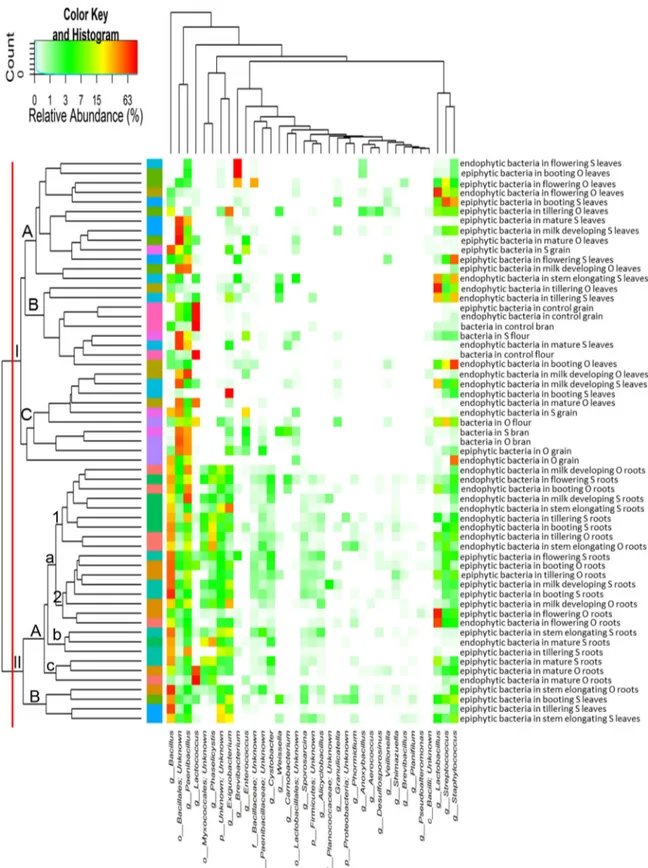
FIG 2 Heat map summarizing the relative abundances of the 35 most dominant genera in DNA samples directly extracted from epiphytic and endophytic
bacteria found on hypogeous organs (roots), epigeous organs (leaves and spikes), and processed wheat (grain, chaff, bran, and flour) of the Odisseo (O) and Saragolla (S) varieties. Hypogeous and epigeous organs were analyzed at the tillering, stem elongation, booting, flowering, milk development, and physiological maturity stages. Commercial grain, bran, and flour were also analyzed as a control. The color key defines the percentages of OTUs in the samples.
Firmicutes Dynamics in Durum Wheat
on November 30, 2016 by guest
http://aem.asm.org/
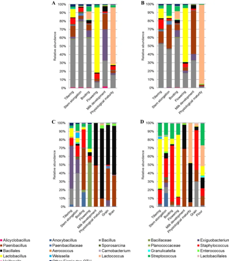
FIG 3 Relative abundance of epiphytic (A and C) and endophytic (B and D) Firmicutes genera found in hypogeous organs (roots) (A and B), epigeous organs
(leaves and spikes), and processed wheat (grain, bran, and flour) (C and D) of Odisseo. Hypogeous and epigeous organs were analyzed at the tillering, stem elongation, booting, flowering, milk development, and physiological maturity stages.
on November 30, 2016 by guest
http://aem.asm.org/
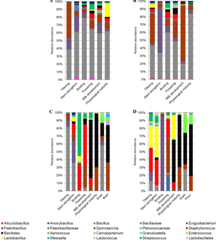
FIG 4 Relative abundance of epiphytic (A and C) and endophytic (B and D) Firmicutes genera found in hypogeous organs (roots) (A and B), epigeous organs
(leaves and spikes), and processed wheat (grain, bran, and flour) (C and D) of Saragolla. Hypogeous and epigeous organs were analyzed at the tillering, stem elongation, booting, flowering, milk development and physiological maturity stages.
Firmicutes Dynamics in Durum Wheat
on November 30, 2016 by guest
http://aem.asm.org/
statistical tests indicated a significant influence of roots and leaves
or spikes on the microbial diversity (FDR
⬍ 0.050). Leaves did not
differ (P
⬎ 0.050) from spikes and processed wheat samples.
Core Firmicutes microbiome. According to alpha- and
beta-diversity, and considering the 35 most dominant genera of all
samples, roots and leaves or spikes were distributed in two clusters
(
Fig. 2
). Exceptions were the epiphytic bacteria belonging to
til-lering, stem elongation, and booting of Saragolla wheat. With few
exceptions, epiphytic and endophytic bacteria grouped in
differ-ent subclusters (cluster IA versus clusters IB and IC; cluster IIAa1
versus cluster IIAa2). Samples corresponding to endophytic
bac-teria found in the last phenological stages (milk development and
physiological maturity) clustered together with processed wheat
samples of both cultivars (cluster IC). A core Firmicutes
micro-biome, defined at the genus level and occurring in
⬎98% of the
samples analyzed was constituted by Bacillus, Exiguobacterium,
Lactobacillus, Paenibacillus, Staphylococcus, and Streptococcus.
Be-sides these genera, OTUs belonging to genera Enterococcus and
Lactococcus occurred in
⬎85% of the samples.
Among Firmicutes, Bacillus was mainly found at the
endo-phytic and, especially, epiendo-phytic levels of roots throughout the
wheat life cycle, whereas it was only occasionally detected in leaves
and spikes (
Fig. 3
and
4
). Paenibacillus was found at the
endo-phytic and epiendo-phytic levels of roots regardless of phenological
stage. However, the relative abundance of Paenibacillus was lower
than that of Bacillus in more than 60% of root samples. In
con-trast, in samples of leaves and spikes, from flowering to
physiolog-ical maturity, Paenibacillus was more frequently detected than
Ba-cillus. Exiguobacterium was found in roots throughout the wheat
life cycle, whereas it was rarely detected in leaves. However, it was
the dominant Firmicutes genus at the epiphytic level of Odisseo
leaves at tillering (
Fig. 3C
) and at the endophytic level of Saragolla
leaves at the booting stage (
Fig. 4D
). Bacillales were among the
dominant OTUs at milk development, at physiological maturity,
and in processed wheat, especially for Saragolla.
At the endophytic level, Lactobacillus was the dominant
Firmi-cutes genus at the flowering stage of Odisseo, both in roots and in
spikes (
Fig. 3B
and
D
). Lactobacillus dominated also in roots at the
FIG 5 Relative abundance of endophytic (END) and epiphytic (EPI) Lactobacillus plantarum within the OTUs belonging to lactic acid bacteria found on
hypogeous organs (roots) (A and B), epigeous organs (leaves and spikes), and processed wheat (grain, bran, and flour) (C and D) of Odisseo (A and C) and Saragolla (B and D). Hypogeous and epigeous organs were analyzed at the tillering, stem elongation, booting, flowering, milk development, and physiological maturity stages.
on November 30, 2016 by guest
http://aem.asm.org/
epiphytic level during flowering (
Fig. 3A
). Lactococcus dominated
in roots (endophytic and epiphytic levels) and spikes (endophytic
level) at physiological maturity. Lactococcus was the lactic acid
bacterium mostly present in grain at the endophytic level and
dominated in flour together with Streptococcus. The genus
Lacto-bacillus was found at a lower level in Saragolla than in Odisseo
wheat (
Fig. 4
). At the flowering stage, Streptococcus was the
dom-inant lactic acid bacterium in roots and spikes. Lactobacillus was
the dominant lactic acid bacterial genus only at the endophytic
level of spikes during milk development (
Fig. 4D
) and at the
epi-phytic level of roots during physiological maturity (
Fig. 4A
).
En-terococcus was the dominant genus of lactic acid bacteria in grain
at the endophytic level and dominated in flour. Lactococcus
dom-inated in commercial grain and in the related bran and flour
sam-ples (see Table S3 in the supplemental material). At the species
level, L. plantarum was the dominant species of Lactobacillus in
almost all samples (
Fig. 5
; see also Table S3). Other lactic acid
bacteria (Aerococcus viridans, Carnobacterium maltaromaticum,
Enterococcus faecalis, Lactobacillus brevis, Lactobacillus fermentum,
Lactobacillus gasseri, Lactobacillus iners, Lactococcus garviae, and
Lactococcus lactis) were variously identified.
Cell density of endophytic presumptive lactic acid bacteria.
Selective media (mMRS and SDB) were used to detect endophytic
presumptive lactic acid bacteria (
Fig. 6
). Lactic acid bacteria were
detected in all plant organs of Odisseo and Saragolla durum wheat
at different phenological stages, with cell densities varying from
ca. 0.5 to 8.5 log CFU g
⫺1. SDB was the medium showing the
highest cell densities. Roots of Odisseo showed a higher cell
den-sity of presumptive lactic acid bacteria during tillering, stem
elon-gation, and booting stages than did Saragolla. Roots showed the
highest cell density of presumptive lactic acid bacteria during the
flowering and physiological maturity stages of Odisseo and
Sara-golla durum wheat, respectively. Leaves and spikes showed the
highest cell density during booting for Saragolla and flowering
and physiological maturity for Odisseo.
Isolation and phenotypic identification of endophytic lactic
acid bacteria. Presumptive lactic acid bacteria were isolated (10
colonies for roots or leaves/spikes in each phenological stage) and
tested for the ability to acidify the culture medium. All
Gram-positive, catalase-negative, acidifying rods or cocci were identified
by Biolog AN microplates. L. plantarum was identified in all plant
organs at all different phenological stages. Other lactic acid
bacte-ria (Lactobacillus helveticus, Lactobacillus coryniformis,
Lactobacil-lus delbrueckii, L. sanfranciscensis, Weissella confusa, Weissella
mi-nor, Streptococcus anginosus, Leuconostoc citreum, and Pediococcus
parvulus) were variously identified (data not shown). Within
lac-tobacilli, L. plantarum dominated in roots and samples of leaves
and spikes together with L. coryniformis until the flowering stage.
L. plantarum, L. helveticus, and L. delbrueckii dominated in both
roots and spikes during the milk development and physiological
maturity stages of Odisseo and Saragolla wheat (data not shown).
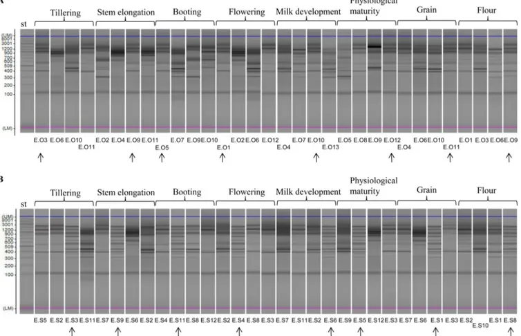
Genotypic characterization of endophytic L. plantarum
strains. Since L. plantarum was the only lactic acid bacterial
spe-cies identified at all phenological stages, RAPD-PCR analysis was
applied to track the strains of this species. All isolates identified as
L. plantarum were characterized by using single primer P4, P7, or
FIG 6 Cell numbers of endophytic presumptive lactic acid bacteria enumerated on mMRS and SDB in hypogeous (roots) and epigeous (leaves and spikes) organs
of Odisseo (A and B, respectively) and Saragolla (C and D, respectively) durum wheat during the life cycle from tillering to flour.
Firmicutes Dynamics in Durum Wheat
on November 30, 2016 by guest
http://aem.asm.org/
M13. Several isolates of L. plantarum at different phenological
stages of growth of Odisseo (
Fig. 7A
; see also Fig. S3A and S4A in
the supplemental material) and Saragolla (
Fig. 7B
; see also Fig.
S3B and S4B) wheat as well as in the processed wheat samples
(grain and flour) (
Fig. 7
; see also Fig. S3 and S4) showed the same
profile.
Correlation between lactic acid bacteria, environmental
temperature, and wheat organ a
w. OTU correlation was
investi-gated considering Firmicutes genus level (
Fig. 8
) taxonomic
as-signments and significant correlations at an FDR of
⬍0.050.
Lac-tobacillus showed positive correlation with Anoxybacillus,
Staphylococcus, Streptococcus, and Weissella. Lactobacillus was
neg-atively correlated mainly with Bacillus and Paenibacillus.
Weis-sella, Lactococcus, and Enterococcus showed positive correlation
with many genera, including other lactic acid bacteria (Aerococcus,
Granulicatella, and Carnobacterium). Cell density of presumptive
lactic acid bacteria, relative abundance of OTU belonging to lactic
acid bacterial genera, environmental temperature, and wheat
or-gan a
wdata were analyzed using PCA. The discrimination of
sam-ples between tillering, stem elongation, and booting and milk
de-velopment, physiological maturity, grain, chaff, flour, and bran
was evident (see Fig. S5 in the supplemental material). The cell
density of presumptive lactic acid bacteria, Lactococcus and
En-terococcus, was positively correlated (r
⬎ 0.5) with environmental
temperature. Lactobacillus showed a higher positive correlation
(r
⫽ 0.72) with a
wthan did Lactococcus and Enterococcus.
DISCUSSIONThis study used the 16S rRNA gene-based high-throughput
se-quencing approach targeting DNA to describe the ecology
dy-namics of Firmicutes and, especially, lactic acid bacteria during
growth of durum wheat plants. Epiphytic and endophytic
Firmi-cutes were analyzed to track the main source of contaminating
lactic acid bacteria from plant to flour. Previously, lactic acid
bac-teria were found to be components of the epiphytic and/or
endo-phytic microbiota of forage crops and roots of Lolium perenne
(
28–30
). The biodiversity of Firmicutes was higher in roots than in
leaves and spikes. At the contact zone with soil, plants host a
dis-tinctive root-associated bacterial microbiota, which is believed to
have a positive role in plant nutrition and health (
48
). Diverse
bacterial populations were described for the plant rhizosphere and
phyllosphere. Beta-diversity indices showed that the composition
of Firmicutes in durum wheat spikes was similar to that of
pro-cessed grains. Bacillus, Exiguobacterium, Paenibacillus,
Staphylo-coccus, Lactobacillus, StreptoStaphylo-coccus, EnteroStaphylo-coccus, and Lactococcus
were the core genera of wheat plant and processed samples. All the
above-mentioned Firmicutes were previously identified in wheat
flour (
16
,
18
,
49
), strengthening the hypothesis that the wheat
FIG 7 Representative randomly amplified polymorphic DNA-PCR (RAPD-PCR) profiles of Lactobacillus plantarum isolated at the endophytic level from leaves
and spikes, grain, or flour of Odisseo (A) and Saragolla (B) durum wheat. Primer M13 was used for RAPD-PCR analysis. A 2-log DNA ladder (0.1 to 10.0 kb) was used as a molecular size standard (st). Capillary electrophoretic profiles were singly acquired by MultiNA. Strains isolated from different phenological stages and processed wheat and showing similar RAPD-PCR profiles are indicated by arrows.
on November 30, 2016 by guest
http://aem.asm.org/
plant microbiota affected the microbial composition of the flour.
Based on the results of this study, the spike or grain microbiota
strongly contaminated the related flour. When flour is used as the
base for producing sourdough, the most adapted microorganisms
(mainly consisting of lactobacilli) are selected, leading to mature
sourdough, whose microbiota greatly differs from that of flour
(
18
). Dominant lactic acid bacteria and spore-forming bacteria in
wheat flour could affect the quality of bread and other baked
goods.
Within the core microbiota of wheat plant and processed
sam-ples, the relative abundance of each genus was affected by the plant
organs, the cultivars, and the phenological stages. Previously, it
was shown that microorganisms can randomly emerge from
neighboring environmental ecosystems, but their survival is
reg-ulated by the plant (
50
). Overall, Bacillus was the most abundant
genus in roots, especially at the epiphytic level. In this study,
Bacil-lales were found as dominant OTUs in the Saragolla flour. Besides
Bacillus, other Firmicutes commonly found in the rhizosphere,
such as Paenibacillus and Exiguobacterium, promote plant growth
(
51
). Exiguobacterium acetylicum, inoculated on wheat seeds,
pos-itively affected plant growth during the first phenological stages
(
52
). In this study, Exiguobacterium was found as the dominant
genus at the endophytic level of roots and leaves, at the stem
elon-gation and booting stages, respectively, of Saragolla wheat.
Recently, a relatively high number and diversity of lactic acid
bacteria was demonstrated in agricultural soils (
53
).
Culture-de-pendent, denaturing gradient gel electrophoresis (DGGE), and
16S rRNA gene sequencing analyses revealed the following main
genera: Lactococcus, Leuconostoc, Weissella, Lactobacillus, and
En-terococcus (
53
,
54
). Saragolla showed a lower relative abundance of
FIG 8 Significant correlations between lactic acid bacterial OTUs (genus level) found on hypogeous organs (roots), epigeous organs (leaves and spikes), and
processed wheat (grain, chaff, bran, and flour) of Odisseo and Saragolla. Hypogeous and epigeous organs were analyzed at the tillering, stem elongation, booting, flowering, milk development, and physiological maturity stages. The colors of the scale bar denote the nature of the correlation, with 1 indicating a perfectly positive correlation (red) and⫺1 indicating a perfectly negative correlation (green) between two microbial orders, families, or genera. Only significant correlations (FDR⬍ 0.05) are shown.
Firmicutes Dynamics in Durum Wheat
on November 30, 2016 by guest
http://aem.asm.org/
OTUs belonging to Lactobacillus in roots, leaves, and spikes than
did Odisseo durum wheat. Saragolla had the fastest increase of
OTUs belonging to lactic acid bacteria (highest abundance for
Streptococcus) in roots and, especially, at epiphytic levels (leaves
and spikes) during booting. At the endophytic level (spikes),
Sara-golla showed the highest number of OTUs belonging to
Lactoba-cillus during milk development. However, the relative abundance
of Lactobacillus markedly decreased at physiological maturity. The
influence of pesticides, farming practice, and atmosphere also on
the composition of the microbial communities should not be
ex-cluded. Since the accelerated flowering stage of Saragolla, the
en-vironmental conditions differed between the phenological stages
of the two cultivars. The lactic acid bacterial composition of spikes
directly affected the bacterial community of processed wheat
sam-ples. As hypothesized, the OTUs found as endophytic bacteria of
grains (Lactobacillus, Lactococcus, Enterococcus, and Streptococcus)
were also found in the flour. L. plantarum was the only species
identified in both cultivars at all the phenological stages.
Com-mercial processed wheat grain (used as the control) harbored a
high relative abundance of endophytic Lactococcus, which
re-flected on the related flour. Some strains of Lactococcus lactis
showed significant biocontrol activity and/or direct plant growth
promotion (
55
). Recently, it was shown that the inoculum of
wheat roots with L. plantarum decreased the oxidative stress (
56
).
Lactobacilli were shown to be effective against biotic stresses, due
to their high antagonistic activity against plant pathogens (
57
).
With the exception of L. plantarum, which is known to be a
mem-ber of the sourdough microbiota, other lactic acid bacteria are
present only as intermediate organisms in spontaneous
sour-dough until more characteristic microorganisms become
pre-dominant (
16
).
As shown by cultivation on mMRS and SDB, the cell density of
presumptive lactic acid bacteria differed depending on the cultivar
and/or the phenological stage. Saragolla durum wheat, which had
an accelerated flowering stage, showed a faster increase of the cell
density of presumptive lactic acid bacteria in leaves and spikes
during booting than did Odisseo wheat. According to the 16S
rRNA gene-based high-throughput sequencing, L. plantarum was
the endophytic lactic acid bacterial species found in both cultivars
at all the phenological stages, grain, and flour. This finding
rein-forced the concept that L. plantarum has a strong ecological or
metabolic adaptability to different habitats (
58
,
59
). Mesophilic
lactobacilli (e.g., L. coryniformis) were mainly isolated in the first
phenological stages. In contrast, thermophilic lactobacilli (L.
hel-veticus and L. delbrueckii) were the highest in the milk
develop-ment and physiological maturity stages.
Overall, the relative abundance of lactic acid bacterial genera
was affected by other Firmicutes genera, environmental
tempera-ture, and a
wof plant organs. Lactobacilli showed the highest
pos-itive correlation with a
w. This finding could explain the strong
decrease of OTU belonging to Lactobacillus during milk
develop-ment (Odisseo) and physiological maturity (Saragolla). At these
stages, Lactobacillus was mainly replaced by Streptococcus,
Lacto-coccus, and Enterococcus. With the goal of decreasing chemical
treatments (e.g., fertilizers, herbicides, and fungicides),
microor-ganism-based commercial products were used to promote plant
growth and to combat biotic disease (
50
,
60
).
The results of this study highlight that the wheat microbiota
differed from those of the grains and flour. Only a few
microor-ganisms identified in wheat plants play a role in sourdough.
Fur-ther experimental approaches, e.g., experiments involving
com-petition of wheat isolates with sourdough isolates, are necessary to
understand whether the L. plantarum strains from wheat grains
dominate in mature sourdoughs.
ACKNOWLEDGMENT
This work was partially funded by the Istituto Agronomico Mediterraneo (IAM) Bari, project FOODING.
REFERENCES
1. Ikeda DM, Weinert E, Chang KCS, McGinn JM, Miller SA,
Kelii-hoomalu C, DuPonte MW. August 2013. Natural farming: lactic acid
bacteria. Sustain Agric 2013:1– 4.
2. Arkhipova TN, Anokhina NL. 2009. Effects of wheat plant inoculation with cytokinin-producing microorganisms on plant growth at increasing level of mineral nutrition. Russ J Plant Physiol 56:814 – 819.http://dx.doi .org/10.1134/S1021443709060119.
3. Zhao Y, Selvaraj JN, Xing F, Zhou L, Wang Y, Song H, Tan X, Sun L,
Sangare L, Folly YM, Liu Y. 2014. Antagonistic action of Bacillus subtilis
strain SG6 on Fusarium graminearum. PLoS One 9:e92486.http://dx.doi .org/10.1371/journal.pone.0092486.
4. Bulgarelli D, Schlaeppi K, Spaepen S, Ver Loren van Themaat E,
Schulze-Lefert P. 2013. Structure and functions of the bacterial
microbi-ota of plants. Annu Rev Plant Biol 64:807– 838.http://dx.doi.org/10.1146 /annurev-arplant-050312-120106.
5. De Vuyst L, Vrancken G, Ravyts F, Rimaux T, Weckx S. 2009. Biodi-versity, ecological determinants, and metabolic exploitation of sourdough microbiota. Food Microbiol 26:666 – 675.http://dx.doi.org/10.1016/j.fm .2009.07.012.
6. Gobbetti M, Rizzello CG, Di Cagno R, De Angelis M. 2014. How the sourdough may affect the functional features of leavened baked goods. Food Microbiol 37:30 – 40.http://dx.doi.org/10.1016/j.fm.2013.04.012. 7. Valerio F, De Bellis P, Di Biase M, Lonigro SL, Giussani B, Visconti A,
Lavermicocca P, Sisto A. 2012. Diversity of spore-forming bacteria and
identification of Bacillus amyloliquefaciens as a species frequently associ-ated with the ropy spoilage of bread. Int J Food Microbiol 156:278 –285. http://dx.doi.org/10.1016/j.ijfoodmicro.2012.04.005.
8. Gobbetti M. 1998. Interactions between lactic acid bacteria and yeasts in sourdoughs. Trends Food Sci Technol 9:267–274.http://dx.doi.org/10 .1016/S0924-2244(98)00053-3.
9. Vogel RF, Knorr R, Müller MRA, Steudel U, Gänzle MG, Ehrmann M. 1999. Non dairy lactic fermentations: the cereal world. Antonie Van Leeu-wenhoek 76:403– 411.http://dx.doi.org/10.1023/A:1002089515177. 10. Gobbetti M, De Angelis M, Corsetti A, Di Cagno R. 2005. Biochemistry
and physiology of sourdough lactic acid bacteria. Trends Food Sci Technol
16:57– 69.http://dx.doi.org/10.1016/j.tifs.2004.02.013.
11. Gobbetti M, Corsetti A, Rossi J. 1994. The sourdough microflora. Inter-actions between lactic acid bacteria and yeasts: metabolism of amino acids. World J Microbiol Biotechnol 10:275–279.
12. De Vuyst L, Schrijvers V, Paramithiotis S, Hoste B, Vancanneyt M,
Swings J, Kalantzopoulos G, Tsakalidou E, Winy M. 2002. The
biodiversity of lactic acid bacteria in Greek traditional wheat sour-doughs is reflected in both composition and metabolite formation. Appl Environ Microbiol 68:6059 – 6069. http://dx.doi.org/10.1128 /AEM.68.12.6059-6069.2002.
13. Meroth CB, Walter J, Hertel C, Brandt MJ, Hammes WP. 2003. Mon-itoring the bacterial population dynamics in sourdough fermentation processes by using PCR-denaturing gradient gel electrophoresis. Appl En-viron Microbiol 69:475– 482. http://dx.doi.org/10.1128/AEM.69.1.475 -482.2003.
14. Scheirlinck I, Van der Meulen R, Van Schoor A, Vancanneyt M, De
Vuyst L, Vandamme P, Huys G. 2007. Influence of geographical origin
and flour type on diversity of lactic acid bacteria in traditional Belgian sourdoughs. Appl Environ Microbiol 73:6262– 6269.http://dx.doi.org/10 .1128/AEM.00894-07.
15. Minervini F, Di Cagno R, Lattanzi A, De Angelis M, Antonielli L,
Cardinali G, Cappelle S, Gobbetti M. 2012. Lactic acid bacterium and
yeast microbiotas of 19 sourdoughs used for traditional/typical Italian breads: interactions between ingredients and microbial species diversity. Appl Environ Microbiol 78:1251–1264.http://dx.doi.org/10.1128/AEM .07721-11.
on November 30, 2016 by guest
http://aem.asm.org/
16. Ercolini D, Pontonio E, De Filippis F, Minervini F, La Storia A,
Gobbetti M, Di Cagno R. 2013. Microbial ecology dynamics during rye
and wheat sourdough fermentation. Appl Environ Microbiol 79:7827– 7836.http://dx.doi.org/10.1128/AEM.02955-13.
17. Scheirlinck I, Van der Meulen R, De Vuyst L, Vandamme P, Huys G. 2009. Molecular source tracking of predominant lactic acid bacteria in traditional Belgian sourdoughs and their production environments. J Appl Microbiol 106:1081–1092. http://dx.doi.org/10.1111/j.1365-2672 .2008.04094.x.
18. Minervini F, Lattanzi A, De Angelis M, Celano G, Gobbetti M. 2015. House microbiotas as sources of lactic acid bacteria and yeasts in tradi-tional Italian sourdoughs. Food Microbiol 52:66 –76.http://dx.doi.org/10 .1016/j.fm.2015.06.009.
19. Van der Meulen R, Scheirlinck I, Van Schoor A, Huys G, Vancanneyt
M, Vandamme P, De Vuyst L. 2007. Population dynamics and
metabo-lite target analysis during laboratory fermentations of wheat and spelt sourdoughs. Appl Environ Microbiol 73:4741– 4750.http://dx.doi.org/10 .1128/AEM.00315-07.
20. Scheirlinck I, Van der Meulen R, Van Schoor A, Vancanneyt M, De
Vuyst L, Vandamme P, Huys G. 2008. Taxonomic structure and stability
of the bacterial community in Belgian sourdough ecosystems as assessed by culture and population fingerprinting. Appl Environ Microbiol 74: 2414 –2423.http://dx.doi.org/10.1128/AEM.02771-07.
21. Weckx S, Van der Meulen R, Allemeersch J, Huys G, Vandamme P, Van
Hummelen P, De Vuyst L. 2010. Community dynamics of bacteria in
sourdough fermentations as revealed by their metatranscriptome. Appl Environ Microbiol 76:5402–5408.http://dx.doi.org/10.1128/AEM .00570-10.
22. Weckx S, Van der Meulen R, Maes D, Scheirlinck I, Huys G,
Vandamme P, De Vuyst L. 2010. Lactic acid bacteria community
dynamics and metabolite production of rye sourdough fermentations share characteristics of wheat and spelt sourdough fermentations. Food Microbiol 27:1000 –1008. http://dx.doi.org/10.1016/j.fm.2010 .06.005.
23. Su MSW, Oh PL, Walter J, Gänzle MG. 2012. Intestinal origin of sourdough Lactobacillus reuteri isolates as revealed by phylogenetic, ge-netic, and physiological analysis. Appl Environ Microbiol 78:6777– 6780. http://dx.doi.org/10.1128/AEM.01678-12.
24. Du Toit M, Dicks LMT, Holzapfel WH. 2003. Identification of het-erofermentative lactobacilli isolated from pig faeces by numerical anal-ysis of total soluble cell protein and RAPD patterns. Lett Appl Micro-biol 37:12–16.http://dx.doi.org/10.1046/j.1472-765X.2003.01334.x. 25. De Angelis M, Siragusa S, Berloco M, Caputo L, Settanni L, Alfonsi G,
Amerio M, Grandi A, Ragni A, Gobbetti M. 2006. Selection of potential
probiotic lactobacilli from pig feces to be used as additives in pelleted feeding. Res Microbiol 157:792– 801.http://dx.doi.org/10.1016/j.resmic .2006.05.003.
26. Groenewald WH, Van Reenen CA, Todorov SD, Du Toit M, Corli
Witthuhn RC, Holzapfel WH, Dicks LMT. 2006. Identification of lactic
acid bacteria from vinegar flies based on phenotypic and genotypic char-acteristics. Am J Enol Vit 57:519 –525.
27. Alfonzo A, Ventimiglia G, Corona O, Di Gerlando R, Gaglio R,
Fran-cesca N, Moschetti G, Settanni L. 2013. Diversity and technological
potential of lactic acid bacteria of wheat flours. Food Microbiol 36:343– 354.http://dx.doi.org/10.1016/j.fm.2013.07.003.
28. Zhang JG, Cai Y, Kobayashi R, Kumai S. 2000. Characteristics of lactic acid bacteria isolated from forage crops and their effects on si-lage fermentation. J Sci Food Agric 80:1455–1460.http://dx.doi.org/10 .1002/1097-0010(200008)80:10⬍1455::AID-JSFA667⬎3.0.CO;2-C. 29. Pang H, Tan Z, Qin G, Wang Y, Li Z, Jin Q, Cai Y. 2012. Phenotypic and
phylogenetic analysis of lactic acid bacteria isolated from forage crops and grasses in the Tibetan Plateau. J Microbiol 50:63–71.http://dx.doi.org/10 .1007/s12275-012-1284-5.
30. Gaggìa F, Baffoni L, Di Gioia D, Accorsi M, Bosi S, Marotti I, Biavati
B, Dinelli G. 2013. Inoculation with microorganisms of Lolium perenne
L.: evaluation of plant growth parameters and endophytic colonization of roots. N Biotechnol 30:695–704.http://dx.doi.org/10.1016/j.nbt.2013.04 .006.
31. Herbert S, Bouchet B, Riaublanc A, Dufour E, Gallant DJ. 1999. Multiple fluorescence labelling of proteins, lipids and whey in dairy prod-ucts using confocal microscopy. Lait 79:567–575.http://dx.doi.org/10 .1051/lait:1999646.
32. Kinkel LL, Wilson M, Lindow SE. 1996. Utility of microcosm studies for
predicting phylloplane bacterium population sizes in the field. Appl En-viron Microbiol 62:3413–3423.
33. Suda W, Nagasaki A, Shishido M. 2009. Powdery mildew-infection changes bacterial community composition in the phyllosphere. Microbes Environ 24:217–223.http://dx.doi.org/10.1264/jsme2.ME09114. 34. Stock JD, Hirano SS. 1991. Location of phyllosphere bacteria on or within
leaves of field-grown snap bean plants. Phytopathology 81:1222. 35. Idris R, Trifonova R, Puschenreiter M, Wenzel WW, Sessitsch A. 2004.
Bacterial communities associated with flowering plants of the Ni hyper-accumulator Thlaspi goesingense. Appl Environ Microbiol 70:2667–2677. http://dx.doi.org/10.1128/AEM.70.5.2667-2677.2004.
36. Minervini F, De Angelis M, Di Cagno R, Pinto D, Siragusa S, Rizzello
CG, Gobbetti M. 2010. Robustness of Lactobacillus plantarum starters
during daily propagation of wheat flour sourdough type I. Food Microbiol
27:897–908.
37. Muh¨ling M, Woolven-Allen J, Murrell JC, Joint I. 2008. Improved group-specific PCR primers for denaturing gradient gel electrophoresis analysis of the genetic diversity of complex microbial communities. ISME J 2:379 –392.http://dx.doi.org/10.1038/ismej.2007.97.
38. Zhang J, Kobert K, Flouri T, Stamatakis A. 2014. PEAR: a fast and accurate Illumina paired-end reAd mergeR. Bioinformatics 30:614 – 620. http://dx.doi.org/10.1093/bioinformatics/btt593.
39. Edgar RC. 2010. Search and clustering orders of magnitude faster than BLAST. Bioinformatics 26:2460 –2461.http://dx.doi.org/10.1093 /bioinformatics/btq461.
40. Edgar RC. 2013. UPARSE: highly accurate OTU sequences from micro-bial amplicon reads. Nat Methods 10:996 –998.http://dx.doi.org/10.1038 /nmeth.2604.
41. Edgar RC, Haas BJ, Clemente JC, Quince C, Knight R. 2011. UCHIME improves sensitivity and speed of chimera detection. Bioinformatics 27: 2194 –2200.http://dx.doi.org/10.1093/bioinformatics/btr381.
42. Andreotti R, Perez de Leon AA, Dowd SE, Guerrero FD, Bendele
KG, Scoles GA. 2011. Assessment of bacterial diversity in the cattle
tick Rhipicephalus (Boophilus) microplus through tag-encoded pyrose-quencing. BMC Microbiol 11:6.http://dx.doi.org/10.1186/1471-2180 -11-6.
43. Kline L, Sugihara TF. 1971. Microorganisms of the San Francisco sour dough bread process. II. Isolation and characterization of undescribed bacterial species responsible for the souring activity. Appl Microbiol 21: 459 – 465.
44. Ahmed W, Sawant S, Huygens F, Goonetilleke A, Gardner T. 2009. Prevalence and occurrence of zoonotic bacterial pathogens in surface wa-ters determined by quantitative PCR. Water Res 43:4918 – 4928.http://dx .doi.org/10.1016/j.watres.2009.03.041.
45. De Angelis M, Corsetti A, Tosti N, Rossi J, Corbo MR, Gobbetti M. 2001. Characterization of non-starter lactic acid bacteria from Italian ewe cheeses based on phenotypic, genotypic and cell wall protein analyses. Appl Environ Microbiol 67:2011–2020.http://dx.doi.org/10.1128/AEM .67.5.2011-2020.2001.
46. Siragusa S, Di Cagno R, Ercolini D, Minervini F, Gobbetti M, De
Angelis M. 2009. Taxonomic structure and monitoring of the
domi-nant population of lactic acid bacteria during wheat flour sourdough type I propagation using Lactobacillus sanfranciscensis starters. Appl Environ Microbiol 75:1099 –1109. http://dx.doi.org/10.1128/AEM .01524-08.
47. Suchodolski JS, Dowd SE, Wilke V, Steiner JM, Jergens AE. 2012. 16S rRNA gene pyrosequencing reveals bacterial dysbiosis in the duo-denum of dogs with idiopathic inflammatory bowel disease. PLoS One
7:e39333.http://dx.doi.org/10.1371/journal.pone.0039333.
48. Schlaeppi K, Dombrowski N, Oter RG, van Themaat EVL,
Schulze-Lefert P. 2014. Quantitative divergence of the bacterial root microbiota in
Arabidopsis thaliana relatives. Proc Natl Acad Sci U S A 111:585–592.http: //dx.doi.org/10.1073/pnas.1321597111.
49. Rizzello CG, Cavoski I, Turk J, Ercolini D, Nionelli L, Pontonio E, De
Angelis M, De Filippis F, Gobbetti M, Di Cagno R. 2015. Organic
cultivation of Triticum turgidum subsp. durum is reflected in the flour-sourdough fermentation-bread axis. Appl Environ Microbiol 81:3192– 3204.http://dx.doi.org/10.1128/AEM.04161-14.
50. Berlec A. 2012. Novel techniques and findings in the study of plant mi-crobiota: search for plant probiotics. Plant Sci 193–194:96 –102. 51. Vessey JK. 2003. Plant growth promoting rhizobacteria as biofertilizers.
Plant Soil 255:571–586.http://dx.doi.org/10.1023/A:1026037216893. 52. Selvakumar G, Joshi P, Nazim S, Mishra PK, Kundu S, Gupta HS.
Firmicutes Dynamics in Durum Wheat
on November 30, 2016 by guest
http://aem.asm.org/
2009. Exiguobacterium acetylicum strain 1P (MTCC 8707) a novel bac-terial antagonist from the North Western Indian Himalayas. World J Microbiol Biotechnol 25:131–137. http://dx.doi.org/10.1007/s11274 -008-9874-4.
53. Somers E, Amke A, Croonenborghs A, van Overbeek LS, Vanderleyden
J. 2007. Lactic acid bacteria in organic agricultural soils, abstr 367827.
Abstr Plant Res Int, Montpellier, France.
54. Yang L, Deng Y, Zhang H, Diao Q. 2012. Isolation and characterization of an Enterococcus strain from Tibetan alpine meadow soil. Wei Sheng Wu Xue Bao 52:1421–1426.
55. Shrestha A, Kim EC, Lim CK, Cho S, Hur JH, Park DH. 2009. Biological control of soft rot on chinese cabbage using beneficial bacterial agents in greenhouse and field. Korean J Pestic Sci 13:325–331.
56. Yarullina DR, Asafova EV, Kartunova JE, Ziyatdinova GK, Ilinskaya
ON. 2014. Probiotics for plants: NO-producing lactobacilli protect plants
from drought. Appl Biochem Microbiol 50:166 –168.http://dx.doi.org/10 .1134/S0003683814020197.
57. Stoyanova LG, Ustyugova EA, Netrusov AI. 2012. Antibacterial metab-olites of lactic acid bacteria: their diversity and properties. Appl Biochem Microbiol 48:229 –243.http://dx.doi.org/10.1134/S0003683812030143. 58. Siezen RJ, Tzeneva VA, Castioni A, Wels M, Phan HT, Rademaker
JL, Starrenburg MJ, Kleerebezem M, Molenaar D, van Hylckama Vlieg JE. 2010. Phenotypic and genomic diversity of Lactobacillus
plantarum strains isolated from various environmental niches. En-viron Microbiol 12:758 –773. http://dx.doi.org/10.1111/j.1462-2920 .2009.02119.x.
59. de Melo Pereira GV, Magalhàes KT, Lorenzetti ER, Souza TP, Schwan
RF. 2012. A multiphasic approach for the identification of endophytic
bacterial [sic] in strawberry fruit and their potential for plant growth pro-motion. Microb Ecol 63:405– 417.http://dx.doi.org/10.1007/s00248-011 -9919-3.
60. Wani ZA, Ashraf N, Mohiuddin T, Riyaz-Ul-Hassan S. 2015. Plant-endophyte symbiosis, an ecological perspective. Appl Microbiol Bio-technol 99:2955–2965.http://dx.doi.org/10.1007/s00253-015-6487-3.