Trafficking properties of plasmacytoid
dendritic cells in health and disease
Silvano Sozzani
1, William Vermi
2, Annalisa Del Prete
3and Fabio Facchetti
2 1Department of Biomedical Sciences and Biotechnology, University of Brescia, Viale Europa 11, 25123 Brescia, Italy 2Department of Pathology, University of Brescia, P.le Spedali Civili 1, 25123 Brescia, Italy
3Istituto Clinico Humanitas, via Manzoni 58, 20089 Rozzano; Department of Medical Biochemistry, Biology and Physics, University of Bari, P.le G. Cesare 11, 70124 Bari, Italy
Plasmacytoid dendritic cells (PDCs) represent a subset of
circulating leukocytes characterized by the ability to
release high levels of type I interferon (IFN). Under
homeostatic conditions PDCs are confined to primary
and secondary lymphoid organs. This is consistent
with the restricted profile of functional chemotactic
receptors expressed by circulating PDCs (i.e. CXCR4
and ChemR23). Accumulation of PDCs in non-lymphoid
tissue is, however, observed in certain autoimmune
diseases, allergic reactions and tumors. Indeed, PDCs
are now considered to be involved in the pathogenesis of
diseases characterized by a type I IFN-signature and are
considered as a promising target for new intervention
strategies. Here, current knowledge of the molecular
mechanisms involved in the recruitment of PDCs
under homeostatic and pathological conditions are
summarized.
Background on PDCs: history and function
The cell now recognized as a plasmacytoid dendritic cell
(PDCs) was originally described as a ‘‘lymphoblast’’ on the
basis of its morphological features. It was subsequently
renamed ‘‘T-associated plasma cell’’ due to the abundance
of rough endoplasmic reticulum, ‘‘plasmacytoid T cell’’ by
reason of CD4 expression, and ‘‘plasmacytoid monocyte’’
for the expression of some myelo-monocytic markers in the
absence of B- and T-cell specific antigens
[1]
. The function
of PDCs remained completely unknown until it was
reported that PDCs represent a subset of dendritic cells
(DCs)
[2]
, and that they correspond to the natural type I
interferon (IFN)-producing cells
[3,4]
.
PDCs originate in the bone marrow, from both myeloid
and lymphoid precursors, although myeloid derivation is
predominant, where a dendritic cell (DCs) progenitor gives
rise to PDCs and classical ‘‘myeloid’’ DCs (MDCs)
[5,6]
.
PDCs can differentiate into MDCs after stimulation and
genome-wide analysis showed that the gene expression
profile of PDCs is closer to MDCs than to other
hemato-poietic cells
[7,8]
. The development of both PDCs and
MDCs depends on Fms-like kinase 3 ligand (Flt3L), but
the E2-2 transcription factor is uniquely required for PDCs
differentiation
[9,10]
.
Peripheral human PDCs have a unique cell surface
phenotype: they lack B cell, T cell, myeloid cell, and NK
cell lineage markers, and express CD4, CD123 (IL3Ra),
CD303 (BDCA2), ILT3 and ILT7
[9]
. Among these
mol-ecules, BDCA2 and ILT7 are highly specific for PDCs.
These cell surface receptors use the transmembrane
adaptor protein FceRIg chain, inhibit type I IFN and PDCs
inflammatory chemokine production upon engagement by
antibodies or ligands, and are down-regulated upon PDCs
activation
[11]
. In addition, an anti-CD303 monoclonal
antibody currently represents the most specific marker
to detect PDCs on tissues sections (
Figure 1
)
[12]
.
The dichotomous function of PDCs, i.e. production of
type I IFN and DCs differentiation
[13]
, is associated with
distinct morphological and structural changes, as well as a
shift of the transcriptional machinery
[14,15]
. In
particu-lar, as PDCs differentiate to DCs they lose the capacity to
produce type I IFN and upregulate MHC class I and II and
the T-cell costimulatory molecules CD40, CD80 and CD86
[2,16,17]
. PDCs are the major cellular source of type I IFN
(especially IFN-a)
[3]
, which induces a strong antiviral
state
[17,18]
. The main PDCs pathogen recognition
tors reside in the endosomes and consist of Toll-like
recep-tors (TLR)-7 and -9, whose engagement leads to powerful
type I IFN secretion, followed by DCs differentiation
[15,19,20]
. TLR-7 and TLR-9 respond to viral RNA and
DNA that reach endosomes upon cell infection or
autop-hagy
[15]
. In addition, these receptors can detect self-DNA
in the context of autoimmunity, especially in combination
with proteins (such as the natural antimicrobial peptide
LL37, anti-self DNA antibodies and the High mobility
group box 1/HMGB1 protein), that form complexes with
extracellular self-DNA, preventing its degradation and
promoting the intracellular transport and prolonged
association with endosomes
[15,21]
. The master mediator
of type I IFN production, occurring downstream of TLR-7/9
signalling, is the transcription factor IRF7, whose
consti-tutive expression by PDCs may also explain their robust
production of this cytokine upon activation
[9]
. In addition
to BDCA2 and ILT7, other surface receptors (e.g. NKp44,
DCIR) negatively regulate the amplitude of the type I IFN
response by PDCs, while others behave as activators (e.g.
FCgRIIa/CD32 and CD300a/c).
In addition to type I IFN, PDCs produce other cytokines,
including TNF-a IL-6, and CXCL8
[17,22]
, and
inflamma-tory chemokines such as CXCL9, CXCL10, CCL3, CCL4,
and CCL5
[20,23]
. Thus, PDCs influence cells of both
innate and adaptive immune responses
[17]
. Type I IFN
Review
might have an autocrine role on PDCs, consequently
amplifying type I IFN secretion and inducing a state
refractory to viral infections
[16,20]
. PDCs-derived type I
IFN also regulates cell function including long-term
cell survival and memory helper 1 polarization, CD8+
T-cell cytolytic activity and IFN-g production
[20]
. PDCs also
increase NK cell-mediated cytotoxicity and IFN-g
pro-duction, induce differentiation and maturation of MDCs
and, together with IL-6, induce the differentiation of B
lymphocytes into immunoglobulin-secreting plasma cells.
Finally, inflammatory chemokines can attract activated T
cells to sites of inflammation
[20]
. Interestingly, it has been
shown that PDCs can induce apoptosis of tumour and
virus-infected cell lines, either directly by secreting TRAIL
(TNF-related apoptosis-inducing ligand) upon activation,
or indirectly via the effect of type I IFN on other cytotoxic
cells
[24,25]
. PDCs contain abundant granzyme B, but the
role of this enzyme as a cytotoxic molecule remains elusive,
because PDCs, in contrast to other cytotoxic cells, lack the
pore-forming perforin
[1,26]
.
Freshly isolated human PDCs are poor
antigen-present-ing cells, but upon activation by viruses, CpG, IL-3 and
CD40L they acquire full DCs morphology and phenotype,
presenting antigen to CD4+ T cells, and cross-priming of
CD8+ T cells, albeit less efficiently than classical MDCs
[17,27–29]
. The DCs features of PDCs in vitro are not
demonstrated easily on tissue PDCs
[1]
, possibly results
from loss of specific PDCs markers (e.g. BDCA-2/CD303)
upon activation and differentiation, or the lack of a
com-plete maturation state by tissue PDCs. More recent data
have shown that PDCs, especially when unstimulated or
alternatively
stimulated,
or
in
certain
anatomical
locations, act as tolerogenic instead of immunogenic cells.
This may occur via different mechanisms, such as secretion
of granzyme B (GrB) to impair T-cell proliferation
[30]
or by
production
of
indoleamine-2,3-dioxygenase
(IDO)
to
promote regulatory T-cell generation
[31,32]
.
Taken together, these observations indicate that PDCs
are flexible in directing T cell responses and support the
idea that PDCs and MDCs are not just quantitatively
different, but complementary, exerting coordinated and
independent control of T cell proliferation and
differen-tiation that may provide fine tuning and plasticity to
immune responses
[27,29]
.
Molecules involved in PDCs trafficking
Pioneering work by Cella and colleagues suggested that
PDCs could migrate directly from the blood compartment
into lymph nodes, across high endothelial venules (HEV)
[3]
(
Figure 1
). This hypothesis was subsequently
con-firmed, in the mouse. Indeed, PDCs express lymph node
homing molecules such as L-selectin and PSGL1, the
counter-ligand of P- and E-selectins
[33,34]
. In addition,
PDCs express CXCR4, the receptor for CXCL12, which is a
homeostatic chemokine expressed by HEV
[3,33,35–37]
.
L-selectin and CXCL12 are apparently central in inducing
PDCs migration, as shown by the reduced number of PDCs
in the secondary lymphoid organs of mice defective for
L-selectin and DOCK2, a molecule involved in CXCR4
sig-nalling in PDCs
[34,38]
. CXCL12 may also have a role in
the development of PDCs in bone marrow stromal cell
niches
[39]
. Under reactive conditions, additional
mol-ecules are involved in mouse PDCs homing to lymph nodes,
such as E-selectin and the chemokine receptors CCR5 and
CXCR3
[36,37,40]
. Consistent with in vivo data, mouse
PDCs migrate in vitro in response to CCL3 and CCL5, and
to CXCL9 and CXCL10, the respective CCR5 and CXCR3
ligands. PDCs also express the chemokine receptor CCR9
and migrate to the CCR9 ligand CCL25 to home to the
small intestine
[41]
(
Figure 2
).
PDCs purified from human blood express an extensive
profile of chemotactic receptors including the CC
chemo-kine receptors, CXCR3 and CXCR4
[35]
. However, with the
exception of CXCR4, these receptors are apparently
non-functional
in
transwell-based
chemotaxis
assays
[35,42,43]
. Although unable to directly induce PDCs
migration, CXCR3 ligands increase the chemotactic
response to CXCL12
[42,43]
. Furthermore, CXCR3 ligands
[(Figure_1)TD$FIG]
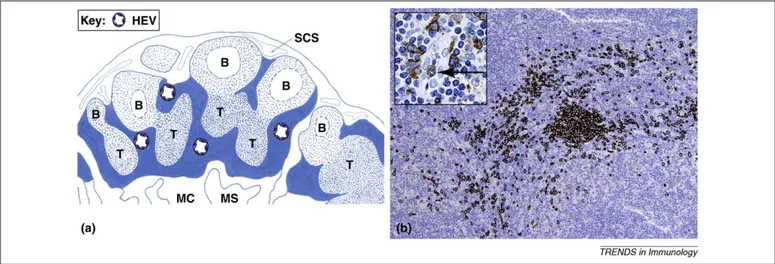
Figure 1. Localization and detection of PDCs in human lymph nodes. (a) PDCs localize in lymph node compartments (filled blue) typically located in areas surrounding B follicles (B) in the cortex and T nodules (T) in the paracortex. In these areas, PDCs localize in proximity to high endothelial venules (HEV); (SCS: subcapsular sinuses, MC: medullary cord; MS: medullary sinuses). (b) PDCs are specifically detected in formalin fixed and paraffin embedded tissues using an anti-BDCA2/CD303 monoclonal antibody (moAb). The figure shows BDCA2-positive PDCs distributed as clusters and dispersed cells; the inset shows a single PDCs within the lumen of a high endothelial venule. BDCA-2 is not expressed by other cells, and this completely circumvents the issue of specificity encountered when other antigens are targeted. For instance, anti-CD123 moAb, largely used in the past to stain PDCs in tissue sections, cross-reacts with other cell types, such as endothelial cells, sinus lining cells and activated macrophages (e.g. epitheliod macrophages in granulomas).
are fully competent in inducing PDCs adhesion and
migration when immobilized on heparan sulphates
expressed by endothelial cells
[33]
. Two recent reports
showed that PDCs purified from patients with chronic
hepatitis C, or exposed in vitro to IFN-a, acquire the ability
to respond to CCR2, CCR5 and CXCR3 ligands
[44,45]
.
These results suggest that under appropriate stimulatory
conditions, additional chemokine receptors may
be
involved in human PDCs recruitment. In line with this
hypothesis, leukemic PDCs express a wider profile of
functional chemokine receptors than the normal
circulat-ing counterpart
[46]
. In addition to chemokines, adenosine
and F2L, two agonists released by damaged tissues at the
site of inflammation, have the ability to induce PDCs
migration through the engagement of the adenosine
re-ceptor A1 and FPR3 (the formyl peptide rere-ceptor formerly
known as FPRL2), respectively
[47,48]
. It was also
reported that like MDCs, PDCs have functional receptors
for the anaphylatoxins C3a and C5a
[49]
. Finally, IL-18
induces the migration of PDCs and promotes the
differen-tiation of Th1 lymphocytes
[50]
. All these data suggest
that, in addition to chemotactic cytokines, signals
associ-ated with inflammation and tissue damage may contribute
to recruitment of PDCs to pathological tissues.
Recruitment of PDCs to non-lymphoid tissues is observed
in certain pathological conditions, such as autoimmune
diseases (i.e. lupus erythematosus disease, psoriasis and
rheumatoid arthritis)
[1,51,52]
, allergic diseases (i.e. contact
dermatitis and nasal mucosa polyps)
[53]
and in tumors
[2,54]
(
Table 1
). However, the mechanisms underlying this
effect are elusive. Recently, we and others have
character-ized chemerin, the ligand of ChemR23, as a new chemotactic
factor for PDCs (
Box 1
)
[55,56]
. Chemerin is expressed by
HEV in reactive lymph nodes and by activated dermal blood
vessels in autoimmune skin lesions
[55,57,58]
. Therefore,
current evidence strongly implicates the
ChemR23/che-merin axis in the regulation of human PDCs trafficking to
lymph nodes and to pathological tissues.
Of note, although the migration of blood PDCs to
sec-ondary lymphoid organs and to peripheral tissues has so
far attracted the most attention, there is evidence that
PDCs may also enter lymph nodes via the lymphatic route
[59]
; further studies are needed to address this issue in
humans.
Accumulation of PDCs in human diseases: recent
advances
Similar to other human leukocytes, PDCs can undergo
neoplastic transformation (
Box 2
) and accumulate during
some inflammatory pathological conditions
[1,27]
.
Pathol-ogists can easily detect human PDCs on archival tissues
using anti-CD303 as a marker and can screen for their
[(Figure_2)TD$FIG]
Figure 2. Trafficking properties of circulating PDCs. Blood PDCs constitutively express high levels of L-selectin, CXCR4 and ChemR23. The ligands for these receptors are expressed by high endothelial venules (HEV); therefore, these molecules may account for the homeostatic recruitment of circulating PDCs to lymph nodes. Under inflammatory conditions other molecules become involved in this process, such as PSGL-1, the ligand for E-selectin, b1 and b2 integrins and the chemokine receptors CCR5 and CXCR3. CCR7 is upregulated following activation, and is likely to be involved in the migration of mature PDCs through the engagement of its ligands CCL19 and CCL21, presented by HEV. Under pathological conditions, PDCs are also recruited to peripheral tissues; the molecules involved in this process have not been characterized directly. However, PDCs are known to migrate in response to chemokines and other chemotactic factors that are produced, or upregulated, during inflammation. In the mouse, CCR9 and its ligand CCL25, are responsible for the recruitment of PDCs to the small intestine both under homeostatic and inflammatory conditions. The different colors indicate the molecules involved in homeostatic (blue) and inflammatory (red) PDCs trafficking; also, constitutive molecules may be upregulated during inflammatory reactions. In the blood vessel, adhesion molecules and their respective counter-receptors are indicated in red or blue, chemotactic factors and their receptors in green. Evidence for the involvement of the different molecules is sometimes related only to mouse or human work; see the text for details and related references.
occurrence in large-scale clinical studies. A number of
pathological conditions can be characterized by the
pre-sence of PDCs (
Table 1
). In this section we summarize
findings for several of these pathologies where
abnormal-ities in PDCs are thought to contribute to disease.
Autoimmune inflammatory dermatoses as a paradigm
of PDCs-associated disease models
PDCs are normally absent from the skin. However, they
accumulate in some inflammatory dermatoses, where they
organize local immune responses
[60]
. The best
character-ized examples of these conditions are lupus erythematosus
(LE)
[61]
and psoriasis
[62]
. Conversely, in other skin
diseases, such as atopic dermatitis, PDCs are not recruited
to the inflamed tissue
[53]
. In LE and psoriasis, cutaneous
accumulation of PDCs is associated with the local
acti-vation of the chemerin/ChemR23 axis (
Box 1
)
[55,57,63]
. In
particular, chemerin is strongly induced in keratinocytes,
dermal vessels and fibroblasts, whereas skin infiltrating
PDCs strongly express ChemR23. The kinetics and
distri-bution of cutaneous PDCs infiltration is different in these
two pathological conditions. In psoriasis, PDCs infiltration
is restricted largely to the dermis, and predominantly
found in early phases of the disease
[57,62]
. In contrast,
in LE skin lesions PDCs persist during the entire spectrum
of the disease
[63]
and are located, not only in the dermis,
but also at the dermo-epidermal junction in areas of
epi-thelial cell damage. These findings suggest a novel view of
the role of these cells in skin autoimmunity, where PDCs
might coordinate the dermal immune reaction and sustain
epithelial damage via secretion of cytotoxic molecules. In
fact, cytotoxic damage is a major pathological event in the
‘‘interface’’ dermatitis found in LE, and similar changes,
Table 1. Pathological conditions characterized by PDCs tissue infiltration1,12,27.
Disease Tissues
Accumulation of ‘‘pathogenetic’’ PDCs with increased function
Lupus erythematosus Skin and kidney
Lichen planus Skin and mucosa
Psoriasis Skin
Rheumatoid arthritis Synovial tissue
Nasal allergy Nasal mucosa
Accumulation of PDCs with defective function
Melanoma Primary and metastatic tumor site
Carcinomas (ovary, breast, head and neck) Primary tumor site
Accumulation of PDCs with anti-viral/ anti-tumor response
Human Herpes Virus infection Skin
Human Papilloma Virus infection Skin
Molluscum Contagiosum Virus infection Skin
EBV-infection (Hydroa vacciniformis) Skin
Imiquimod-treated carcinomas, melanoma and viral infection Skin
Accumulation of PDCs (function unknown)
Kikuchi’s lymphadenitis Lymph node, skin
Hodgkin’s lymphoma Lymph node
Castleman’s disease, hyalin vascular type Lymph node
Granulomas (infectious, sarcoidosis) Lymph node
Cutaneous Pseudolymphomas Skin
Cutaneous Marginal Zone Lymphomas Skin
Decrease in number/function of PDCs
Human Immunodeficiency Virus infection Peripheral Blood
Hepatitis C Virus infection Peripheral Blood
Solid tumors and haematologic malignancies Peripheral Blood
A wide spectrum of human diseases including infection, cancer and autoimmunity is associated with accumulation of PDCs in lymphoid and peripheral tissues, or reduction of PDCs in peripheral blood. For many of these diseases, compelling evidence supports a pathogenic role of PDCs accumulation, mainly related to either the increase or the reduction of PDCs function. Alternatively, PDCs accumulation might exert an adjuvant immune function, as in viral infection, and in Imiquimod-treated cancers. In many other pathologies, information available is still limited and PDCs function largely unknown.
Box 1. The chemerin/ChemR23 axis in inflammation
ChemR23 is a serpentine Gi-coupled receptor originally cloned by Parmentier and colleagues[91]. ChemR23 is expressed by antigen-presenting cells, such as MDCs, PDCs and macrophages, and by blood monocytes and NK cells [55,58,92]. In contrast to other chemotactic receptors, human ChemR23 maps to chromosome 12, and shows little homology with other receptors of the same family (e.g. 38%, 36% and 35% with C3aR, C5aR and FPR1, respectively). In vivo, ChemR23 is expressed in the lymph node by interfollicular MDCs and PDCs, by sinus macrophages and by germinal center macrophages. In inflamed skin, ChemR23 expression is mostly restricted to MDCs and PDCs[55]. Chemerin, the only natural ligand so far identified for this receptor, was originally purified from ovarian cancer ascites and rheumatoid synovial fluids, and corre-sponds to the product of the Tig-2 gene. This protein contains a cystatin fold, and, similar to other family members, it is produced as an inactive precursor that is activated through proteolytic cleavage of the C-terminus by proteases released from neutrophils and proteases generated by the coagulation cascade or produced by bacteria [93]. In vivo, chemerin is produced by HEV and sparse perivenular spindle cells in the interfollicular area of the lymph node, and in inflamed skin, by activated blood endothelial cells, fibroblasts, and epithelial and mast cells. The anti-inflammatory lipid Resolvin E1 was shown to exert its function through interaction with ChemR23. In addition, chemerin proteolytically generated peptides possess anti-inflammatory activity in vivo[94,95]. There-fore, ChemR23 may also possess anti-inflammatory activities, as recently suggested in experiments testing a mouse model of lung inflammation [96]. CCRL2, an orphan serpentine receptor binds chemerin at the N-terminal in the absence of receptor signalling and internalization, and presents chemerin to ChemR23-positive cells in vitro[56]. This finding discloses a new potential role of chemerin in leukocyte recruitment. Chemerin was recently shown to be pro-duced by adipocytes and to act as an autocrine differentiation and activation factor. Therefore the potential role of chemerin in the metabolism of adipose tissue represents a new area of active research[97].
albeit to a lesser extent, are found in lichen planus, where
PDCs co-localize at the dermo-epidermal junction with NK
cells
[58]
. These observations point toward a role for PDCs
as effector cells. In all these dermatoses, production of high
levels of type I IFN by PDCs is pathogenic. In LE, type I
IFN is induced in response to nucleic acid-containing
immunocomplexes internalized through Fc receptors. In
psoriasis, a recently reported mechanism of PDCs
acti-vation is based on the production of the endogenous
anti-microbial peptide LL37 by damaged keratinocytes. LL37 is
able to bind and convert self-DNA or self-RNA into potent
TLR-dependent PDCs triggers
[64,65]
. Remarkably, LL37
is induced strongly in the lesional epidermis of psoriasis
biopsies
[66]
. Therefore, locally activated PDCs acquire the
ability to release large amounts of type I IFN, that in turn
sustains B and T cell autoimmune responses.
Tumor-associated PDCs: new players in cancer
immunity
Type I IFN plays a prominent role in limiting tumor cell
growth, and several human cancers are treated with type I
IFN
[67]
. However, the role of PDCs in the control of tumor
cell proliferation is still questionable. In a clinical study,
TLR-dependent activation of PDCs contributed to rejection
of cutaneous tumors. In this condition, PDCs produced
type I IFN, expressed TRAIL and localized together with
T cells and NK cells
[25]
. Interestingly, a new cell
popu-lation of CD11c
+myeloid DCs endowed with tumoricidal
potential was identified in these lesions
[25]
. The relevance
of PDCs in cancer immunosurveillance has been recently
shown in mice
[68,69]
, where TLR9-activated PDCs lead to
the regression of subcutaneous B16 melanoma tumors, by
orchestrating the sequential activation of NK cells, MDCs
and CD8
+T cells.
The characterization of PDCs has been performed in a
variety of human neoplasms
[54,70–72]
. However, the
potential impact of PDCs for cancer immunity is based
largely on in vitro studies of circulating PDCs, while PDCs
detectable at the tumour site (tumor-associated PDCs,
PDCs) are poorly characterized. Available data on
TA-PDCs indicate that they are defective in type I IFN
pro-duction, and secrete indoleamine 2, 3-dioxygenase (IDO)
[73]
. It should be noted, however, that the possibility that
PDCs may produce IDO has been questioned
[20]
. The
production of type I IFN in vitro can be inhibited by
cross-linking surface receptors such as CD303, NKp44 and ILT7
[20]
. The identity and tissue source of the natural ligands
for CD303 and NKp44 remain elusive. In contrast, ILT7
ligands, including BSTA2, are expressed by several human
cancer cell lines, and inhibit strongly the production of type
I IFN by activated PDCs
[11,74]
. A novel mechanism was
proposed recently, where PDCs may exert tolerogenic,
instead of immunogenic, functions. In fact, it was reported
that IL-10 can induce human circulating PDCs to suppress
T-cell proliferation through the secretion of GrB
[30]
. This
finding is of interest given the high amounts of IL-10
produced in the tumour microenvironment and might
explain the pro-tumorigenic effect of this cytokine observed
in some experimental conditions
[75]
.
In summary, properly activated PDCs are naturally
endowed with anti-tumor activity, but the tumor
micro-environment can subvert this property. There are some
data on the migration mechanisms adopted by TA-PDCs,
with the CXCR4/CXCL12 axis being involved in the
migration to ovarian cancer and melanoma
[54,71]
. The
availability of new experimental models and new
strat-egies for depleting PDCs in vivo will be extremely helpful
in defining the trafficking properties and the role of PDCs
in tumour growth.
Modulation of leukocyte recruitment by PDCs
The production of chemokines by DCs subsets represents
an important level of immuneregulation
[76,77]
. As
men-tioned, the potential of PDCs to regulate leukocyte
recruit-ment is mostly related to the ability of these cells to
respond to viral infection with high levels of type I IFN
production. HSV1-activated PDCs recruit T and NK cells
through the production of CXCL9, CXCL10 and CCL4, and
CD40-engagement further strengthens this response
[78,79]
. Exposure of PDCs to HIV-1 surface components
triggers the secretion of the CCR5 ligands CCL3 and
CCL4, suggesting a role of PDCs in limiting viral spreading
[80]
. In the lung, PDCs are among the earliest
inflamma-tory cells to enter the bronchoalveolar space following
influenza A virus infection
[81]
, and support the
recruit-ment of CXCR3
+and CCR5
+effector T cells through the
local secretion of CXCL10 and CCL5
[82]
. A protective role
for C5a in allergic asthma has been linked to a shift in the
MDC/PDCs ratio toward PDCs, with an accompanying
decrease in CCL17 production
[83]
. Furthermore, PDCs
depletion exacerbates respiratory syncytial virus lung
immunopathology
[84]
. In indirect acute lung injury, PDCs
are thought to control the recruitment of inflammatory
Box 2. Tumors derived from PDCs
Proliferation of PDCs in tumors occurs under two distinct clinico-pathological conditions: one is composed of morphologically mature PDCs that are barely distinguishable from their normal counterparts; the other, defined as blastic PDCs neoplasms (BPDCN)
[98], consists of PDCs showing an immature morphology. De novo expression of antigens such as CD2, CD5 and CD7 has been reported in both conditions, but a more profound aberrant phenotype is typically found in BPDCN, with frequent loss of classical PDCs markers (such as CD68 and GrB) and expression of CD56 and TdT proteins [12]. Both conditions can be associated with myeloid neoplasms. In mature PDCs, proliferation is a constant finding[99], and the myeloid neoplasm is generally represented by chronic myelomonocytic leukemia. The clonal relationship between PDCs and myeloid leukaemia is supported by FISH analysis that demon-strated identical cytogenetic abnormalities in the two cell popula-tions[99,100]. On the other hand, only ten to twenty per cent of BPDCN cases are associated with a myeloid neoplasm, generally consisting of an acute or chronic leukemia with monocytic differentiation that may be associated with an underlying myelo-dysplastic syndrome. This myeloid leukemia can precede, coexist or develop subsequently to BPDCN.
Mature PDCs tumors manifest as nodular accumulations of PDCs, especially in lymph nodes and bone marrow, while BPDCN typically presents with multiple skin lesions that rapidly spread to bone marrow and multiple organs. The peculiar skin tropism of PDCs in BPDCN has been related to their expression of the neural cell adhesion molecule CD56 and of the skin homing receptor CLA. In addition, leukemic PDCs express the chemokine receptors CXCR3, CXCR4, and CCR7, whose respective ligands CXCL9, CXCL12, and CCL19 are concomitantly found in cutaneous sweat glands and follicle epithelium[46].
monocytes through the regulation of lung CCL2 production
[85]
. Also, PDCs-induced CXCL8 secretion might impact the
recruitment of neutrophils into the lung during chronic
obstructive pulmonary disease
[86]
. PDCs-derived CXCL10
was also shown to be responsible for recruiting CXCR3
+T
cells to cutaneous LE lesions
[51]
, and the cerebrospinal
fluid of patients with neuropsychiatric lupus
[87]
. In this
context, PDCs have also been implicated in autoimmune
diseases by promoting Th17 immune responses through the
production of IL1b and IL23p19
[88,89]
. Moreover, by
secreting CCL4, PDCs might be involved in regulatory T
cell recruitment to the tumor site, representing an
additional mechanism for controlling immune function
[90]
.
Concluding remarks
More than fifty years since the first description of the
‘‘enigmatic’’ cell population now known as PDCs, many
aspects of PDCs biology still await clarification. The
mech-anisms that govern the in vivo distribution of these cells
under homeostatic conditions remain elusive, and the
biological role of PDCs in many diseases in which they
are regularly detected is poorly understood. Paradoxically,
more information is available on the involvement of PDCs
in human inflammatory and neoplastic conditions than in
experimental models, hampering the correct
understand-ing of the molecular basis of PDCs activation. The new and
more specific reagents now available to detect PDCs in
human and mouse tissues will be instrumental in defining
the role of PDCs in immune responses. Furthermore, the
possibility to generate mice selectively depleted of PDCs
will allow conceptual models about PDCs function to be
probed in vivo. Therefore, it is likely that in the next few
years we will be able to evaluate in a critical manner the
available literature and to advance our current knowledge
on the biological function of PDCs.
Acknowledgements
Limitations of space preclude extensive citation of the literature; we apologize to those whose work is not mentioned in this review. This work was supported by AIRC (Associazione Italiana per la Ricerca sul Cancro); Istituto Superiore di Sanita` (I.S.S.), Ministero dell’Istruzione, dell’Universita` e della Ricerca (M.I.U.R.), and Fondazione Berlucchi (Brescia, Italy).
References
1 Facchetti, F. et al. (2003) The plasmacytoid monocyte/interferon producing cells. Virchows Arch. 443 (6), 703–717
2 Grouard, G. et al. (1997) The enigmatic plasmacytoid T cells develop into dendritic cells with interleukin (IL)-3 and CD40-ligand. J. Exp. Med. 185 (6), 1101–1111
3 Cella, M. et al. (1999) Plasmacytoid monocytes migrate to inflamed lymph nodes and produce large amounts of type I interferon. Nat. Med. 5 (8), 919–923
4 Siegal, F.P. et al. (1999) The nature of the principal type 1 interferon-producing cells in human blood. Science 284 (5421), 1835–1837 5 Liu, K. et al. (2009) In vivo analysis of dendritic cell development and
homeostasis. Science 324 (5925), 392–397
6 Naik, S.H. et al. (2007) Development of plasmacytoid and conventional dendritic cell subtypes from single precursor cells derived in vitro and in vivo. Nat. Immunol. 8 (11), 1217–1226
7 Reizis, B. (2010) Regulation of plasmacytoid dendritic cell development. Curr. Opin. Immunol. 22 (2), 206–211
8 Robbins, S.H. et al. (2008) Novel insights into the relationships between dendritic cell subsets in human and mouse revealed by genome-wide expression profiling. Genome Biol. 9 (1), R17
9 Cao, W. (2009) Molecular characterization of human plasmacytoid dendritic cells. J. Clin. Immunol. 29 (3), 257–264
10 Cisse, B. et al. (2008) Transcription factor E2-2 is an essential and specific regulator of plasmacytoid dendritic cell development. Cell 135 (1), 37–48
11 Cao, W. et al. (2009) Regulation of TLR7/9 responses in plasmacytoid dendritic cells by BST2 and ILT7 receptor interaction. J. Exp. Med. 206 (7), 1603–1614
12 Jegalian, A.G. et al. (2009) Plasmacytoid dendritic cells: physiologic roles and pathologic states. Adv. Anat. Pathol. 16 (6), 392–404 13 Soumelis, V. and Liu, Y.J. (2006) From plasmacytoid to dendritic cell:
morphological and functional switches during plasmacytoid pre-dendritic cell differentiation. Eur. J. Immunol. 36 (9), 2286–2292 14 Cao, W. and Liu, Y.J. (2007) Innate immune functions of plasmacytoid
dendritic cells. Curr. Opin. Immunol. 19 (1), 24–30
15 Gilliet, M. et al. (2008) Plasmacytoid dendritic cells: sensing nucleic acids in viral infection and autoimmune diseases. Nat. Rev. Immunol. 8 (8), 594–606
16 Cella, M. et al. (2000) Plasmacytoid dendritic cells activated by influenza virus and CD40L drive a potent Th1 polarization. Nat. Immunol. 1 (4), 305–310
17 Liu, Y.J. (2005) IPC: professional type 1 interferon-producing cells and plasmacytoid dendritic cell precursors. Annu. Rev. Immunol. 23, 275–306
18 Ito, T. et al. (2006) Specialization, kinetics, and repertoire of type 1 interferon responses by human plasmacytoid predendritic cells. Blood 107 (6), 2423–2431
19 Ito, T. et al. (2005) Plasmacytoid dendritic cell precursors/type I interferon-producing cells sense viral infection by Toll-like receptor (TLR) 7 and TLR9. Springer Semin. Immunopathol. 26 (3), 221–229 20 Swiecki, M. and Colonna, M. (2010) Unraveling the functions of plasmacytoid dendritic cells during viral infections, autoimmunity, and tolerance. Immunol. Rev. 234 (1), 142–162
21 von Landenberg, P. and Bauer, S. (2007) Nucleic acid recognizing Toll-like receptors and autoimmunity. Curr. Opin. Immunol. 19 (6), 606–610 22 Decalf, J. et al. (2007) Plasmacytoid dendritic cells initiate a complex chemokine and cytokine network and are a viable drug target in chronic HCV patients. J. Exp. Med. 204 (10), 2423–2437
23 Penna, G. et al. (2002) Cutting edge: differential chemokine production by myeloid and plasmacytoid dendritic cells. J. Immunol. 169 (12), 6673–6676
24 Chaperot, L. et al. (2006) Virus or TLR agonists induce TRAIL-mediated cytotoxic activity of plasmacytoid dendritic cells. J. Immunol. 176 (1), 248–255
25 Stary, G. et al. (2007) Tumoricidal activity of TLR7/8-activated inflammatory dendritic cells. J. Exp. Med. 204 (6), 1441–1451 26 Rissoan, M.C. et al. (2002) Subtractive hybridization reveals the
expression of immunoglobulin-like transcript 7, Eph-B1, granzyme B, and 3 novel transcripts in human plasmacytoid dendritic cells. Blood 100 (9), 3295–3303
27 Colonna, M. et al. (2004) Plasmacytoid dendritic cells in immunity. Nat. Immunol. 5 (12), 1219–1226
28 Hoeffel, G. et al. (2007) Antigen crosspresentation by human plasmacytoid dendritic cells. Immunity 27 (3), 481–492
29 Villadangos, J.A. and Young, L. (2008) Antigen-presentation properties of plasmacytoid dendritic cells. Immunity 29 (3), 352–361 30 Jahrsdorfer, B. et al. (2010) Granzyme B produced by human plasmacytoid dendritic cells suppresses T-cell expansion. Blood 115 (6), 1156–1165
31 Manches, O. et al. (2008) HIV-activated human plasmacytoid DCs induce Tregs through an indoleamine 2,3-dioxygenase-dependent mechanism. J. Clin. Invest. 118 (10), 3431–3439
32 Moseman, E.A. et al. (2004) Human plasmacytoid dendritic cells activated by CpG oligodeoxynucleotides induce the generation of CD4+CD25+ regulatory T cells. J. Immunol. 173 (7), 4433–4442 33 Kohrgruber, N. et al. (2004) Plasmacytoid dendritic cell recruitment
by immobilized CXCR3 ligands. J. Immunol. 173 (11), 6592–6602 34 Nakano, H. et al. (2001) CD11c(+)B220(+)Gr-1(+) cells in mouse lymph
nodes and spleen display characteristics of plasmacytoid dendritic cells. J. Exp. Med. 194 (8), 1171–1178
35 Penna, G. et al. (2001) Cutting edge: selective usage of chemokine receptors by plasmacytoid dendritic cells. J. Immunol. 167 (4), 1862–1866
36 Diacovo, T.G. et al. (2005) Adhesive mechanisms governing interferon-producing cell recruitment into lymph nodes. J. Exp. Med. 202 (5), 687–696
37 Yoneyama, H. et al. (2004) Evidence for recruitment of plasmacytoid dendritic cell precursors to inflamed lymph nodes through high endothelial venules. Int. Immunol. 16 (7), 915–928
38 Gotoh, K. et al. (2008) Differential requirement for DOCK2 in migration of plasmacytoid dendritic cells versus myeloid dendritic cells. Blood 111 (6), 2973–2976
39 Kohara, H. et al. (2007) Development of plasmacytoid dendritic cells in bone marrow stromal cell niches requires CXCL12-CXCR4 chemokine signaling. Blood 110 (13), 4153–4160
40 Yoneyama, H. et al. (2005) Plasmacytoid DCs help lymph node DCs to induce anti-HSV CTLs. J. Exp. Med. 202 (3), 425–435
41 Wendland, M. et al. (2007) CCR9 is a homing receptor for plasmacytoid dendritic cells to the small intestine. Proc. Natl. Acad. Sci. U. S. A. 104 (15), 6347–6352
42 Krug, A. et al. (2002) IFN-producing cells respond to CXCR3 ligands in the presence of CXCL12 and secrete inflammatory chemokines upon activation. J. Immunol. 169 (11), 6079–6083
43 Vanbervliet, B. et al. (2003) The inducible CXCR3 ligands control plasmacytoid dendritic cell responsiveness to the constitutive chemokine stromal cell-derived factor 1 (SDF-1)/CXCL12. J. Exp. Med. 198 (5), 823–830
44 Cicinnati, V.R. et al. (2009) Mycophenolic acid impedes the antigen presenting and lymph node homing capacities of human blood myeloid dendritic cells. Transplantation 88 (4), 504–513
45 Cicinnati, V.R. et al. (2008) Altered chemotactic response of myeloid and plasmacytoid dendritic cells from patients with chronic hepatitis C: role of alpha interferon. J. Gen. Virol. 89 (Pt 5), 1243–1253 46 Bendriss-Vermare, N. et al. (2004) In situ leukemic plasmacytoid
dendritic cells pattern of chemokine receptors expression and in vitro migratory response. Leukemia 18 (9), 1491–1498
47 Devosse, T. et al. (2009) Formyl peptide receptor-like 2 is expressed and functional in plasmacytoid dendritic cells, tissue-specific macrophage subpopulations, and eosinophils. J. Immunol. 182 (8), 4974–4984
48 Schnurr, M. et al. (2004) Role of adenosine receptors in regulating chemotaxis and cytokine production of plasmacytoid dendritic cells. Blood 103 (4), 1391–1397
49 Gutzmer, R. et al. (2006) Human plasmacytoid dendritic cells express receptors for anaphylatoxins C3a and C5a and are chemoattracted to C3a and C5a. J. Invest. Dermatol. 126 (11), 2422–2429
50 Kaser, A. et al. (2004) Interleukin-18 attracts plasmacytoid dendritic cells (DC2 s) and promotes Th1 induction by DC2 s through IL-18 receptor expression. Blood 103 (2), 648–655
51 Farkas, L. et al. (2001) Plasmacytoid dendritic cells (natural interferon- alpha/beta-producing cells) accumulate in cutaneous lupus erythematosus lesions. Am. J. Pathol. 159 (1), 237–243 52 Lande, R. et al. (2004) Characterization and recruitment of
plasmacytoid dendritic cells in synovial fluid and tissue of patients with chronic inflammatory arthritis. J. Immunol. 173 (4), 2815–2824
53 Wollenberg, A. et al. (2002) Plasmacytoid dendritic cells: a new cutaneous dendritic cell subset with distinct role in inflammatory skin diseases. J. Invest. Dermatol. 119 (5), 1096–1102
54 Vermi, W. et al. (2003) Recruitment of immature plasmacytoid dendritic cells (plasmacytoid monocytes) and myeloid dendritic cells in primary cutaneous melanomas. J. Pathol 200, 255–268 55 Vermi, W. et al. (2005) Role of ChemR23 in directing the migration of
myeloid and plasmacytoid dendritic cells to lymphoid organs and inflamed skin. J. Exp. Med. 201 (4), 509–515
56 Zabel, B.A. et al. (2008) Mast cell-expressed orphan receptor CCRL2 binds chemerin and is required for optimal induction of IgE-mediated passive cutaneous anaphylaxis. J. Exp. Med. 205 (10), 2207–2220 57 Albanesi, C. et al. (2009) Chemerin expression marks early psoriatic
skin lesions and correlates with plasmacytoid dendritic cell recruitment. J. Exp. Med. 206 (1), 249–258
58 Parolini, S. et al. (2007) The role of chemerin in the colocalization of NK and dendritic cell subsets into inflamed tissues. Blood 109 (9), 3625–3632
59 Pascale, F. et al. (2008) Plasmacytoid dendritic cells migrate in afferent skin lymph. J. Immunol. 180 (9), 5963–5972
60 Meller, S. et al. (2009) Chemokines in the pathogenesis of lichenoid tissue reactions. J. Invest. Dermatol. 129 (2), 315–319
61 Banchereau, J. and Pascual, V. (2006) Type I interferon in systemic lupus erythematosus and other autoimmune diseases. Immunity 25 (3), 383–392
62 Nestle, F.O. et al. (2005) Plasmacytoid predendritic cells initiate psoriasis through interferon-alpha production. J. Exp. Med. 202 (1), 135–143
63 Vermi, W. et al. (2009) Cutaneous distribution of plasmacytoid dendritic cells in lupus erythematosus. Selective tropism at the site of epithelial apoptotic damage. Immunobiology 214 (9–10), 877–886
64 Ganguly, D. et al. (2009) Self-RNA-antimicrobial peptide complexes activate human dendritic cells through TLR7 and TLR8. J. Exp. Med. 206 (9), 1983–1994
65 Lande, R. et al. (2007) Plasmacytoid dendritic cells sense self-DNA coupled with antimicrobial peptide. Nature 449 (7162), 564–569 66 Frohm, M. et al. (1997) The expression of the gene coding for the
antibacterial peptide LL-37 is induced in human keratinocytes during inflammatory disorders. J. Biol. Chem. 272 (24), 15258–15263 67 Dunn, G.P. et al. (2006) Interferons, immunity and cancer
immunoediting. Nat. Rev. Immunol. 6 (11), 836–848
68 Liu, C. et al. (2008) Plasmacytoid dendritic cells induce NK cell-dependent, tumor antigen-specific T cell cross-priming and tumor regression in mice. J. Clin. Invest. 118 (3), 1165–1175
69 Rajagopal, D. et al. (2010) Plasmacytoid dendritic cell-derived type I interferon is crucial for the adjuvant activity of Toll-like receptor 7 agonists. Blood 115 (10), 1949–1957
70 Kim, R. et al. (2007) Potential functional role of plasmacytoid dendritic cells in cancer immunity. Immunology 121 (2), 149–157 71 Zou, W. et al. (2001) Stromal-derived factor-1 in human tumors
recruits and alters the function of plasmacytoid precursor dendritic cells. Nat. Med. 7 (12), 1339–1346
72 Hartmann, E. et al. (2003) Identification and functional analysis of tumor-infiltrating plasmacytoid dendritic cells in head and neck cancer. Cancer Res. 63 (19), 6478–6487
73 Fallarino, F. et al. (2007) Tryptophan catabolism in IDO+ plasmacytoid dendritic cells. Curr. Drug Metab. 8 (3), 209–216 74 Tsukamoto, N. et al. (2009) Impairment of plasmacytoid dendritic
cells for IFN production by the ligand for immunoglobulin-like transcript 7 expressed on human cancer cells. Clin. Cancer Res. 15 (18), 5733–5743
75 Lin, W.W. and Karin, M. (2007) A cytokine-mediated link between innate immunity, inflammation, and cancer. J. Clin. Invest. 117 (5), 1175–1183
76 Sozzani, S. (2005) Dendritic cell trafficking: more than just chemokines. Cytokine Growth Factor Rev. 16 (6), 581–592
77 Piqueras, B. et al. (2006) Upon viral exposure, myeloid and plasmacytoid dendritic cells produce 3 waves of distinct chemokines to recruit immune effectors. Blood 107 (7), 2613–2618 78 Megjugorac, N.J. et al. (2004) Virally stimulated plasmacytoid
dendritic cells produce chemokines and induce migration of T and NK cells. J. Leukoc. Biol. 75 (3), 504–514
79 Bendriss-Vermare, N. et al. (2005) Virus overrides the propensity of human CD40L-activated plasmacytoid dendritic cells to produce Th2 mediators through synergistic induction of IFN-g and Th1 chemokine production. J. Leukoc. Biol. 78 (4), 954–966
80 Del Corno, M. et al. (2005) Human immunodeficiency virus type 1 gp120 and other activation stimuli are highly effective in triggering alpha interferon and CC chemokine production in circulating plasmacytoid but not myeloid dendritic cells. J. Virol. 79 (19), 12597–12601
81 Wolf, A.I. et al. (2009) Plasmacytoid dendritic cells are dispensable during primary influenza virus infection. J. Immunol. 182 (2), 871– 879
82 Iparraguirre, A. et al. (2008) Two distinct activation states of plasmacytoid dendritic cells induced by influenza virus and CpG 1826 oligonucleotide. J. Leukoc. Biol. 83 (3), 610–620
83 Kohl, J. et al. (2006) A regulatory role for the C5a anaphylatoxin in type 2 immunity in asthma. J. Clin. Invest. 116 (3), 783–796 84 Smit, J.J. et al. (2006) Plasmacytoid dendritic cells inhibit pulmonary
immunopathology and promote clearance of respiratory syncytial virus. J. Exp. Med. 203 (5), 1153–1159
85 Venet, F. et al. (2010) Plasmacytoid dendritic cells control lung inflammation and monocyte recruitment in indirect acute lung injury in mice. Am. J. Pathol. 176 (2), 764–773
86 Mortaz, E. et al. (2009) Cigarette smoke attenuates the production of cytokines by human plasmacytoid dendritic cells and enhances the release of IL-8 in response to TLR-9 stimulation. Respir. Res. 10, 47 87 Santer, D.M. et al. (2009) Potent induction of IFN-a and chemokines by autoantibodies in the cerebrospinal fluid of patients with neuropsychiatric lupus. J. Immunol. 182 (2), 1192–1201
88 Isaksson, M. et al. (2009) Plasmacytoid DC promote priming of autoimmune Th17 cells and EAE. Eur. J. Immunol. 39 (10), 2925– 2935
89 Yu, C.F. et al. (2009) Human plasmacytoid dendritic cells support Th17 cell effector function in response to TLR7 ligation. J. Immunol. 184 (3), 1159–1167
90 Penna, G. et al. (2007) 1,25-Dihydroxyvitamin D3 selectively modulates tolerogenic properties in myeloid but not plasmacytoid dendritic cells. J. Immunol. 178 (1), 145–153
91 Samson, M. et al. (1998) ChemR23, a putative chemoattractant receptor, is expressed in monocyte-derived dendritic cells and macrophages and is a coreceptor for SIV and some primary HIV-1 strains. Eur J. Immunol. 28 (5), 1689–1700
92 Wittamer, V. et al. (2003) Specific recruitment of antigen-presenting cells by chemerin, a novel processed ligand from human inflammatory fluids. J. Exp. Med. 198 (7), 977–985
93 Du, X.Y. and Leung, L.L. (2009) Proteolytic regulatory mechanism of chemerin bioactivity. Acta Biochim. Biophys. Sin. (Shanghai) 41 (12), 973–979
94 Arita, M. et al. (2005) Stereochemical assignment, antiinflammatory properties, and receptor for the omega-3 lipid mediator resolvin E1. J. Exp. Med. 201 (5), 713–722
95 Cash, J.L. et al. (2008) Synthetic chemerin-derived peptides suppress inflammation through ChemR23. J. Exp. Med. 205 (4), 767–775 96 Luangsay, S. et al. (2009) Mouse ChemR23 is expressed in dendritic
cell subsets and macrophages, and mediates an anti-inflammatory activity of chemerin in a lung disease model. J. Immunol. 183 (10), 6489–6499
97 Lago, F. et al. (2007) The emerging role of adipokines as mediators of inflammation and immune responses. Cytokine Growth Factor Rev. 18 (3–4), 313–325
98 Facchetti, F., Jones, D.M., Petrella, T., ed. (2008) Blastic plasmacytoid dendritic cell neoplasm, Edited by Swerdlow S.H., Campo, E., Harri, N.L., Jaffe, E.S., Pileri, S.A., Stein, H., Thiele, J., Vardiman, J.W., Lyon, IARC, pp. 145–147
99 Vermi, W. et al. (2004) Nodal and extranodal tumor-forming accumulation of plasmacytoid monocytes/interferon-producing cells associated with myeloid disorders. Am. J. Surg. Pathol. 28 (5), 585–595 100 Pileri, S.A. et al. (2007) Myeloid sarcoma: clinico-pathologic, phenotypic and cytogenetic analysis of 92 adult patients. Leukemia 21 (2), 340–350