UNIVERSITÀ DEGLI STUDI DI ROMA
"TOR VERGATA"
FACOLTA' DI MEDICINA E CHIRURGIA
DOTTORATO DI RICERCA IN
FISIOPATOLOGIA SPERIMENTALE
CICLO XXII
Placenta Growth Factor is a Survival Factor for Human
Malignant Mesothelioma Cells
Docente Guida Dottoranda
Dott.ssa LOREDANA ALBONICI ELENA DOLDO
Coordinatore:
Prof. RENATO LAURO
CONTENTS
ABSTRACT p.4 2. INTRODUCTION... p.5 1.1. Aethipathogenesis... p.6 1.1.1. Effect of asbestos... p.8 1.1.2. Chromosome alterations... p.8 1.1.3. Gene mutation ... p.9 1.1.4. DNA methylation... p.9 1.1.5. Invasiveness... p.9 1.1.6. Activation of telomerase... p.9 1.1.7. Cell proliferation... p.9 1.1.8. Association with SV-40 ... p.10 1.2. Histopathology ... p.11 1.3. Angiogenesis... p.12 1.4. Angiogenetic factors... p.13 2. MATERIALS AND METHODS...p.212.1. Materials... p.21 2.2. Cell Cultures... p.21 2.3. RNA analysis ... p.21 2.4. Western Blotting analysis... p.22
2.5. PlGF ELISA... p.22 2.6. Immunohistochemistry ... p.23
2.7. Cell growth assay... p.24 2.8. Cell survival assay... p.24 2.9. Statistical analysis... p.24
3. RESULTS...p.25 3.1. Expression of PlGF, VEGF and their cognate receptors and co-receptors in
cultured MM cells... p.25 3.2. In vitro release of PlGF by MM cells ... p.25
3.3. Expression of PlGF, VEGF and their cognate receptors in human mesothelium
and MM tissue ... p.26 3.4. Effect of PlGf-2 on MM cell proliferation ... p.27
3.5. Assessment of biochemical responsiveness of MM to PlGF-2... p.27 3.6. PlGF withdrawal reduces MM cell survival and induces cell death... p.28 4. DISCUSSION ...p.29 FIGURES AND TABLES ...p.33
REFERENCES...p.43
ABSTRACT
Placenta growth factor (PlGF) is a key regulator of pathological angiogenesis and its overexpression has been linked to neoplastic progression. To assess whether PlGF could have a role in malignant mesothelioma (MM), we analyzed the expression of PlGF, VEGF, and their cognate receptors (VEGF-R1 and VEGF-R2) and coreceptors (neuropilin-1 and neuropilin-2) in MM cell lines as well as in resected MM tissues, hyperplastic/reactive mesothelium and normal mesothelium. MM cell cultures expressed both ligands and the associated receptors to a variable extent and released different amounts of PlGF. As assessed by immunohistochemistry, PlGF expression was switched on in hyperplastic/reactive compared to normal mesothelium. Moreover, 73 and 94% of MM tissues overexpressed PlGF and VEGF-R1, respectively (p<0.05). Administration of recombinant PlGF-2 did not elicit a significant stimulation of MM cell growth, while it was associated with a transient phosphorylation of Akt, suggesting that PlGF-2 could activate downstream effectors of cytoprotective and anti-apototic signals via VEGF-R1 in MM cells. Indeed, the administration of an anti-PlGF antibody was found to cause a significant reduction of MM cell survival. In conclusion, our data demonstrate that, by acting as a survival factor, PlGF can play a role which goes beyond the stimulation of angiogenesis in MM. This evidence could help the rational design of new therapeutic interventions for this aggressive tumor.
1.
INTRODUCTIONMalignant Mesothelioma (MM) is a highly aggressive tumor that arises from the mesothelial linings of the chest and abdominal cavities, pleura, peritoneum, pericardium and the tunica vaginalis testis. Mesothelium functions as a non-adesive surface for the internal organs and as a selective barrier that regulates the transport of molecules and cells between the circulation and the body cavities. Mesothelium is supported by a layer of connective tissue consisting of collagen and elastin fibers interspersed with fibroblasts, mononuclear phagocytes, lymphocytes, capillaries and lymphatics. The mesothelial cells are the progenitor cells of mesothelioma (Bielefeldt-Ohmann et al, 1996).
The most frequent location of the MM is the pleura (>90%), followed by the peritoneum (6-10%). Mesotheliomas arising from pericardium and tunica vaginalis are very rare (Sekido Y, 2009; Schure et al, 2006; Robinson & Lake, 2005). MM is responsible for approximately 15000-20000 deaths annually worldwide (Zervos MD, 2008). All MMs are much more common in males because of a strong association between the tumor and occupational asbestos exposure. The link between mesothelioma and asbestos date at 1960 since several publications have documented mesotheliomas in various occupational communities including shipyard workers, insulators, gas mask manufactures and asbestos cement workers (Robinson & Lake, 2005; Montjoy et al, 2009). The annual incidence increases 3,5 fold for men and 1,4 fold for women.
Approximately 70% of cases are associated with documented asbestos exposure, although the lifetime risk for MM among workers exposed to asbestos is thought to be as high as 8% to 13%. No direct correlation exists between the incidence of MM and the duration of asbestos exposure (Astoul P, 1999).
This suggests that additional factors such as SV40 infection and genetic predispositions can render some individuals more susceptible to asbestos carcinogenicity (Krocynska B, 2006). The peak in the deaths per year is expected in the 2020. This peak is caused by the long latency period for the development of MM as many as 30 to 40 years after the exposure. After 2020 MM rates are expected to decrease because of new legislation aimed to reduce asbestos exposure in the workplace and the general environment (Astoul P, 1999).
1.1 Aetiopathogenesis
There is irrefutable evidence linking asbestos exposure to the subsequent development of MM. About 80%-90% of MM appears to be related to prior asbestos exposure (Greillier and Astoul, 2008). It occurs in men exposed to asbestos in the workplace and sometimes in their family members or in people who live near mines. The remaining 20% of men who develop MM have no history of exposure to asbestos, and there is usually no excess of mineral fibers in their lungs (Mossman et al., 1997). Asbestos encompasses a heterogeneous group of hydrated fibrous silicates. These can be divided in two mineral families: serpentine and amphibole, and have very different propensities for causing MM. Chrysotile or white asbestos is the main member of the serpentine family and is used in more than 90% of industrial application in most western countries. The amphibole family contains several members: crocidolite (blue) and amosite (brown), which previously have been used commercially; anthophyllite and tremolite which generally have not been used commercially but can occur as trace minerals in Chrysotile and talc deposits. The studies have shown a clear-cut difference in MM potency between the different asbestos fiber types:
Crocidolite>amosite/tremolite>>>chrysotile. Chrysotile exposure alone rarely causes MM. The migration of asbestos to a site proximate to serosal membranes is important in the pathogenesis of MM and it is largely determined by fiber size. Fiber diameter has an inverse relationship with mesothelioma risk, in fact fiber greater than 8 µm are mainly associated with MM. Both Chrysotile and amphibole fibers have been identified in the visceral and parietal pleura. The mechanism by which asbestos reaches the peritoneum is speculative. Asbestos fibers have been shown to penetrate the gastrointestinal wall in animals experimentally fed asbestos and asbestos bodies have been seen within some peritoneal mesothelioma cases. The ingestion of asbestos fibers can occur directly by contaminated food products or swallowing expectorated sputum-rich in asbestos may be one mechanism of transport of asbestos to the peritoneum. Other possible routes for fiber transmission involve asbestos permeation of diaphragmatic stomata or via haematogenous and lymphatic channels (Attanoos & Gibbs, 1997). Numerous studies have demonstrated that asbestos exposure enhances the risk of MM. In fact, crocidolite induces human mesothelial cells to release TNF-α and to express the TNF-α receptor. TNF-α is a proinflammatory cytokine and a major inducer of NF-kB, a key regulator of
oncogenesis. In animal models asbestos induces the expression of monocyte chemoattractant protein-1 (MCP-1) expression by mesothelial cells. MCP-1 favors the macrophage inflammatory response within the pleural space that follows asbestos exposure. Macrophages phagocytize asbestos but are unable to digest these fibers. Possibly, because they are damaged by asbestos, these macrophages release TNF-α and other cytokines. TNF-α can induce either cell death or, more frequently, increase cell survival via NF-kB activation. Activation of NF-kB promotes cellular proliferation and inhibits apoptosis, favoring cancer development because mesothelial cells with asbestos– induced DNA damage divide rather than die and these genetic damages can be sufficient to develop into malignant MM (Yang et al., 2006). MM cells are assumed to undergo neoplastic transformation as a result of the activation of the NF-kB pathway (Toyooka et al., 2008).
Asbestos fibers also have an intrinsic redo activity and contain ferrous iron, which catalyze reactions generating active oxygen intermediates on the fiber surface. In the tissues several asbestos fiber types can produce reactive oxygen free radicals from hydrogen peroxide, a common product of intermediary tissue metabolism. Crocidolite has a great surface-area and higher ferrous iron content compared to Chrysotile. Consequently crocidolite is more biologically active in the generation of free radicals (Toyokuni S, 2009). In MM asbestos appear to act as a complete carcinogen. Asbestos can generate active oxygen free-radicals by direct or indirect mechanisms. The first involves an iron catalyzed reaction on the fiber surface. The second requires the physical interaction of asbestos fibers with phagocytic cells. Macrophages and neutrophils are known to liberate oxygen free radicals via the respiratory burst mechanism following asbestos phagocytosis and neutrophils can initiate arachidonic acid metabolism and generate active oxygen species via the cycloxygenase and lypoxygenase pathway. The long latency period associated with MM suggests that multiple cumulative genetic, cytotoxic and proliferative events occur during the tumourigenesis process. Active oxygen intermediates can possibly partecipate in the oncogenic process via several different mechanisms. They may directly or indirectly interact with chromosomal material affecting mutational change. In vitro studies have shown that asbestos-induced oxidants can target membranes, affect lipid peroxydation and thereby liberate peroxyl radicals potentiating the free radical damage. In addition, both free radical-induced
genotoxicity and lipid peroxydation may serve to modulate oncogene and oncosuppressor gene expression and product function and thereby may influence tumor development at several different stages (Toyokuni S, 2009).
In MM have been reported numerous alterations:
1.1.1 Effect of asbestos.
The clinical manifestations of MM are thought to arise as result of a “build-up” of many molecular alterations. Asbestos fibers show increased expression of the proto-oncogenes c-fos and c-jun. The Fos family together with Jun family members form the AP-1 transcription factor complex which is localized in the nucleus where it binds to the promoter and enhancer regions of target genes resulting in cell proliferation and transcription.These are thought to be the initial intranuclear alterations caused by asbestos (Milde-Langosch K, 2005; Kovary & Bravo, 1991; Heintz et al, 1993; Zanella et al, 1996).
1.1.2 Chromosome alterations
Alterations at the chromosome level include especially amplifications and deletions. Deletions are more frequent than amplifications. A particularly high frequency of homo-deletion is seen in the 1p21-22 and 3p21 chromosome region which causing a homo-deletion of RASSF1A, tumor suppressor genes important in the neoplastic transformation process (Destro et al, 2008; Toyooka et al, 2008), raising a high frequency of deletions of p16 and p14 genes and a loss of expression of their proteins (Musti et al, 2006; Kobayashi N et al, 2008). The mechanism of the p16 alterations and malignant transformation has been found to involve the loss of P16 protein, which causes a breakdown of the cell-cycle control mechanism by inhibiting the phosphorylation of retinoblastoma (Rb) protein, which controls the cell-cycle. In addition the loss protein result in the activation of mdm2 protein, a p53 ubiquitin ligase, and this activation is thought to be linked to the destabilization of p53 protein, thus causing alterations in cell-cycle control (Lowe & Sherr, 2003).
Trisomy 7 is another frequent chromosomal change identified in MM. In fact, chromosome 7 is the site of the sis-1 oncogene encoding platelet-derived growth factor-α
(PDGF-α chain) and the proto-oncogene HER-1 which encode for the epidermal growth factor receptor (EGF-R) (Sekido Y, 2009).
1.1.3 Gene mutation
Mutation in genes, such as ras, p53, p16, RB, Wilm’s tumor gene and the neurofibromatosis gene are thought to be involved in MM development (Zervos MD et al., 2008). Alterations in p53 and RB are rare, it is assumed that these alterations are unnecessary, because even if p53 and RB genes are of wild type, they do not function because of the p14 and p16 deletions (Toyooka S, 2008).
1.1.4 DNA methylation
Gene inactivation by epigenetic alterations has been established as a crucial mechanism that satisfies Knudson’s hypothesis in which both alleles of a tumor suppressor gene (TSG) must be inactivated for carcinogenesis. Promoter methylation and the associated event of histone deacetylation are epigenetic changes in chromatin structure that cause gene silencing without altering the DNA sequence (Toyooka S, 2008).
1.1.5 Invasiveness
MM is a locally invasive tumor, distal metastasis sometimes occur in advanced cases. The matrix metalloproteinase (MMPs) MMP-1, MMP-2 and MMP-9, which are activated by AP-1 complex, are known to be related to the invasion of MM (Kroczynska B et al, 2006; Toyooka et al, 2008).
1.1.6 Activation of telomerase
Telomerase activation, which is thought to be responsible of immortalization, is increased in malignant tumor cells. This activity is very high in MM. The expression of hTERT, which is closely associated with telomerase activity, has been found in 90% of MM cases (Cakir et al, 2006; Villa et al, 2008).
1.1.7 Cell proliferation
In MM, cell proliferation increases as a result of the autocrine and paracrine function of growth factors. Changes in oncogene status can lead to the production of numerous
growth factors including epatocyte growth factor (HGF), epidermal growth factor (EGF), platelet derived growth factor (PDGF), transforming growth factor-ß (TGF-β), insulin-like growth factor (IGF). At the same time, high level of expression of receptors for these growth factors such as EGF-R, c-Met and IGF-R1 have been found (Toyooka et al., 2008). Cytokines within angiogenic pathways are also involved in MM and include interleukin 6 (IL-6), interleukin 8 (IL-8), fibroblast growth factors (FGF), vascular endothelial growth factors (VEGFs), platelet-derived growth factor (PDGF) (Salgado et al, 1999; Zervos et al, 2008).
1.1.8 Association with SV-40.
Simian virus-40 is a DNA transforming virus that infects rhesus macaque monkeys without making animals sick. It was isolated from polio vaccines produced in the USA and distributed in several countries between 1954 and 1963. SV-40 has been found to be oncogenic in animals with the development of fibrosarcoma, leukemia, lymphoma, osteosarcoma and MM. The mechanism of SV-40 tumorigenesis is related to the properties of large T antigen (Tag) and small t antigen (tag). Tag promotes cell-cycle progression by binding and inactivating the function of p53 and Rb family tumor suppressor proteins. Inhibition of p53 prevents apoptosis in SV40-infected cells and ensuing could drive to an immortal transformed clone (Carbone et al, 1997; De Luca et al, 1997). Additionally, the complex Tag-p53 binds to the promoter of the insulin-like growth factor-1 (IGF-1), causing the release of IGF-1 and the increased expression of its receptor. IGF-1 directly promotes malignant cell growth. Therefore the binding between Tag and p53 accomplishes two critical functions in cellular transformation, in fact, when p53 is inactive, cells with DNA damage cannot enter in apoptosis, and instead, these cells can divide and propagate the genetic damage (Garcea & Imperiale, 2003). Additionally, normal p53 is able to activate the transcription of molecules with antiangiogenic function such as Thrombospondin-1 (TSP-1) that could prevent the tumor progression. (Dameron et al, 1994).
Furthermore, Tag induces the phosphorylation of the Met oncogene and stimulates the production of the HGF receptor (Cacciotti P et al, 2001). Finally, SV-40 positive MM have higher AKT activity that promotes cell proliferation and cell survival (Altomare & Testa, 2005). Instead, the tag inactivates protein phosphatase 2 (PP2A) altering the
function and the activity of numerous viral and cellular proteins, therefore reinforcing mitogenic extracellular stimuli (Rivera et al. 2008).
1.2 Histopathology
There are three main histological types of diffuse MM: Epithelioid, sarcomatoid/fibrous and biphasic or mixed type. The majority of mesotheliomas have epithelial (50%), 10% have sarcomatoid and the remaing mesotheliomas have mixed histotype (Zervos et al, 2008).
Epithelioid histotype comprises mesothelial cells arranged in tubulo-papillary or trabecular formations. The neoplastic mesothelial cells line fibrohyaline papillae and have uniform cuboidal cells with large nuclei and prominent nucleoli. They mainly express cytocheratine molecules (Attanoos and Gibbs, 1997).
Sarcomatoid/fibrous histotype is characterized by spindle cells arranged in fascicles or sheets, resembling fibrosarcoma. Sarcomatous MM typically has more mitotic figures, necrosis, and cytological atypia than epithelial MM. In addition, fibroblast-like cells predominantly express vimentin molecules. Biphasic or mixed histotype comprises both epithelioid and fibroblast-like cells. Similarly, chemical characterization of MM cells also indicates intermediate differentiation between epithelial and sarcomatoid elements, with only differences between cells phenotypes. Epithelioid MM is most common and has a better prognosis than biphasic and sarcomatoid ones. (Tsao AS et al., 2009). A dominance of sarcomatoid phenotype usually is associated with a less differentiated and hence a more aggressive tumor (Hjerpe & Dobra, 2008). Mesothelioma develops locally, sometimes for a long time, before invading surrounding organs. Median survival time ranges from 12 to 17 months. The 5-year survival rate is less than 5%. At the time of presentation with mesothelioma, poor prognosis is indicated by thrombocytosis; fever of unknown origin, sarcomatous histology or mixed histology; age of more than 65 years. Clinically detectable secondary lesions in bone, subcutaneous and brain sites are uncommon, while there is an involvement of the controlateral pleura or lung (Astoul, 1999).
1.3 Angiogenesis
A growing tumor, after attaining a size of a few millimeters in diameter, requires the induction of new capillaries for further expansion of the tumor cell population (Folkmann J, 1990; Liotta et al., 1991) and is limited to cells exhibiting the angiogenic phenotype. When angiogenesis is absent or blocked experimental tumors range in diameter from 0,2 to 2 mm, which corresponds to about 105-106 cells ensuing tumor dies necrosis (Folkmann J, 2006). The occurrence of new blood vessels suggests that tumors release diffusible activators of angiogenesis that signal a normally quiescent vasculature to begin capillary sprouting. Angiogenesis is the process of new blood vessel formation from pre-existing vascular network by capillary sprouting. During this process, mature endothelial cells divide and are incorporated into new capillaries. In adult humans most endothelial cells are quiescent, however there is an increase rate of endothelial cell mitosis and angiogenesis during wound healing and tissue repair, ovarian corpus luteum formation and placental development establishing pregnancy (Ferrara N., 2004). Angiogenesis is regulated by both endogenous pro-angiogenic factors and anti-angiogenic factors. Under most physiological conditions in mature animals, the action of negative regulators predominates and angiogenesis is quiescent. Under certain pathological conditions, for example during tumor progression, the vasculature undergoes the so-called “angiogenic switch”, the action of positive regulators predominates and angiogenesis is active (Folkman & Hanahan, 1991). Both physiologic and tumor angiogenesis are regulated by a host of growth factors in the microenvironment, some of which, such as VEGF are highly specific for endothelial cells, while others, such as basic fibroblast growth factor (bFGF) and the matrix metalloproteinases (MMPs), have a much broader range of action. Activating factors can be produced by the tumors themselves, by the surrounding tissue, or by infiltrating macrophages and fibroblasts. The majority of the activating compounds exert their actions through endothelial cell surface receptors, for which they serve as ligands, ultimately leading to secretion of additional angiogenic factors. In addition, hypoxia, hypoglycemia and mechanical stress can serve as stimuli (Rosen LS, 2002). In the case of the matrix metalloproteinases, the stimulation is thought to reflect the proteolysis of basement membrane constituents, such as heparan sulfate proteoglycans (HSP), and the consequent release of sequestered growth factors (Pupa et al, 2002). Cytokines like interleukins (IL6 and 8) released by the endothelial cells, regulate
angiogenesis with an autocrine loop effect. Angiogenesis in tumor is an intricate process that involves interactions between regulatory and effector molecules and it is a critical factor in the progression and metastasis of solid tumors. Currently, many angiogenic molecules have been postulated to be released from tumor-associated inflamatory cells, extracellular matrix or tumor cells per se, which support and stimulate neoangiogenesis. Important among these molecules are TNF-α, TGF-β, the VEGF family, acidic and basic FGF (Relf et al, 1997). Many genetic changes that underlie the transformation to the malignant state, such as activation of oncogenes and loss of tumor suppressor genes, are also capable of inducing the angiogenic switch (Rak et al, 2000). Once initiated, tumor angiogenesis not only permits the growth of the primary tumor, but the nascent blood vessels also offer a route for metastatic spread of individual cancer cells (Li et al, 2000). Metastatic tumors, which are derived from transformed cells that have undergone many of the genetic changes underlying the angiogenic switch, have the potential for rapid growth for the earliest stages (Ferrara N, 2004). The link between angiogenesis and tumor progression has been provided with intratumoral microvascular density (IMD) which is related to a poor prognosis in human tumors. Although MM demonstrates a higher IMD than colon and breast tumors in this tumor the IMD has an independent prognostic value (Kumar-Singh S. et al., 1997; Gasparini & Harris, 1995; Vermeulen et al, 1995). Consistently, MM presents with minimal central necrosis, despite its huge size. The primary cause of fatality in MM is related to the propensity of the tumor cells to invade locally, although metastatic spread is also not uncommon, unlike other solid tumors where metastasis is most commonly seen (Kumar-Singh S. et al., 1997). In MM metastasis are more common after surgery and, at the autopsy, metastatic diffusion is observed in 50% of patients (Astoul, 1999).
1.4 Angiogenic factors
The VEGF family plays an integral role in angiogenesis, lymphangiogenesis and vasculogenesis. The human VEGF family consists at least of five members: VEGF (or VEGF-A), VEGF-B, VEGF-C, VEGF-D and Placenta growth factor (PlGF). Each of these proteins contains a signal sequence that is cleaved during biosynthesis. Moreover, alternative splicing of their corresponding pre-mRNA generates multiple isoforms of VEGF, VEGF-B and PlGF. Several members of VEGF family, that has an aminoacid
basic sequence, bind to HSP on the plasma membrane and in the extracellular matrix. VEGF consists of nine isoforms (VEGF-121, 165, 189, 206 aminoacids) and other less frequent splice variants that result from alternative splicing of a pre-mRNA transcribed from a single gene that contains eight exons (Hoeben et al, 2004; Tischer et al, 1991; Guttmann-Raviv et al, 2006). VEGF165 is the predominant isoform followed by 189 and
121 residue molecules. It is a homodimeric protein of 45 kDa, it is in part secreted and, in part, matrix-bounded. VEGF189 and VEGF206 are basic, with a high affinity for heparin
and remain sequestered in the extracellular matrix, presumably to HSP. VEGF121 is acid,
does not bind heparin and is secreted and readily diffusible. The matrix sequestered forms may be released by enzymatic action, either through the action of heparinase or through cleavage by plasmin to release a diffusible fragment. The actions of VEGF-165 involve the activation of proteinase cascades, including that leading to plasmin generation, so the consequent plasmin-mediated release of matrix-bound VEGF isoforms provides an amplification mechanism (Dvorak et al, 1999; Ferrara & Davis-Smyth, 1997). VEGF is especially a potent mitogenic and chemo attractant for vascular endothelial cells (Ferrara et al, 1997) and acts as survival factor for endothelial cells through the inhibition of apoptosis. This action is mediated through the induction of expression of anti-apoptotic proteins bcl-2 and A1, regulation of phosphatidylinositol 3-kinase/AKT (PI3K/AKT) pathway, increased phosphorylation of focal adhesion kinase and stimulation of endothelial cell production of NO and prostaglandin-I2 (Ferrara N, 2001; Zachary I, 2001). In addition to promoting division of endothelial cells, VEGF also has an important role in modulating their migration to sites of angiogenesis (Rousseau et al., 2000).
VEGF mediates the secretion and the activation of enzymes involved in degrading the extracellular matrix; while decreases levels of tissue inhibitors of metalloproteinases 1 and 2 (Lamoreaux et al, 1998).
VEGF is essential for the mobilization of bone-marrow-derived endothelial precursors in promoting vascularization (Asahara et al, 1999), and promotes vascular endothelial cells and monocyte mobility (Waltenberger et al, 1994; Barleon et al, 1996).
VEGF selectively and reversibly permeabilizes endothelium to plasma and plasma proteins without leading to injury (Dvorak HF, 2005; Senger et al, 1990). Other actions of VEGF include increasing vascular permeability (Senger et al, 1983); up regulation of hexose transport into endothelial cells (Pekala et al, 1990) and induction of tissue factor
(Mechtcheriakova et al, 2001). VEGF is expressed in the majority of cancers in fact has a central role in tumor growth and metastasis. VEGF promotes the development of tumor vasculature, moreover many tumors express VEGF receptors, so that VEGF can act as paracrine factor, leading to feedback loop not only through the stimulation of vascularization, but also through direct action on the tumor cells themselves. It can both promote the growth of transformed cell lines in vitro, (Masood R. et al, 2001) and act as a survival factor for cancer cells through enhanced expression of the antiapoptotic factors bcl-2 (Harmey et al, 2002) and survivin (Tran et al, 2002). VEGF-mediated inhibition of dendritic-cell differentiation and infiltration (Gabrilovich et al, 1998) that has been observed in gastric carcinoma tissues, also suggests that VEGF causes reduced immune surveillance of tumors. Most importantly the report that VEGF is essential for vascularization at the early stages of tumor formation by transformed cells implies that this growth factor is a key promoter of metastasis (Saito et al, 1998). Elevated levels of VEGF may also contribute to increased resistance to chemotherapy or endocrine therapy, moreover VEGF status has proved to be a value in predicting the effectiveness of radiotherapy, chemotherapy and hormonal therapy (Toi et al, 2001; Poon et al, 2001). VEGF actions are mediated through binding two protein-tyrosine kinase receptors (Roskoski R. Jr, 2007) VEGF-R1/Flt-1, VEGF-R2/KDR (whose murine form is known as Flk-1). Activation of these receptors triggers the phosphorylation of a multitude of proteins that are active in signal transduction cascades (Ferrara et al, 2003). VEGF-R1 participates in cell migration; it has an important role in monocyte chemotaxis and promotes recruitment of circulating endothelial precursor cells from bone marrow (Hattori et al., 2002). VEGF-R1 binds VEGF, VEGF-B and PlGF. Its expression is increased in various tumors, correlates with disease progression and can predict poor prognosis, metastasis and recurrent disease in humans. VEGF-R1 has a weaker tyrosine-kinase activity than VEGF-R2 (Fischer et al, 2008), but it has a higher affinity for VEGF than VEGF-R2, this suggests that VEGF-R1 binds and inhibits VEGF actions, acting as a decoy receptor by preventing VEGF binding to VEGF-R2 (Park et al, 1994; Shibuya et al 2006).
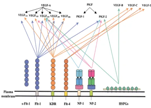
Figure 1. Vascular endothelial growth factors and VEGFRs family members.
The VEGF family binds to three transmembrane receptors, VEGF-R1/Flt-1, VEGF-R2/KDR and VEGF-R3/Flt-4, leading to the formation of VEGF-R homodimers and heterodimers. A soluble form of VEGF-R1/Flt-1 has also been characterized and it appears to be an important modulator for the placental vasculature. VEGFs as well as VEGF-Rs bind to co-receptors such as heparan sulfate proteoglicans and neuropilins. VEGFs and related receptors and co-receptors regulate angiogenesis, vasculogenesis, lymphangiogenesis, inflammatory responses and carcinogenesis.
In addition, modulatory actions are also exerted by a soluble form of VEGF-R1 (sVEGF-R1/sFlt-1), however the precise biological role of sVEGF-R-1 remains to be elucidated yet. Consistent with this model, loss of VEGF-R1 in mice causes embryonic lethality because of vascular overgrowth of endothelial cells, leading disorganization and dysfunction of the vasculature. Surprisingly, loss of the tyrosine kinase domain of VEGF-R1 alone produces a nearly healthy phenotype with normal vasculatures. This suggests that a membrane-anchored and a soluble VEGF-R1 might coordinately regulate angiogenic activity ensuring the development of healthy vasculature during embryonic growth (Cao Y, 2009).
VEGF-R2 is the predominant mediator of VEGF-stimulated endothelial cell migration, proliferation, survival and enhanced vascular permeability (Gille et al, 2001; Bernatchez et al, 1999). VEGF-R2 null mice die between embryonic days 8,5-9,5 as result of defects in the development of hematopoietic and endothelial precursors ( Roskoski Jr, 2007). Other two non enzymatic receptors called neuropilin-1 (NP-1) and neuropilin-2 (NP2) have been found in a wide variety of adult human tissue and in several types of tumors where they play a crucial role in tumor progression (Bielenberg et al, 2006; Ellis LM, 2006). The neuropilins may mediate tumor growth by enhancing angiogenesis or by directly influencing tumor cells per se. Neuropilins are transmembrane non-protein-tyrosine kinase receptor for the semaphorin/collapsin family of neuronal guidance mediators and the VEGF family. Neuropilins act as co-receptors with VEGF-R1 and VEGF-R2 and also function as receptors for VEGF isoforms independently of VEGF-Rs. VEGF165, PlGF-2 and both isoforms of VEGF-B bind to NP-1 (Migdal et al, 1998;
Makinen et al, 1999). VEGF145, VEGF165, PlGF-2 and VEGF-C bind to NP-2
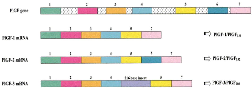
(Gluzman-Poltorak et al, 2000; Karkkainen et al, 2001). Both neuropilins function as receptors for PlGF-2 binding, but not PlGF-1 or -3. The binding of PlGF-2 to neuropilin-1 may perhaps potentiate the binding of PlGF-2 to the VEGF-R1 in a similar manner. The interaction of PlGF-2 with neuropilin-1 could affect other responses of endothelial cells such as cell-cell interactions or blood vessel organization in vivo (Migdal et al., 1998). Another member of VEGF family is PlGF. It is a homodimeric glycoprotein that shares a 42% amino acid sequence identity with VEGF (Maglione et al, 1991). The PlGF gene contains seven exons and expresses four isoforms: PlGF-1 (PlGF131), PlGF-2 (PlGF152),
PlGF-3 (PlGF203) and PlGF-4 (PlGF224) due to alternative mRNA splicing of the PlGF
primary transcripts (Maglione et al, 1993; Yang et al, 2003).
Apart from size, the PlGF isoforms differ in terms of both their secretion properties and their binding affinities. PlGF-1 is a dimeric protein, composed of 131 amino acid residues for monomer; PlGF-2 consists of 170 amino acid residues prior to signal peptide and also contains a highly basic 21 amino acid insertion that results in high heparin-binding affinity and HSP of the extracellular matrix; PlGF-3 contains a sequence of 216 nucleotides; PlGF-4 consists of the same sequence as PlGF-3 plus a heparin-binding domain that was previously thought to be present only in PlGF-2 (Yang et al, 2003).
Figure 2. Splicing variants of the human PlGF gene.
Description of the three splicing variants of the human gene encoding for PlGF mRNA. The gene contains seven exons. PlGF-1 and PlGF-3 lack exon 6 (which is constituted of 21 basic amino acid residues). PlGF-3 shows a 216 bp insertion between exons 4 and 5. The length of each splicing isoform encodes the protein mature isoform.
PlGF isoforms only bind VEGF-R1, but not VEGF-R2. As VEGF-A, PlGF-2 additionally binds NP-1 and NP-2. PlGF-1 is a chemotactic factor and may contribute to the recruitment of mesenchymal progenitor cells (MPC) in the context of fracture healing, bone formation and remodeling (Fiedler et al, 2005). Additionally PlGF-1 can form heterodimers with VEGF, so VEGF is sequestered and its activities are impaired (Schomberg et al, 2007). PlGF was originally identified in the placenta, during early embryonic development (Maglione et al, 1993; Khaliq et al, 1996) and is expressed in several other organs including the heart, lung, thyroid, skeletal muscle and adipose tissue (Persico et al, 1999). Under pathological conditions, PlGF abundance is elevated in various cell types, including vascular endothelial cells, smooth muscle cells, keratinocytes, hematopoietic cells, retinal pigment epithelial cells and many different tumor cells (Cao et al., 1996; Fischer et al, 2007; Failla et al, 2000; Yonekura et al, 1999). PlGF induces various biological effects in vitro and in vivo by affecting a wide range of different cell types. PlGF can stimulate vessel growth and maturation directly by affecting endothelial and mural cells, as well as indirectly by recruiting pro-angiogenic cell types (Carmeliet P, 2003). Indeed, PlGF stimulates the growth, migration and survival of endothelial cells (Fischer et al, 2007; Adini et al, 2002; Ziche et al, 1997), increases the proliferation of fibroblasts and smooth-muscle cells, induces vasodilatation
and stimulates collateral vessel growth (Bellik et al, 2005; Yonekura et al, 1999). It also promotes the recruitment and maturation of angiogenesis-competent myeloid progenitors to growing sprouts and collateral vessels (Luttun et al, 2002; Hattori et al, 2002; Rafii et al, 2003). PlGF also activates and attracts macrophages that release angiogenic and lymphangiogenic molecules and inhibits the differentiation of dendritic cells (Selvaraj et al, 2003). PlGF is a survival factor for monocytes, it is able to protect endothelial cells from apoptosis in a similar manner as VEGF-A by inducing of antiapoptotic genes such as survivin (Adini et al, 2002).
Several mechanisms might explain the role of PlGF in pathological, but not physiological angiogenesis. PlGF and VEGF-R1 are minimally expressed in adult quiescent vasculature, but are markedly up regulated during pathological conditions (Carmeliet et al, 2001; Oura et al, 2003). Moreover, PlGF plasma levels and intratumoral expression has been found to correlate with tumor stage, vascularity, recurrence, metastasis and survival in different types of cancer (Chen et al, 2004; Marcellini et al, 2006). Further, in vivo anti-PlGF treatment was able to inhibit tumor growth without affecting healthy vessels by reducing the infiltration of angiogenic macrophages and severe tumor hypoxia, and thus preventing the switch on of the angiogenic rescue program responsible for resistance to VEGF- Receptors inhibitors (Fischer et al, 2007). On the other hand, PlGF could play a negative role in tumor angiogenesis through the formation of PlGF/VEGF heterodimers. These heterodimers are naturally produced by both normal and tumor cells in culture (Di Salvo et al, 1995; Cao et al, 1996), binding and activating VEGFR-2, although with reduced affinity compared to VEGF homodimers (Cao et al, 1996). Therefore, some authors suggest that the overexpression of PlGF in tumor tissue could result in inhibition of VEGF-mediated tumor angiogenesis because of the augmented formation of less active PlGF/VEGF heterodimers and the ensuing depletion of VEGF homodimers (Eriksson et al, 2002; Schomberg et al, 2007; Xu et al, 2006). In contrast to the negative regulation of VEGF-A function when PlGF and VEGF are produced in the same cell population, PlGF can also potentiate VEGF-induced angiogenic activity when both factors are produced in different cells. PlGF homodimers may potentially compete with VEGF-A homodimers for VEGF-R1 binding, indirectly rendering more VEGF molecules available for binding to VEGF-R2, which would thereby increase pro-angiogenic and vasculogenic signals. Another mechanism by which PlGF may enhance
VEGF induced vascular function is through activation of VEGF-R1 by PlGF, which in turn may lead to phosphorylation and transactivation of VEGF-R2 (Autiero et al, 2003). Intriguingly, VEGF-R1 and VEGF-R2 form heterodimers in blood vessel endothelial cells and, in theory, VEGF-R1/VEGF-R2 heterodimers could bind VEGF-PlGF heterodimers (Mac Gabhann & Popel, 2007). Currently, the biological functions mediated by VEGF-R1/VEGF-R2 heterodimers are not understood, because it is impossible to separate the responses of receptor heterodimers from those homodimers within cells and in vivo angiogenesis models.
Because the biological role and signaling mechanisms mediating cellular action of PlGF on cell behavior have not been fully elucidated yet, this study aims at assessing the expression of PlGF, along with other vascular growth factor and their cognate receptors involved in the angiogenic process, by MM cells in vitro and in vivo and at evaluating the effect of PlGF on growth and survival of cultured MM cells.
2. MATERIALS AND METHODS
2.1 Materials
Oligonucleotide primers were purchased from Invitrogen (Milan, Italy). The primary antibody for VEGF-R1 (sc-316), VEGF-R2 (sc-504), NP-1 (sc-5541), NP-2 (sc-7242), VEGF (sc-507) and PlGF (sc-1880) were obtained from Santa Cruz Biotechnology (Santa Cruz, CA, USA); anti-phospho-AKT (Ser473) and anti-AKT were from Cell Signaling Technology (Danvers, MA, USA), anti-β actin was from Sigma Aldrich (Milan, Italy). PlGF Quantikine ELISA Immunoassay kit was from R&D Systems (Abingdon, UK). Human recombinant PlGF-2 and human recombinant VEGF were from ReliaTech (Braunschweig, Germany). The phosphatidylinositol 3-kinase (PI3-K) inhibitors Wortmannin and LY294002 were from Calbiochem (EMD Chemical Inc. Darmstadt, Germany) and used at nontoxic and specific concentrations (50 nM and 20 µM, respectively).
2.2 Cell cultures
A panel of seven human pleural MM cell lines with different histological features has been used in the present study. The cell lines were: H-Meso-1, MM-B1, MM-F1 (Pass H. et al., 1995) Ist-Mes1 (Orengo AM et al., 1999), Mero 25, Mero 48a and Mero 84 (Versnel MA et al., 1989). H-Meso-1, Ist-Mes1, Mero 25 and Mero 84 are of epithelioid histotype, MM-B1 and Mero 48a of biphasic, while MM-F1 of fibrous histotype. The cell lines were maintained in RPMI 1640 medium supplemented with 10% heat-inactivated fetal bovine serum (FBS), 1% L-glutamine and 50 µg/ml gentamycin (complete medium), all from Invitrogen. Primary Human Umbilical Vein Endothelial Cells (HUVECs), used as control, were purchased from Clonetics (Cambrex Bio Science, Walkersville, MD) and cultured in EGM-2 BulletKit according to manufacturer’s instructions. HUVECs were used between the fourth and seventh passages.
2.3 RNA analysis
Total RNA was extracted from cells with Trizol (Invitrogen) according to the manufacturer’s instructions. One µg of total RNA was reverse-transcribed using 200U of Superscript III, 10mM DTT, 20U of RNAse inhibitor, 2.5µM of random hexamers, 1mM
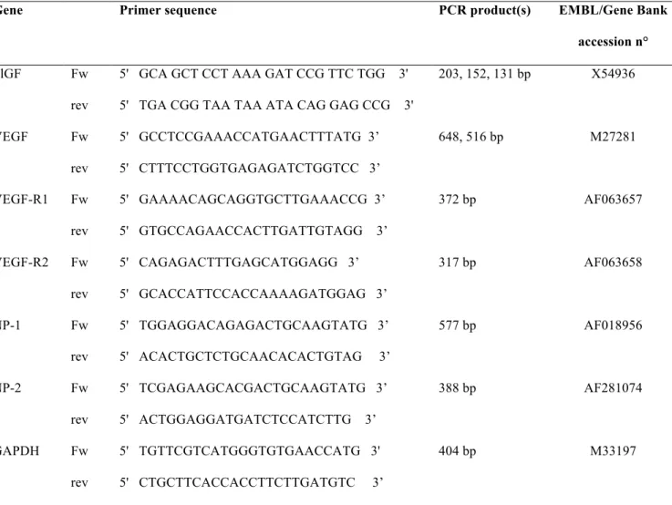
of each dNTP (all from Invitrogen) in a final volume of 20 µl. Reactions were performed at 42°C for 60 min. The cDNA product (1µl) was amplified with 1U of Platinum Taq DNA polymerase (Invitrogen) in a final volume of 50 µl containing 200 µM of each dNTP and 50 pmol of each primer. The sequences of the primers are listed in Table I. Amplification consisted of 30 sec at 94°C, 30 sec at 65°C and 30 sec at 72°C for 30 cycles, preceded by a first step of 2 min at 94°C to allow activation of the enzyme and followed by a final extension at 72°C for 7 min. Primers were designed to span over different exons in order to avoid amplification of contaminating genomic DNA. Moreover, primers for VEGF and PlGF were designed to detect each of the described alternative splicing isoforms. The identity of the amplicons was confirmed by sequencing.
2.4 Western blot analysis
Cellular extracts were obtained from HUVECs and MM cell lines by using a lysis buffer containing 50mM Tris-HCl (pH 7.4), 150mM NaCl, 1% Nonidet-P40, 1% Triton X-100, 1mM Na3VO4, 1mM NaF, 1mM PMSF and protease inhibitor cocktail from
Sigma-Aldrich. Proteins (50-75 µg/ lane) were separated on 4-8% or 12% NuPage gels (Invitrogen) according to the molecular weight of the antigen analyzed, and blotted onto nitrocellulose. The membranes were then treated as previously reported (Bei R. et al., 2006). The immunoblots were developed with the Super Signal chemiluminescence reagent (Pierce, Rockford, IL, USA).
2.5 PlGF ELISA
PlGF levels in serum-free media conditioned by HUVEC or by MM cell lines for 24-72 hours were measured using a Quantikine ELISA Immunoassay kit (R&D) according to manufacturer’s instructions. In our hands, this assay showed a lower limit of sensitivity of 15.6 pg/ml; further, according to the manufacturer the kit displays cross-reactivity with VEGF/PlGF heterodimers of 5% only. The concentration of PlGF in the conditioned media was normalized to 106 cells.
The results have been reported as mean ± standard error (SE) values calculated from four independent assays, each performed in triplicate.
2.6 Immunohistochemistry
Tissues were obtained according to ethical guidelines of the institutions (Tor Vergata University, Rome, Italy and A.O. San Camillo-Forlanini, Rome, Italy) after informed consent of the patients. Nineteen cases of resected pleural MM, including 14 cases with epithelial, 3 with biphasic and 2 with sarcomatous histotype were analyzed. The immunohistochemical analysis was also performed on 5 biopsies of normal mesothelium (NM) and on 7 reactive/hyperplastic mesothelium (RM/HM) specimens. NM biopsies were from patients with no history of pleural-pulmonary disease. RM/HM biopsies were from patients with non-neoplastic diseases.
Formalin-fixed paraffin-embedded sections (5µm) were mounted on sylane-coated slides. Immunohistochemistry (IHC) was performed with UltraTek HRP kit (ScyTek, Logan, UH) according to manufacturer’s instructions. Briefly, the sections were deparaffinized in xylen and rehydrated in a series of diluted ethanol. After antigen retrieval in a microwave oven for 10 min in 10mM citrate buffer (pH 6.0), endogenous peroxydase activity was blocked with methanol containing 3% H2O2. Primary antibodies were incubated in moist
chamber at 4°C overnight. After washing, the sections were incubated with biotinylated polyvalent IgGs followed by HRP-labelled streptavidin and visualized using 3-amino-9-ethylcarbazole (AEC) chromogen in H2O2 as substrate. Finally, the slides were
counterstained with Mayer’s haematoxylin. Non immune goat or rabbit serum was used as negative control, while the endothelium within the specimens was used as internal positive control.
The immunostained sections were independently evaluated by two pathologists, who recorded immunostaining distribution in mesothelial cells, MM cells, stroma, endothelial and inflammatory cells. Mesothelial and MM cell immunostaining was additionally scored for intensity using a semi quantitative system: 0, no staining; 1+, weak staining; 2+, moderate staining; and 3+, strong staining. The percentage of cells with positive staining was assessed independently and ranked into three categories: +, <30% positive rate; ++, 30–60% positive rate; and +++, >60% positive rate. Over expression was considered by a percentage of positive cells ≥ 60% with an intensity of the immunostaining of 2+/3+. No reactivity was observed with rabbit or goat control IgGs.
2.7 Cell growth assays
For cell proliferation studies, HUVEC and MM cells were seeded at 5 x 10³ cells/well in 96-well plates, allowed to adhere overnight and then starved in medium devoid of serum and containing 0.2% BSA with 20 or 50ng/ml of human recombinant PlGF-2. Media and treatments were renewed after 48 hours and after a total of 72 hours cell proliferation was quantified by the MTS-based assay (CellTiter 96 AQueous Cell Proliferation assay, Promega Corporation, Madison, WI) performed according to the manufacturer’s instructions. The MTS-based assay is a colorimetric method in which the conversion of the Owen’s reagent into a soluble formazan can occur only in metabolically active cells and is directly proportional to the number of living cells in culture (Buttke TM et al., 1993). All experiments were performed in quadruplicate and repeated a minimum of three times.
2.8 Cell survival assays
The effect of PlGF withdrawal from cell cultures was evaluated using the commercially available goat anti-human PlGF antibody reported in the Materials section. The cells were seeded at low density in 96-well plates and grown in complete medium with or without 10 µg/ml of anti-PlGF antibody or normal goat IgGs as control. Fresh antibodies were added daily. Cell survival was evaluated by the MTS-based assay (Promega) after 24, 48 and 72 hours from the beginning of treatment. The percentage survival of the cell cultures receiving the anti-PlGF antibody was calculated by normalization of their O.D. values to those of the cells receiving normal goat IgGs, whose survival was arbitrarily set at 100 %. The experiments were performed in triplicate and repeated a minimum of three times.
2.9 Statistical analysis
For continuous variables, Student’s two-tailed t-test was employed to compare calculated means. Proportions among categorical variables (growth factors and receptors over expression, as assessed by IHC, in RM/HM vs NM, MM vs NM and MM vs RM/HM) were analyzed by the two-tailed Fisher’s exact test. Statistical significance was set at p < 0.05.
3. RESULTS
3.1 Expression of PlGF, VEGF, and their cognate receptors and co-receptors in cultured MM cells
The expression of PlGF, VEGF and their cognate receptors and co-receptors by cultured MM cells was investigated both at the mRNA and protein level using a panel of seven established MM cell lines, HUVECs were used as positive control. As assessed by RT-PCR, all MM cell lines expressed the three PlGF splicing isoforms (PlGF-1, PlGF-2, and PlGF-3), and two main VEGF (VEGF121 and VEGF165) isoforms. Further, MM cell lines
exhibited different levels of transcripts for both VEGF-R1 and VEGF-R2 and for the NP-1 co-receptor, as well as significant levels of transcripts for NP-2 (Figure NP-1A).
As determined by Western blotting, the lysates from three out of seven MM cell lines (H-Meso-1, Mero 25 and MM-B1) displayed PlGF levels similar to that found in HUVECs, while lower levels of PlGF were detected in lysates from MM-F1 and Mero 48a cells. Conversely, the cytokine was not detectable in Mero 84 and Ist-Mes 1, although these cell lines were positive for PlGF transcripts by RT-PCR analysis (Figure 1B). Western blot analysis confirmed that VEGF and VEGF receptors and co-receptors were expressed in all cell lines (Figure 1B).
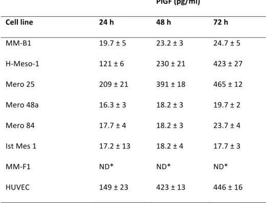
3.2 In vitro release of PlGF by MM cells
As determined by ELISA, two out of seven MM cell lines (H-Meso-1 and Mero 25) released high amounts of PlGF. Indeed, the serum-free media conditioned by these cell lines for 72 hours contained about 400 pg/ml of cytokine, an amount similar to that found in HUVECs conditioned media. On the other hand, the amount of PlGF released by Mero 48a, MM-B1, Mero 84 and Ist-Mes 1 was much lower, after 72 hours of culture being in the range of 20 pg/ml (Table II). PlGF was not detectable in the media conditioned by MM-F1 cells, in spite of the positivity shown by the corresponding cell lysates in Western blot analysis (Table II, Figure 1B). Remarkably, the media conditioned by MM-F1 cells lacked PlGF immunoreactivity even when concentrated to one-tenth of the original volume or when 1 µg/ml heparin, which competes with the PlGF-2-retaining HSPG of the cell membrane, was added to MM-F1 cultures. These results indicate that the secretion of PlGF is at least poorly efficient in this cell line.
Additionally, the possibility that culture conditions could affect PlGF release was further analyzed. However, results similar to those described so far were obtained in the conditioned media of MM cells grown in the presence of 10% of FBS (not shown).
3.3 Expression of PlGF, VEGF and their cognate receptors in human mesothelium and MM tissues.
To verify whether the results obtained by using established MM cell lines could be representative of the tumor condition in vivo, the expression of PlGF was investigated by IHC in 19 cases of resected pleural MM. Furthermore, in order to unravel whether PlGF expression could be modulated upon neoplastic transformation of the mesothelial cell, immunohistochemical analysis was also performed on 5 biopsies of normal mesothelium (NM). For comparison 7 reactive/hyperplastic mesothelium (RM/HM) specimens were included in the study. Both NM and RM/HM biopsies were from patients with non-neoplastic diseases. Representative IHC pictures are reported in Figure 2. PlGF immunostaining was not detectable in flattened NM. On the other hand, IHC revealed that VEGF was over expressed in 2 out of 5 NM specimens, whereas the cognate receptors and co-receptors were expressed at low levels in all 5 NM specimens (Table III). The expression levels of PlGF and VEGF receptors and co-receptors were found to be significantly higher in MM as compared to NM (Figure 2, Table III). Indeed, PlGF was over expressed in 14 out of 19 MM samples (74%; p = 0.0059 vs NM), VEGF-R1 in 18/19 (94%; p = 0.00014 vs NM), VEGF-R2 in 12/19 (63%; p = 0.037 vs NM), NP-1 in 16/19 (84%; p = 0.0013 vs NM) and NP-2 1n 14/19 (74%; p = 0.0059 vs NM). Conversely, although VEGF appeared over expressed in 16/19 MM samples (84%), its expression levels were not found to be significantly different in MM vs NM (Figure 2, Table III). Therefore, while statistical significance of tumor-specific over expression applied to all receptors (p<0.05 in MM vs NM), only PlGF over expression appeared significant about the two ligands (p<0.05 in MM vs NM) (Figure2, Table III). On the other hand, due to a variable over expression of both ligands and receptors in plump cuboidal mesothelial cells of RM/HM specimens, the respective levels of expression were not statistically different in MM vs RM/HM (Table III). Of note, besides MM cells, mesothelial and endothelial cells, immunoreactivity for both PlGF and VEGF was also detected in some fibroblasts and inflammatory cells residing in MM and RM/HM.
Finally, it was observed that both growth factors and receptors were more often over expressed in epithelioid and biphasic than in sarcomatoid MM.
3.4 Effect of PlGF-2 on MM cell proliferation
The finding that MM tissues were strongly positive for PlGF and its receptor (VEGF-R1) and co-receptors (NP-1 and NP-2), and that several MM cell lines secreted PlGF and expressed its receptors suggested that PlGF might act on MM cells through an autocrine stimulation loop. To verify this hypothesis, the MM cell growth was evaluated upon the presence of exogenously added recombinant human PlGF-2, the only PlGF isoform able to bind both the co-receptors NP-1 and NP-2 and HSPGs (Migdal M. et al., 1998), (Mamluk R. et al., 2002).
To avoid a possible mitogenic synergism between PlGF-2 and serum-containing growth factors or cytokines, HUVECs and MM cells were grown in the absence of serum under continuous exposure to 20 – 50 ng/ml of PlGF-2 over a period of 72 hours. As illustrated in Figure 3 panel A, PlGF-2 had no significant effects on MM cell proliferation. Unlike MM cell proliferation, the proliferation of HUVECs showed an increase of about 25% after 72 hours of PlGF-2 treatment (p<0.05), demonstrating that the limited effects of PlGF-2 on MM cells are not due to a reduced biological activity of the growth factor.
3.5 Assessment of biochemical responsiveness of MM cells to PlGF-2
Next, we sought to investigate whether the administration of exogenous PlGF-2 was able to activate downstream effectors of proliferative and cytoprotective signals. To this end, it was analyzed the ability of PlGF-2 to induce phosphorylation of Akt, a protein kinase that is activated via growth factor receptor stimulation in a PI3-K-dependent manner (Nishi J. et al., 2008). Thus, H-Meso-1 and MM-B1 cell lines, which showed the highest and the lowest levels of PlGF release, respectively, were serum-starved for 24 hours, stimulated with 50 ng/ml of PlGF-2 and lysed at different time points over a time course of 90 min from stimulation. Akt phosphorylation was then determined by Western blotting using an anti-phospho-Akt (S473) antibody. The results reported in Figure 3 panel B; show that PlGF-2 was effective in promoting Akt phosphorylation, although with striking differences in the two cell lines. Indeed, in H-Meso-1 cells the phosphorylation of Akt showed a biphasic course, as reported in other systems (Marino
M. et al, 2003), exhibiting a transient increase after 1 minute from PlGF-2 stimulation, followed by a decline and by a later increase at 30 min. Conversely, in MM-B1 cells Akt phosphorylation was detected after 5 minutes from stimulation and declined thereafter, being undetectable after 30 min. PlGF-2-induced AKT phosphorylation was completely abolished in cells pretreated for 2 hours with the PI3-K inhibitors Wortmannin and LY294002. These results indicate that PlGF-2 can elicit survival signals via VEGF-R1 in MM cells.
3.6 PlGF withdrawal reduces MM cell survival and induces cell death
To unravel the role exerted by PlGF in MM, it was assessed whether the treatment with an anti-PlGF antibody which is able to neutralize PlGF activity could affect the survival of MM cultures. In order to rule out a possible cross-reactivity of the anti-PlGF antibody with VEGF, which is known to exert autocrine effects on MM cells (Strizzi L. et al., 2001), the specificity of the anti-PlGF antibody was first verified by Western blotting. Immunoblot analysis, performed either in denaturing (Figure 3C) or non-denaturing (Figure 3D) conditions, demonstrated reactivity of the neutralizing antibody with recombinant PlGF but not with recombinant VEGF. Thus, MM cell lines were grown in complete medium under daily exposure to the anti-PlGF antibody or non-specific IgGs as control. The withdrawal of PlGF from the culture medium induced a time-dependent decrease of cell survival in all cell lines (Figure 3E). In particular, after 72 hours of treatment, H-Meso-1 and Mero 25 cell cultures showed a residual survival of 78 and 67%, respectively (p<0.05). It is worthy of note that H-Meso-1 and Mero 25 are the cell lines which secreted the highest amounts of PlGF. Further, a reduction of cell survival of about 50% was observed in Mero 48a, Mero 84, Ist-Mes 1 (p<0.005) and MM-F1 (p<0.05), whereas a residual survival of 25% was observed in MM-B1 cultures (p<0.05). As assessed by light microscopy, MM cultures treated with the anti-PlGF antibody were characterized by a strong reduction of viable cells and by a high proportion of dying cells, demonstrating that PlGF withdrawal was effective in inducing MM cell death (Figure 3F).
4. DISCUSSION
The therapeutic inhibition of tumor progression requires a detailed definition of the different pro-angiogenic growth factors produced by tumor cells and an understanding of their relative biological activity. The ability of MM and mesothelial cells to produce and respond to VEGF is well established (Strizzi et al, 2001; Catalano et al, 2003). By contrast, to our knowledge, the expression and biological functions of PlGF in MM have not been investigated in previous studies. In this report we provide evidence, for the first time, that expression of PlGF is a common feature of pleural MM and that it is associated with expression of its cognate receptor and co-receptors both in vitro and in vivo.
All MM cell lines tested expressed PlGF at the mRNA level, whereas all but one (MM-F1) secreted moderate to high levels of PlGF into the medium. Since MM-F1 cell lysates displayed positivity for PlGF in western blot analysis, the apparent lack of PlGF immunoreactivity in the corresponding conditioned medium might indicate that the secretion of PlGF is poorly efficient in this cell line or that these cells preferentially release VEGF/PlGF heterodimers (Cao et al, 1996) which cannot be detected by the ELISA kit used in this study. Conversely, although PlGF was not detectable in Mero 84 and Ist-Mes 1 cell lysates, the corresponding conditioned media contained detectable amounts of this cytokine, indicating that PlGF release is highly efficient in these cell lines. Altogether, these findings suggest that expression and release of PlGF by MM cells are independently regulated events. Further, albeit the PlGF degree release appeared to be more frequent among cell lines displaying the highest growth rate than among cell lines that grow more slowly, this observation could not be applicable to the MM-F1 cell line which exhibits a very high growth rate.
Our study also demonstrates NP-1 and NP-2 expression in all MM cell lines as well, although the transcript level for NP-1 appear to be lower than NP-2. This result is consistent with other reports wherein neoplasms that are not of epithelial origin, such as MM, express less NP-1 than NP-2 (Bielenberg et al, 2006). Further, the expression of one or both neuropilins have been correlated with tumor progression and/or poorer prognosis in many types of cancer (Ellis ML, 2006).
According to our immunohistochemical data, PlGF expression appears to be induced upon neoplastic transformation of mesothelial cells. PlGF up-regulation in tumor cells
has also been reported to occur in colorectal, gastric and breast cancer (Chen et al, 2004), cervical squamous cell carcinoma (Kodama et al, 1997), and melanoma (Marcellini et al, 2006). Interestingly, mononuclear phagocytes positive for PlGF were detected in both RM/HM and MM, indicating that the inflammatory infiltrate can also represent a source of PlGF in these tissues. In a diabetic wound closure model PlGF was required for the recruitment of monocytes and macrophages to the closing wound (Cianfarani et al, 2006), and in an ischemic hind limb model PlGF administration increased the number of macrophages around collateral side branches (Luttun et al, 2002). Therefore, the release of PlGF by MM cells could drive the recruitment of macrophages in the tumor microenvironment and, in turn, the recruited macrophages could contribute to tumour progression by releasing PlGF and promoting tumour angiogenesis (Odorisio et al, 2002; Oura et al, 2003; Marcellini et al, 2006; Condeelis & Pollard, 2006; Coussens & Werb, 2002). Although the simultaneous presence of VEGF-R1, NP-1 and NP-2 on MM cells suggests that PlGF may act in an autocrine manner to regulate MM cell growth, the addition of PlGF-2 to the culture medium was ineffective in eliciting a significant growth response in MM cultures. Still, we demonstrate that PlGF-2 administration induced a rapid yet transitory activation of Akt in MM cells. Akt is a widely expressedkinase that, activated through a PI3-K-dependent mechanism, promotes cell proliferation and survival (Song et al, 2005). Consistent with the effect of PlGF-2 on the activation of Akt, PlGF withdrawal from culture media was found to significantly reduce the proportion of viable MM cells and to induce MM cell death. While the above results represent the first evidence for a functional role of PlGF-2/VEGF-R1 in mediating survival of MM cells, PlGF-2/VEGF-R1 neutralization has been previously found to inhibit growth and metastatization of different tumor models without affecting normal tissues (Fisher et al, 2007; Taylor & Goldenberg, 2007).
Although the biological role and signaling mechanisms mediating the effect of PlGF on cell behavior has not been fully elucidated yet, we (Barillari et al, 1998) and others (Autiero et al, 2003; Carmliet et al, 2001) have proposed feasible mechanisms by which PlGF could affect tumor growth and progression. We demonstrated that the basic residues of PlGF-2 compete for the binding of VEGF and basic fibroblast growth factor (bFGF) to the HSPGs of the cell surface and to the extracellular matrix, thus inhibiting the sequestering of VEGF and bFGF and maintaining them in a highly diffusible form (Cao
et al, 1996). Besides, PlGF could synergize with VEGF either directly by displacing VEGF from VEGF-R1 and thus making more VEGF available to bind and activate VEGF-R2 (Autiero et al, 2003) or indirectly by a mechanism of transphosphorylation between VEGF-R1 and VEGF-R2 (Carmeliet et al, 2001). PlGF also up regulates the expression of VEGF, bFGF, matrix metalloproteinases (MMPs) and other angiogenic factors (Fisher et al, 2008; Marcellini et al, 2006; Barillari et al, 1998). Further, PlGF might enhance the angiogenic response to VEGF by forming VEGF/PlGF heterodimers (DiSalvo et al, 1995; Cao et al, 1996), which have been detected in tumors and are up-regulated by hypoxia in vivo (Cao et al, 1996). In this regard it has to be highlighted that, while several studies have documented that PlGF expression by cancerscorrelates with recurrence, metastasis,and mortality (Chen et al, 2004; Marcellini et al, 2006), in other studies it has been reported that PlGF/VEGF heterodimers display a reduced activity as compared to VEGF homodimers and that PlGF over expression can inhibit tumor growth, angiogenesis and metastatization through the augmented formation of PlGF/VEGF heterodimers and ensuing depletion of the more active VEGF homodimers (Eriksson et al, 2002; Schomber et al, 2007). Remarkably, the same authors reporting that PlGF/VEGF heterodimers can have inhibitory effects on angiogenesis when produced by tumor cells in vivo, previously reported that the heterodimers are active and pro-angiogenic (Cao et al, 1996; Eriksson et al, 2002). It is of note that in two studies reporting the inhibitory activity of PlGF/VEGF heterodimers, the heterodimers occurred between VEGF and the less active form of PlGF (PlGF-1) (Eriksson et al, 2002; Schomber et al, 2007), and that in an additional study performed using an orthotopic mouse model, where the heterodimers occurred between VEGF and PlGF-2, the majority of tumor tissue was composed of tumor cells which did not express or expressed undetectable levels of VEGF receptors (Xu et al, 2006).
Conversely, in our study both the MM cell cultures and the tumor tissues were composed of tumor cells which express not only VEGF-R1 and VEGF-R2, but also the relative co-receptors NP-1 and NP-2.
Interestingly, one of the mechanisms by which PlGF elicits biological responses is the induction of FosB and c-fos expression in endothelial cells and monocytes as effectively as VEGF, indicating that FosB and c-fos may play a role in mediating biological actions of PlGF via VEGF-R1 (Holmes & Zachary, 2004). Besides, it has been reported that
asbestos stimulates the expression of c-fos and c-jun mRNA in mesothelial cells in a dose-dependent fashion (Heintz et al, 1993; Ramos-Nino et al, 2002). The co-existence of different stimuli, such as asbestos fibres and PlGF, concurring to the activation of the early response gene pathway, might lead to the persistent induction of AP-1 transcription factors in mesothelial cells and to the chronic stimulation of mesothelial cell proliferation. Although, further studies are required to determine the precise mechanism by which PlGF could affect tumor growth, collectively, the evidences provided in the present study indicate that PlGF can act as a survival factor in MM and suggest that this cytokine may represent a molecular target for novel antitumor approaches against MM.
Table I. Sequence of primers used for PCR.
Gene Primer sequence PCR product(s) EMBL/Gene Bank
accession n°
PlGF Fw
rev
5' GCA GCT CCT AAA GAT CCG TTC TGG 3' 5' TGA CGG TAA TAA ATA CAG GAG CCG 3'
203, 152, 131 bp X54936 VEGF Fw rev 5' GCCTCCGAAACCATGAACTTTATG 3’ 5' CTTTCCTGGTGAGAGATCTGGTCC 3’ 648, 516 bp M27281 VEGF-R1 Fw rev 5' GAAAACAGCAGGTGCTTGAAACCG 3’ 5' GTGCCAGAACCACTTGATTGTAGG 3’ 372 bp AF063657 VEGF-R2 Fw rev 5' CAGAGACTTTGAGCATGGAGG 3’ 5' GCACCATTCCACCAAAAGATGGAG 3’ 317 bp AF063658 NP-1 Fw rev 5' TGGAGGACAGAGACTGCAAGTATG 3’ 5' ACACTGCTCTGCAACACACTGTAG 3’ 577 bp AF018956 NP-2 Fw rev 5' TCGAGAAGCACGACTGCAAGTATG 3’ 5' ACTGGAGGATGATCTCCATCTTG 3’ 388 bp AF281074 GAPDH Fw rev 5' TGTTCGTCATGGGTGTGAACCATG 3' 5' CTGCTTCACCACCTTCTTGATGTC 3’ 404 bp M33197
Figure 1.
Expression of PlGF, VEGF and their cognate receptors in malignant mesothelioma cell lines. A) Electrophoretic pattern of PlGF, VEGF, VEGF-R1, VEGF-R2, NP-1, NP-2 and GAPDH RT-PCR products from HUVEC and MM cell lines. PlGF primer pairs generated products of the expected size for PlGF-1 (131 bp), PlGF-2 (152 bp) and PlGF-3 (203 bp) transcript isoforms in HUVEC as well as in all MM cell lines; VEGF primer pairs generated products of the expected size for VEGF121 (516 bp) and VEGF165 (648 bp)
transcriptisoforms. Further, MM cell cultures variably expressed transcripts for VEGF-R1 (392 bp product), VEGF-R2 (477 bp product), NP-1 (577 bp product) and NP-2 (388 bp product). The ubiquitously expressed gene GAPDH was included as control of the amplifiability of cDNA from respective RNAsamples. PCR ctrl: template-free control of PCR reaction. B) Immunoblot analysis. PlGF was detected in five out of seven MM cell lines whereas, despite the positivity in RT-PCR, it was not detected in Ist-Mes 1 and Mero 84. All MM cell lines constitutively express VEGF and VEGF-Rs and co-receptors. Membranes were reprobed with anti-β-actin to ensure equal loading and transfer of samples. HUVECs were used as control in all experiments.
Table II. PlGF levels in serum‐free media conditioned by MM cell lines or by HUVECs for 24‐ 72 h. PlGF (pg/ml) Cell line 24 h 48 h 72 h MM‐B1 19.7 ± 5 23.2 ± 3 24.7 ± 5 H‐Meso‐1 121 ± 6 230 ± 21 423 ± 27 Mero 25 209 ± 21 391 ± 18 465 ± 12 Mero 48a 16.3 ± 3 18.2 ± 3 19.7 ± 2 Mero 84 17.7 ± 4 18.2 ± 3 23.7 ± 4 Ist Mes 1 17.2 ± 13 18.2 ± 4 17.7 ± 3 MM‐F1 ND* ND* ND* HUVEC 149 ± 23 423 ± 13 446 ± 16 *ND, not detectable