Technical Report CoSBi 06/2007
Simulating a faulty mechanism of protein
folding in the pathogenesis of familial
Parkinson’s disease
Paola LeccaThe Microsoft research - University of trento Centre for Computational and Systems Biology
This is the preliminary version of a paper that will appear in Online Journal of Bioinformatics 9 (1):30-43, 2008
Simulating a faulty mechanism of protein folding in
the pathogenesis of familial Parkinson’s disease
Paola Lecca
Abstract
A growing body of evidence suggests that the accumulation of mis-folded proteins in brain tissues is a crucial event in the Parkinson’s disease neurodegeneration. Pathogenic mutations may directly induce abnormal protein conformations or compromise the ability of the cel-lular machinery to detect and degrade misfolded proteins. Although the recent explosion in the rate of discovery of genetic defects linked to Parkinson’s disease (PD) have provided tangible clues to the neurobi-ology of the disorder, they have provided neither direct explanation for the disease process or its key biochemical mechanism. The aim of the work is to provide quantitative models for in silico experiments, that can help the researchers either to elucidate important and still elusive aspects of the Parkinson’s disease or to design new wet-experiments.
Here we present three stochastic models of a faulty mechanism of protein re-folding and degradation of misfolded proteins. Our mod-els are specified in biochemical stochastic π-calculus and are based on what is currently known about the genetic mutations causing PD. The expressive capabilities of this formalism in the description of parallel and competive nature of biochemical interactions make it particularly suitable for modeling the intricate mechanism of proteins folding, folding and eventually degradation. Furthermore, the simulation re-sults point out those kinetic quantitative parameters, whose variations lead to significant changes in the capability of the system to react to the accumulation of dangerous proteins.
1
Introduction
Parkinson’s disease (PD) is a chronic, progressive movement disorder, that results from the degeneration of dopamine-producing nerve cells in the sub-stantia nigra. Dopamine is a neurotransmitter that stimulates motor neu-rons, those nerve cells that control the muscles. When dopamine production is depleted, the motor system nerves are unable to control movement and coordination. Parkinson’s disease patients have lost 80% or more of their dopamine-producing cells by the time symptoms appear. The inherited forms of Parkinson are relatively rare but may provide clues to the biologi-cal origins of the more common forms of the disease. More specifibiologi-cally, the rationale for studying the rare genetic forms of PD is the expectation that the phenotypic similarity between the genetic and sporadic forms indicates that they share important pathogenic mechanisms, and as consequence, that genetic information will help focus research on a key biochemical pathway responsible for the disease.
Although the scientists have not yet found the exact cause of PD, recent years have seen an explosion in the rate of discovery of genetic defects linked to the pathogenesis of PD. In the mid-1990s three missense mutations in the gene encoding α-synuclein were identified as a possible cause of a dominantly inherited PD [18]. None of these mutations has been found in sporadic PD or individuals without the disease. At the end of 1990s another protein, whose mutation has been associated to PD, has been identified: the parkin. Loss-of-function mutations in the gene encoding parkin cause a recessively inherited form of PD, that usually occurs under age 40 [7].
The causes of these mutations are believed to result from the exposure to environmental toxins, for example pesticides, that inhibits dopamine pro-duction and produce free radicals and oxidation damages. The effect of the pathogenic α-synuclein is the inhibition of the degradation of misfolded pro-teins [11, 18], whose accumulation in the dopaminergic neurons is the deter-mining factor of their death. However, how mutant α-synuclein and parkin variants exactly produces neurotoxicity remains unknown, in part because the theoretical and experimental efforts made to understand the proteins function and the reasons of the block of the proteolitic machinery are just at the beginning. At the present there are no available mathematical or computational models or computer simulated experiment that can aid the researchers to unravel the molecular basis of the neurodegenerative processes of PD. This work proposes two gene-based models of PD and a hypotheti-cal model that relates the onset of the disease to an insufficient quantity of chaperones. The gene-based models concern PD caused by dominant mu-tations in the α-synuclein gene and PD caused by recessive mumu-tations in the parkin gene. The hypothetical model intends to suggest a third possible mechanism in which the presence of an insufficient quantity of chaperones promotes the accumulation of misfolded proteins. All the three models has
been specified in biochemical stochastic π-calculus [15] and simulated with SpiM (Stochastic PI-Machine) [13]. The choice of such a formalism is moti-vated by the capability of expressing the parallel and concurrent nature of biological interaction, especialliy at molecular level and the modularity and the adaptability of a biological system as well. Common approaches as those based on differential equations revealed to be not so expressive, especially for handling concurrency and adaptability, i. e. the ability of the system to reconfigure itself in rensponse to environmental stimuli (see [9, 10] for a detailed comparison of the π-calculus with ordinary differential equation formalism).
2
Mechanisms of neurodegeneration
In patients with inherited PD, pathogenic mutations are though to cause dis-ease directly by inducing abnormal and toxic protein conformations [2] or indirectly by interfering with the processes that normally target for degrada-tion the misfolded proteins. The triggers for disfuncdegrada-tional protein metabolism may be oxidative stress. The tissue content of oxidized proteins, which may misfold, increases with age [1], and neurons may be susceptible because they are postmitotic. At the present a definitive diagnosis of PD can only be made by autopsy, and it is based on the presence of intraneuronal proteic inclu-sions called Lewy bodies (LBs) and on the loss of nigrostriatal dopaminergic neurons In PD caused by dominant mutations of α-synuclein, LBs contain a significant amount of the oxidatively modified variant of this protein. The mutations in the gene encoding parkin protein cause a recessively inherited form of PD. Pathologically, this form of familial PD is associated with a loss of nigrostriatal neurons, but LBs are not typically observed [3]. The ability of the cell to handle misfolded proteins is expressed by some complexes of macromolecules, called chaperones.
Molecular chaperones interact with unfolded or partially folded protein subunits, e.g. nascent chains emerging from the ribosome, or extended chains being translocated across sub-cellular membranes. They prevent inappropriate association or aggregation of exposed hydrophobic surfaces and direct their substrates into productive folding, transport or degrada-tion pathways. Essential for viability and cell survival, the expression of the molecular chaperones is often increased by cellular stress.
In the healthy cells, if a protein does not assume the correct 3D shape, or a cellular stress induces a right-folded protein to assume a wrong fold-ing, the chaperones re-shape it correctly. In the case in which the protein is not correctly refolded, the cellular proteasome - a system designated to the digestion of cell wastes - degrades it before the faulty protein can cause damages. First the protein parkin attaches molecules of ubiquitin to the misfolded protein; once the ubiquitin is bound to the faulty protein, it
sig-2 mRNA SYNTHETIZED PROTEIN CHAPERONE SYNTHETIZED PROTEIN CHAPERONE MISFOLDED PROTEIN
MISFOLDED PROTEIN RIGHT FOLDED PROTEIN
MISFOLDED PROTEIN RIGHT FOLDED PROTEIN
zzz 1 3 UBIQUITIN MISFOLDED PROTEIN PROTEASOME INACTIVE ACTIVE PROTEASOME Signal to proteasome PARKIN UBIQUITIN MISFOLDED PROTEIN SYNUCLEIN MUTATED α
Block the signal UBIQUITIN MUTATED PARKIN RIGH PARKIN CORRECTLY WORKING
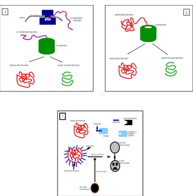
Figure 1: Pathogenesis of PD induced by mutant α-synuclein: 1. the in-tereation of a nascent protein with a chaperone can results in a right-folded protein or in a misfolded protein; 2. the chaperone attempts to re-fold the faulty protein and the result can be again a right-folded protein or a mis-folded protein; 3. therefore,the mismis-folded protein is drapped by the ubiquitin transported by the parkin protein. A mutant variant of the parkin is not able to transport the ubiquitin on the misfolded protein. The mutant α-synuclein inhibits the activation of the proteasome by the ubiquitin. The mutant α-synuclein seems to be proteasome-proof, but the model presented in this paper takes into account an eventual attempt of the proteasome to attack the faulty α-synuclein. The outcomes of the interactions between the nascent linearprotein and the chaperone, as well as of the interaction between the mutant α-synuclein and the proteasome are stochastically de-termined by the reaction probabilities derived from the kinetic reaction rates accordingly to the Direct Gillespie algorithm [6]
.
nals to the proteasome of decomposing the protein into its amino-acids, that will be employed somewhere else in the cell. A mutation of the gene of the
α-synuclein gives rise to a proteasome-proof variant of α-synuclein, that
per-turbatively interferes with the communication between ubiquitin and pro-teasome [11]. If the ubiquitin can not enable the activity of the propro-teasome, the misfolded protein can not be degraded. Fig. 1 depicts the mechanisms
of neurodegeneration triggered by the mutant variant of α-synuclein. Some chaperones are non-specific, and interact with a wide variety of polypeptide chains, but others are restricted to specific targets. With aging, the ability of cells to induce a variety of chaperones is impaired as is the activity of the proteasome. Proteasomal disfunctions and the accumulations of misfolded proteins are involved in a vicious cycle, with excess of misfolded proteins inhibiting an already compromised proteasome.
Finally, the mutations of the encoding for parkin, produce a variant of parkin unable to transport the molecules of ubiquitin on the misfolded proteins, that being untargeted, can not be recognized by the proteasome for the degradation.
3
Modeling and simulating in SpiM
The network of interactions that transform a linear protein, turned out by the translation of its messenger RNA in the ribosome, into a functional 3D spatial structure, has been specified in the formalism of biochemical
π-calculus.
This abstract formal language, initially developed for the specification of concurrent computational processes, revealed particularly suitable to de-scribe biological molecular systems [9, 8, 10, 15]. A mathematical theory of concurrent processes has been built in the π-calculus [12]. In this cal-culus, each process, belonging to a set of interacting processes, is defined by its potential communication activities and may be composed either se-quentially or concurrently with other processes. Communications occur via channels, indicated by their names. The π-calculus process algebra is an expressive and efficient formal language for modeling biochemical processes. In such systems, multiple processes interact with each other on complemen-tary shared communication channels by sending and receiving messages in a synchronized way. The π-calculus can be used to model a system of in-teracting bio-molecules, treating molecules and their individual domains as computational processes, where their complementary structural and chemi-cal determinants correspond to communication channels. Moreover chemichemi-cal interaction and subsequent modification coincide with communication and channel transmission. Finally, the simulation of the dynamic behavior of the system is defined by the operational semantics of the language.
A significant extension of the π-calculus was realized by Priami [14], by developing a stochastic variant of the original operational semantics of the calculus. This stochastic variant introduces the possibility to assign differ-ent rates to each involved biochemical reaction, from which it is possible to derive the probability of occurrence, and from there the reaction wait-ing time, of the different reactions. On the basis of that information it is then possible to implement a race condition, establishing which is the next
reaction and when it occurs.
To summarize and to introduce the essential notation, the most basic process form is a choice P = P1 + · · · + PN, among zero or more actions
exhibited by the processes P s composing the sumP. The simplest process is the deadlock, that is a process that can do nothing (usually denoted by 0. An action π can be an output x(n), or an input x(m), or a delays τ that the process can perform. x is the channel through which the output message n is sent. A process R defined by R = π.R0, is a process prefixed
by an unguarded action, i. e. a process offering to perform the action π. Once this action is performed the process R changes to the state defined by the process R0. Two processes P and Q can be combined using parallel
composition P |Q. Moreover, another basic operator of stochastic π-calculus is the new operator (indicated by ν followed by name or a set of channel names). It allows the creation of fresh channels. A fresh channels is a channel different from any other channel defined in the system. If a fresh name is defined in the body of a process, that channels a private channel for that process, i. e. it lives in the scope of that process. The delays represent a single communication on a fresh channels, that correspond to an internal evolution of the process. While sending and receiving messages on shared channels represent bi-molecular reactions, delay actions correspond to mono-molecular reactions of to changes of state or conformation of a given chemical. Table 1 lists the axioms of the semantics of biochemical stochastic
π-calculus.
In this paper the simulations were obtained using the Stochastic Pi Ma-chine (SpiM), which is the most recent simulator for the biochemical stochas-tic π-calculus. The simulator in extensively described in [13]. Here we briefly recall that SpiM simulates a given process P by firstly converting the process to a corresponding simulator data structure, consisting of a list of compo-nents A = P1, . . . ,PN. These list is processed by a procedure based on the Gillespie algorithm [6], that stochastic determines the next interaction channel x and the corresponding reaction time τ . Once an interaction chan-nel x has been chosen, the simulator randomly selects from the list A a component of the formP+x(m).P , containing an input on channel x, and a different component of the form P0+x(n).Q containing an output on x. The selected components can then interact in such a way that P is replaced by P {n/m} (i. e. in the body of P the variable m is replaced by n) and Q remains unchanged. Finally, the summationsP and P0 are discarded and the P {n/m} and Q are added to the remainder of the list A.
Accordingly to the stochastic formulation of the chemical kinetics de-veloped by Gillespie, for a reaction µ, the propensity aµ is a function of its
kinetic rate rµ and the number of individual potential copies of reactions µ
involving the same reactant species. As stated in the following equation, the reaction propensity is the product of the reaction ”rate” and the number of unique reactant combinations.
aµ=
rµ× (#A) × (#B) for bi-molecular reactions1
rµ×(#A)×(#A−1)2 for homodimerization
rµ× #A for monomolecular reaction
where #A and #B are the numbers of reactants of species A and B in the elementary reaction µ. The Gillespie algorithm calculates explicitly
which reaction occurs next and when it occurs. Both question are answered
probabilistically by specifying the probability P (µ, τ ) = aµexp(−τ
P
jaj)dτ
that the reaction is µ and it occurs a time τ . P (µ, τ ) can be expressed as the product of two distributions: the distribution for reactions P (µ) =
aµ/
P
jaj, and th distribution for times P (τ )dτ = (
P
jaj) exp(−τ
P
jaj)dτ .
The algorithm chooses a reaction according to P (µ), and the time step τ according to an exponential with parameterPjaj.
SpiM simulator uses the notion of channel activity to compute the re-action/communication propensity. The activity of channel x in a list L of processes is defined by Actx(L) = £ Inx(L) × Outx(L) ¤ − Mixx(L)
where Inx(L) and Outx(L) are the number of unguarded inputs and
out-puts on channel x in L, respectively, and Mixx(L) is the sum of Inx(Pi) × Outx(
P
i) for each summation
P
i in L. By subtracting Mixx(L) from the
product of the number of inputs and outputs on x, an eventual interac-tion between an input and an output belonging to the same summainterac-tion is avoided.
Finally, the dynamic behavior of a process is driven by a race condition. All activities enabled attempt to proceed, but only the fastest one succeeds. The fastest activity is different on successive attempts because durations are random variables. The continuity of the probabilistic distribution ensures that the probability that two activities end simultaneously is zero. Further-more, exponential distributions enjoy the memoryless property: the time at which a certain transition occurs is independent on the time at which it ever occurred before. Therefore there is no need to record the time elapsed to reach the current state.
3.1 First model: misfolded protein accumulation induced by mutant α-synuclein
The system of bio-molecules and cellular structures driving the proteins folding has been implemented as process SYSTEM, given by the parallel composition of 8 processes, that represent their homonymous molecules: LINEAR PROTEIN, RIBOSOME, RNA α SYNUCLEIN, CHAPERONE, PROTEASOME,
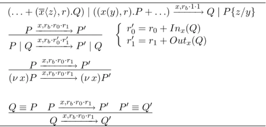
(. . . + (xhzi, r).Q) | ((x(y), r).P + . . .) x,rb·1·1 −−−−→ Q | P {z/y} P x,rb·r0·r1 −−−−−−→ P0 P | Q−−−−−−→ Px,rb·r00·r01 0 | Q ½ r0 0 = r0+ Inx(Q) r0 1 = r1+ Outx(Q) P x,rb·r0·r1 −−−−−−→ P0 (ν x)P x,rb·r0·r1 −−−−−−→ (ν x)P0 Q ≡ P P x,rb·r0·r1 −−−−−−→ P0 P0 ≡ Q0 Q x,rb·r0·r1 −−−−−−→ Q0
Table 1: Rules of the semantics of biochemical stochastic π-calculus. A reaction is implemented by the three parameters rb, r0 and r1, where rb
represents the basal rate, and r0 and r1 denote the quantities of interacting
molecules, and are compositionally computed using the two functions Inx
and Outx defined below. These two functions inductively count the number
of receive and send operations on channel x. The first axiom of the BioSpi reduction semantics corresponds to usual reactions involving two different molecules, the second rule corresponds to homo-dimerization reactions, in-volving the same molecular species. The third rules states that if a process
P evolves into a process P0 through a communication on channel x, the
restriction of channel x to the scope of process P does not affect the transi-tion of P into P0. Finally, the fourth axiom states that if P is structurally
congruent to Q and P evolves into P0, that is structurally congruent to Q0,
then Q evolves into Q0.
a faulty non deterministic operation of the chaperones system, whose twofold interaction with a protein can result in a right-folded protein or in a mis-folded one according to the value of the associate reaction rate. In this model we assume that the rate of interaction between a chaperone and a linear protein is equal to the rate of a a typical fast protein folding: 1
µs−1. CHAPERONE is the process abstracting the cellular chaperone and
LINEAR PROTEIN is the one representing the linear chain of amino-acids derived by the translation of the messenger RNA. The linear protein process “’physically binds” to the chaperone by sending a private name to chaperon to the process CHAPERONE via the shared channel bind to chaperone. The result of this interaction is the complex PROTEIN CHAPERONE, that can undergo two internal modifications: one resulting in a right-folded pro-tein, that in this model is a deadlock process, and the other resulting in a misfolded protein. We have called MISFOLDED’ the misfolded protein generated by the interaction between chaperone and linear protein, and MISFOLDED” the misfolded protein generated by the interaction between
chaperone and MISFOLDED’. MISFOLDED” is thus the process represent-ing the protein that has been not correctly re-folded even durrepresent-ing a second interaction with a chaperone. Therefore, this misfolded protein is targeted by parkin according to the following sequence of reactions: parkin performs first a physical binding with ubiquitin and then with the misfolded protein. The the sub-component ubiquitin of the trimer formed by parkin, ubiquitin, and misfolded protein, sends a signal to proteaseome to activate it. The physical binding of the parkin with the ubiquitin is modeled as the send-ing from the process PARKIN to the process UBIQUITIN of the private name parkin ubiquitin on the public channel bind to ubiquitin. After bind-ing, PARKIN changes to PARKIN BOUND and, analogously, UBIQUITIN changes to UBIQUITIN BOUND. The formation of the trimer including parkin, ubiquitin and misfolded protein is modeled as two sequential physi-cal bindings on the public channels to misfold1 and to misfold2. In partic-ular, PARKIN BOUND interacts with MISFOLDED” on to misfold1 and UBIQUITIN BOUND interacts with MISFOLDED” on to misfold2.
In our model we hypothesize that the process STRESS, representing the oxidative cellular stress, interferes with the translation of the tran-script of the α-synuclein gene. The messenger RNA of the α-synuclein (RNA A SYNUCELIN) after the interaction with RIBOSOME on the chan-nel bind to ribosome can either be correctly translated (i. e. it performs a communication on channel translate and it does not have any other evolu-tion) or it receive a signal from STRESS on channel stress and transforms into MUTATED A SYNUCLEIN.
The sub-part MISFOLDED” UBIQUITIN of the trimer midolfed protein-ubiquitin-parkin is defined as a choice: it can either communicate with the process PROTEASOME via the channel to proteasome and activate it for the degradation or it can receive a signal from MUTATED A SYNUCLEIN on the channel signal from synuclein and, consequently, remains intact.
The specification takes into account the attempts to the proteolitic ma-chinery of the cell to degrade the mutant variant of the α-synuclein, by defining the behavior of the process MUTATED A SYNUCLEIN as a choice between the communication with MISFOLDED” UBIQUITIN and the com-munication on the channel degrade, representing the eventuality of a degra-dation. The kinetic rates and the initial amoutn of reacting molecules are listed in Table 3.
The Figs. 2 (A)-(G) show the time evolution of the amount of MIS-FOLDED’ (solid line) and MISFOLDED” (dotted line) for different values of the initial number of stress processes (Ns) and stress signaling rate (rs).
The plots (A)-(C)-(E)-(G) were generated by fixing at 10 µs−1 the value of
the stress signaling rate and varying the number of processes STRESS by 10, 100, 200, and 1000 respectively. On the contrary, the plots (B)-(D)-(F)-(H) were generated by fixing at 100 the amount of processes STRESS and
SY ST EM := LIN EAR P ROT EIN | P ARKIN | U BIQU IT IN RN A A SY N U CLEIN | RIBOSOM E | ST RESS
P ROT EASOM E | CHAP ERON E
Interaction between parkin and ubiquitin
P ARKIN := (ν parkin ubiquitin)bind to ubiquitinhparkin ubiquitini.P ARKIN BOU N D P ARKIN BOU N D :=
(ν tm1)to misf old1htm1i.P ARKIN BOU N D M ISF OLDED00(tm1)
P ARKIN BOU N D M ISF OLDED00(tm1) := tm1
U BIQU IT IN := bind to ubiquitin(pu).U BIQU IT IN BOU N D(pu)
U BIQU IT IN BOU N D(pu0) :=
(ν tm2)to misf old2htm2i.U BIQU IT IN BOU N D M ISF OLDED00(pu0, tm2)
U BIQU IT IN BOU N D M ISF OLDED00(pu00, tm20) :=
pu00.M ISF OLDED00 U BIQU IT IN (tm20)
M ISF OLDED00 U BIQU IT IN (tm200) :=
to proteasome.degrade + signal f rom synuclein.M ISF OLDED00 U BIQU IT IN (tm200)
Interaction between chaperone and protein
LIN EAR P ROT EIN :=
(νto chaperon)bind to chaperonehto chaperoni.P ROT EIN CHAP ERON E(to chaperone)
P ROT EIN CHAP ERON E(to chaperone0) := to chaperon0.M ISF OLDED0+ to chaperon0
M ISF OLDED0:= (ν to chaperonebind to chaperonM ISF OLDED CHAP ERON E
M ISF OLDED CHAP ERON E := to chaperon.M ISF OLDED00+ to chaperon
M ISF OLDED00:=
to misf old1(tm10).to misf old2(tm200
).M ISF OLDED00 P ARKIN U BIQU IT IN
M ISF OLDED P ARKIN U BIQU IT IN := tm1.parkin ubiquitin.P ARKIN CHAP ERON E :=
bind to chaperon(to chaperone00).CHAP ERON E BOU N D(to chaperone00)
CHAP ERON E BOU N D(to chaperone000) := to chaperone000.CHAP ERON E
Translation of protein, perturbation from the process STRESS and communication with proteasome
P ROT EASOM E := to proteasome.ACT IV E P ROT EASOM E ACT IV E P ROT EASOM E :=!degrade.P ROEASOM E
RN A A SY N U CLEIN := bind to ribosome.(translate + stress.M U T AT ED A SY N U CLEIN ) M U T AT ED A SY N U CLEIN :=
signal f rom synuclein.M U T AT T ED A SY N U CLEIN + degrade RIBOSOM E := bind to ribosome.translate.RIBOSOM E
ST RESS := stress.ST RESS
Table 2: Stochastic π-calculus specification of a model of a faulty mechanism of protein folding and degradation. If the concent of a send or receive action is not specified, it means it is non relevant.
considering four different values of the stress signaling rate: 0.01, 1, 100, 1000 µs−1, respectively. The proteins MISFOLDED’ that are not degraded become proteins MISFOLDED”. In Figs. 2 (C)-(E)-(G), after about 5 µs−1,
0 2 4 6 8 10 12 0 1 2 3 4 5 6 7 8 9 10 Number of processes Time (micro-sec) MISFOLDED’ MISFOLDED’’ ) (A) 0 2 4 6 8 10 12 0 1 2 3 4 5 6 7 8 9 10 Number of processes Time (micro-sec) MISFOLDED’ MISFOLDED’’ (B) 0 2 4 6 8 10 12 0 1 2 3 4 5 6 7 8 9 10 Number of processes Time (micro-sec) MISFOLDED’ MISFOLDED’’ (C) 0 2 4 6 8 10 12 14 0 1 2 3 4 5 6 7 8 9 10 Number of processes Time (micro-sec) MISFOLDED’ MISFOLDED’’ (D) 0 2 4 6 8 10 12 14 0 1 2 3 4 5 6 7 8 9 10 Number of processes Time (micro-sec) MISFOLDED’ MISFOLDED’’ (E) 0 2 4 6 8 10 12 0 1 2 3 4 5 6 7 8 9 10 Number of processes Time (micro-sec) MISFOLDED’ MISFOLDED’’ (F) 0 2 4 6 8 10 12 0 1 2 3 4 5 6 7 8 9 10 Number of processes Time (micro-sec) MISFOLDED’ MISFOLDED’’ (G) 0 2 4 6 8 10 12 14 0 1 2 3 4 5 6 7 8 9 10 Number of procsses Time (micro-sec) MISFOLDED’ MISFOLDED’’ (H) Figure 2: (A) rs = 10 µs−1, Ns = 10; (B) rs = 0.01 µs−1, Ns = 100; (C) rs = 10 µs−1, Ns = 100; (D) rs = 1.0 µs−1, Ns = 100; (E) rs = 10 µs−1, Ns = 200; (F) rs = 100.0 µs−1, N s = 100; (G) rs = 10 µs−1, Ns = 1000;
(H) rs = 1000.0 µs−1, Ns = 100. The rates used in these simulations has
been taken from [4].
with the increase of the number of processes STRESS. In Figs. 2 (D)-(F)-(H) the number of MISFOLDED” decreases independently of the rate of perturbation. Fig. 2 (A) indicates that if Ns ≤ 10 and rs = 10.0 µs−1,
the MISFOLDED” proteins are rapidly degraded after about 4.4 µs−1, as
a scarce STRESS, even if its rate of perturbation is significant, the cell is still able to respond to the accumulation of faulty proteins. These results suggest that the intensity of cellular stress causing the α-synuclein muta-tions is more significant than the rate at which the stress interferes with the translation of this protein. It means that the probability of producing mu-tated α-synuclein proteins essentially depends on the amount of stress that interacts with the cell system. A particular attention should be payed to the plot of Fig. 2 (B), showing the simulation obtained with 100 processes STRESS and a stress rate of 0.01 µs−1. With such a rate, after 6 µs, we do not observe the rapid decrease in the number of MISFOLDED”, as we can observe in the plots (D)-(F)-(H). Most probably, this fact may reveal the existence of “threshold” phenomena in the onset of the degradation of the misfolded proteins. On the basis of these simulations, the proteolitic machinery seems be activated only for stress signaling rate greater at least than 0.01 µs−1.
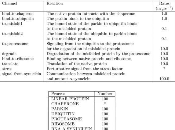
Channel Reaction Rates
(in µs−1)
bind to chaperon The native protein interacts with the chaperone 1.0
bind to ubiquitin The parkin binds to the ubiquitin 1.0
to misfold1 The bound state of the parkin to ubiquitin binds
to the misfolded protein 0.1
to misfold2 The bound state of the ubiquitin to parkin binds
to the misfolded protein 0.1
to proteasome Signaling from the ubiquitin to the proteasome
for the degradation of misfolded protein 10.0 degrade Degradation of the misfolded protein by the proteasome 10.0 bind to ribosome Binding between native protein and ribosome 10.0
translate Translation of the native protein 10.0
stress Perturbative signal from the stress factor *
signal from synuclein Communication between misfolded protein
and mutant α-synuclein 100.0
Process Number LINEAR PROTEIN 100 CHAPERONE * PARKIN 100 UBIQUITIN 100 PROTEASOME 100 RIBOSOME 100 RNA A SYNUCLEIN 100 STRESS *
Table 3: Channels rates and number of processes (molecules) used in the models. The fields marked with the symbol ”‘*” is different in the three considered models (see Fig. 2).
3.2 Second model: misfolded protein accumulation induced by mutant parkin
The mutant parkin does not transfer the ubiquitin to the misfolded protein, that remains untargeted for the degradation. In order to model this situation the specification showed in Table 2 has been modified in the following way. The process PARKIN still communicates with the process UBIQUITIN to realize the binding, but the dimer UBIQUITIN BOUND formed by this interaction, is now a deadlock process, unable to do any other reactions. Thence, in this model the process PROTEASOME is not necessary, because the reactions of polyubiquitination do not occur any more.
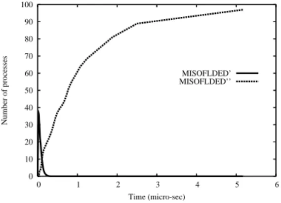
Fig. 3 shows that the number of misfolded proteins derived by a wrong refolding process, presents a rapid increase within the first 5 µs. After that time the number stabilizes about 100.
0 10 20 30 40 50 60 70 80 90 100 0 1 2 3 4 5 6 Number of processes Time (micro-sec) MISOFLDED’ MISOFLDED’’
Figure 3: Number of non correctly refolded proteins in PD induced by mu-tant parkin. The curve of MISFOLDED’ zeros before 5 µs, indicating that the production of MISFOLDED” starts since the beginning of the simu-lation and increases as the square root of the time, without giving to the proteosomal mechanism of the cell any chance to react.
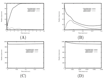
3.3 Third model: misfolded protein accumulation as func-tion of chaperones number
The aim of this experiment is to investigate a possible relationship between the number of chaperones and the ability of the cell of degrading misfolded proteins in the case in which mutant α-synucleins are operating in the sys-tem. In particular, the purpose is to validate the thesis for which the greater the number of chaperones is, the lower the rate of decreasing is and con-sequently the more efficient the ability of the cell to respond to misfolded proteins. By varying the number of instances of process CHAPERONE and setting to 1000 the instances of process STRESS, the plots of Figs. 4 and 5 are obtained.
0 5 10 15 20 25 30 35 40 0 1 2 3 4 5 6 7 8 9 10 Number of processes Time (micro-sec) CHAPERONES MISFOLDED’ MISFOLDED’’
Figure 4: Variation of number of chaperones and wrongly refolded proteins in PD induced by mutant α-synuclein. The initial number of chaperones is 10. This simulation shows that this number is not adequate to defend the cell from the increasing of faulty proteins.
0 20 40 60 80 100 0 1 2 3 4 5 6 7 8 9 10 Number of processes Time (micro-sec) CHAPERONES MISFOLDED’ MISFOLDED’’ (A) 0 20 40 60 80 100 0 0.05 0.1 0.15 0.2 Number of processes Time (micro-sec) CHAPERONES MISFOLDED’ MISFOLDED’’ (B) 0 200 400 600 800 1000 0 0.05 0.1 0.15 0.2 Number of processes Time (micro-sec) CHAPERONES MISFOLDED’ MISFOLDED’’ (C) 0 200 400 600 800 1000 0 0.001 0.002 0.003 0.004 0.005 Number of processes Time (micro-sec) CHAPERONES MISFOLDED’ MISFOLDED’’ (D)
Figure 5: Variation of number of chaperons and wrongly refolded proteins in PD induced by mutant α-synuclein. The initial number of chaperones is 100 in the plot (A) and 1000 in the plot (C). The plots (B) and (D) are a zoom of the plots (A) and (C) to better visualize the time behavior of the processes in the first 0.2 µs−1 and 0.005 µs−1, respectively. A sufficiently
large number of chaperones seem to ensure the cell the possibility to activate the proteasomes and consequently to decrease the number of faulty proteins. The plots of Fig. 4 indicates that starting with an initial number of 10 chaperones, the curve of MISFOLDED” has a wide pick decreasing after 4
µs and zeroing about 10 µs, exactly when the number of MISFOLDED”
starts to increase. The simulations showed in Fig. 5 (A)-(B) indicate that with an initial number of 100 chaperones, the system is only partially able to control the number of misfolded proteins. Namely, the number of MIS-FOLDED” zeroes only after 6 µs and, unlike the case of Fig. 4, the number of MISFOLDED” is significantly greater than zero since the beginning of the simulation. On the contrary, the curves of Fig. 5 (C)-(D) shows that with 1000 instances of chaperones, the number of misfolded proteins remains null during all the simulation time. This results confirm what very recent wet experiments are starting to recognize, i. e. that a therapeutically induced increase of chaperones can enhance the cellular environment for protein fold-ing and stability. In particular, in a recent review D. F. Smith, L. Whitesell and E. Katsanis [17] provided an overview of protein misfolding as a basis for disease and have provided a prospective look at pharmacological ap-proaches that may help to prevent or resolve protein-folding problems. In this review they asserted that specific mechanisms for inducing production and accumulation of intracellular chemical chaperones are potentially useful for preventing and correcting protein misfolding; perhaps drugs will be dis-covered that serve this purpose. Smith et al. recall also that an alternative approach would be to administer, by tissue perfusion, nontoxic chemical chaperones that could be taken up by cells. The feasibility of this approach has been already examined in cell culture systems.
4
Conclusions and future directions
Any advance in understanding the genetic basis of neurodegeneraive disor-ders like parkinsonism can open up new lines of investigation. Over the past decade, converging lines of research revealed that the common pathogenic mechanism underlying many of neurodegenerative diseases is the aggrega-tion and the deposiaggrega-tion of misfolded proteins leading to progressive central nervous system degeneration. This process develops insidiously over the lifetime of an individual, even though they do not manifest clinically until middle or late life. The cause of this prolonged preclinical phase is not com-pletely understood, by it certainly points out the requirement for progressive damages to specific brain regions prior to clinical manifestation of the dis-ease, as well as the unfavorable kinetics of protein misfolding [5]. Cells have adapted sophisticated quality-control mechanisms to protect against the accumulation of misfolded and aggregated proteins. Molecular chap-erones promote the correct protein folding and the proteins that remain misfolded are degraded by the ubiquitin-proteasome system. Furthermore, genetic mutations of α-synuclein and parkin cause an abnormal processing of misfolded proteins that overwhelms the quality-control system of the cell. In this context, a computational model of the mechanisms regulating protein
processing can facilitate the development of rationally designed therapies to treat and prevent these disorders. Moreover, this work has showed how modeling and simulation can be used also to reveal similarities and differ-ences between the effects of different causes. This kind of knowledge can be the starting point to project new pharmacological strategy to defeat the disease attacking it from different points and with different methods [11]. A long term goal could be the creation of drugs inspired to the activity of the chaperones [17]. Our model showed, in particular, that a scarce number of chaperones has the same effects of the perturbation introduced by the faulty
α-synuclein and parkin, and that there may exist “threshold” phenomena
on the rates of toxic chemical absorption triggering the beginning of protein aggregation. Such an information can be used to design the guidelines of new wet-experiments.
Our model does not take into account the possible interactions between parkin and α-synuclein, because at the present they are poorly understood. Therefore, a future direction to extend our work will be the addition of such information. Finally, we remark that the usage of the biochemical π-calculus language in the specification of our model revealed noticeably adapt to de-scribe the concurrent and parallel nature of the reaction involved in the protein processing. Unlike the most common language of differential equa-tion, it offers a new point of view of a biological system by switching from the direct modeling the dynamics of the system component to the modeling of the system components them selves. This new paradigm allows the ex-pression of the compositional and modular nature typical of the biochemical networks.
References
[1] K. B. Beckam and B. N. Ames, The free radicals theory of aging
ma-tures, Physiol. Rev. 78, 547-581, 1998
[2] R. Bussel Jr. and D. Eliezer, Residual structure and dynamics in
Parkinson’s disease-associated mutants of alpha-synyclein, J. Biol.
Chem., 276, 45996-46003, 2001
[3] W. Dauer and S. Przedborski, Parkinson’s Disease: Mechanisms and
Models, Neuron, Vol. 39, pp. 889-909, September 11, 2003
[4] W. A. Eaton, V. Munoz, S. J. Hagen, G. S. Jas, L. J. Lapidus, E. R. Henry and J. Hofrichter, Fast kinetics and mechanisms in protein
folding, Annu. Rev. Biophys. Biomol. Struct, 29: 327-59, 2000.
[5] M. S. Forman, J. Q. Trojanowsky and V. M-Y Lee,
Neurodegenera-tive diseases: decade of discoveries paves the way to therapeutic brek-throughs, Nature Medicine, vol. 10, n. 10, October 2004.
[6] D. T. Gillespie, Exact stochastic simulation of coupled chemical
reac-tions., J. Phys. Chem. 81, pp. 2340-2361, 1977
[7] P. J. Kahle and C. Haass, How does parkin ligate ubiquitin to
Parkin-son’s disease?, EMBO report 2004.
[8] C. Kuttler, J. Niehren and R. Blossey, Gene Regulation in the Pi
Cal-culus: Simulating Cooperativity at the Lambda Switch. Proceedings of
BioConcur 2004.
[9] P. Lecca and C. Priami, Cell cycle control in eukaryotes: a biospi model, Technical Report DIT-03-045, University of Trento, 2003.
[10] P. Lecca, C. Priami, P. Quaglia, B. Rossi, C. Laudanna and G. Costantin. A stochastic process algebra approach to simulation of
au-toreactive lymphocyte recruitment. SIMULATION: Transactions of the
society for modelling and simulation international , vol. 80, 273-288, 2004.
[11] A. M. Lozano and S. K. Kalia, Nuovi orizzonti per la cura del Parkinson, Le Scienze (italian edition of Scientific American), October 2005 [12] R. Milner, Communicating and Mobile Systems: the π-calculus,
Cam-bridge University Press, 1999.
[13] A. Phillips and L. Cardelli, A Correct Abstract Machine for the
Stochas-tic Pi-calculus, In Procs. of BioConcur 2004.
[14] C. Priami, Stochastic π-calculus, The Computer Journal, 38, vol. 6, 578-589, 1995.
[15] A. Regev, W. Silverman, and E. Shapiro, Representation and simulation
of biochemical processes using the pi-calculus process algebra, In Procs of
the Pacific Symposium of Biocomputing (PSB2001), 6: 459-470, 2001. [16] M. Y. Sherman and A. L. Goldberg, Cellular defenses against unfolded
proteins: a cell biologist thinks about neurodegenerative diseases,
Neu-ron 29, 15-32, 2001.
[17] D. F. Smith, L. Whitesell, and E. Katsanis, Molecular chaperones:
bi-ology and prospects for pharmacological intervention, Pharmacological
Reviews vo. 50 No. 4, 1998.
[18] M. Vila and S. Przedborski, Genetic clues to the pathogenesis of