Impact of b-hexachlorocyclohexane on
human cellular biochemistry and
environmental remediation strategies.
PhD course in Biochemistry PhD course in Sciences agronomiques et ingénierie biologique
PhD candidate:
Elisabetta Rubini
XXXIII cycle
PhD tutors:
Prof. Fabio Altieri
Prof. David Cannella
Supervisor:
Prof. Margherita Eufemi
Index
1. Organochlorine pesticides (OCPs) ... 4
1.1. Introduction. ... 4
1.2. OCPs toxicology ... 5
1.2.1. Endocrine Disruption. ... 6
1.2.2. Activation of AhR pathway. ... 8
1.2.3. Oxidative stress and mitochondrial dysfunction. ... 9
1.3. OCPs activation pathways. ... 10
2. Signal Transducer and Activator of Transcription 3 (STAT3) ... 11
2.1. The STAT protein family. ... 11
2.1.1. STAT3 activation. ... 13
2.1.2. STAT3 non-canonical activation. ... 14
2.1.3. STAT3 alternative post-translational modifications. ... 15
2.2. STAT3 and cancer. ... 17
2.3. STAT3 and energy metabolism. ... 18
3. Hexachlorocyclohexane ... 19
3.1. Physicochemical properties. ... 19
3.2. Production and environmental impact. ... 20
3.3. Hexachlorocyclohexane in Italy. ... 22
4. Remediation strategies for environmental contamination ... 23
4.1. Environmental remediation: an overview. ... 23
4.1.1. Environmental remediation technologies. ... 23
4.1.2. Bioremediation vs Chemical remediation. ... 25
4.2. Hexachlorocyclohexane environmental degrading approaches. ... 26
4.3. Fenton’s Reactions. ... 28
4.3.1. Fenton’s Reaction applied to the removal of hexachlorocyclohexane. ... 29
5. Research Aims ... 31
6. Results: the multifaceted effects of β-HCH on human cells. ... 34
6.1. β-HCH molecular activation pathways. ... 34
6.2. β-HCH: small molecule, big impact. ... 37
6.2.1. β-HCH as an endocrine disrupting chemical. ... 37
6.2.2. β-HCH activates AhR pathway. ... 40
6.2.3. Impact of b-HCH on oxidative stress and energy metabolism. ... 42
6.3. b-HCH induces malignant transformation in BEAS-2B cells. ... 46
6.3.1. Effects of β-HCH on cells viability. ... 46
6.3.2. b-HCH activation pathway in BEAS-2B cells. ... 47
6.3.3. Impact of b-HCH on cell morphology. ... 48
6.3.4. b-HCH induces EGF secretion. ... 50
6.3.5. b-HCH effects on apoptosis and cell cycle. ... 52
6.3.6. b-HCH induces H2A.X phosphorylation in BEAS-2B cells. ... 54
6.3.7. b-HCH induces oxidative stress in BEAS-2B cells. ... 55
6.3.8. b-HCH induces an increase in Ki67-positive cells. ... 56
6.4. b-HCH promotes chemoresistance in H358 cells. ... 57
6.4.1. Ongoing experiments. ... 63
6.5. Material and Methods. ... 64
6.5.1. Cell cultures. ... 64
6.5.2. Protein extraction and immunoblotting. ... 66
6.5.3. Dot Blot. ... 67
6.5.4. Immunofluorescence. ... 67
6.5.5. RNA extraction and RT-qPCR. ... 68
6.5.6. Reactive Oxygen Species (ROS) detection. ... 69
6.5.7. Determination of apoptosis. ... 69
6.5.8. Cell cycle analysis. ... 69
6.5.9. Statistical analysis. ... 70
6.5.10. Determination of GSH/GSSG ratio. ... 70
6.5.11. Determination of lactate/pyruvate ratio. ... 70
6.5.12. Primary antibodies ... 71
6.5.13. Primers ... 72
7. Results: b-HCH degradation. ... 73
7.1. Experimental background. ... 73
7.2. b-HCH degradation: method fine-tuning. ... 74
7.3. Materials and methods. ... 77
8. Results: STAT3 in prostate cancer progression. ... 79
8.1. STAT3 in prostate cancer. ... 79
8.2. Study of STAT3 PTMs pattern in prostate cancer. ... 79
8.3. STAT3 in prostate cancer energy metabolism. ... 83
9. Conclusions. ... 85
Appendix ... 86
1. Organochlorine pesticides (OCPs)
1.1. Introduction.
Environmental pollution represents one of the most pressing problems in industrialized countries and in recent years has raised concerns and doubts also from the scientific perspective. A growing number of epidemiological observational studies, carried out on population at risk, correlated the exposure to environmental chemicals with the incidence of several pathological conditions, ranging from metabolic to cardiological and reproductive diseases, until the development of cancers1. These evidences have made more urgent the
need for further investigations on the biological mechanism at the basis of pollutants’ toxicity.
In particular, the review statistics show that 40% of all pesticides are organochlorines (OCPs)2, constituting a substantial source of contamination
all over the world. The evaluation of the impact of these chemicals on human health draw a lot of attention in the scientific community. OCPs belong to a large class of organic compounds catalogued by the Stockholm Convention as “POPs” (Persistent Organic Pollutants). The list of banned chemicals includes dioxins and their derivatives, hexachlorocyclohexane, polychlorinated biphenyls, and aldrin, whereas many other similar substances are subjected to restrictions3.
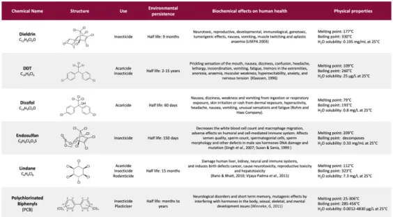
OCPs have a related chemical structure, showing chlorine-substituted aliphatic or aromatic rings; their hazardousness is mostly due to shared physicochemical properties such as lipophilia and energetic stability, responsible for the high environmental persistence and bioaccumulation potential of these molecules. Table 1 summarizes the principal compounds representative of OCPs and their features, comprising the biochemical effects on human health4.
Table 1. Major organochlorine pesticides, their chemical structures, common uses, environmental
persistence, biochemical effects and physical properties. Adapted from Jayaraj R. et al, 2016.
1.2. OCPs toxicology
The overuse or misuse of pesticides is adversely affecting both environmental and human health: in fact, only a small percentage of these compounds (0.3%) goes into the target, while the rest goes somewhere else5. Many pesticides have
been identified as endocrine-disrupting chemicals (ECDs)6. A recent report
commissioned by the European Parliament’s Committee on Petitions (PETI), and also taken up by The Lancet Oncology7, entitled "Endocrine Disruptors:
from Scientific Evidence to Human Health Protection”8 noted that EDCs have
been linked to several cancers, including breast, prostate, vaginal, and thyroid cancers9-10- 11.
1.2.1. Endocrine Disruption.
Due to their physical characteristics and chemical structure, EDCs can mimic or block the transcriptional activation elicited by naturally circulating hormones, thus inducing an imbalance in intracellular homeostasis.
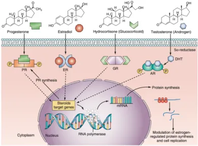
Steroid hormones are a large class of lipophilic molecules that act on a variety of target sites and closely regulate many physiological functions. In particular, sexual and reproductive development is closely regulated by androgens, oestrogens, and progestins12. Upon secretion, hormones blood levels increase
by several orders of magnitude and then rapidly return to the basal concentration when the stimulus stops. Hormones have a short half-life in human body and after having served their biological purposes are inactivated by enzymatic systems. Increasing or decreasing steroid metabolism could contribute to the negative effects of EDC13. Steroid Hormone Receptors (SHR)
function as hormone dependent nuclear transcription factors. Upon entering the cell by passive diffusion, the hormone (H) binds the receptor, which is subsequently released from heat shock proteins, and translocates to the nucleus. There, the receptor undergoes dimerization, binds specific DNA sequences, called Hormone Responsive Elements or HREs, and recruits a number of coregulators that facilitate gene transcription14.
Figure 1. Schematic diagram of signal transduction pathways for clones of steroid hormones and their
effects on protein-regulated synthesis and cellular replication.
Figure 2 outlines the key characteristics of ECDs, according to La Merril et
al.15; these parameters represent the functional properties of agents that alter
hormones action and comprise the major mechanisms by which the endocrine system can be disrupted.
1.2.2. Activation of AhR pathway.
Exogenous substances that come into contact with the human body are referred to as xenobiotics, which bring together a wide range of non-physiological and structural divergent chemicals including organochlorine pollutants. Without a metabolic transformation, xenobiotics would reach a toxic concentration; for this reason, the organism has developed different detoxification mechanisms resulting in the induction of metabolizing enzymes. Organochlorine pollutants may bind and modulate several endocrine receptors, one for all the Aryl Hydrocarbon Receptor (AhR)16. AhR is considered the xenobiotic sensor par
excellence but is also a converging point for many physiological intracellular
processes17. From a biochemical point of view, AhR is a cell nuclear receptor
that acts as ligand-activated transcription factor involved in the recognition and metabolism of xenobiotics, leading to the activation of cytochrome P450 enzymes needed for their clearance from the body18. In its inactive state, AhR
is complexed with the 90 kDa heat shock protein AhR-interacting protein (AIP), p23 and SRC. This chaperone complex keeps AhR in the cytosol, preventing its proteasomal degradation and keeping it in a high-affinity state for its ligands. Upon agonist binding, AhR and some components of the chaperone complex translocate to the nucleus, where AhR binds DNA-responsive elements to control gene expression. After exerting its function, AhR is addressed to the ubiquitin-proteasome system and is degraded 19(figure
Figure 3. AhR activation pathway.
1.2.3. Oxidative stress and mitochondrial dysfunction.
There are other reported mechanisms linking pesticides exposure to chronic diseases, such as mitochondrial dysfunction and oxidative stress induction. A particular subgroup of ECDs, referred as metabolism-disrupting chemical (MDC), can specifically affect energy homeostasis. Mitochondria are best known as the powerhouse of the cell for their crucial role in the oxidative phosphorylation and energy conversion, but these organelles are also involved in other intracellular events such as hormones secretion, generation of reactive oxygen species and cell death. Numerous studies have reported that MDC-induced mitochondrial dysfunction is characterized by perturbations in mitochondrial bioenergetics, biogenesis and dynamics, activation of the mitochondrial pathway of apoptosis and excessive ROS production. ROS are physiologically formed in cells as a consequence of both oxidative biochemical reactions and external factors, but, when overproduced, they can become harmful. There is substantial evidence that environmental pollution induces oxidative stress via increased intracellular steady-state levels of ROS such as
AHRR - AHR repressor ARNT - AHR nuclear translocator 2 XRE- xenobiotic response element
O2•− and H2O2 and, under these conditions, the endogenous antioxidants may
be unable to encounter ROS formation. This situation may cause cellular damage by peroxidation of membrane lipids, inactivation of enzymes, cross-linking and breakdown of DNA 20-21.
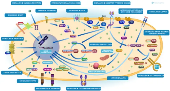
1.3. OCPs activation pathways.
Several signalling pathways underlie the toxic effects of organochlorine pesticides, involving the regulation of cell growth, survival, proliferation, migration, invasion, apoptosis, and anticancer drug resistance22. For some of
the most popular OCPs (i.e. TCDD, lindane, PCBs, bisphenol A, dieldrin) the
Comparative Toxicogenomic Database (http://ctdbase.org), which provides
information about the exposure effects of a wide range of chemicals integrated with functional and pathway data, reveals common intracellular transduction cascades mediated by several different proteins, included STAT3.
2. Signal Transducer and Activator of Transcription 3 (STAT3)
2.1. The STAT protein family.
The STAT (Signal Transducer and Activator of Transcriptions) protein family is a group of ubiquitously expressed intracellular transcription factors involved in the regulation of a variety of critical functions such as cell differentiation, proliferation, apoptosis, angiogenesis, metastasis, and immune responses23.
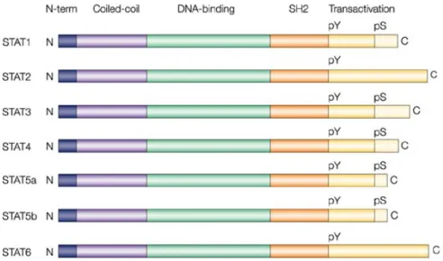
Seven STAT members have been identified - STAT1, STAT2, STAT3, STAT4, STAT5A, STAT5B, and STAT6 - and multiple isoforms have also been found. STATs can be generally activated by a series of extracellular signalling molecules (i.e. cytokines, growth factors, hormones) and they can modulate the transcription of responsive genes, leading to downstream phenotypic effects. Despite their functional differences, all seven STAT proteins share common structural elements (figure 5).
Stat2 and Stat6 proteins consist of approximately 850 amino acid residues, whereas the other five members are 750-800 amino acids in length.
Each STAT protein includes several conserved domains that contribute to protein biological behavior:
• The N-terminal protein-protein interaction domain (PPID) mediates interaction between neighboring STAT proteins (or other coregulatory proteins) and contributes to cooperative binding of STAT dimers on DNA, leading to the formation of stabilized tetramers;
• The DNA binding domain is involved in DNA binding, recruitment of coactivators, and the transcription of STAT target genes;
• The Src-homology 2 (SH2) domain is necessary for STATs dimerization and their recruitment to phosphorylated receptors. In fact, differences in STAT SH2 domains determine the selectivity for the target receptors;
• A critical tyrosine residue is located near the SH2 domain and is required for SH-phosphotyrosine interaction between monomeric STATs to form dimers;
• The C-terminal transcriptional activation domain (TAD) is involved in the communication with transcriptional complexes. In addition, the carboxy-terminal domain contains a site of serine phosphorylation (pS) that enhances transcriptional activity in some STATs 24-25-26.
As STATs regulate fundamental biological processes in normal cells, their alteration has a deep biological impact on intracellular homeostasis. In particular, STAT3 has been frequently reported constitutively overexpressed and/or hyperactivated in a variety of tumor types27, suggesting its essential
2.1.1. STAT3 activation.
Like other STATs proteins, STAT3 is a latent cytoplasmic transcription factor that translocate into the nucleus upon cytokines or growth factors stimulation. Following the interaction between a ligand and its receptor, STAT3 is activated through the phosphorylation at the critical tyrosine residue 705 that triggers STAT3 dimerization via the reciprocal phosphotyrosine-SH2 interactions. Then, dimeric STAT3 translocates into the nucleus where regulates the transcription of responsive target genes28.
Activation of STAT3 through the phosphorylation at Y705 residue is referred to as STAT3 canonical pathway and can be mediated by multiple upstream inputs. There are receptors present on plasma membrane, like gp130 receptor, that lacks intrinsic tyrosine kinase activity and recruits cytoplasmic kinases like JAK family including JAK1, JAK2, JAK3 and TYK2. On the other hand, there are receptors like EGFR, PDGFR, Her2, FGFR, VEGFR, IGFR and HGFR with intrinsic tyrosine kinase activity that itself phosphorylates Y705 residue and induces STAT3 activation. Apart from receptors present on plasma membrane there are cytoplasmic kinases also that can activate STAT3 signaling like Bcr-Abl fusion protein, Src kinase family and Bone Marrow X-linked (BMX) kinase. Multiple ligands like cytokines (IL6, LIF, OSM, L-10, and IL-11) and growth factors (EGF, PDGF and CSF-1) are known to stimulate STAT3 signaling29.
2.1.2. STAT3 non-canonical activation.
Besides its well-described canonical signaling, STAT3 can be subjected to the phosphorylation at the serine residue 727, located in the carboxy-terminal transcriptional activation domain, responsible for STAT3 functions independent of the canonical phosphorylation at Y705. In particular, pS727STAT3 seems to be essential for important mitochondrial activities and is
required for the optimal transcriptional induction of a subset of target genes 30.
Specifically, mitochondrial STAT3 was shown to preserve optimal ETC activity, increase membrane polarization and ATP production, and enhance the activity of lactate dehydrogenase, probably by interacting with ETC complexes I and II 31. The Ser727 phosphorylation is a more-complex regulated
modification because different activation signals lead to serine phosphorylation by different kinases, including ERK1, ERK2, p38, JNK and MAP kinases. Ser727phosphorylation at the transactivating domain is
considered a secondary event after Y705 phosphorylation and is required for the maximal transcriptional activity of STAT332.
2.1.3. STAT3 alternative post-translational modifications.
Post-translational modifications (PTMs) come in various forms and a single type of PTM can be responsible for very different biological functions, depending on the modification site and the context of the signaling pathway. Chemical modifications are reversible and can include the addition of functional groups (as phosphorylation, acetylation) or redox-based modifications (as S-glutathionylation). Phosphorylation is one of the most common and studied PTMs and constitutes an extremely important regulation mechanism in several cellular processes, as many enzymes and receptors are activated and deactivated via phosphorylation/dephosphorylation determined by the interplay of specific kinases and phosphatases33. Protein acetylation has
a prevalence and significance that rival those of phosphorylation; it is involved in crucial intracellular phenomena and can govern relevant protein properties such as enzymatic activity, localization, stability, or interactions with other molecules34 . Instead, the redox-sensitive proteome can be post-translationally
modified through disulfide linkages between the tripeptide glutathione and cysteine residues within proteins, primarily promoted by oxidative stress35.
PTMs can function as sensors, allowing cells to quickly and dynamically adapt to changes in environmental conditions. STAT3 can be decorated with many PTMs that are able to modulate its functions. In response to cytokines and growth factors signaling, STAT3 can be acetylated (Ac) on multiple lysine (K)
residues by the CBP/p300 histone acetyltransferase; in particular, K685 acetylation is important for the formation of STAT3 dimer and transcription enhancement at certain genes, perhaps independent of tyrosine phosphorylation36. Then, STAT3 can become glutathionylated on multiple
cysteine residues, impairing its transcriptional activity, either under conditions of oxidative stress or downstream of IL-6 signaling, which can raise reactive oxygen species (ROS) levels37.
2.2. STAT3 and cancer.
STAT3 is a converging point for many intracellular signaling pathways and an-ever growing number of publications demonstrated its constitutive hyperactivation in nearly every human cancer (neck, brain, breast, liver, lung, kidney, pancreas, prostate, ovary cancer, and multiple myeloma, as well as acute myeloid leukemia)38. Accumulating evidence support the critical role of
STAT3 in malignant transformation and carcinogenesis. In fact, constitutive STAT3 activation is required for cellular processes that include proliferation, survival, inflammation, invasion, metastasis and angiogenesis, all supporting tumor initiation and progression toward a more aggressive phenotype 39.
2.3. STAT3 and energy metabolism.
STAT3 is a pleiotropic protein and, in addition to its canonical functions, has been reported to be a master regulator of energy metabolism in both its nuclear and mitochondrial form. In particular, STAT3 activation appears to participate, together with PKM2 (pyruvate kinase isoform 2) and HIF-1a (Hypoxia-inducible factor 1a), in the positive feedback loop which is involved in the Warburg Effect40. This loop enhances aerobic glycolysis and proliferation:
oxygen deprivation or oncogenes, up-regulating 1α and increasing HIF-1a activity, lead to increased levels of the pyruvate kinase PKM2 isoform; in turn, this enhances HIF-1a transcriptional activity and directly phosphorylates STAT3; closing the loop, activated STAT3 up-regulates HIF-1α expression41.
3. Hexachlorocyclohexane
3.1. Physicochemical properties.
Hexachlorocyclohexane (HCH) is a chlorinated cyclic saturated hydrocarbon extensively used as a commercial pesticide starting from the late 1940s.
Figure 10. Lindane production process
Hexachlorocyclohexane is almost universally synthetized through the photochlorination of benzene, resulting in a mixture of isomers (a,b, g, d, e) that structurally differ in the axial and equatorial orientation of the chlorine atoms with respect to the cyclohexane carbon ring (figure 11).
Figure 11 and Table 2. Chemical structure of HCH isomers and physicochemical properties.
Boiling points for b- and d- HCH were measured at 0.5 mmHg and 0.36 mmHg respectively.
The raw product from the chlorination of benzene contains about 14% g- HCH and 86% of inactive isomers, i.e. a: 65-70%, b: 7-10%, g: 14-15%, d: approximately 7%, e: 1-2%, and 1-2% other components. Among these
isomers, only g-HCH has specific insecticidal properties and it is possible to extract and purify the active g-HCH. If the purity is more than 90% in terms of g-HCH, it is to referred as Lindane42. The different HCH isomers and their
physicochemical properties are shown in figure 6 and table 2.
3.2. Production and environmental impact.
The production and application of lindane during the last 7 decades have resulted in environmental contamination at global scale. For each tonne of lindane 8–12 tonnes of waste HCH isomers were produced, and the production of approximately 600,000 t of lindane has therefore led to 4.8 to 7.2 million tonnes of HCH/POPs waste. These waste isomers were mostly buried in uncontrolled dumps at many sites around the world43 ; the stockpiles and the
large contaminated sites can be categorized as “mega-sites”.
Figure 13. HCH residuals in France.
Considering the magnitude of the problem, α-, β-, and γ-HCH were banned in several countries, including Italy (Regulation EC No 850/2004), and were designated as Persistent Organic Pollutants in the Stockholm Convention in May 2009 44-45.
Compared to other HCH isomers, β-HCH has stronger lipophilic properties and is most stable from the physical and metabolic point of view due to the equatorial position of all the six chlorine atoms in the chair cyclohexane conformation; this stability is reflected in the environmental and biological persistence of this isomer.
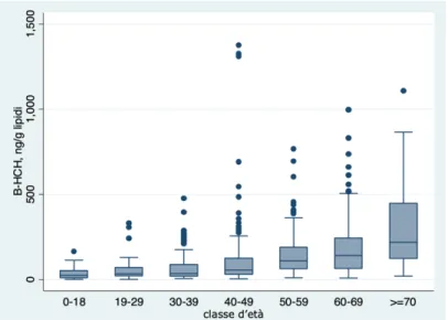
3.3. Hexachlorocyclohexane in Italy.
The company SNIA-BDP (Società di Navigazione Italo Americana Bombrini Parodi Delfino) located in Colleferro, a town in the south of Rome, the Central Italy, was the only lindane manufacturer in Italy since the 50’s. In 2005, during a random national survey on chemical contamination of raw cow’s milk, products from farms located in the rural area of Valle del Sacco, Colleferro, were found to be polluted with β-HCH as a result of the illegal disposal of chemical waste produced by the close industrial conglomerate46. Considering
the contamination of the food chain, an epidemiological investigation on the population at risk in three towns (Colleferro, Gavignano, Segni) near to the offending site was established in 2006 and is still ongoing (VEGA 2016)47.
Results of the survey demonstrated high β-HCH serum levels among the 690 subjects participating in the surveillance program, with a β-HCH amount below the limit of quantification observed only in seven people; in addition, β-HCH serum concentration increases with age particularly in patients with more than 50 years, and becomes even higher among people over 70 years48.
4. Remediation strategies for environmental contamination
4.1. Environmental remediation: an overview.
The modernization in productive technologies, together with an increase of the agricultural sector, is reflected by an excessive use of pesticides and fertilizers to satisfy the requirements of an ever-growing world population49 .
According to the World Health Organization (WHO), 23% of all global deaths are linked to environmental pollution, amounting to roughly 12.6 million deaths a year50; this scenario makes more urgent the need to select appropriate
remediation approaches based on a careful analysis of physical, chemical and biological factors affecting each contaminated site.
Environmental remediation is defined as the process of removing contaminants from soil, surface and groundwater in order to preserve living systems and the environment against further deterioration for a sustainable future51. The
remediation is generally carried out on soil or water media and may be conducted separately or together, depending on the type and extent of the pollution.
4.1.1. Environmental remediation technologies.
The presence in the soil and groundwater of contaminants in concentrations far over the tolerance threshold represents a high potential health as well as ecological risk. For this reason, the optimization of different remediation methodologies could offer a valid tool to clean up polluted sites in relation to the characteristics of the environmental media. In broad terms, technical principles for remediation can be divided into physical, chemical and biological processes. These techniques may be applied in the contaminated area (in situ), or by removing the soil from the contaminated area, treating it in a specific treatment complex, and restoring the treated soil to its original place
(ex situ). The primary action approach for contaminated sites is the containment technique, consisting in the use of barriers in order to avoid the migration of the pollutants to neighboring soil and to inhibit the flow of clean water through the contaminated area. Containment is achieved by the installation of low-permeable or impermeable cutoff walls that minimize the risk for further contamination52.
Biological techniques are based on the bioremediation principle that involves the use of microorganism (i.e. bacteria, fungi) for the removal and/or degradation of hazardous chemicals from various environmental media. Microorganisms used in decontamination purposes have often the capability to degrade almost all organic contaminants; the transformation of these substances, in fact, provide the microorganisms with both carbon atoms and electrons, which are beneficial for their own growth and reproduction53.
Another promising method among biological techniques is the phytoremediation, an in situ and clean procedure based on the use of some species of plants with the ability to accumulate or degrade specific organic pollutants. Phytoremediation and bioremediation cannot be viewed separately, since plants constantly interact with microorganism that sometimes establish close associations or symbiotic relationships 54.
A hybrid remediation tool between chemical and biological remediation technologies is constituted by biosurfactants. In general, surfactants are amphipathic compounds that have a hydrophobic moiety directed towards the surface and a hydrophilic portion orientated towards the solution. These amphiphilic molecules can reduce the surface tension at air/water or oil/water interfaces. Surfactants produced by microorganisms are extracellular metabolites referred to as biosurfactants and their biotechnological properties allow their application in the environmental field for xenobiotic
biodegradation and bioremediation55 (i.e. Deep Horizon oil spill remediation
Corexit 9500s – DOSS)56. New ecofriendly biosurfactants with the advantage
of owning biodegradable or biological-derived dispersant molecules have recently been developed (i.e. rhamnolipid, sophorolipid, and surfactin)57.
These biosurfactants are in general less harmful, if not harmless at all, to the environment: in fact, they can be degraded immediately after their applications, being less persistent contrary to DOSS in Corexit58.
And last but not least, chemical methods are employed to remediate the contaminating substances that have been hoarded in the soil by adding chemicals or solvents into the polluted site in order to convert them into less toxic and less harmful products59.
4.1.2. Bioremediation vs Chemical remediation.
The success of a remediation process requires an optimized development and selection of strategies capable of responding to the specific conditions of the polluted sites. However, each technology has advantages and disadvantages, making it necessary to synergistically integrate different approaches, such as the chemical and the biological one 60.
Chemical methods can offer a cost-effective and fast remediation compared to slow bioremediation process, but sometimes their use can compromise the quality of the environmental media, thus limiting their large-scale application. On the other hand, biological remediation has been reported as an efficient approach with long-term attenuation benefits but is usually unable to quickly remove highly persistent and toxic pollutants. In addition, the microbial degradation is limited to environments where specialized microbial populations are competitive61-62. In light of these considerations, the
combination of different methods could be beneficial to achieve a higher removal efficiency and to address the limitation of each technique.
4.2. Hexachlorocyclohexane environmental degrading approaches.
Several technologies have been investigated as potential remediation solutions for hexachlorocyclohexane contaminated sites, but the characteristics and stability of this molecule enhance its resistance towards degradation, making each effort quite challenging. This imposes the need to develop efficient large-scale systems to achieve proper HCH breakdown strategies.
The microbial degradation of HCH is attracting increasing attention and an ever-growing number of studies reported potential microorganisms for the efficient bioremediation of HCH-polluted environments, in particular for the g-isomer 63. The key reaction during microbial degradation of halogenated
compounds is the removal of the halogen atom, i.e., dehalogenation of the organic halogen. During this step, the halogen atoms, usually responsible for the toxicity of this molecule, are most commonly replaced by a hydrogen or a hydroxyl group. Halogen removal reduces both recalcitrance to biodegradation and the risk of forming toxic intermediates during subsequent metabolic steps. Lindane can be biodegraded under both aerobic and anaerobic conditions, but it is generally mineralized only under aerobic conditions 64-65.
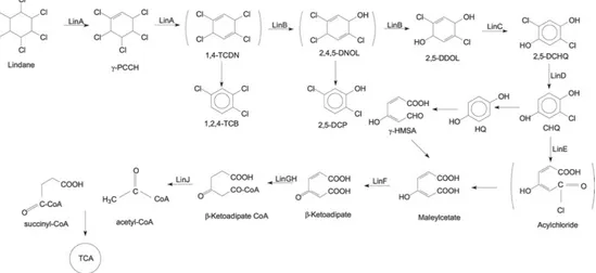
The aerobic degradation pathway of lindane summarized in figure 15 was extensively studied in Sphingobium japonicum UT26 66. In this pathway,
lindane is converted to 1,2,4-trichlorobenzene (1,2,4-TCB), 2,5-dichlorophenol (2,5-DCP), and 2,5-dichlorohydroquinone (2,5-DCHQ) by the enzymatic activities of dehydrochlorinase (LinA), halidohydrolase (LinB), and dehydrogenase (LinC). The degradation of lindane to 2,5-DCHQ is referred to as an upstream pathway, which is further metabolized through the downstream
pathway 2,5-DCHQ is converted to β-ketoadipate by reductive dechlorinase (LinD), ring-cleavage dioxygenase (LinE), and maleylacetate reductase (LinF). Researchers documented β-ketoadipate as a marker metabolite for the degradation of compounds containing aromatic rings. The intermediate β-ketoadipate is further converted to succinyl-coenzyme A (CoA) and acetyl-CoA by succinyl-acetyl-CoA: 3-oxoadipate acetyl-CoA transferase (LinGH) and β-ketoadipyl CoA thiolase (LinJ). Both these compounds are metabolized in the tricarboxylic acid (TCA) cycle. In addition, other studies reported that lindane can be metabolized by microorganisms to produce pentachlorocyclohexene (PCCH), 3,4,5,6-tetrachloro-1-cyclohexene (TCCH), pentachlorobenzene (PCB), or trichlorobenzene (TCB) 67.
Figure 15. Proposed mechanism for HCH aerobic degradation.
Bacteria have been found to be capable of bioremediating HCH through chemical and physical interactions that lead to structural changes or complete degradation of the target molecule. A wide range of bacteria were reported to degrade lindane at different rates and a number of lindane-degrading bacteria strains have been identified and screened68. Fungal biodegradation is also
Fungi, in fact, possess non-specific extracellular enzymatic systems, which include lignin peroxidases, manganese peroxidases and laccases, that can catalyze several reactions toward a wide range of substrates69. In particular,
laccases are blue multicopper oxidases constitutively expressed in many plants and fungi and are involved in the catabolism of organic pollutants. These enzymes, in fact, are able to catalyze the oxidation of diphenols and aromatic amines by removing an electron and a proton from a hydroxyl group 70. As
regards the physicochemical techniques, some traditional approaches, such as coagulation, flocculation, membrane separation or adsorption on activated carbon, only do a phase transfer of the pollutant. A modern oxidation technology like the photo-Fenton process has been applied for the degradation of several classes of pesticides and refractory compounds. Generally, Fenton’s process involves application of iron salts and hydrogen peroxide to produce hydroxyl radicals. This reaction is spontaneous and can occur with or without the influence of light71.
4.3. Fenton’s Reactions.
Among the oxidative technologies, one of the best known and most developed is the Fenton’s reaction. This chemical process was reported for the first time in 1894 by the French scientist H.J. Fenton, who discovered the oxidizing potential of Fe2+/H2O2 systems at pH 2-3. Under the activation of Fe2+, medium
containing hydrogen peroxide (H2O2) can produce reactive hydroxyl radicals
(•OH) with a strong electron-capturing ability, allowing them to attack most
organic groups without selectivity72. The Fenton’s reaction is expressed by:
Fe2+ + H
-It has many advantages such as high performance, simplicity (operated at room temperature and atmospheric pressure for substrates oxidation, no energy input is required as a catalyst or to activate H2O2) and non-toxicity73.
However, Fenton’s reaction still has some disadvantages including high operating costs, limited optimum pH range (pH~3), large volume of iron sludge produced, and difficulties in recycling the homogeneous catalyst (Fe2+)74. In order to overcome these practical limitations, research efforts have
been focused on finding suitable catalysts other than iron to generate •OH from H2O2. In this regard, Fenton-like systems were developed by substituting the
iron with other metals at a low oxidation state. In terms of its reactivity toward H2O2, copper shows very similar redox properties to those of iron, with the
non-negligible difference that Cu2+/H2O2 Fenton-like system reaches the
maximum efficiency over a broader pH range (near-neutral or neutral) compared to the Fe2+/H
2O2, which works only under acidic conditions 75-76.
The reaction of the copper-dependent •OH production is reported: Cu2+ + H
2O2 ® Cu+ + •OH + OH
-Another extensively used Fenton’s process is the so-called Photo-Fenton, in which the combination of H2O2, together with iron and UV radiation, boosts
the production of more hydroxyl radicals, thus enhancing the degradation rate of substrates. The use of solar irradiation has opened a high application potential for large-scale solar photochemical installations and specific reviews on this topic have been published77.
4.3.1. Fenton’s Reaction applied to the removal of hexachlorocyclohexane.
Over the last decades, Fenton’s reaction has emerged as a valid environmental remediation technology and has been successfully applied for the degradation of several classes of pesticides and persistent chemicals78-79-80.
Taking into consideration the potential of Fenton’s reaction, some studies focused on the application of Fe2+/H2O2 based oxidation systems for the
breakdown of the insecticide g-HCH.
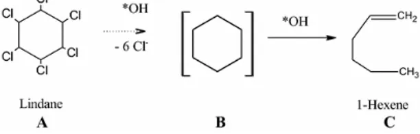
Begum et al81 reported the complete dechlorination of lindane with the
formation of 1-hexene using Fenton’s reagent (Fe2+/H2O2) in aqueous phase at
pH= 3. The proposed degradation pathway is showed below:
Nitoi et al71 evaluated the oxidation efficiency of UV/Fe2+/H2O2 systems on
lindane degradation, suggesting the following photooxidation process:
Figure 16. Mechanism for lindane degradation proposed by Nitoi et al. Lindane is progressively
dechlorinated until its breakdown to CO2 and H2O2, passing through intermediates with less chlorine atoms. Reaction intermediates were identified by GC.
5. Research Aims
The presented work was carried out on two parallel research lines in order to achieve a more comprehensive overview on β-HCH, which is one of the most widespread and, at the same time, poorly studied among organochlorine pesticides. For this reason, it appears to be absolutely essential to deepen the knowledge of β-HCH biological impact starting with the possible triggering of signaling pathways crucial for intracellular homeostasis. β-HCH can be absorbed by humans through contamination of the food chain and it is then bioaccumulated in adipose tissue (10 to 30 times higher than isomer γ), with a slow elimination time from the body (5 times lower than other isomers)82; on
the basis of its characteristics, β-HCH is suspected to be harmful to humans. Virtually, all the insecticidal properties resided in HCH have meant its recent accreditation by the International Agency for Research on Cancer (IARC) as carcinogenic to humans: there is sufficient evidence in humans for lindane’s carcinogenicity in non-Hodgkin lymphoma83. Others frequently reported
effects are related to neurological disorders, endocrine disruption, reproductive disorders, cardiovascular effects and cancer84-85-86. However, despite its
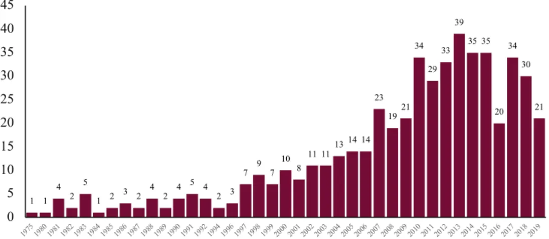
worldwide distribution, knowledge of β-HCH effects on human health is controversial and limited to studies in workers employed in the use and production of this pesticide87. By typing “b-hexachlorocyclohexane” into
PubMed search engine, the results-by-year timeline shows 450 publications spanning from 1975 to 2019 (table 3); after adding the word “disease” to narrow the field and focus on β-HCH biological consequences, the number of available papers is reduced to 63. Conversely, literature outcomes for lindane provide 6594 scientific papers, of which 694 are found including the keyword “disease” in the search criteria. Such a difference between the two analogue
compounds could be explained by the fact that β-HCH is considered a waste by-product without insecticidal activity.
Table 3. Timeline by year (from 1975 to 2019) of results for “b-hexachlorocyclohexane” extrapolated from PubMed search engine.
Even so, β-HCH is potentially one of the main contributors to the so-called civilization diseases, which are pathological conditions (i.e. cancer, neurodegenerative diseases, metabolic disorders) mostly linked to exogenous factors rather than to an intrinsic impairment of human physiological processes. For this purpose, a panel of human continuous cell lines corresponding to different tissues (i.e. liver, lungs, prostate, breast), both normal and transformed, were tested with 10 µM β-HCH. The experimental concentration of the pesticide was chosen averaging across all the plasma concentration values detected in patients under the biomonitoring study carried out in the Valle del Sacco, in order to reproduce the real exposure conditions. After evaluating the effects of β-HCH on cellular viability, different types of analysis were performed to identify the biomolecules, together with the transduction cascades, involved in the molecular responses to β-HCH.
1 1 4 2 5 1 2 3 2 4 2 4 5 4 2 3 7 9 7 10 8 11 1113 14 14 23 1921 34 29 33 39 35 35 20 34 30 21 0 5 10 15 20 25 30 35 40 45 197519801981198219831984198519861987198819891990199119921994199619971998199920002001200220032004200520062007200820092010201120122013201420152016201720182019
Furthermore, on the basis of the biological behavior reported in scientific literature for other organochlorine pesticides, the potential role of β-HCH as a contributor in tumor initiation and progression was inspected. On the other hand, the environmental persistence of β-HCH still represents an open question for the presence of massive illegal repositories all around the world. For this reason, β-HCH degradation through a copper-based Fenton-like method was explored by setting up a HPLC protocol under different experimental conditions. The process focused on the quantitative degradation of the parental β-HCH, since the detection of its breakdown products or transformed molecules would need a mass-spectrometry for their qualitative characterization.
6. Results: the multifaceted effects of β-HCH on human cells.
6.1. β-HCH molecular activation pathways.
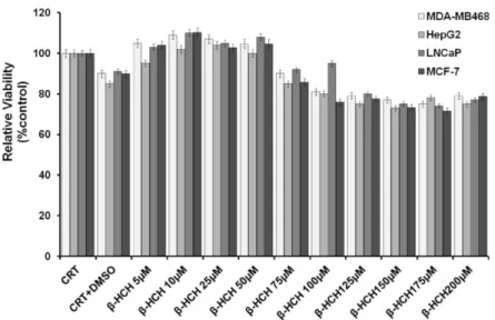
β-HCH was tested on a panel of cell lines representing different human tumor types associated with the expression and activation of specific membrane and membrane associated tyrosine kinase receptors: human breast cancer MDA-MB468 (EGFR+), human hepatoma HepG2 (JAK2+), human prostate cancer
LNCaP (AR+), and human breast cancer MCF-7 (Her2+). The experimental
concentration of β-HCH (10 μM) was extrapolated from both the environmental-epidemiological investigation carried out on the exposed population living in the “Valle del Sacco”46 and from previous in vitro
studies88. First of all, the impact of β-HCH on cell proliferation and viability
was verified on all the selected cell lines. No appreciable reduction in cell viability was observed upon cells exposure to 10 μM β-HCH, which, on the contrary, exhibited some proliferative effects.
Figure 17. Viability assay performed on the selected cell lines at different β-HCH concentrations,
A reduction of cell viability can be observed only at higher β-HCH concentrations, as showed in figure 17.
Subsequently, experiments were performed on all four cell lines for identifying β-HCH activation pathways. Results from western blot and RT-qPCR analysis revealed the central role of STAT3 as a hub protein in the intracellular responses triggered by β-HCH by means of its canonical phosphorylation at Y705, also confirmed by using pharmacological inhibitors for each cell line-specific STAT3-mediated signaling. Then, the involvement of STAT3 in cellular energy metabolism was explored. In fact, STAT3 is a well-established master regulator in the metabolic shift toward aerobic glycolysis (Warburg Effect) by interplaying with protein partners such as PKM2 (Pyruvate Kinase M2) and HIF-1a (Hypoxia-inducible Factor 1a)40. Investigations carried out
on all the selected cell lines show that, as a consequence of pY705-dependent canonical activation, STAT3 can also be phosphorylated at the serine residue 727, referred to as non-canonical pathway. The phosphorylation at S727 of STAT3 is a hallmark of oxidative stress conditions and is related to HIF-1a upregulation, together with an increased nuclear localization of PKM2. All these activated functions require an increased level of energy, and thus a burst in oxidative respiration, resulting in a ROS overproduction. Under the tested conditions, β-HCH induces a slight increase in ROS levels together with a decrease in GSH/GSSG ratio, testifying an alteration in cellular redox homeostasis. In conclusion, experimental outcomes suggest the involvement of STAT3 in β-HCH-induced toxicity through both its canonical and non-canonical pathways. Therefore, STAT3 may regulate the cellular responses to β‐HCH, switching from an acute to chronic phase, and may be responsible for the progression of the tumor into an advanced clinical stage, as attested by the occurrence of a metabolic shift towards the aerobic glycolysis.
Figure 18. The hub role of STAT3 in the intracellular signaling network triggered by b-hexachlorocyclohexane. The canonical pY705-STAT3 activation occurs at different time points through a cell-line specific manner on the basis of the overexpression and activation status of the membrane and membrane associated tyrosine kinase receptors typical of each cell line. Activated STAT3 initiates and sustains a vicious cycle with the two protein partners HIF-1a and PKM2, responsible for a rewiring in cellular energy metabolism aimed to satisfy the growth requirements of a more aggressive tumor phenotype. Following phosphorylation at Y705, STAT3 is also phosphorylated at S727 which is involved in the cellular responses to b-HCH-induced oxidative stress conditions (Rubini et al. 2018).
All the reported results are discussed in detail in the scientific article entitled “STAT3, a Hub Protein of Cellular Signaling Pathways, Is Triggered by
β-Hexachlorocyclohexane” and published on the International Journal of
6.2. β-HCH: small molecule, big impact.
After identifying β-HCH activation pathways, the next step is to unravel the molecular mechanisms underlying β-HCH toxicity. Taking into account the tight correlation between structure and function of biomolecules, it is conceivable to hypothesize that β-HCH may:
1. act as an endocrine-disrupting chemical by interfering with hormone cascades;
2. interact with the Aryl Hydrocarbon Receptor (AhR), the xenobiotic sensor par excellence;
3. induce oxidative stress, consequently affecting energy homeostasis and metabolism;
4. cause DNA damage.
To clarify whether β-HCH at the exposure concentration value could be able to trigger the above-mentioned processes, experiments were performed on HepG2 (hepatocellular carcinoma) and LNCaP (prostate cancer) cell lines, which have already been employed as a model to demonstrate the hub role of STAT3 in β-HCH-induced molecular responses.
6.2.1. β-HCH as an endocrine disrupting chemical.
Many pesticides, classified as “endocrine disruptors”, can mimic or block the transcriptional activation elicited by naturally circulating hormones because of their physicochemical characteristics and chemical structure.
Information regarding a possible role of β-HCH as an endocrine disrupting chemical are scarce and controversial, therefore shedding light on this mechanism could provide a further element to draw the toxicological profile of this substance. To understand whether β-HCH can interact with the Androgen Receptor (AR) signaling in the guise of agonist or antagonist, an
effective experimental approach consists in following AR nuclear translocation by immunoblot and immunofluorescence upon treatment of LNCaP (prostate cancer AR+) cells with 10 µM β-HCH for 4 hours.
To confirm the impact of β-HCH on AR-signaling, samples were subjected to a pre-treatment step in the presence of the chemotherapeutic agent bicalutamide, an AR competitive inhibitor approved for prostate cancer therapy under the trade name Casodex90.
Results from both western blot and immunofluorescence analysis highlighted the capability of bicalutamide to block AR nuclear translocation induced by testosterone and β-HCH, providing extra evidence of β-HCH endocrine-disrupting potential (figure 18).
To check if β-HCH could also activate AR from a transcriptional point of view and affect the expression of AR-target genes, the mRNA level of PSA (Prostate Specific Antigen) was evaluated through RT-qPCR (figure 19). Apart from being a biomarker of choice for prostate cancer, PSA is an AR-dependent gene and therefore constitutes a good candidate to verify AR activity as a transcription factor91.
Figure 19. Cellular distribution of AR followed by immunofluorescence in LNCaP cells. CTR: control
untreated cells; Test: cells subjected to a 4-hours stimulation with 30 nM testosterone; β-HCH: cells subjected to a 4-hours stimulation with 10 µM β-HCH; Bic: cells treated overnight with 120 nM bicalutamide. Samples referring to the images in the third and fifth rows of the panel (“Test + Bic” and “β-HCH + Bic”) were pretreated overnight with 120 nM bicalutamide and then subjected to a 4-hours stimulation with β-HCH or testosterone. AR nuclear localization induced by both β-HCH and testosterone, as evidenced by the images in the second and fourth rows, results inhibited by bicalutamide
Figure 20. mRNA expression levels for PSA were analyzed by RT-qPCR. The exposure of LNCaP cells
for 4 h to 10 µM β-HCH or 30 nM testosterone results in a two-fold PSA overexpression compared to the control untreated cells. Overnight pretreatment with 120 nM bicalutamide largely prevents the increase in PSA mRNA level. PSA expression values were normalized to β-actin as a housekeeping gene and expression levels of untreated cells were set to 1. CTR: control untreated cells; Test: cells subjected to a 4-hours stimulation with 30 nM testosterone; β-HCH: cells subjected to a 4-hours stimulation with 10 µM β-HCH; Bic: cells treated overnight with 120 nM bicalutamide. Statistically significant differences (**p<0.01) are marked with asterisks.
6.2.2. β-HCH activates AhR pathway.
To demonstrate the capability of β-HCH to activate AhR genomic pathway, AhR nuclear localization was verified through both immunoblotting and immunofluorescence performed on nuclear extracts obtained from LNCaP and HepG2 cells exposed to β-HCH and pre-treated or not with the AhR antagonist CH22319192. Obtained results highlight the nuclear localization of AhR upon
Figure 21. Cellular distribution of AhR followed by immunofluorescence in LNCaP (A) and HepG2 (B)
cells. CTR: control untreated cells; β-HCH: cells after 4h of 10 µM β-HCH stimulation; β-HCH + CH223191: cells after 2h pre-incubation with 150 nM CH223191 followed by 4h of 10 µM β-HCH stimulation.
6.2.3. Impact of b-HCH on oxidative stress and energy metabolism.
The capability of OCPs of inducing the formation of reactive oxygen species (ROS), responsible for the establishment of an overall oxidative stress condition, has been well-established. A recently published article provided evidence that 20 µM β-HCH can induce a substantial ROS increase in HOSE ovary cells93. To confirm this outcome, ROS production was quantified by
performing CellRox assay on both LNCaP and HepG2 cells treated with 10 µM β-HCH for 6 hours. A significant intensification of the fluorescence after β-HCH stimulation occurs, thus indicating an enhanced ROS production.
Figure 22. ROS production detected by CellROX assay. The histogram shows an approximately
two-fold increase in fluorescence intensity in samples treated with 10 µM β-HCH for 6 hours compared to untreated cells. 75 µM Tert-Butyl Hydroperoxide (t-BuOOH) was used as a positive control for ROS induction. Statistically significative differences (**p<0.01) are marked with asterisks.
Taking into account that glutathione redox status constitutes another reliable biomarker of oxidative stress94, the same samples were subjected to
measurement of the GSH/GSSG ratio. Results reported in figure 23 show a marked increase in the glutathione oxidized form (GSSG), with a consequent decrease in GSH/GSSG ratio, demonstrating the induction of oxidative stress
in response to β-HCH. The establishment of an overall oxidative stress condition is often associated with a reprogramming of cellular bioenergetics; for this reason, additional studies are needed to evaluate the extent to which an imbalance in redox homeostasis is reflected in energy metabolism. Highly aggressive tumors are likely to display a particular metabolic condition known as aerobic glycolysis or Warburg Effect, characterized by the preferential conversion of pyruvate to lactate, rather than to acetyl-CoA, even in normoxia95. In this context, the potential impact of β-HCH on cell metabolism
was inspected by determining the lactate/pyruvate ratio in the culture media of cells treated or not with β-HCH. As is clear from figure 23, lactate is predominant in stimulated samples, attesting the influence of β-HCH molecular action on cellular metabolic rewiring.
Figure 23. Impact of β-HCH on glutathione redox state and cell metabolism. The increase in glutathione oxidized form (GSSG), with a consequent decrease in GSH (reduced glutathione)/GSSG ratio, proves that 10 µM β-HCH can induce oxidative stress after 6 h treatment in both LNCaP and HepG2 cells. Analysis were carried out on 106cells. In addition, a sharp increase in Lactate/Pyruvate ratio is detectable
in the culture media of both LNCaP and HepG2 cells stimulated with 10 µM β-HCH for 6 h, demonstrating an enhancement in lactate production induced by β-HCH. All the differences between control and β-HCH treated samples are statistically significant.
6.2.4. b-HCH induces DNA damage.
The relationship between DNA damage and sustained exposure to environmental pollutants is widely described in the scientific literature and is probably linked to the redox signaling triggered by OCPs96. For some
pesticides, the processes leading to alterations in the cellular homeostasis are partially understood, but commonly recognized mechanisms include their enzymatic conversion to secondary reactive products, depletion of cellular antioxidant defenses and/or impairment of antioxidant enzyme functions97.The
phosphorylation of histone H2AX at serine 139 is a post-translational modification that constitutes a solid and versatile endpoint to investigate the genotoxic potential of a chemical98. LNCaP and HepG2 cells were stimulated
with β-HCH or tert-butyl Hydroperoxide (t-BuOOH) as positive control99 and
nuclear fractions subjected to immunoblotting. With respect to the untreated control, H2AX results phosphorylated following β-HCH exposition in both the considered cell lines.
Figure 24. β-HCH induces the phosphorylation of H2AX at Serine 139. Nuclear protein extracts were
obtained from both LNCaP and HepG2 exposed to 10 µM β-HCH for 4 hours or 75 µM t-BuOOH for 1 hour used as a positive control for DNA damage.
Reported results are further discussed in the paper entitled
“β-Hexachlorocyclohexane: A Small Molecule with a Big Impact on Human Cellular Biochemistry” published in Biomedicines100.
6.3. b-HCH induces malignant transformation in BEAS-2B cells.
The correlation between synthetic chemicals and carcinogenesis was originally identified in the middle 1700s101, but it was only in 1915 that Yamagiwa
published the first experimental study on cancer pathogenesis in association with environmental contaminants exposure102. Although the carcinogenicity of
the most popular OCPs has currently been established103, significant gaps still
remain in the knowledge of less renowned compounds such as b-HCH. To investigate whether also β-HCH could trigger cellular malignant transformation toward cancer development, experiments were performed on human continuous cell line BEAS-2B (normal bronchial epithelium) in order to evaluate both the possibleeffects of this pollutant on tumour initiation.
6.3.1. Effects of β-HCH on cells viability.
First of all, cell viability was assessed by performing MTT assay on BEAS-2B cells after 24 hours of exposure to increasing concentration of β-HCH. Cells exposure up to 25 µM concentration revealed >100% cell viability; in addition, 10 µM β-HCH (selected working concentration) exhibits some proliferative effects in accordance with what has been previously demonstrated for other cell lines. However, exposure to β-HCH at high concentration (>25 µM) results toxic. Results are reported in figure 26.
Figure 26. MTT assay on BEAS-2B cells at β-HCH concentrations ranging from 2 µM up to 200 µM.
6.3.2. b-HCH activation pathway in BEAS-2B cells.
In our previous work89, it has been demonstrated that b-HCH can activate
different signaling pathways in a cell-line specific manner. To identify the intracellular cascade triggered by b-HCH in BEAS-2B cells, a time course assay was performed. Samples were treated with 10 µM b-HCH at different time points ranging from 5 minutes to 6 hours and various hypothesis were tested. Immunoblotting carried out on total protein extracts revealed that b-HCH can induce ERK-1/2 phosphorylation already after 5 minutes of treatment (figure 27). 0 20 40 60 80 100 120 140 CTRL Solve nt 0,1 % β-HC H 2 µM β-HC H 10 µ M β-HC H 25 µ M β-HC H 50 µ M β-HC H 100 µM β-HC H 200 µM Ce ll viability % (r elative to CTRL)
Figure 27. Time course assay on BEAS-2B cells. Cells were treated with 10 µM b-HCH from 5 minutes up to 6 hours. The phosphorylation of ERK-1/2 occurs after 5 minutes of incubation, as clearly demonstrated by western blot. ERK-1/2 phosphorylation levels were measured on the total amount of ERK-1/2 present in each sample and normalized to the control.
6.3.3. Impact of b-HCH on cell morphology.
The impact of b-HCH on BEAS-2B cell morphology was assessed by following for 48 hours the growth of cells exposed to 10 µM b-HCH. As showed in figure 28, treated BEAS-2B cells appear to assume a different growth path on the well surface compared to the control, suggesting a b-HCH-induced morphological change.
Figure 28. β-HCH-induced morphological changes in BEAS-2B cells. Cells were seeded at 300’000
cells/well and, after 24 hours, were exposed to 10 µM b-HCH for 48 hours. The different arrangement of cells induced by b-HCH is evidenced in a black dotted box. Selected images are representative of three independent experiments.
To confirm this observation, cells were immunoassayed with the mesenchymal marker vimentin. Vimentin is a structural protein that plays an important role in the dynamic organization of cytoskeleton, maintaining cellular integrity and providing resistance against stress104. In recent years, vimentin has been
considered a predominant intermediate filaments protein in the cytoplasm of mesenchymal cells and, for this reason, has been identified as a mesenchymal marker of epithelial-mesenchymal transition105. Cells were treated with 10 µM
b-HCH for 48 hours and analyzed by immunofluorescence. Compared to the control, cells exposed to b-HCH exhibit a punctuated pattern for vimentin, testifying a rearrangement of the protein typical of morphological changes in spreading cells106-107.
Figure 29. HCH induces a rearrangement in vimentin organization. Cells were exposed to 10 µM
β-HCH for 48 hours and subjected to immunofluorescence analysis using a specific antibody against vimentin. White arrows indicate the punctuated pattern assumed by vimentin in treated cells, testifying a morphological change. Selected images are representative of three independent experiments and were captured under the same acquisition parameters.
6.3.4. b-HCH induces EGF secretion.
The Epidermal Growth Factor (EGF) plays an important role in the regulation of in vitro cells growth. In particular, due to its capability to enhance cell proliferation, EGF levels correlate with tumor initiation and progression108.
Considering that a slight increase in cells viability was observed through the MTT assay upon stimulation with 10 µM b-HCH, the possible b-HCH-induced release of EGF was investigated. After treating cells for 48 hours with 5 and 10 µM b-HCH, conditioned medium was collected and an aliquot of 500 µL was subjected to dot blot using a specific antibody against EGF (figure 30). A calibration curve was constructed by loading a known amount of standard EGF on the membrane. Were loaded, respectively: 0.1 µg, 0.2 µg, 0.5 µg, 1 µg and 2 µg of EGF.
Figure 30. Calibration curve of the dot-blot assay obtained with pure EGF. The average of densitometric
analysis was plotted against the loaded amount of EGF: 0.1 µg, 0.2 µg, 0.5 µg, 1 µg and 2 µg. R² = 0,998 0 10.000.000 20.000.000 30.000.000 40.000.000 50.000.000 60.000.000 70.000.000 80.000.000 90.000.000 0 0,5 1 1,5 2 2,5 De ns it y µg loaded EGF standard
As showed in figure 31 and supported by densitometric analysis, an increase in EGF secretion occurs in the culture medium of cells stimulated with 48 hours with both 5 and 10 µM b-HCH compared to the control.
Figure 31. Dot blot analysis for EGF secretion in BEAS-2B conditioned medium after a 48 hours
treatment with 5 and 10 µM b-HCH. An aliquot of 500 µL of medium from stimulated BEAS-2B cells was loaded on a nitrocellulose membrane and EGF was detected using a specific primary antibody through immunoblotting. The obtained signal was compared by densitometric analysis to the standard and EGF amount was estimated. Statistically significative differences (*p<0.05; **p<0.01) are marked with asterisks. 0 0,2 0,4 0,6 0,8 1 1,2 1,4 CTRL β-HCH 5 µM β-HCH 10 µM µg of EG F in 500 µL
µg of EGF in BEAS conditioned medium
**
6.3.5. b-HCH effects on apoptosis and cell cycle.
In order to confirm the abovementioned results, the antiapoptotic activity of b-HCH on BEAS-2B cells was investigated by performing the Annexin V-FITC assay by flow cytometry. Cells were subjected to a 24 hours stimulation with 10 µM b-HCH and the chemotherapeutic agent camptothecin was used as a positive control for the apoptosis at a final concentration of 10 µM109.The
graphics and the histogram reported in figure 32 clearly show that there are no significant differences between b-HCH and the control sample, confirming that b-HCH is not an apoptosis inducer.
Figure 32. Annexin V-FITC assay performed on BEAS-2B cells treated with b-HCH. Cells were exposed for 24 hours to 10 µM b-HCH and 10 µM camptothecin was used as a positive control for
apoptosis induction. Results show that b-HCH did non induce apoptosis. Statistically significative
differences (**p<0.01) are marked with asterisks.
0 50000 100000 150000 200000 250000 300000 350000 400000 450000 CTRL β-HCH Camptothecin Annexin V -FITC
**
On the light of these outcomes, the impact of b-HCH on cell cycle was evaluated. The cell cycle is a 4-stage process consisting of Gap1 (G1), synthesis (S-phase), Gap2 (G2) and mitosis (M). Cell cycle progression plays an important role in cell proliferation and its alteration has been acknowledged as one of the hallmarks of cancer110. Cells were exposed for 48 hours to 10 µM
b-HCH and then were analyzed by flow cytometry following staining with propidium iodide. The histogram and the relative graphics reported in figure 33 demonstrate that b-HCH induces a slight increase in the percentage of cells in G2M phase. Since a checkpoint is operational in the late G2 phase to allow the repair of damaged DNA111, the increase in G2M peak is not necessarily
reflected in a cell cycle arrest, but it could mean a delay in the cell cycle duration due to the establishment of repair processes that prevent cells from entering mitosis.
Figure 33. Effects of b-HCH on cell cycle profile in BEAS-2B cells. Exposition to 10 µM b-HCH for 48 hours induces an increase in the percentage of propidium iodide-stained cells in G2M phase. This result could relate with the establishment of repair mechanisms that prevent cells from entering mitosis. Statistically significative differences (**p<0.01) are marked with asterisks.
β-HCH CTRL
6.3.6. b-HCH induces H2A.X phosphorylation in BEAS-2B cells.
Some studies already established the susceptibility of BEAS-2B to DNA damage induced by environmental pollutants112. In addition, there is strong
evidence of a correlation between H2A.X phosphorylation and the activation of the G2M checkpoint113. To verify whether also b-HCH can induce DNA
damage on BEAS-2B through phosphorylation of histone H2AX at serine 139, cells were treated with 10 µM b-HCH at different time points and protein extracts were analyzed by western blot. Cells treated with 75 µM t-BuOOH for 1 hour were used as a positive control. As is clear from the blot reported in figure 34, phosphorylated H2A.X is detectable in samples treated with b-HCH after 12, 24, 48 and 72 hours.
Figure 34. b-HCH induces the phosphorylation of H2A.X at Serine 139 in BEAS-2B cells. Total protein extracts were obtained from cells exposed to 10 µM β-HCH at different time points or 75 µM t-BuOOH for 1 hour used as a positive control for DNA damage. An Alexa647-conjugated primary antibody was
used to detect pS139H2A.X.
tBuOOH
10 µM β-HCH
CTRL 6h 12h 24h 48h 72h