www.nature.com/scientificreports
The intrinsically disordered
E-domains regulate the IGF-1
prohormones stability, subcellular
localisation and secretion
Giosuè Annibalini
1, Serena Contarelli
1, Mauro De Santi
1, Roberta Saltarelli
1,
Laura Di Patria
1, Michele Guescini
1, Anna Villarini
2, Giorgio Brandi
1, Vilberto Stocchi
1&
Elena Barbieri
1,3Insulin-like growth factor-1 (IGF-1) is synthesised as a prohormone (proIGF-1) requiring enzymatic activity to yield the mature IGF-1. Three proIGF-1s are encoded by alternatively spliced IGF-1 mRNAs: proIGF-1Ea, proIGF-1Eb and proIGF-1Ec. These proIGF-1s have a common IGF-1 mature sequence but different E-domains. The structure of the E-domains has not been resolved, and their molecular functions are still unclear. Here, we show that E-domains are Intrinsically Disordered Regions that have distinct regulatory functions on proIGF-1s production. In particular, we identified a highly conserved N-glycosylation site in the Ea-domain, which regulated intracellular proIGF-1Ea level preventing its proteasome-mediated degradation. The inhibition of N-glycosylation by tunicamycin or glucose starvation markedly reduced proIGF-1Ea and mature IGF-1 production. Interestingly, 2-deoxyglucose, a glucose and mannose analogue, increased proIGF-1Ea and mature IGF-1 levels, probably leading to an accumulation of an under-glycosylated proIGF-1Ea that was still stable and efficiently secreted. The proIGF-1Eb and proIGF-1Ec were devoid of N-glycosylation sites, and hence their production was unaffected by N-glycosylation inhibitors. Moreover, we demonstrated that alternative Eb- and Ec-domains controlled the subcellular localisation of proIGF-1s, leading to the nuclear accumulation of both proIGF-1Eb and proIGF-1Ec. Our results demonstrated that E-domains are regulatory elements that control IGF-1 production and secretion.
Insulin-like growth factor-1 (IGF-1) is a growth factor with multiple roles in various aspects of normal and patho-logical growth and differentiation1,2. The translation of the IGF-1 gene gives rise to an immature IGF-1 peptide,
which has a signal peptide at the 5′ end of the gene, a core region and a C-terminal E-domain extension. The passage of the polypeptide into the endoplasmic reticulum (ER) removes the signal peptide, while the nascent IGF-1 prohormone (proIGF-1) is emerging, retaining the E-domain. Conversion of proIGF-1 to mature peptide requires the endoproteolytic cleavage of the E-domain by proprotein convertases, such as furin, which processes proproteins at highly conserved, unique pentabasic motif3,4.
Due to alternative splicing of terminal exon 5 of the IGF-1 gene, three distinct 1s might exist: proIGF-1Ea, proIGF-1Eb and proIGF-1Ec3–5. These prohormones have the same IGF-1 mature sequence of 70 amino
acids (aa) but different E-domains. In particular, the human Ea-domain is composed of 35 aa; the first 16 aa of Ea-domain are common in all E-domains, while 19 aa are unique to this isoform. The human Ea-domain contains a potential N-glycosylation site, N92, which follows the consensus sequence motif for N-glycosylation, NX(S/T) (where X can be any amino acid except proline). Accordingly, both unglycosylated proIGF-1Ea (11.7 kDa) and glycosylated proIGF-1Ea (~17–22 kDa) were found in normal and IGF-1-overexpressing cells6,7. The human
Eb- and Ec-domains contain the 16 common aa and 61 and 24 additional isoform-specific aa respectively, with a predicted molecular weight of 16.5 kDa for proIGF-1Eb and 12.5 kDa for proIGF-1Ec. The human Eb- and Ec-domains lack potential N-linked glycosylation consensus sequences3,4.
1Department of Biomolecular Sciences, University of Urbino Carlo Bo, 61029, Urbino, Italy. 2Research Department,
Fondazione IRCCS Istituto Nazionale dei Tumori, 20133, Milan, Italy. 3IIM, Interuniversity Institute of Myology, 61029,
Urbino, Italy. Correspondence and requests for materials should be addressed to G.A. (email: [email protected]) Received: 28 February 2018
Accepted: 24 May 2018 Published: xx xx xxxx
the degree of disorder across the IGF family varies significantly13. Despite the fact that amino acid sequences of
E-domains are less conserved than those of mature IGF-1, we demonstrated that the disordered propensity of E-domains has been strongly conserved5. In fact, IDRs can tolerate a higher number of mutations without
sub-stantial loss of flexibility and function15,16.
IDRs may facilitate the regulation of protein function through various mechanisms. For example, owing to their conformational flexibility, IDRs have a high propensity to undergo posttranslational modifications, such as acetylation, glycosylation, methylation, or phosphorylation15,17. IDRs might also control protein half-life by
efficiently engaging proteins to the proteasome18,19. Moreover, studies have identified IDRs as enriched in the
alternatively spliced protein segments, indicating that protein isoforms may display functional diversity due to the alteration of tissue-specific and species-specific modules within these regions20.
In this study, we analysed the structural proprieties of proIGF-1s, using a combination of bioinformatics anal-yses and limited proteolysis. Site-direct mutagenesis and inhibition of N-glycosylation were used to evaluate the role of glycosylation on proIGF-1s regulation in terms of stability and secretion. Finally, we investigated the role of alternative E-domains on proIGF-1s subcellular localisation. Our results show that the alternative disordered E-domains affect distinct aspects of proIGF-1s regulation including protein stability, localisation and secretion. Thus, E-domains may represent novel targets to control proIGF-1s and, by extension, mature IGF-1 production.
Results
Disorder propensity of Human proIGF-1s.
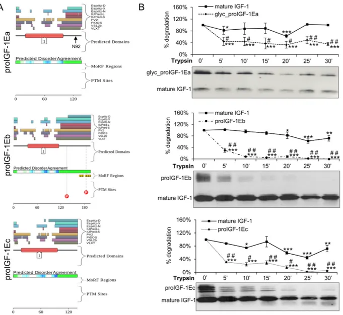
We used the D2P2 platform (http://d2p2.pro/) and limitedproteolysis to predict intrinsically disordered regions of human proIGF-1Ea (ENSP00000416811), proIGF-1Eb (ENSP00000302665) and proIGF-1Ec (ENSP00000376638)21,22 (Fig. 1). Figure 1A shows the plot generated by the
D2P2 platform: this analysis showed that the mature IGF-1 is mostly ordered, while all E-domains were predicted
to contain disordered residues. Moreover, the Eb-domain also contains two predicted molecular recognition features (MoRFs) and two phosphorylation sites.
Subsequently, we used limited proteolysis to identify the regions of the polypeptide chain mostly prone to proteolysis and thus the sites of high flexibility or local unfolding22 (Fig. 1B). The supernatant of HEK293 cells
enriched with glycosylated proIGF-1Ea, proIGF-1Eb, proIGF-1Ec and mature IGF-1 were digested with trypsin, loaded on SDS-PAGE gels and probed with the anti-mature IGF-1 antibody. As shown in Fig. 1B, all proIGF-1s were sensitive to trypsin digestion while mature IGF-1 was significantly more resistant. Digestion of mature IGF-1 required long term incubation with trypsin (i.e. >45 minutes) (Supplementary Fig. S1B). Similar results were obtained using proteinase K digestion of proIGF-1Ea (Supplementary Fig. S1C). These data show that proIGF-1s are composed of both protein structural domain, i.e. the mature IGF-1, and intrinsically disordered regions, i.e. the C-terminal E-domains.
Intracellular IGF-1 is mainly expressed as proIGF-1Ea, not mature IGF-1.
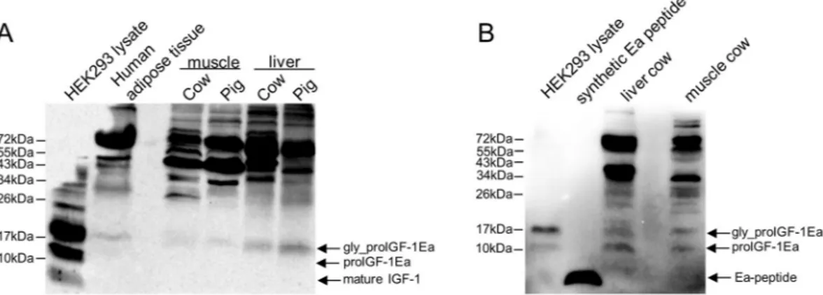
Using RT-PCR analyses, we previously demonstrated that skeletal muscles, adipose tissues and liver of several mammalian species mainly expressed the IGF-1Ea isoform, which represents about 90% of IGF-1 transcripts5. The first goal of the presentstudy was to examine the protein forms endogenously produced in these tissues. Immunoblotting of protein lysates using the anti-mature IGF-1 antibody showed a distinct ~17 kDa band, most likely representing glyco-sylated proIGF-1Ea, in all samples analysed (Fig. 2A). Notably, the band corresponding to mature IGF-1 (~7 kDa) was not found in naïve tissues, in agreement with Durzynska, J. et al.6.
To further confirm the presence of proIGF-1s, we used specific antibody directed against the common E-domain region of proIGF-1s (RSVRAQRHTD). The antibody specificity towards E-domain region was checked using HEK293 cells overexpressing IGF-1 isoforms (Supplementary Fig. S2). As shown in Fig. 2B, two bands of ~12 kDa and ~17 kDa were detected with the anti E-domain antibody, corresponding respectively to the ular weight of unglycosylated and glycosylated proIGF-1Ea. As expected, no band corresponding to the molec-ular size of Ea-domain (~4 kDa) was detected in the lysate of HEK293 cells overexpressing IGF-1Ea or tissues confirming that the E-domain was not cleaved intracellularly from the IGF-1 mature protein. Subsequently, we moved to a cell-based system to improve IGF-1 detection and to control the IGF-1 isoforms produced. We recently demonstrated that in HEK293 cells over-expressing IGF-1 isoforms the proIGF-1s are the main forms produced intracellularly, and for proIGF-1Ea both unglycosylated (~12 kDa) and glycosylated (~17 kDa) forms were detected7. Hence the tissue expression pattern of IGF-1 was recapitulated in our HEK293 cell-based
tran-sient gene expression system.
We next examined the effects of E-domains on proIGF-1s stability and secretion, starting with the predomi-nant isoform produced in normal tissues, i.e. the proIGF-1Ea.
www.nature.com/scientificreports/
Glycosylation is necessary to stabilise proIGF-1Ea and regulate mature IGF-1 secretion.
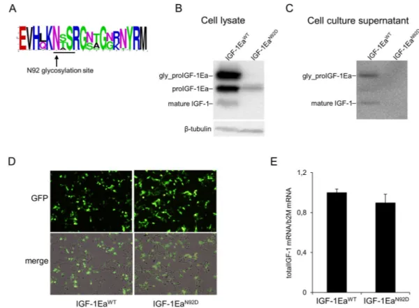
Multiple sequence alignment of vertebrate Ea-domain showed that the N-glycosylation site of proIGF-1Ea has been con-served from teleosts to mammals (Fig. 3A). This strong evolutionary conservation led us to hypothesise that glycosylation could play an important role in the regulation of proIGF-1Ea.Site-direct mutation of this glycosylation site (IGF-1EaN92D) resulted in a ~12 kDa band, corresponding to
the size of unglycosylated proIGF-1Ea (Fig. 3B). Western blotting analysis showed that the intracellular level of unglycosylated proIGF-1Ea was significantly lower in IGF-1EaN92D-transfected HEK293 cells compared
to wild-type IGF-1Ea (IGF-1EaWT) (Fig. 3B). Moreover, contrary to IGF-1EaWT, the IGF-1EaN92D-transfected
Figure 1. Evaluation of the intrinsic disorder propensity of human proIGF-1s predicted using the D2P2
platform (A) and limited proteolysis (B). (A) Analysis of the intrinsically disordered regions of human proIGF-1Ea (ENSP00000416811), proIGF-1Eb (ENSP00000302665) and proIGF-1Ec (ENSP00000376638) sequences by the D2P2 platform (http://d2p2.pro/). The red box corresponds to the insulin-like domain; the
green-and-white bar in the middle of the plot shows the predicted disorder agreement among nine predictors, with the green parts corresponding to the portions of sequence where at least 75% of the predictors agreed. Yellow bars show the location of the predicted disorder-based binding sites (molecular recognition features, MoRFs), whereas red circles at the bottom of the plot show the location of putative phosphorylation sites. Position of the N-glycosylation site of Ea-domain (N92) is also indicated. (B) Limited proteolysis of proIGF-1s. Cell culture supernatants of IGF-1Ea-, IGF-1Eb- or IGF-1Ec-transfected HEK293 cells were concentrated using Amicon Ultra 3 K centrifugal filters and incubated with 0.2 μM trypsin at 37 °C for different times. Reactions were removed over a time-course and the digested products were loaded on 12% SDS-PAGE and analysed by western blotting with an anti-mature IGF-1 antibody. Results are means ± SEM (n = 3). Repeated measures ANOVA,
#(p < 0.01) and ##(p < 0.0001) significantly different compared to mature IGF-1; *(p < 0.05), **(p < 0.001)
and ***(p < 0.0001) significantly different compared to the 0-minute time point. Cropped blots are shown. Uncropped blots are presented in Supplementary Fig. S1A.
HEK293 cells did not secrete IGF-1 (both glycosylated proIGF-1Ea and mature IGF-1) (Fig. 3C). Notably, similar GFP fluorescence intensity (Fig. 3D) and total IGF-1 mRNA (Fig. 3E; p = 0.314) were found in IGF-1EaWT- and
IGF-1EaN92D-transfected HEK293 cells, ruling out the possibility that the marked reduction in the protein levels
between the two constructs was due to different transfection efficiency. Thus, these data suggest that glycosyla-tion of proIGF-1Ea is required for efficient IGF-1 producglycosyla-tion and secreglycosyla-tion. Subsequently, we wondered whether direct inhibition of N-glycosylation by tunicamycin (Tun) might interfere with IGF-1 production. Notably, the band corresponding to glycosylated proIGF-1Ea disappeared after treatment with Tun (Fig. 4A). Moreover, the analysis of cell culture supernatants of the IGF-1Ea-transfected HEK293 cells showed that Tun treatment com-pletely abrogated the glycosylated proIGF-1Ea secretion and markedly reduced the secretion of mature IGF-1 (Fig. 4B). The marked reduction of glycosylated proIGF-1Ea after Tun treatment was not due to general suppres-sion of transcription, as shown by total IGF-1 mRNA quantification (Supplementary Fig. 4C), or general protein synthesis inhibition, as shown by co-transfection of GFP (Supplementary Fig. 4D). Notably, conditioned media from IGF-1Ea-transfected HEK293 cells treated with Tun was unable to activate the IGF-1 receptor (IGF-1R) and downstream phosphorylation of ERK1/2 and AKT of MCF-7 breast cancer cells (Fig. 4C).
Similar results were obtained by blocking N-glycosylation by glucose withdrawal although the effect was less specific since glucose starvation also slightly decreased cell number and GFP-dependent fluorescence (Supplementary Fig. 5).
Collectively, these results documented that the interference with Ea-domain glycosylation resulted in a dra-matic decrease of intracellular proIGF-1Ea level and hence proIGF-1Ea and mature IGF-1 secretion.
The turnover of unglycosylated proIGF-1Ea is faster than glycosylated proIGF-1Ea and depends
on proteasome activity.
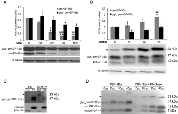
The inhibition of proIGF-1Ea glycosylation by mutation of the glycosylation site (proIGF-1EaN92D) (Fig. 3B), Tun treatment (Fig. 4A) or glucose starvation (Supplementary Fig. 5A), did notdeter-mine a concomitant increase of unglycosylated 1Ea. One possibility is that the unglycosylated proIGF-1Ea is rapidly degraded. Thus, we next sought to examine the role of glycosylation in the stability of proIGF-proIGF-1Ea. In presence of cycloheximide (CHX), a protein synthesis inhibitor, the turnover rate of unglycosylated proIGF-1Ea was faster than glycosylated one (Fig. 5A). To verify the involvement of 26 S proteasome machinery on ungly-cosylated proIGF-1Ea degradation, we subsequently treated IGF-1Ea-transfected HEK293 cells with proteasome inhibitor MG132. As shown in Fig. 5B, we found an increase of unglycosylated proIGF-1Ea, while glycosylated proIGF-1Ea was only marginally affected by the proteasome inhibitor. These results demonstrated that unglyco-sylated proIGF-1Ea was unstable and degraded faster than glycounglyco-sylated proIGF-1Ea.
Besides the increase of unglycosylated proIGF-1Ea, treatment with MG132 also promoted an accumulation of a band of ~23 kDa (indicated with an asterisk in Fig. 5B). This band was approximately twice the molecular weight of the unglycosylated proIGF-1Ea monomer (11.7 kDa) and was detected with both the anti-mature IGF-1 antibody (Fig. 5B) and the anti-E-domain antibody (Supplementary Fig. S6E). Thus, we hypothesised that inhi-bition of the proteasome leads to unglycosylated proIGF-1Ea accumulation and dimerisation. In support of this hypothesis, we found that proteasome inhibition by MG132 increased the cytoplasmic level of the ~23 kDa pro-tein (Fig. 5C). Moreover, PNGase deglycosylation of the supernatant of HEK293 cells enriched with glycosylated proIGF-1Ea increased both the unglycosylated proIGF-1Ea and the putative ~23 kDa dimer in the cell culture
Figure 2. Immunoblotting of several mammalian tissues using an antibody directed against mature IGF-1
sequence (A) or common region of E-peptides (B). (A) Protein lysates (80 μg) were subjected to 12% SDS-PAGE and immunoblotted with anti-mature IGF-1 antibody. A band at a molecular weight around 17 kDa, most likely representing glycosylated proIGF-1Ea, was detected in all the tissue samples tested. A cell lysate of HEK293 overexpressing IGF-1Ea was used has a positive control. The band corresponding to mature IGF-1 (~7 kDa) was not found in tissues and was detectable in HEK293 overexpressing IGF-1Ea only after long exposure of the blots. (B) Immunoblotting of liver and muscle cow lysate using an antibody directed against E-domains. Two bands at a molecular weight around 12 kDa and 17 kDa were detected in all the tissue samples tested, most likely representing the unglycosylated and glycosylated proIGF-1Ea respectively. No band at a molecular weight of Ea peptide (~4 kDa) was detected. A cell lysate of HEK293 overexpressing IGF-1Ea and synthetic human Ea peptide were used as positive controls.
www.nature.com/scientificreports/
supernatants (Fig. 5D). Taken together, these data showed that addition of N-glycan on N92 site of Ea-domain prevented the degradation of proIGF-1Ea by the proteasome, probably overcoming the folding limitation of Ea-domain and the tendency of unglycosylated proIGF-1Ea to self-aggregate.
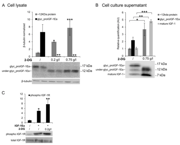
The glucose analogue, 2-deoxyglucose, rescues the secretion defect of IGF-1 under low glucose
conditions.
Due to its inhibitory activity on glycolysis, 2-deoxyglucose (2-DG) has been shown to selectively kill cancer cells23,24. Because of its similarity to mannose, 2-DG is also known to interfere with N-linkedglyco-sylation24–26. Notably, increasing evidence links 2-DG effects to the interference with glycosylation instead of
glycolysis inhibition27–29. Prompted by this, we investigated whether 2-DG interfered with proIGF-1Ea
glycosyl-ation. IGF-1Ea-transfected HEK293 cells were grown in low glucose medium (0.65 g/L) in presence of increasing concentration of 2-DG (Fig. 6). As shown in Fig. 6A, the band of glycosylated proIGF-1Ea (~17 kDa) disappeared after the addition of 0.2 g/L or 0.75 g/L of 2-DG. Notably, we also found a dose-dependent accumulation of a ~12 kDa band, both intracellularly (Fig. 6A) and extracellularly (Fig. 6B). Both the anti-mature IGF-1 antibody (Fig. 6A) and the anti-E-domain antibody (Supplementary Fig. 7D) recognised this ~12 kDa band. Thus, it is likely that the 12kD protein was not a non-specific product that appears after 2-DG treatment. Moreover, cell cul-ture supernatant from IGF-1Ea-transfected HEK293 cells treated with 2-DG fully activated the IGF-1R of MCF-7 breast cancer cells (Fig. 6C). Thus, these data indicate that 2-DG interfered with the proIGF-1Ea glycosylation leading to an accumulation of a ~12 kDa protein, probably representing an aberrant under-glycosylated form of proIGF-1Ea, which was stable and efficiently secreted. These results are in line with a recent study showing that the effect of 2-DG on protein glycosylation was dose-dependent and, at doses similar to those used in the pres-ent study, 2-DG increased mannose incorporation into cellular glycoproteins instead of inhibits glycosylation26.
Further studies are needed to identify the glycan structure of proIGF-1Ea that appears after 2-DG treatment and to understand how 2-DG rescues the secretion defect of proIGF-1Ea under low glucose conditions.
Figure 3. Conservation of the N-glycosylation site of Ea-domain (N92) (A) and effect of site-direct mutation of
this N-glycosylation site (IGF-1EaN92D mutant) on intracellular (B) or extracellular (C) proIGF-1Ea production.
Comparison of transfection efficiency between wild-type (IGF-1EaWT) and IGF-1EaN92D constructs (D and E).
(A) WebLogo of Ea-domain sequences obtained from UniProt database. The relative frequency plots of amino acids of 250 E-domain sequences obtained from UniProt database is shown. The consensus sequence motif for N-glycosylation NX(S/T) (where X can be any amino acid except proline) is underline. The WebLogo was produced using the web server at http://weblogo.berkeley.edu/logo.cgi. (B and C) Transfection of HEK293 cells with IGF-1EaWT or IGF-1EaN92D. IGF-1EaWT and IGF-1EaN92D were transiently expressed in HEK293 cells.
After 24 h the cell lysates (B) and cell culture supernatants (C) were analysed by western blot using an antibody directed against mature IGF-1 sequence. There was no significant difference in GFP fluorescence intensity (10 × magnification) (D) and total IGF-1 mRNA quantity (p = 0.314) (E) between the IGF-1EaWT and
The presence of the alternative Eb- and Ec-domains hampers the proIGF-1Eb and proIGF-1Ec
sensitivity to N-glycosylation inhibitors and determines the nuclear accumulation of
prohor-mones.
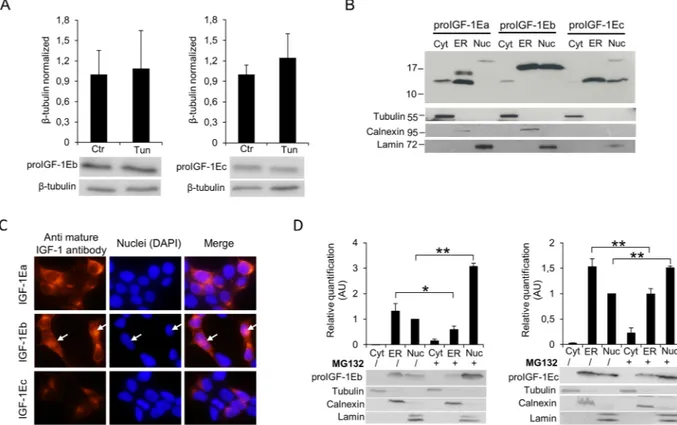
The proIGF-1Eb and proIGF-1Ec ran at the expected molecular weight of ~16.5 kDa and ~12.5 kDa in SDS-PAGE gels respectively, suggesting that these prohormones did not undergo posttranslational modification (Supplementary Fig. S2)7. Accordingly, unlikely proIGF-1Ea, proIGF-1Eb and proIGF-1Ec did not contain anypotential glycosylation sites and migrated at the same molecular weight after treatment of HEK293 cells with Tun (Fig. 7A) or no glucose media (Supplementary Fig. S8C). Noteworthy, unlikely proIGF-1Ea, also the intracel-lular levels of proIGF-1Eb and proIGF-1Ec were unaffected by Tun treatment (Fig. 7A). Hence, the presence of the Eb- or Ec-domains, instead of Ea-domain, completely abrogated the response of proIGF-1s to glycosylation inhibitors.
Notably, previous studies demonstrated that both proIGF-1Eb and proIGF-1Ec might localise in the nucleus30–32. This led us to hypothesise that disordered Eb- and Ec-domains might control subcellular
locali-sation of proIGF-1Eb and proIGF-1Ec. To test this hypothesis, we investigated the subcellular localilocali-sation of proIGF-1s by cytosol, ER and nucleus isolations (Fig. 7B) and immunofluorescence (Fig. 7C). In line with previ-ously published studies30–32, we found that proIGF-1Eb and proIGF-1Ec partially accumulated in the nucleus of Figure 4. Effect of tunicamycin (Tun) treatment on proIGF-1Ea glycosylation. (A and B) IGF-1Ea was
transiently expressed in HEK293 cells in the presence of 0.1 µg/ml of Tun. After 24 h the cell lysates (A) and cell culture supernatants (B) were analysed by western blot and relative expression level of glycosylatyed proIGF-1Ea, unglycosylated proIGF-1Ea and mature IGF-1 was calculated. The band at a molecular weight of ~17 kDa, corresponding to glycosylated proIGF-1Ea, disappeared in presence of Tun in cell lysates (A) and in cell culture supernatants (B). The band corresponding to mature IGF-1 (~7 kDa) was markedly reduced in cell culture supernatants after Tun treatment (B). (C) Phosphorylation of IGF-1R, AKT and ERK1/2 after treatment of MCF-7 cells with cell culture supernatants from IGF-1Ea-transfected HEK293 cells treated with Tun. The phosphorylation of the IGF-1R pathway was markedly reduced by Tun treatment. β-tubulin was used as a loading control for the cell lysates. Results are means ± SEM (n = 3); T-test or a one-way ANOVA was used to evaluate statistical significance (*p < 0.01, **p < 0.0001). Cropped blots are shown. Uncropped blots are presented in Supplementary Fig. S4.
www.nature.com/scientificreports/
HEK293-transfected cells, while proIGF-1Ea was mainly localised into the ER (Fig. 7B and C). Notably, proIGF-1Eb and proIGF-1Ec did not appear to be accumulated in the cytosol, and the molecular weight of proIGF-proIGF-1Eb or proIGF-1Ec in the ER and nuclear fractions corresponded to that of proIGF-1s without signal peptide (5.3 kDa) (Fig. 7B). These data suggest that proIGF-1Eb and proIGF-1Ec translocate to the nucleus from the ER and not from the cytoplasm. Accordingly, treatment with the proteasome inhibitor MG132 increased the nuclear accu-mulation of proIGF-1Eb and proIGF-1Ec, while their cytoplasmic level remained undetectable (Fig. 7D). Several substrates of the nuclear ubiquitin-proteasome system have been identified to date, showing that the nuclear ubiquitin-proteasome system is a key quality-control mechanism that rapidly eliminates unfolded or damaged proteins33. In this regard, the nuclear degradation of proIGF-1Eb and proIGF-1Ec might represent a mechanism
for controlling the intracellular level of these highly disordered proteins; however, further studies are needed to clarify this point.
Discussion
The mechanism by which IGF-1 production is regulated remains largely unknown4. In this study, we show the
functional role of disordered E-peptides in the regulation of proIGF-1s production.
It is now well established that most peptide hormones and growth factors are initially synthesised as prohor-mones that are converted to active forms by endoproteolysis at specific sites34. Accordingly, the growth factor
IGF-1 is produced as prohormone which contains a C-terminal domain, i.e. the E-domain, cleaved by furin con-vertase3. Evidence has been provided that only a small portion of proIGF-1 cleavage occurs intracellularly; hence
most proIGF-1 might be converted to mature form at the cell surface membrane or extracellularly6,7. The results
of our study confirm these findings showing that intracellular IGF-1 is mainly expressed as proIGF-1, not mature IGF-1, both in vitro and in vivo (Figs 2 and S2).
The amino acid composition of mature IGF-1 markedly differs compared to E-domains5,35. In particular, the
E-domains were enriched in disorder-promoting amino acids, and by bioinformatic analyses, we demonstrated that the E-domains are IDRs, which were also confirmed by the limited proteolysis approach (Figs 1 and S1C).
Figure 5. Analysis of unglycosylated and glycosylated proIGF-1Ea turnover. (A and B) IGF-1Ea was transiently
expressed in HEK293 cells in the presence of 25 μg/ml of protein synthesis inhibitor cycloheximide (CHX) (A) or 10 μM of the proteasome inhibitor MG132 (B) in a time-course experiment. Cells were collected at different time points, and relative expression level of glycosylated and unglycosylated proIGF-1Ea was calculated. After MG132 treatment intracellular accumulation of a ~23 kDa band was found (indicated with an asterisk in B), probably representing unglycosylated proIGF-1Ea dimer. (C) Cytosol (Cyt) and endoplasmic reticulum (ER) isolations of IGF-1Ea-transfected HEK293 cells treated with MG132. The band at a molecular weight around 23 kDa was detected only after MG132 treatment and only in the cytosolic fraction. (D) Deglycosylation of proIGF-1Ea enriched media using the N-Glycosidase F (PNGase F). The PNGase treatment determined an accumulation of the band at a molecular weight around 23 kDa probably corresponding to unglycosylated pro-IGF-1Ea dimer. Results are means ± SEM (n = 3). Repeated measures ANOVA, #(p < 0.05), ##(p < 0.01) and ###(p < 0.0001) significantly different compared to glycosylated proIGF-1Ea; *(p < 0.01) and **(p < 0.0001)
significantly different compared to the 0-minute time point. β-tubulin was used as a loading control for the cell lysates and the cytosol separation; calnexin was used as a control for the ER separation. Cropped blots are shown. Uncropped blots are presented in Supplementary Fig. S6.
Therefore, the proIGF-1s consist of two different parts: the mature IGF-1, with a well-organised structure, and the flexible C-terminal E-domains. Notably, we verified that the disorder features of E-domains are conserved both across species and for all the three E-domains generated by alternative splicing5.
Many studies show that the conformational plasticity associated with intrinsic disorder provides IDRs with a complementary functional repertoire of ordered domains15,17, especially if these IDRs are at the protein termini36.
Therefore, we next focused on the potential functional role of C-terminal Ea-, Eb- and Ec-domains.
The Ea-domain contains a highly conserved N-glycosylation site (N92) (Fig. 3A), which is heavily glycosylated with sugars comprising over 30% of the total mass of the proIGF-1Ea (Fig. 3B; see also6,7). Here, we
demon-strated that the inhibition of N-glycosylation by site-directed mutagenesis (Fig. 3B), Tun treatment (Fig. 4A) or glucose withdrawal (Supplementary Fig. S5A), blocked the intracellular production of glycosylated proIGF-1Ea. The secretion of glycosylated proIGF-1Ea and mature IGF-1 was also substantially affected by the inhibition of N-glycosylation (Figs 3C, 4B and S5B). Accordingly, the conditioned media from IGF-1Ea-transfected cells treated with Tun were unable to activate the IGF-1R pathway in human MCF-7 breast cancer cell line (Fig. 4C). It is important to highlight that the unglycosylated proIGF-1Ea was not accumulated after N-glycosylation inhi-bition. In this line, we demonstrated that unglycosylated proIGF-1Ea had a faster turnover rate compared to glycosylated proIGF-1Ea (Fig. 5A). The proteasome inhibitor MG132 partially rescued the accumulation defect of unglycosylated proIGF-1Ea, but also markedly increased the production of a ~23 kDa band, probably repre-senting unglycosylated proIGF-1Ea dimer (Fig. 5B,C and D). N-glycosylation influences the folding and traffick-ing of many glycoproteins ensurtraffick-ing protein solubility and minimistraffick-ing aggregation37–39. Accordingly, our work
indicates that addition of N-glycan on Ea-domain prevents the degradation of proIGF-1Ea by the proteasome, probably overcoming the folding limitation of unglycosylated proIGF-1Ea and its entry into the ER-associated
Figure 6. Effect of 2-Deoxyglucose (2-DG) on glycosylated proIGF-1Ea production. (A and B) IGF-1Ea was
transiently expressed in HEK293 cells growth in low glucose medium (0.65 g/L) with or without 2-DG. After 24 h the cell lysates (A) and cell culture supernatants (B) were analysed by western blot and relative expression level of glycosylated proIGF-1Ea, unglycosylated proIGF-1Ea and mature IGF-1 was calculated. The band at a molecular weight around 17 kDa, corresponding to glycosylated proIGF-1Ea, disappeared in the presence of 2-DG in the cell lysates (A) and in the cell culture supernatants (B). 2-DG treatment determined also an intracellular (A) and supernatants (B) accumulation of a ~12 kDa band, probably representing an under-glycosylated proIGF-1Ea form. (C) Phosphorylation of IGF-1R after treatment of MCF-7 cells with conditioned media from IGF-1Ea-transfected HEK293 cells treated with 2-DG. Phosphorylation of IGF-1R increased both in 2-DG-treated and untreated cells. β-tubulin was used as a loading control for the cell lysates. Results are means ± SEM (n = 3); T-test or a one-way ANOVA was used to evaluate statistical significance (*p < 0.05, **p < 0.01 and ***p < 0.0001). Cropped blots are shown. Uncropped blots are presented in Supplementary Fig. S7.
www.nature.com/scientificreports/
degradation (ERAD) pathway40,41. This finding has interesting functional implications since the IGF-1Ea isoform
is widely expressed in normal tissues as well as in various tumour cells5,8,10,42. Thus, interfering with proIGF-1Ea
glycosylation provides a means to regulate IGF-1 production. In this regard, we also investigated the effect of 2-DG on proIGF-1Ea glycosylation. 2-DG, a non-metabolizable glucose analogue, is one of the most frequently used antiglycolytic agents and it has come under increasing scrutiny as a therapeutic agent, especially in can-cer24. However, since 2-DG also mimics mannose, might also interfere with N-linked glycosylation24,26. In fact,
recent studies showed that interference with N-linked glycosylation rather than glycolysis as the predominant mechanism by which 2-DG inhibits cancer cell growth under normoxia27–29. Here, we demonstrated that 2-DG
interferes with 1Ea glycosylation preventing the formation of the normal, highly glycosylated proIGF-1Ea (~17 kDa) (Fig. 6A). However, 2-DG also determined a concomitant dose-dependent increase of a ~12 kDa protein that probably represents an aberrant under-glycosylated proIGF-1Ea form. Notably, this protein was sta-ble and efficiently secreted (Fig. 6B). Accordingly, despite the loss of highly glycosylated proIGF-1Ea after the 2-DG treatment, the condition media of these cells fully activated the IGF-1R of human breast cancer cell line MCF-7 (Fig. 6C). Thus, these data suggested that 2-DG interfere with normal proIGF-1Ea glycosylation process leading to an accumulation of an under-glycosylated form which is still able to fold and secreted by the cells. These results are in line with a recent study showing that the main effect of low, non-toxic and pharmacologically relevant concentration of 2-DG was to increase the incorporation of mannose on the glycan structure instead of inhibiting glycosylation26. Thus, 2-DG alters the glycosylation process favouring the synthesis of glycoproteins
with incomplete or defective oligosaccharide chains. Taking advantage of the increased glucose uptake that occurs in most tumours, treatment with 2-DG might represent a new diagnostic and therapeutic tool to discriminate
Figure 7. Effect of inhibition of N-glycosylation using Tun on IGF-1Eb (A, left panel) and IGF-1Ec (A, right
panel) production and subcellular localisation of proIGF-1s in untreated (B and C) or MG132 treated HEK293 cells (D). (A) IGF-1Eb or IGF-1Ec were transiently expressed in HEK293 cells in the presence of 0.1 µg/ml of Tun. After 24 h the cell lysates were analysed by western blot and relative expression level of proIGF-1Eb (left panel) and proIGF-1Ec (right panel) was calculated. The molecular weight and the expression level of proIGF-1Eb (p = 0.8) and proIGF-1Ec (p = 0.6) were unaffected by Tun treatment. Results are means ± SEM (n = 3); T-test was used to evaluate statistical significance. (B) Subcellular localization of IGF-1 isoforms analysed by cytosol, ER and nucleus isolations or (C) immunofluorescence staining of IGF-1Ea-, IGF-1Eb- or IGF-1Ec-transfected HEK293 cells. ProIGF-1Ea was mainly localised in the cytosol fraction (unglycosylated proIGF-1Ea) and the ER fraction (both unglycosylated and glycosylated proIGF-1Ea) while proIGF-1Eb and proIGF-1Ec were mainly localised in the nuclear fraction and ER. (D) Subcellular localisation of proIGF-E1b (left panel) or proIGF-1Ec (right panel) after treatment with 10 μM of the proteasome inhibitor MG132 for 6 h. Results are means ± SEM (n = 3); a two-way ANOVA was used to evaluate statistical significance (*p < 0.01 and **p < 0.001). β-tubulin was used as a loading control for the cell lysates and the cytosol separation; calnexin as a control for the ER separation and lamin as a control for the nucleus separation. DAPI nuclear stain (blue). Cropped blots are shown. Uncropped blots are presented in Supplementary Fig. S8.
obstacle for their correct folding and traffic through the ER. Accordingly, we previously demonstrated that the secretion of 1 (both pro1s and mature 1) was significantly lower in the cell overexpressing IGF-1Eb or IGF-1Ec isoforms compared to IGF-1Ea-transfected cells7. Furthermore, the inhibition of proteasome
by MG132 determined nuclear accumulation of proIGF-1Eb and proIGF-1Ec (Fig. 7D) demonstrating that the production of these prohormones was controlled through their subcellular localization.
In conclusion, our data suggested that alternative E-domains act as flexible tails controlling proIGF-1s and mature IGF-1 production. In particular, we demonstrated that N-linked glycosylation regulates the stability and secretion of proIGF-1Ea, probably ensuring proper prohormone folding and favouring its passage through the secretory pathway. Interference with proIGF-1Ea N-glycosylation (e.g. mutation of N-glycosylation site or Tun, glucose starvation and 2-DG treatment) directly affects protein IGF-1 level. The splice variants IGF-1Eb and IGF-1Ec encode the alternative proIGF-1Eb and proIGF-1Ec forms, which were insensitive to modulation of glycosylation. Notably, the Eb- and Ec- disordered tails promoted the nuclear accumulation of proIGF-1Eb and proIGF-1Ec, and thus directly affected the efficiency of proIGF-1s secretion.
Thus, disordered E-domains play a crucial role in the structure, regulation, localisation and functioning of IGF-1.
Methods
Tissue sampling and cell cultures.
Human and animal tissues were previously obtained and analysed for mRNA extraction and IGF-1 isoforms quantification as described in Annibalini et al.5. From the same tissuesstored at −80 °C, about 30 mg were used for protein extraction and subsequent western blotting analysis. Freshly frozen normal human adipose (2 males and 2 females, mean age 52 +/− 12 years) samples were provided by the complex structure of biomarkers (DOSMM) of National Cancer Institute of Milan. All subjects provided written informed consent before archival tissue samples. Pig (Sus scrofa; 3 males) and cow (Bos Taurus; 3 females) tis-sues were collected at local slaughter during routine meat inspection. The age of the animals ranged from 2 to 5 years. The study was conducted and reported in accordance with standards for reporting of diagnostic accuracy (STARD) requirements. The experimental protocols were in accordance with the Guide for the Care and Use of Laboratory Animals by Ministero della Sanità D.L. 116 (1992) and approved by the University of Urbino “Carlo Bo” Committee. The HEK293 and MCF-7 cell lines were obtained from the American Type Culture Collection (ATCC, Rockville, MD, USA). The cells lines were cultured in DMEM media supplemented with 10% fetal bovine serum, 2 mM L-glutamine, 1x MEM Non-essential Amino Acid Solution, 0.1 mg/ml streptomycin and 0.1 U/L penicillin. Cells were maintained in a humidified incubator (5% CO2) at 37 °C. All cell culture materials were
purchased from Sigma-Aldrich (St. Louis, MO, USA).
Protein extraction and western blotting analysis.
Tissues and cells were processed for western blot analysis as previously reported7. Briefly, protein extracts were prepared by homogenizing with a Polytronhomog-enizer (KINEMATICA AG, Switzerland) in lysis buffer containing: 20 mM HEPES (pH 7.9), 25% v/v glycerol, 0.42 M NaCl, 0.2 mM EDTA, 1.5 mM MgCl2, 0.5% v/v Nonidet P-40, 1 mM DTT, 1 mM Naf, 1 mM Na3VO4,
and 1 × complete protease inhibitor cocktail (Roche Diagnostics Ltd, Mannheim, Germany). The lysates were frozen and thawed twice and clarified by centrifugation at 12000 rpm for 10 minutes at 4 °C. Protein concentra-tion in each sample was determined using the Bradford colourimetric assay. An equal amount of total proteins were fractionated by SDS-PAGE on a 15% polyacrylamide gel and then transferred to PVDF or nitrocellulose membranes (Bio-Rad Laboratories Inc). The membranes were incubated overnight at 4 °C with the primary anti-bodies directed towards: IGF-1 (1:2000; no. 500P11), purchased from Peprotech (Rocky Hill, New Jersey, USA), proIGF-1s (1:2000; no. PA5-19382) purchased from Invitrogen (Carlsbad, California, USA), β-tubulin (1:2000; no. 2146), lamin A/C (1:2000; no. 4777), calnexin (1:2000; no. 2679) phospho-IGF-1 Receptor β (1:2000; no. 3024), IGF-1 Receptor β (1:2000; no. 3027), phospho-p44/42 (ERK1/2) (1:2000; no. 9101), p44/42 (ERK1/2) (1:2000; no. 9102), phospho-Akt (Ser473) (1:2000; no. 9271) and Akt (1:2000; no. 9272) were purchased from Cell Signaling Technology (Beverly, MA, USA). Membranes were washed and incubated with appropriate secondary HRP-conjugated antibodies (Bio-Rad Laboratories Inc) at room temperature for 1 hour. After TBS-T washing, protein bands were visualised using Clarity Western ECL Substrate (Bio-Rad Laboratories Inc) and were quanti-fied using Fluor-S MAX System (Bio-Rad Laboratories Inc) equipped with Quantity One software. β-tubulin was used for normalisation.
Cell culture transfection assays.
Transient cell transfection was carried out with TransIT-X2®
Transfection Reagent (Mirus Bio, Madison, WI, USA) with plasmid constructs containing sequences encodingwww.nature.com/scientificreports/
proIGF-1s as previously described7. Each plasmid contained DNA encoding the class 1 IGF-1 48-amino acid
signal peptide, the mature 70-amino acid IGF-1 peptide, the first 16 aa common in all E-domains, and C-terminal sequences encoding either the Ea (19 aa), the Eb (61 aa) or the Ec (24 aa) domain. Where indicated, to interfere with N-glycosylation, cells were grown in glucose-depleted medium or treated with 0.1 µg/ml of Tunicamycin (Tun) (Sigma-Aldrich, St. Louis, MO, USA) or 2-Deoxyglucose (2-DG) (Sigma-Aldrich, St. Louis, MO, USA) for 24 h. The supernatants of HEK293-transfected cells treated with Tun or 2-DG were also used to treat the MCF-7 cells for 1 h to evaluate their effects on IGF-1R, AKT and ERK1/2 phosphorylation. Afterwards, the MCF-7 cells were washed with PBS and lysed for western blotting analysis. Where indicated, the protein synthesis inhibitor cycloheximide (CHX) (Sigma-Aldrich) and the proteasome inhibitor MG132 (Sigma-Aldrich) were added to HEK293 cells cultured with a final concentration of 25 μg/ml and 10 μM respectively in a time-course experiment. The cells were then collected at indicated time points, lysed and prepared to western blotting analysis.
The efficiency of transfection was estimated by GFP-dependent fluorescence and by real-time RT-PCR for total IGF-1 mRNA levels at 24 hours after transfection, as previously described7.
Deglycosylation of proIGF-1Ea was performed by incubation of proIGF-1Ea enriched media with 2500 U of PNGase F (NewEngland Biolabs) for 3 hours at 37 °C, according to manufacturer’s recommendations. Aliquot of proIGF-1Ea supernatant incubated with equal volume of PNGase assay reaction buffer without the enzyme PNGase F was used as a control.
Limited proteolysis.
Supernatants of IGF-1Ea-, IGF-1Eb- or IGF-1Ec-transfected HEK293 cells were con-centrated using an Amicon Ultra 3 K centrifugal filter unit (Merck Millipore, Billerica, MA, USA) and subjected to limited proteolysis. Limited proteolysis was performed by enzymatic digestion at 37 °C adding bovine trypsin (Sigma-Aldrich, Italy) or proteinase K (Sigma-Aldrich, Italy) to protein extract at a ratio 1:100 enzyme/substrate (w/w). Reactions were removed over a time-course, and the digested products were quenched with SDS sample buffer before SDS-PAGE and analysed by western blotting. The quantification of bands intensity was normalised to the “no trypsin” samples of each set.Conservation of Ea-domain N-glycosylation site.
BLASTP was used to align protein sequence of human Ea-domain (EVHLKNASRGSAGNKNYRM) against the entire UniProt database using E-threshold of 0.0145. The obtained UniProt hits (250 sequences with a minimal sequence identity of ∼60%) were aligned usingMUSCLE46, and WebLogo (http://weblogo.berkeley.edu/logo.cgi) was used to create relative frequency plots.
Site-direct mutagenesis.
Site-direct mutagenesis was used to generate the proIGF-1EaN92D mutantlacking the glycosylation site. The composition of PCR mutagenesis reactions run on an Applied Biosystems SimpliAmp Thermal Cycler were as follows: 25 μl of 2x Platinum SuperFi PCR Master Mix (Thermo Fisher); 10 μM of each primer (N92D-F 5′-ATTTGAAGGACGCAAGTAGAGGGAG-3′; 5′-N92D-R
TTGCGTCCTTCAAATGTACTTCCTTC-3′) 1 ng plasmid contained DNA encoding proIGF-1Ea and H2O to
50 μl. Following PCR, the reactions were incubated with 1 μl (20 U) of AnzaTM 10 DpnI (Invitrogen) for 1 h to
selectively digest the methylated parent plasmids, and the resulting PCR products were purified with GenElute PCR Clean-Up Kit (Sigma-Aldrich). 2 μl of the purified PCR reactions were transformed into electrocompetent E. coli XL1-Blue cells with selection for resistance to ampicillin (100 μg/ml), and successful mutagenesis was con-firmed by sequencing of plasmid DNA.
Subcellular localisation analysis.
The cytosol, ER and nucleus isolations were performed as described in47. Briefly, cells were treated with permeabilisation buffer for 5 minutes and then were centrifuged at 3000 g for5 min to collect the cytosol fraction in the soluble fraction. The pellet was then washed, subjected to lysis buffer for 5 minutes and centrifuged again at 3000 g for 5 min to collect the ER in the soluble fraction, whereas the pellet represented the nucleus fraction. Finally, all the samples were clarified at 7500 g for 10 minutes to remove cell debris and transferred to clean tubes for further analysis.
The HEK293 cells subjected to immunofluorescence were seeded in 4-well chamber slide at a density of 5 × 10⁴ cells/well, incubated overnight and transfected for IGF-1 isoforms expression as previously described.
After overnight incubation, cells were fixed with 4% paraformaldehyde for 15 minutes, permeabilised with 0.2% TritonX-100, blocked with 5% of goat serum and incubated overnight at 4 °C with the anti-mature IGF-1 antibody (Prepotech). Next, cells were incubated 1 hour with an anti-rabbit-PE conjugated antibody, stained with DAPI, mounted with Fluoreshield (Sigma) and photographed with a fluorescence microscope (ZEISS AxioVert A.1).
Statistical analysis.
Data are represented as mean ± SEM of at least three independent experiments. Statistical analyses were performed using repeated measures ANOVA or one-way ANOVA as appropriate, fol-lowed by Bonferroni’s multiple comparison post hoc tests. A p-value < 0.05 was considered statistically significant.Data availability.
The authors declare that the data supporting the findings of this study are available within the paper and its Supplementary Information files or upon reasonable request.References
1. Dyer, A. H., Vahdatpour, C., Sanfeliu, A. & Tropea, D. The role of Insulin-Like Growth Factor 1 (IGF-1) in brain development, maturation and neuroplasticity. Neuroscience 325, 89–99, https://doi.org/10.1016/j.neuroscience.2016.03.056 (2016).
2. Christopoulos, P. F., Msaouel, P. & Koutsilieris, M. The role of the insulin-like growth factor-1 system in breast cancer. Mol Cancer
14, 43, https://doi.org/10.1186/s12943-015-0291-7 (2015).
3. Barton, E. R. The ABCs of IGF-I isoforms: impact on muscle hypertrophy and implications for repair. Appl Physiol Nutr Metab 31, 791–797, https://doi.org/10.1139/h06-054 (2006).
Oncotarget 8, 79003–79011, https://doi.org/10.18632/oncotarget.20743 (2017).
12. Barton, E. R., DeMeo, J. & Lei, H. The insulin-like growth factor (IGF)-I E-peptides are required for isoform-specific gene expression and muscle hypertrophy after local IGF-I production. J Appl Physiol (1985) 108, 1069–1076, https://doi.org/10.1152/ japplphysiol.01308.2009 (2010).
13. Redwan, E. M., Linjawi, M. H. & Uversky, V. N. Looking at the carcinogenicity of human insulin analogues via the intrinsic disorder prism. Sci Rep 6, 23320, https://doi.org/10.1038/srep23320 (2016).
14. Lieutaud, P. et al. How disordered is my protein and what is its disorder for? A guide through the “dark side” of the protein universe.
Intrinsically Disord Proteins 4, e1259708, https://doi.org/10.1080/21690707.2016.1259708 (2016).
15. Babu, M. M. The contribution of intrinsically disordered regions to protein function, cellular complexity, and human disease.
Biochem Soc Trans 44, 1185–1200, https://doi.org/10.1042/BST20160172 (2016).
16. Brown, C. J., Johnson, A. K., Dunker, A. K. & Daughdrill, G. W. Evolution and disorder. Curr Opin Struct Biol 21, 441–446, https:// doi.org/10.1016/j.sbi.2011.02.005 (2011).
17. Dunker, A. K., Bondos, S. E., Huang, F. & Oldfield, C. J. Intrinsically disordered proteins and multicellular organisms. Semin Cell Dev
Biol 37, 44–55, https://doi.org/10.1016/j.semcdb.2014.09.025 (2015).
18. Ciechanover, A. Intracellular protein degradation: from a vague idea through the lysosome and the ubiquitin-proteasome system and onto human diseases and drug targeting. Neurodegener Dis 10, 7–22, https://doi.org/10.1159/000334283 (2012).
19. Pauwels, K., Lebrun, P. & Tompa, P. To be disordered or not to be disordered: is that still a question for proteins in the cell? Cell Mol
Life Sci 74, 3185–3204, https://doi.org/10.1007/s00018-017-2561-6 (2017).
20. Buljan, M. et al. Tissue-specific splicing of disordered segments that embed binding motifs rewires protein interaction networks.
Mol Cell 46, 871–883, https://doi.org/10.1016/j.molcel.2012.05.039 (2012).
21. Oates, M. E. et al. D(2)P(2): database of disordered protein predictions. Nucleic Acids Res 41, D508–516, https://doi.org/10.1093/ nar/gks1226 (2013).
22. Fontana, A., de Laureto, P. P., Spolaore, B. & Frare, E. Identifying disordered regions in proteins by limited proteolysis. Methods Mol
Biol 896, 297–318, https://doi.org/10.1007/978-1-4614-3704-8_20 (2012).
23. Boutrid, H. et al. Targeting hypoxia, a novel treatment for advanced retinoblastoma. Invest Ophthalmol Vis Sci 49, 2799–2805,
https://doi.org/10.1167/iovs.08-1751 (2008).
24. Xi, H., Kurtoglu, M. & Lampidis, T. J. The wonders of 2-deoxy-D-glucose. IUBMB Life 66, 110–121, https://doi.org/10.1002/iub.1251
(2014).
25. Parodi, A. J. Protein glucosylation and its role in protein folding. Annu Rev Biochem 69, 69–93, https://doi.org/10.1146/annurev. biochem.69.1.69 (2000).
26. Ahadova, A., Gebert, J., von Knebel Doeberitz, M., Kopitz, J. & Kloor, M. Dose-dependent effect of 2-deoxy-D-glucose on glycoprotein mannosylation in cancer cells. IUBMB Life 67, 218–226, https://doi.org/10.1002/iub.1364 (2015).
27. Kurtoglu, M. et al. Under normoxia, 2-deoxy-D-glucose elicits cell death in select tumor types not by inhibition of glycolysis but by interfering with N-linked glycosylation. Mol Cancer Ther 6, 3049–3058, https://doi.org/10.1158/1535-7163.MCT-07-0310 (2007). 28. Liu, H. et al. Conversion of 2-deoxyglucose-induced growth inhibition to cell death in normoxic tumor cells. Cancer Chemother
Pharmacol 72, 251–262, https://doi.org/10.1007/s00280-013-2193-y (2013).
29. Gu, L. et al. Low dose of 2-deoxy-D-glucose kills acute lymphoblastic leukemia cells and reverses glucocorticoid resistance via N-linked glycosylation inhibition under normoxia. Oncotarget 8, 30978–30991, https://doi.org/10.18632/oncotarget.16046 (2017). 30. Peng, Q. et al. The nuclear localization of MGF receptor in osteoblasts under mechanical stimulation. Mol Cell Biochem 369,
147–156, https://doi.org/10.1007/s11010-012-1377-9 (2012).
31. Tan, D. S., Cook, A. & Chew, S. L. Nucleolar localization of an isoform of the IGF-I precursor. BMC Cell Biol 3, 17 (2002). 32. Durzynska, J., Wardzinski, A., Koczorowska, M., Gozdzicka-Jozefiak, A. & Barton, E. R. Human Eb peptide: not just a by-product
of pre-pro-IGF1b processing? Horm Metab Res 45, 415–422, https://doi.org/10.1055/s-0032-1331699 (2013).
33. von Mikecz, A. The nuclear ubiquitin-proteasome system. J Cell Sci 119, 1977–1984, https://doi.org/10.1242/jcs.03008 (2006). 34. Chretien, M. How the prohormone theory solved two important controversies in hormonal and neural Peptide biosynthesis. Front
Endocrinol (Lausanne) 4, 148, https://doi.org/10.3389/fendo.2013.00148 (2013). 35. Hede, M. S. et al. E-peptides control bioavailability of IGF-1. Plos One 7, e51152 (2012).
36. Uversky, V. N. The most important thing is the tail: multitudinous functionalities of intrinsically disordered protein termini. FEBS
Lett 587, 1891–1901, https://doi.org/10.1016/j.febslet.2013.04.042 (2013).
37. Helenius, A. & Aebi, M. Roles of N-linked glycans in the endoplasmic reticulum. Annu Rev Biochem 73, 1019–1049, https://doi. org/10.1146/annurev.biochem.73.011303.073752 (2004).
38. Skropeta, D. The effect of individual N-glycans on enzyme activity. Bioorg Med Chem 17, 2645–2653, https://doi.org/10.1016/j. bmc.2009.02.037 (2009).
39. Mitra, N., Sinha, S., Ramya, T. N. & Surolia, A. N-linked oligosaccharides as outfitters for glycoprotein folding, form and function.
Trends Biochem Sci 31, 156–163, https://doi.org/10.1016/j.tibs.2006.01.003 (2006).
40. Smith, M. H., Ploegh, H. L. & Weissman, J. S. Road to ruin: targeting proteins for degradation in the endoplasmic reticulum. Science
334, 1086–1090, https://doi.org/10.1126/science.1209235 (2011).
41. Tsai, B., Ye, Y. & Rapoport, T. A. Retro-translocation of proteins from the endoplasmic reticulum into the cytosol. Nat Rev Mol Cell
Biol 3, 246–255, https://doi.org/10.1038/nrm780 (2002).
42. Pelosi, M., Alfo, M., Martella, F., Pappalardo, E. & Musaro, A. Finite mixture clustering of human tissues with different levels of IGF-1 splice variants mRNA transcripts. BMC Bioinformatics IGF-16, 289, https://doi.org/10.1186/s12859-015-0689-7 (2015).
43. Pickard, A., Durzynska, J., McCance, D. J. & Barton, E. R. The IGF axis in HPV associated cancers. Mutat Res Rev Mutat Res 772, 67–77, https://doi.org/10.1016/j.mrrev.2017.01.002 (2017).
www.nature.com/scientificreports/
44. Vassilakos, G., Philippou, A. & Koutsilieris, M. Identification of the IGF-1 processing product human Ec/rodent Eb peptide in various tissues: Evidence for its differential regulation after exercise-induced muscle damage in humans. Growth Horm IGF Res 32, 22–28, https://doi.org/10.1016/j.ghir.2016.11.001 (2017).
45. Magrane, M. UniProt Knowledgebase: a hub of integrated protein data. Database (Oxford) 2011, bar009, https://doi.org/10.1093/ database/bar009 (2011).
46. Edgar, R. C. MUSCLE: multiple sequence alignment with high accuracy and high throughput. Nucleic Acids Res 32, 1792–1797,
https://doi.org/10.1093/nar/gkh340 (2004).
47. Jagannathan, S., Nwosu, C. & Nicchitta, C. V. Analyzing mRNA localization to the endoplasmic reticulum via cell fractionation.
Methods Mol Biol 714, 301–321, https://doi.org/10.1007/978-1-61779-005-8_19 (2011).
Acknowledgements
This research was supported by the Italian Ministry of Health, “Ricerca Finalizzata 2011” (grant number: GR-2011-02348423) and “Progetti di ricerca DISB 2017”, granted by the Department of Biomolecular Sciences, University of Urbino Carlo Bo, Italy. We thank Dr. F. Berrino for his careful and critical reading of our discussion paper.
Author Contributions
G.A., S.C. and M.D.S. conceived and designed the experiments. G.A., S.C., M.D.S., R.S. and L.D.P. performed the experiments. G.A., M.G. and E.B. analysed the data and performed the statistical analysis. G.A. wrote the paper. A.V., G.B., V.S. and E.B. provided overall direction to the project and revised the manuscript. All authors reviewed, discussed and approved the manuscript.
Additional Information
Supplementary information accompanies this paper at https://doi.org/10.1038/s41598-018-27233-3.
Competing Interests: The authors declare no competing interests.
Publisher's note: Springer Nature remains neutral with regard to jurisdictional claims in published maps and
institutional affiliations.
Open Access This article is licensed under a Creative Commons Attribution 4.0 International
License, which permits use, sharing, adaptation, distribution and reproduction in any medium or format, as long as you give appropriate credit to the original author(s) and the source, provide a link to the Cre-ative Commons license, and indicate if changes were made. The images or other third party material in this article are included in the article’s Creative Commons license, unless indicated otherwise in a credit line to the material. If material is not included in the article’s Creative Commons license and your intended use is not per-mitted by statutory regulation or exceeds the perper-mitted use, you will need to obtain permission directly from the copyright holder. To view a copy of this license, visit http://creativecommons.org/licenses/by/4.0/.