anno: 2017 Mese: april Volume: 36 no: 2
rivista: International angiology Cod rivista: Int angiol
lavoro:
titolo breve: HolMIUM laSer In CVD treatMent primo autore: gIaneSInI
pagine: 122-8
citazione: Int angiol 2017;36:122-8
ablate the refluxing vein in a minimally invasive way.
nevertheless, tumescent anesthesia is required, leading
to possible patient discomfort, occasional hematomas
and to extremely rare but serious complications such as
fiber fracture.
6, 7recently, an innovative holmium laser (Hol) has
been introduced to the market, offering the consistent
advantage of not requiring tumescent anesthesia. This
device is able to reduce the great saphenous vein (gSV)
caliber, allowing immediate, subsequent foam
sclero-therapy, even in the largest veins. The strategy is known
e
ndovenous thermal ablation has become a mainstay
in the treatment of saphenous reflux
1and is
rec-ommended as the best treatment option by international
guidelines.
2, 3So far, the main thermal sources for
endo-venous ablation are radiofrequency ablation (rFa) and
endovenous laser ablation (EVLA).
4, 5they are
cur-rently preferred over surgery, because they are safe and
minimally invasive.
2, 3By introducing a fiber into the saphenous vein,
ther-mal energy is delivered intra-luminally and absorbed by
blood and vein wall. The aim of these techniques is to
O R I G I N A L A R T I C L E
Histologic and sonographic features of holmium
laser in the treatment of chronic venous disease
Sergio gIaneSInI
1*, roberta gaFÀ
2, Savino oCCHIonorellI
1, erica MenegattI
1,
anna Maria MalagonI
1, Paolo SPatH
1, giovanni lanZa
2, Paolo ZaMBonI
11Vascular Disease Center, Unit of translational Surgery, Ferrara, Italy; 2Department of Pathology, University of Ferrara, Ferrara, Italy *Corresponding author: Sergio Gianesini, Vascular Disease Center, University of Ferrara, Via Aldo Moro 8, Cona, 44123 Ferrara, Italy.
E-mail: [email protected]
a B S t r a C t
BACKGROUND: A new holmium laser (HOL) has been introduced to the market. The device is able to reduce the great saphenous vein (GSV) caliber in a tumescence-free procedure, favoring an effective sclerotherapy of large vessels. Aim of the present investigation is to provide the first in vivo data about the effect of HOL on GSV histology.
METHODS: Six chronic venous disease (C2-5, ep, as, Pr) patients (M:F ratio 1:1; age: 57±8, BMI 24±2 kg/m2) underwent HOL-assisted caliber reduction of the GSV, high-ligation and flush ligation of the incompetent tributaries. Three cm of proximal great saphenous vein not treated by laser and 3 cm of a contiguous segment that was just previously treated by HOL were harvested.
Histological assessments were performed. Patent GSV lumen caliber was assessed at the mid-thigh right before, and after the procedure. Peripro-cedural pain was graded by Visual Analogue Scale.
RESULTS: GSV samples after holmium laser therapy showed thickening of the vascular wall with a decreased, yet patent lumen. Immunos-taining demonstrated intact endothelial lining in both the treated and not treated segments. Expansion of collagen fibers was observed in the laser-treated segments. Collagen appeared more homogeneous than in controls, with an amorphous appearance. Laser treated veins showed a reduction in elastic fibers with greater fragmentation. Smooth muscle cells appeared swollen. The caliber of the mid-thigh great saphenous vein lumen decreased from 8.1±0.8 mm to 3.9±0.2 mm (P<0.0001). The average periprocedural pain was 1±0.6.
CONCLUSIONS: HOL significantly reduces the caliber of the GSV. The endothelial lining is spared, while the remaining wall is thickened by a hyalinization-like process.
(Cite this article as: gianesini S, gafà r, occhionorelli S, Menegatti e, Malagoni aM, Spath P, et al. Histologic and sonographic features of holmium laser in the treatment of chronic venous disease. Int Angiol 2017;36:122-8. DOI: 10.23736/S0392-9590.16.03652-X)
Key words: Solid-state lasers - Holmium - Ablation techniques - Laser therapy - Sclerotherapy.
International angiology 2017 april;36(2):122-8 DOI: 10.23736/S0392-9590.16.03652-X © 2016 eDIZIonI MInerVa MeDICa
Online version at http://www.minervamedica.it
COPYRIGHT
©2017 EDIZIONI MINERVA MEDICA
y inter national cop yr ight la ws .
No additional reproduction is author
iz ed. It is per mitted f or persona l use to do wnload and sa v
e only one file and pr
int only one cop
y of this Ar ticle . It is not per mitted to mak e additional copies , either pr
inted or electronic) of the Ar
ticle f or an y pur pose . It is not per mitted to dis tr ib
ute the electronic cop
y of the ar
ticle through online inter
net and/or intr
anet file shar
ing systems
, electronic mailing or an
y other
ticle
.
The use of all or an
y par
t of the Ar
ticle f
or an
y Commercial Use is not per
mitted.
The creation of der
iv
ativ
e w
or
ks from the Ar
ticle is not per
mitted.
The production of repr
ints f
or personal or commercial use is
v e , co v er , o v er la y, obscure , b loc k, or change an y cop yr
ight notices or ter
ms of use wh
ich the Pub
lisher ma y post on the Ar ticle . It is not per mitted to fr ame or use fr
aming techniques to enclose an
y tr ademar k, logo , lisher .
HolMIUM laSer In CVD treatMent gIaneSInI
orcein staining (a specific histochemical staining used
to identify elastic fibers) and Masson trichrome
stain-ing (a histochemical stainstain-ing used to identify collagen
fibers and muscle fibers).
Immunohistochemistry was also employed to verify
the presence of factor VIII — which is normally
ex-pressed in endothelial cells — and of actin — which is
normally expressed in smooth muscle cells.
Patent GSV lumen caliber was assessed at the
mid-thigh right before and after the procedure in the supine
position.
Periprocedural HOL-induced pain was graded by
Visual analogue Scale ranging from 0 (no pain) to 10
(unbearable pain).
15, 16Statistical analysis
InStat GraphPad (GraphPad Software, Inc., La Jolla,
CA, USA) was used for statistical analysis. The data
were expressed as mean ± standard deviation. The
nor-mal distribution of the data was verified by the
Kol-mogorov-Smirnov test. The differences between vein
diameters pre- and post-procedure were performed
us-ing two-tailed paired Student’s t-test. A P value of 0.05
or less was considered statistically significant.
Results
All 6 harvested samples were suitable for
histologi-cal analysis. Gross examination of the GSV after HOL
therapy showed a conspicuous thickening of the
vascu-lar wall with a decreased patent lumen (Figure 1).
On microscopic examination, the vascular wall
showed sub-intimal fibrosis and hypertrophy of the
smooth muscle coat in both Hol treated and not treated
samples, because of the common chronic venous
dis-ease condition.
the endothelial lining remained intact in both groups,
as demonstrated by factor VIII immunostaining
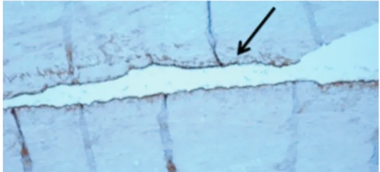
(Fig-ure 2). Various constituents of the vascular wall showed
structural alterations. Compared to the contiguous GSV
tract (Figure 3A) that was not previously shrunken, HOL
samples demonstrated an expansion of the collagen fibers,
with a more homogeneous hyalinization-like amorphous
appearance and a loss of normal fibrillar characteristics
(Figure 3B). In HOL treated samples, orcein staining
demonstrated a significant reduction in elastic fibers with
by the acronym laFoS (laser assisted Foam
Sclero-therapy).
8The specific effect of HOL is strictly related
to the average lower temperature generated by the
de-vice in comparison to traditional rFa and eVla: 42 °C
versus more than 100 °C, respectively.
8-11Endovenous thermal ablation techniques will likely
continue to play a persistent major role among modern
and future phlebology therapeutic options.
Neverthe-less, their mechanistic features on the venous histologic
layers and the related biological effects are described
just by a limited number of papers.
12the aim of the present morphologic investigation is
to provide a detailed description of the effect of Hol
on the venous wall that can be used as a basis for
fu-ture, deeper comprehension of the phenomena
govern-ing the action, and thus the performance of endovenous
devices.
Materials and methods
Six chronic venous disease patients (M/F:1/1;
age:57±8; BMI:24±2) (C
2, 2 cases; C
3, 2 cases; C
4, 1
case; C
5,1 case) (C
2-5, Ep, As, Pr) underwent a single
procedure that included a Hol assisted caliber
reduc-tion of the GSV from the groin downward, a
sapheno-femoral surgical high-ligation and a flush ligation of the
incompetent saphenous tributaries along the leg. The
HOL fiber (800 microns) was inserted directly into the
GSV right above the knee by a 17-G needle. Neither a
guide-wire nor tumescent anesthesia was used.
After positioning the device 3 cm below the
sapheno-femoral junction, HOL was used in short pulse mode
(350 µs), delivering 300 mJ per pulse at a frequency of
7 Hz for a total of 2.1 J/s. HOL was used in 3 repeated
cycles of 2.5 s each, during which the fiber was kept in
the same position, moving distally 1 cm after delivering
16 J/cm.
Subsequently, a high ligation was performed at the
groin under local anesthesia. During the high ligation,
3 cm of proximal GSV not treated by HOL and 3 cm of
the contiguous distal segment that was just previously
treated by HOL were harvested.
all patients signed an informed consent prior to the
procedure. Institutional Review Board approval was
obtained.
For the histopathologic study, multiple sections of the
samples were stained with standard ematoxylin-eosin,
y inter national cop yr ight la ws .
No additional reproduction is author
iz ed. It is per mitted f or persona l use to do wnload and sa v
e only one file and pr
int only one cop
y of this Ar ticle . It is not per mitted to mak e additional copies , either pr
inted or electronic) of the Ar
ticle f or an y pur pose . It is not per mitted to dis tr ib
ute the electronic cop
y of the ar
ticle through online inter
net and/or intr
anet file shar
ing systems
, electronic mailing or an
ticle
.
The use of all or an
y par
t of the Ar
ticle f
or an
y Commercial Use is not per
mitted.
The creation of der
iv
ativ
e w
or
ks from the Ar
ticle is not per
mitted.
The production of repr
ints f
or personal or commercial use is
v e , co v er , o v er la y, obscure , b loc k, or change an y cop yr
ight notices or ter
ms of use wh
ich the Pub
lisher ma y post on the Ar ticle . It is not per mitted to fr ame or use fr
aming techniques to enclose an
lisher
gIaneSInI HolMIUM laSer In CVD treatMent
Discussion
Hol is a device that has been on the market for more
than a decade. Its main application has been in urology
and orthopedics. Nowadays however, it is used in
sev-eral other specialties including gensev-eral surgery,
neuro-surgery, dentistry and phlebology.
8, 13, 14It is a pulsed Ho:YAG laser that uses a wavelength of
2100 nm, thus highly absorbed by water.
15In the phlebology setting, a Hol pulse lasts 350 µs
and delivers from 100 to 500 mJ in cycles of 7 Hz,
cre-ating a peak temperature of 76 °C that is responsible for
the shrinkage.
The average temperature never rises above 42 °C.
Until now, a limited number of histopathological
evaluations have been performed on venous walls
fol-lowing EVLA and RFA treatments.
16-19great fragmentation. Loss of the normal weave was
clear-ly appreciated just in the HOL treated samples and not in
the contiguous venous segment (Figure 4).
Actin immunostaining demonstrated significant
swelling in the HOL samples that was not detected in
the contiguous GSV segments (Figure 5).
After HOL shrinkage, thickening of the media was
immediately detected by intraoperative sonographic
scanning, together with a hyperechoic line at the
bor-der of the media and adventitia (Figure 6A). Even if
shrunken, the venous lumen remained patent and,
be-fore the high-ligation, inhabited by a thoracic pump
ac-tivated flow (Figure 6B). Caliber of the patent mid-thigh
GSV lumen decreased from 8.1±0.8 mm to 3.9±0.2 mm
(P<0.0001) (Figure 6). The average periprocedural pain
was 1±0.6.
Neither minor nor major complications were reported.
Figure 1.—Macroscopic comparative evaluation among GSV not treated by HL (A) and the GSV immediately below that was previously treated by HL (B, C). A clearly thickened wall is detected both at the harvesting (B) and after histological fixation (C).Figure 2.—Brown staining (black arrow) of endothelial cells with factor VIII immunostaining after HL shrinkage of the GSV (4X).
Figure 3.—GSV tract not treated by HL (trichrome staining 2×) (3A) compared to the contiguous GSV segment treated by HL (trichrome staining 2X)(3B). An expansion of the collagen fibers is clearly evident, in particular near the adventitia after the HL shrinkage (arrow, blue staining).
a
B
C
a
B
COPYRIGHT
©2017 EDIZIONI MINERVA MEDICA
y inter national cop yr ight la ws .
No additional reproduction is author
iz ed. It is per mitted f or persona l use to do wnload and sa v
e only one file and pr
int only one cop
y of this Ar ticle . It is not per mitted to mak e additional copies , either pr
inted or electronic) of the Ar
ticle f or an y pur pose . It is not per mitted to dis tr ib
ute the electronic cop
y of the ar
ticle through online inter
net and/or intr
anet file shar
ing systems
, electronic mailing or an
y other
ticle
.
The use of all or an
y par
t of the Ar
ticle f
or an
y Commercial Use is not per
mitted.
The creation of der
iv
ativ
e w
or
ks from the Ar
ticle is not per
mitted.
The production of repr
ints f
or personal or commercial use is
v e , co v er , o v er la y, obscure , b loc k, or change an y cop yr
ight notices or ter
ms of use wh
ich the Pub
lisher ma y post on the Ar ticle . It is not per mitted to fr ame or use fr
aming techniques to enclose an
y tr ademar k, logo , lisher .
venous perforation that was higher in continuous rather
than pulsed lasers: a phenomenon that was linked to the
carbonization and significant heating of the fiber tip.
20, 22In contrast to rFa and eVla, the current
investiga-tion demonstrates that the lower temperature of HOL
spares the endothelium.
20, 21the high temperature produced by eVla and rFa
is associated with macroscopic induration of the vessel.
Microscopic assessments have demonstrated
endothe-lial and media disintegration, smooth muscle cell
swell-ing and transmural thermal lesions.
16, 17, 20, 21Previous investigations have also reported a risk of
Figure 4.—A) Normal representation of elastic fibers (orcein staining 4×) in the segment not treated by HL. B) Significant reduction in elastic fibers with fragmentation and loss of normal weave after HL use in contiguous GSV segment in the same patient (orcein staining 4×). A) GSV segment not treated by HL compared to the contiguous GSV tract that was previously treated by HL. B) Significant swelling of smooth muscle cells is dem-onstrated after HL use by actin immunostaining (4×).
Figure 5.—A) GSV segment not treated by HL compared to the contiguous GSV tract that was previously treated by HL. B) Significant swelling of smooth muscle cells is demonstrated after HL use by Actin immunostaining (4X).
a
B
a
B
y inter national cop yr ight la ws .No additional reproduction is author
iz ed. It is per mitted f or persona l use to do wnload and sa v
e only one file and pr
int only one cop
y of this Ar
ticle
.
It is not per
, either pr
inted or electronic) of the Ar
ticle f or an y pur pose . It is not per mitted to dis tr ib
ute the electronic cop
y of the ar
ticle through online inter
net and/or intr
anet file shar
ing systems
ticle
.
The use of all or an
y par
t of the Ar
ticle f
or an
y Commercial Use is not per
mitted.
The creation of der
iv
ativ
e w
or
ks from the Ar
ticle is not per
mitted.
The production of repr
v e , co v er , o v er la y, obscure , b loc k, or change an y cop yr
ight notices or ter
ms of use wh
ich the Pub
lisher ma y post on the Ar ticle . It is not per mitted to fr ame or use fr
aming techniques to enclose an
lisher
gIaneSInI HolMIUM laSer In CVD treatMent
the Hol-induced vessel caliber reduction paves
the way for more effective sclerotherapy, decreasing
the risks linked to high sclerotherapy drug
concentra-tions.
27, 28Further investigations should assess the
histopatho-logical evolution of this brand new type of shrinkage
over time, together with the eventual biological
conse-quences of an intact endothelium. The HOL-induced
low temperature also brings the great advantage of a
tumescent free and painless office-based procedure.
Another benefit comes from patient feedback in the
ab-sence of any anesthesia, so providing a potential further
reduction of the neurological and thermal complications
that are already diminished by the significantly lower
energy delivered by HOL.
29The saphenous-sparing strategies’ satisfying
recur-rence rate
30, 31together with the mini-invasiveness of
the modern endovenous techniques
32have
progres-sively stimulated pioneering investigations regarding
gSV competence restoration by means of endovenous
devices.
33-35In this environment, HOL offers the chance to switch
from an ablative tool, as in laFoS strategy, to a
po-tential means of GSV flow restoration without the need
of any subsequent foam sclerotherapy. Further
hemo-dynamics investigations on this topic are currently
on-Collagen denatures between 50 °C and 60 °C.
23the circulating blood represents a fundamental factor
in the process of wall cooling.
In fact, during the pulsed HL switched off phase, the
endothelium is cooled by the thermal convection that is
created by venous flow. Conversely, the media layer can
only cool by conduction, a slower phenomenon that
re-quires more time than the same HL switched off phase.
For this reason, the media layer progressively reaches
temperatures above 60 °C, leading to wall damage
de-spite the endothelium sparing.
8, 20, 24The present investigation demonstrates how,
de-spite mild thermal damage, the microscopic features
of the media are similar to those detected after
ef-fective high temperature rFa and eVla: collagen
denaturation, elastic fiber fragmentation, and smooth
muscle cell swelling. At the same time, neither splits
and gaps, nor wall perforations were reported in HOL
samples.
like traditional endovenous thermal devices,
Hol-induced wall damage leads to a chronic rather than
tran-sitory process.
In the present investigation, a consequent lumen
re-duction was detected by intraoperative Doppler
scan-ning. The efficacy of foam sclerotherapy is limited by
GSV caliber.
25, 26Figure 6.—A Venous wall thickening after HL shrinkage. A “double wall sign” is created by the appearance of the adventitia and the hyperechogenic line that is produced after HL use. A narrower but still patent lumen is clearly evident by color-Doppler investigation (B).
a
B
Double wall sign Media thickening
lUMen
g
SV Patent lumen
COPYRIGHT
©2017 EDIZIONI MINERVA MEDICA
y inter national cop yr ight la ws .
No additional reproduction is author
iz ed. It is per mitted f or persona l use to do wnload and sa v
e only one file and pr
int only one cop
y of this Ar ticle . It is not per mitted to mak e additional copies , either pr
inted or electronic) of the Ar
ticle f or an y pur pose . It is not per mitted to dis tr ib
ute the electronic cop
y of the ar
ticle through online inter
net and/or intr
anet file shar
ing systems
, electronic mailing or an
y other
ticle
.
The use of all or an
y par
t of the Ar
ticle f
or an
y Commercial Use is not per
mitted.
The creation of der
iv
ativ
e w
or
ks from the Ar
ticle is not per
mitted.
The production of repr
ints f
or personal or commercial use is
v e , co v er , o v er la y, obscure , b loc k, or change an y cop yr
ight notices or ter
ms of use wh
ich the Pub
lisher ma y post on the Ar ticle . It is not per mitted to fr ame or use fr
aming techniques to enclose an
y tr ademar k, logo , lisher .
DF, et al. Radiofrequency Endovenous ClosureFAST versus Laser Ablation for the Treatment of Great Saphenous Reflux: A Multicent-er, Single-blinded, Randomized Study (RECOVERY Study). J Vasc Interv Radiol 2009;20:752-9.
10. van der Geld CW, van den Bos RR, van Ruijven PW, Nijsten T, Neumann HA, van Gemert MJ. The heat-pipe resembling action of boiling bubbles in endovenous laser ablation. Lasers Med Sci 2010;25:907-9.
11. Malskat WS, Stokbroekx MA, van der Geld CW, Nijsten TE, van den Bos RR. Temperature profiles of 980- and 1,470-nm endovenous laser ablation, endovenous radiofrequency ablation Guess JJ. Endov-enous chemical (and physical) treatments for varices: what’s new? Phlebology 2014;19:45-8.
12. Malskat WS, Poluektova AA, van der Geld CW, Neumann HA, Weiss RA, Bruijninckx CM, et al. Endovenous laser ablation (EVLA): a review of mechanisms, modeling outcomes, and issues for debate. Lasers Med Sci 2014;29:393-403.
13. Wollin TA, Denstedt JD. The holmium laser in urology. J Clin Laser Med Surg 1998;16:13-20.
14. Janis LR, Kravitz RD, Wagner SS. The pulsed holmium: yttrium-alu-minum-garnet laser. Applications to ankle arthroscopy. J Clin Laser Med Surg 1998;16:13-20.
15. Kou L, Labrie D, Chylek P. Refractive indices of water and ice in the 0.65- to 2.5-µm spectral range. Appl Opt 1993;32:3531-40.
16. Vuylsteke M, Van Dorpe J, Roelens J, De Bo T, Mordon S. Endov-enous laser treatment: a morphological study in an animal model. Phlebology 2009;24:166-75.
17. Schmedt CG, Sroka R, Steckmeier S, Meissner OA, Babaryka G, Hunger K, et al. Investigation on radiofrequency and laser (980 nm) effects after endoluminal treatment of saphneous vein insufficiency in an ex-vivo model. Eur J Vasc Endovasc Surg 2006;32:318-25. 18. Proebstle TM, Sandhofer M, Kargl A, Guel D, Rother W, Knop J, et
al. Thermal damage of the inner vein wall during endovenous laser
treatment: key role of energy absorption by intravascular blood. Der-matol Surg 2002;28:596-600.
19. Corcos L, Dini S, De Anna D, Marangoni O, Ferlaino E, Procacci T,
et al. The immediate effects of endovenous diode 808-nm laser in the
greater saphenous vein: morphologic study and clinical implications. J Vasc Surg 2005;41:1018-24.
20. Proebstle TM, Lehr HA, Kargl A, Espinola-Klein C, Rother W, Bethge S, et al. Endovenous treatment of the greater saphenous vein with a 940-nm diode laser: thrombotic occlusion after endolumi-nal thermal damage by laser-generated steam bubbles. J Vasc Surg 2002;35:729-36.
21. Bush RG, Shamma HN, Hammond K. Histological changes occur-ring after endoluminal ablation with two diode lasers (940 and 1319 nm) from acute changes to 4 months. Lasers Surg Med 2008;40:676-9.
22. Kansaku R, Sakakibara N, Amano A, Endo H, Shimabukuro T, Suesishi M. Histological difference between pulsed wave laser and continuous wave laser in endovenous laser ablation. Phlebology 2015;30:429-34.
23. Samouillan V, Dandurand J, Lacabanne C, Stella A, Gargiulo M, Degani a, et al. Analysis of the molecular mobility of collagen and elastin in safe, atheromatous and aneurysmal aortas. Pathol Biol 2012;60:58-65.
24. Zhang Q, Huang SM, Meng LY, Wang XD, Ding JQ. Endovenous holmium laser treatment for varicose veins. Zhonghua Wai Ke Za Zhi 2004;4:1244-6.
25. Blaise s, Bosson JL, Diamand JM. Ultrasound-guided sclerotherapy of the great saphenous vein with 1% vs 3% Polidocanol Foam: a mul-ticentre double-blind randomised trial with 3-year follow up. Eur J Vasc Endovasc Surg 2010;39:779-86.
26. Ceulen RPM, Bullens-Goessens YIJM, Pi-Van De Venne SJA. Out-comes and side effects of duplex-guided sclerotherpay in the treat-ment of great saphenouos veins with 1% vs 3% Polidocanol Foam: results of a randomized controlled trial with 1-year follow up. Derma-tol Surg 2007;33:276-81.
hancement).
Despite significant investigations in endovenous
thermal techniques, there is still a lack of agreement
re-garding their principles of action and consequent
thera-peutic strategies.
36HOL adds a new histopathological scenario to the
thermal induced venous wall evaluations, thus
pro-viding not only a potentially effective tumescent free
therapeutic option, but also a deeper understanding of
pathophysiology.
Conclusions
HOL offers a new option for tumescence-free
treat-ment of varicose veins. The device reduces saphenous
caliber, however investigations on the topic are lacking.
This investigation provides the first in vivo evidences
of the effect of Hol on the endothelium and on the
re-maining vessel wall, as well as the immediate reduction
of vein caliber. The data offer the basis for further study
on the histologic effects durability together with
inves-tigations addressed to increasing the efficacy of present
and future endovenous thermal devices.
References
1. Niedzwiecki G. Endovenous Thermal Ablation of the Saphenous Vein. Semin Intervent Radiol 2005;22:204-8.
2. Gloviczki P, Comerota AJ, Dalsing MC, Eklof BG, Gillespie DL, gloviczki Ml, et al. The care of patients with varicose veins and as-sociated chronic venous diseases: clinical practice guidelines of the Society for Vascular Surgery and the American Venous Forum. J Vasc Surg 2011;53:2S-48S.
3. Wittens C, Davies AH, Bækgaard N, Broholm R, Cavezzi A, Chas-tanet S, et al. Editor’s Choice - Management of Chronic Venous Disease: Clinical Practice guidelines of the european Society for Vascular Surgery (ESVS). Eur J Vasc Endovasc Surg 2015;49:678-737.
4. Min RJ, Zimmet SE, Isaacs MN, Forrestal MD. Endovenous laser treatment of the incompetent greater saphenous vein. J Vasc Interv Radiol 2001;12:1167-71.
5. Holdstock JM, Marsh P, Whiteley MS, Price BA. It is possible to cause damage to a laser fibre during delivery of tumescent anaesthe-sia for endovenous laser ablation (EVLA). Eur J Vasc Endovasc Surg 2008;36:473-6.
6. Holdstock JM, Marsh P, Whiteley MS, Price BA. It is possible to cause damage to a laser fibre during delivery of tumescent anaesthe-sia for endovenous laser ablation (EVLA). Eur J Vasc Endovasc Surg 2008;36:473-6.
7. Lun Y, Shen S, Wu X, Jiang H, Xin S, Zhang J. Laser fiber migration into the pelvic cavity: a rare complication of endovenous laser abla-tion. Phlebology 2015;30:641-3.
8. Frullini A, Fortuna D. Laser assisted foam sclerotherapy (LAFOS): a new approach to the treatment of incompetent saphenous veins. Phle-bologie 2013;66:51-4. y inter national cop yr ight la ws .
No additional reproduction is author
iz ed. It is per mitted f or persona l use to do wnload and sa v
e only one file and pr
int only one cop
y of this Ar
ticle
.
It is not per
, either pr
inted or electronic) of the Ar
ticle f or an y pur pose . It is not per mitted to dis tr ib
ute the electronic cop
y of the ar
ticle through online inter
net and/or intr
anet file shar
ing systems
ticle
.
The use of all or an
y par
t of the Ar
ticle f
or an
y Commercial Use is not per
mitted.
The creation of der
iv
ativ
e w
or
ks from the Ar
ticle is not per
mitted.
The production of repr
v e , co v er , o v er la y, obscure , b loc k, or change an y cop yr
ight notices or ter
ms of use wh
ich the Pub
lisher ma y post on the Ar ticle . It is not per mitted to fr ame or use fr
aming techniques to enclose an
lisher
gIaneSInI HolMIUM laSer In CVD treatMent
32. Nessbitt C, Eifell RK, Coyne P, Badri H, Bhattacharya V, Stansby G. Endovenous ablation (radiofrequency and laser) and foam sclero-therapy versus conventional surgery for great saphenous vein varices. Cochrane Database Syst Rev 2011;(10):CD005624.
33. Ferracani E. Internal laser valvuloplasty and venous remodelling us-ing 1470 laser. Initial experience. Flebologia 2013;39-40.
34. Gianesini S, Menegatti E, Zuolo M, Tessari M, Ascanelli S, Oc-chionorelli S, et al. Short endovenous laser ablation of the great saphenous vein in a modified CHIVA strategy. Veins and Lymphatics 2013;2;e21.
35. Gianesini S, Menegatti E, Zuolo M, Tessari M, Spath P, Ascanelli S,
et al. Laser-assisted strategy for reflux abolition in a modified CHIVA
approach. Veins and Lymphatics 2015;4.
36. Poluektova AA, Malskat WS, van Gemert MJ, Vuylsteke ME, Bru-ijninckx CM, Neumann HA, et al. Some controversies in endovenous laser ablation of varicose veins addressed by optical-thermal math-ematical modeling. Lasers Med Sci 2014;29:441-52.
27. Thomasset SC, Butt Z, Liptrot S, Fairbrother BJ, Makhdoomi KR. Ultrasound Guided Foam Sclerotherapy: Factors Associated with Outcomes and Complications. Eur J Vasc Endovasc Surg 2010;40, 389e392.
28. Breu FX, Guggenbichler, Wollmann JC. 2nd european Conensus Meeting on Foam Sclerotherapy, 28-30 april 2006, tegernsee, ger-many. Vasa 2008;37(Suppl 71):1.
29. Shahid KR, Dellon AL, Amrami KK, Spinner RJ. Sciatic and pero-neal nerve injuries after endovascular ablation of lower extremity varicosities: case reports and review of the literature. Ann Plast Surg 2015;74:64-8.
30. Bellmunt-Montoya S, Escribano JM, Dilme J, Martinez-Zapata MJ. CHIVA method for the treatment of chronic venous insufficiency. Co-chrane Database Syst Rev 2015;6:CD009648.
31. Chan CY, Chen TC, Hsieh YK, Huang JH. Retrospective compari-son of clinical outcomes between endovenous laser and saphenous vein-sparing surgery for treatment of varicose veins. World J Surg 2011;35:1679-86.
Conflicts of interest.—The authors certify that there is no conflict of interest with any financial organization regarding the material discussed in the manuscript. Congresses.—The content of this paper was accepted as a poster presentation at the Annual Meeting American Venous Forum, which was held in Orlando,
Fl, on February 24th-26th, 2016.
Article first published online: February 12, 2016. - Manuscript accepted: February 8, 2016. - Manuscript received: December 10, 2015.
COPYRIGHT
©2017 EDIZIONI MINERVA MEDICA
y inter national cop yr ight la ws .
No additional reproduction is author
iz ed. It is per mitted f or persona l use to do wnload and sa v
e only one file and pr
int only one cop
y of this Ar ticle . It is not per mitted to mak e additional copies , either pr
inted or electronic) of the Ar
ticle f or an y pur pose . It is not per mitted to dis tr ib
ute the electronic cop
y of the ar
ticle through online inter
net and/or intr
anet file shar
ing systems
, electronic mailing or an
y other
ticle
.
The use of all or an
y par
t of the Ar
ticle f
or an
y Commercial Use is not per
mitted.
The creation of der
iv
ativ
e w
or
ks from the Ar
ticle is not per
mitted.
The production of repr
ints f
or personal or commercial use is
v e , co v er , o v er la y, obscure , b loc k, or change an y cop yr
ight notices or ter
ms of use wh
ich the Pub
lisher ma y post on the Ar ticle . It is not per mitted to fr ame or use fr
aming techniques to enclose an
y tr ademar k, logo , lisher .