Dipartimento di scienze Chimiche, Biologiche,
Farmaceutiche e Ambientali
Dottorato di Ricerca in Scienze Chimiche XXIX Ciclo
Transition metal anion complexation agents:
sensors, molecular carriers and acid-base
titrants
Ileana Ielo
Supervisor
Coordinator
Contents
Introduction
... 11. Ammonium based anion receptors ... 3
1.1 Acyclic ammonium receptors ... 3
1.2 Monocyclic ammonium receptors ... 5
1.3. Bicyclic ammonium receptors ... 8
1.4. Polycyclic ammonium receptors ... 12
References ... 16
2. Abiotic guanidinium containing receptors for anionic
species ... 18
2.1. A bicyclic scaffold containing a guanidinium group ... 18
2.2. Guanidinium groups incorporated into thetriethylbenzene scaffold .. 24
2.3. Receptors with covalently attached signalling groups ... 25
References ... 27
3. Imidazolium receptors for the recognition of anions ... 29
3.1. Benzene tripodal systems ... 29
3.2. cyclophane and calix-[n]-imidazolium ... 30
3.3. Cavitand derivative ... 32
3.4. Fluorescent imidazolium systems ... 33
3.5. ferrocenyl imidazolium systems ... 37
References ... 37
4. Amide based anion receptors ... 39
4.1. Coordination Number 3 ... 40 4.2. Coordination Number 4 ... 41 4.3. Coordination Number 5 ... 44 4.4. Coordination Number 6 ... 46 4.5. Coordination Number 9 ... 48 4.6. Linear Anions ... 49 4.7. V-Shaped Anions ... 50 4.8. Trigonal-Planar Anions ... 53 4.9. Tetrahedral anions ... 55
5. Urea and Thiourea based anion receptors ... 61
5.1. Synthesis of N,N’-substituted ureas ... 62
5.2. Receptors containing more urea subunits ... 64
5.3. Thiourea based anion receptors ... 70
References ... 78
6. Pyrrole based anion receptors ... 81
References ... 100
7. Metal based anion receptors ... 102
7.1. Direct interaction of a cationic metal centre(s )... 103
7.2. Metals that organize ligands to enhance anion binding ... 113
7.3 Solvent effect ... 121
7.4 Scorpionate effect ... 123
References ... 128
8. Platinum(II) dithioxamide complexes based receptors ... 131
8.1. The state of art ... 131
8.2. Pt(II)-dithiooxamide -S,S Pt based anion receptors as sensors ... 146
8.2.1. Sensing in the fluid phase: [Pt(H2R2DTO)2]X2 and [Pt(HR2DTO)2] as sensors. ... 146
8.2.2. Sensing in the fluid phase: [(dppf)ClPt(HR2DTO)]Cl and [(dppf)6Cl6P6(HR2DTO)6][Cl]6 as sensors ... 155
8.2.3. Spontaneous Assembly and Characterization of the Hexameric Macrocycles ... 156
8.2.4. Properties of the Hexameric Macrocycles ... 165
8.2.5. Sensing in the solid phase ... 173
References ... 178
9. Pt(II)-dithiooxamide
-S,S Pt based anion receptors as
molecular carriers ... 183
9.1. Transfer of HCl from acqueous fase to chloroform solution containing platinum dithiooxamide complexes ... 183
9.2. Transfer of HCl from acqueous fase to other chlorinated solvents containing platinum dithiooxamide complexes ... 188
9.3. Transfer of HCl between two aqueous phases through bulk organic membranes ... 190
receptors ... 200
10.1. acid-base reactions between hydrohalogenated ion-paired complexes and pyridine ... 200
10.2. acid-base reactions between hydrohalogenated ion-paired complexes and pyridines of different pKa: a study of thermodynamic parameters governing noncovalent interactions ... 207
10.3. Pt(II)-dithiooxamide based anion receptors as self-indicating titrants: determination of amines ... 215
10.4. Pt(II)-dithiooxamide based anion receptors as self-indicating titrants: determination acids in chloroform ... 223
Conclusions ... 226
References ... 226
11. Experimental part... 229
11.1. Preparation of Compounds ... 229
11.2. Equilibrium Constant and Curve fitting ... 236
11.3. Instruments ... 239
Introduction
Studies on the noncovalent coordination of anions may be considered to have started in 1968, with early contributions by Park and Simmons, [1] later developed in particular by Schmidtchen [2] and Lehn. [3] After this pioneering start, there was extensive diffusion in 1990s. [4,5] Since then, design and development of new anion receptors has become a more and more attractive aim.
In fact, a full understanding of the reversible intermolecular interaction between a receptor and its complementary anion paves the way toward the availability of high-sensitivity anion sensors, which is particularly interesting for anionic species that are the cause of serious environmental problems. In such cases, stable and reversible receptor–anion complexes could function as carriers of a pollutant anion and thus to remove it from blood, cells, soil, water, and so on. Furthermore, anion transport by receptor–anion carriers through a lipid bilayer membrane by a receptor that can function as a mobile carrier or may form a channel, could find application in biology and medicine. [6]
Fig. 1 - The structural variety of anions
A proper design of a suitable anion receptor must take into account a series of factors: i) electrostatic binding interactions, substantially ruled by charge to radius ratio; ii) pH window in which the target anion are not yet protonated; iii) geometric complementarity with the shape of the anions to be received. In fact the wide range of geometries possible for an anionic species (Fig. 1) requires a higher degree of design for the complementary accommodation of the anionic guest.[7]
of the solvent in which the designed molecule could work; for example an neutral receptor may only complex anions in aprotic organic solvents, where it can form strong hydrogen bonds with anions. On the contrary a charged receptor has the capacity to bind highly solvated (hydrated) anions in protic solvent media.
Anion receptors that match the above requisites are usually classified as: [8] charged anion receptors, like those based on the presence of ammonium, guanidinium, imidazolium, and triazolium groups; neutral anion receptors, which, in turn, can be further divided into Lewis acid receptors and molecules containing hydrogen-bond-donor (HBD) groups. These latter are essentially based on urea, thiourea, amide, indole, pyrrole, and alcohol moieties.
In the following a survey of the above classes of charged and neutral receptors will be done; the survey not pretends to be exhaustive but provides only adequate illustrative examples of the various classes of receptors.
References
[1] C. H. Park and H. E. Simmonds, J. Am.Chem.Soc., 1968, 90, 2428.
[2] a) F. P. Schmidtchen, Angew. Chem., Int. Ed. Engl. , 1981, 20, 466;b) F. P. Schmidtchen,
Chem. Soc. Rev. 2010, 39, 3916.
[3] J. M. Lehn, Angew. Chem., Int. Ed. Engl., 1988, 27, 89 and references therein.
[4] A. Bianchi, K. Bowman-James and E. Garcia-Espana, Supramolecular Chemistry of
Anions, Wiley-VCH New York 1997,
[5] F. B. Schmidtchen and M. Berger, Chem. Rev. 1997, 97, 1609.
[6] a) G. A. Jeffrey and W. Saenger, Hydrogen Bonding in Biological Structures; Springer-Verlag: Berlin, 1991; b) M. P. Hughes and B. D. Smith, J. Org. Chem. 1997, 62, 4492; c) D. C. Gadsby, Nature 2004, 427, 795–7; d) S. Y. Noskov, S. Berneche and B. Roux,
Nature 2004, 431.
[7] P. A. Gale, N. Busschaert, C. J. E Haynes, L. E. Karagiannidis and I. L. Kirby Chem. Soc.
Rev., 2014, 43, 205.
1. Ammonium based anion receptors
Ammonium based anion receptors may be classified according to their complexity. Then, we can distinguish:
1) Acyclic ammonium receptors 2) Monocyclic ammonium receptors 3) Bicyclic ammonium receptors
1.1 Acyclic ammonium receptors
This kinds of compound are the simplest receptors in terms of design, in that they include alkyl ammonium salts. In the protonated form they can readily complex anions, and do so in biological systems. The diprotonated extended chain diamine and putrescine (1,4-diamino-n-butane), binds H2PO4
in what can be considered as a model for nucleic acid interactions with amines (Fig. 1.1A) [1, 2].
Even simple protonated ethylenediamine can form complexes, as seen in the crystal structure of the ethylenediammonium salt with citrate (Fig. 1.1B).
Fig. 1.1 - (A) Complex of diprotonated putrescine with H2PO4- [9]. (B)
Ethylenediammonium complex with citrate [2].
Some Polyamine anion receptors (figure 1.2). anthrylpolyamines, 1 and 2, exhibit fluorescence capabilities. Interactions with oxo anion groups, carboxylate, phosphate, and sulfate were examined [3]
Fig. 1.2
A later study focused on a variety of linear amines, 3, of varying chain lengths with one or two terminal anthracenes as fluorescence-signaling receptors for ATP, ADP and AMP. The findings indicated that the binding of ATP compared to the other nucleotides was significantly higher (Ks = 8.1-9.9) [4].
The ability of dibenzylated linear amines, 4, to bind [Co(CN)6]3- was also examined [5].
The supramolecular adducts of L1-L5 (Fig. 1.3) with [Co(CN)6]3- have been studied with photochemical methods: The emission of the fully protonated forms of the polyamines L1-L5 were followed as a function of the added concentration of hexacyanocobaltate(III) in 0.15 mol dm-3 NaClO, or NaCI. addition of the anion gives rise to a quenching effect on the emission of the fully protonated form of the L1-L5 receptor, and it has been possible the calculation of association constants.
1.2 Monocyclic ammonium receptors.
Monocylic ammonium receptors can be divided according to molecular complexity of hosted anions.
2.2.1 Simple inorganic anions
Fig. 1.4 - (A) Encapsulation of nitrate in the complex of H464+ .
4NO3- [7]. (Only the
encapsulated nitrate is shown) (B) Layered structure of the nitrate complex of H67A .
membered system [18]aneN6 (Fig. 1.4). Nomenclature for the monocycles will include the ring size in brackets followed by “ane” for saturated carbon chain, followed by the number(s) of heteroatoms. Hence, [18]aneN6 is an membered ring with six nitrogens, the amine corollary to the well-known 18-crown-6.], 5A.
Molecular dynamics studies on 6 and several related systems indicated that in solution solvation effects come into play, and the folded structure is very flexible [7-9].
More recently, some researchers have shifted their synthetic strategies [10] to receptors readily obtainable by simple Schiff base condensations followed by borohydride reductions (Fig. 1.5)
Fig. 1.5 - Synthetic route to polyaza macrocycles and aza cryptands using Schiff base
methodology [18].
Many of these aza monocycles (Route A) also tend to show layered structures, including complexes of 7A with nitrate [11], sulfate [11], bromide [12] and fluoride [13]. An example is the nitrate structure of 7A (Fig. 1.4B), where two ‘layers’ of nitrates are shown.
1.2.2 Organic anions
Macromonocycles have also been found to bind organic ions, including carboxylates and nucleotides [14-18].
A paper by Lehn and co-workers in 1981 described the interaction of three different polyamine macromonocycles, 9-10 (Fig. 1.6), with a series of organic anions (as well as with sulfate, nucleotides and transition metal anionic complexes) [14].
Fig. 1.6
Dicarboxylates could be bound by using ditopic receptors. One method is to attach two monotopic macrocyclic receptors together as was done to obtain 12 (fig. 1.7) [17].
Fig. 1.7
Rather than linking together two separate macrocycles, another strategy for introducing ditopic binding is to place two binding sites at opposite ends of a macrocycle separated by a bridging chain or ‘spacer’, 13 (fig. 1.7) [18].
Large ring polyammonium monocycles with six or more nitrogens form complexes with anionic transition metal complexes in second sphere coordination[14,19,20]. Some examples are given in the figure 1.8.
Fig. 1.8 - Structures of metallate anions with the hexaprotonated [21]aneN10. (A)
Co(CN)63- [19,20,22]; (B) PdCl42- [21]; (C) Pt(CN)42- [20].
1.3. Bicyclic ammonium receptors
The bicyclic polyammonium receptors are also known as azacryptands because of their structural similarity with the early ether-derived cryptands of Lehn and coworkers [23]. In their polyprotonated states, these bicyclic amine-based receptors bind more strongly to anions, even monoanions, compared to monocycles, often by two or more orders of magnitude.
1.3.1. Halides and pseudohalides
The katapinands, 14 (Fig. 1.9), are diaza bicyclic receptors named from a Greek word meaning “to swallow-up! [24]. In the series of varying chain lengths. The field expanded with the introduction of the linear bis-tren azacryptand 15 [25,26], which was found to be exceptionally complementary
for encapsulating the linear azide ion (Fig. 1.8A), with an aqueous stability constant of log Ks = 4.3. Crystallographic studies also indicated halide inclusion within the cavity. Chloride and bromide (Fig. 1.9B) are centrally located. In the fluoride structure a single fluoride sits off-center, closer to one of the tren units (Fig. 1.9C) [26], although it had been speculated that bifluoride could be encapsulated as well.
Fig. 1.9 - Crystal structures of H6186+ with (A) azide (B) bromide, and (C) fluoride
[26].
1.3.2. Oxo anions
In 1998 an encapsulated nitrate was observed crystallographically in an azacryptand, in this instance for 8A (Fig. 1.5) [11,27].
The C3 symmetry of the receptor is perfectly suited to the D3h geometry of nitrate, such that the cavity contains two nitrates, perfectly aligned in an eclipsed conformation (Fig. 1.10B). More recently several nitrate complexes with the hexaprotonated 8C have been isolated, some of which show a single nitrate in the cavity (Fig. 1.10C) [28].
crystallographic verification of internal binding of an oxo anion, however, was reported for a perchlorate encapsulated in the furan cryptand 8B [29].
Likewise, host 8A has been found to be a versatile receptor for tetrahedral species, encapsulating thiosulfate and chromate [30], as well as perchlorate [40] (Fig. 1.10).
Fig. 1.10
Of the tetrahedral species, hydrogen bonding interactions appear to be maximized in the thiosulfate structure, probably due to the longer S-S bond, which can extend farther along the cavity. The apical sulfur is bound by three protonated amines on one side of the bicycle, while each of the three oxygens are held by the other side in a fashion similar to that observed for the nitrate complex of 8A (Fig. 1.10B). The other tetrahedral molecules, chromate and perchlorate, do not appear to be as perfectly accommodated (Fig. 1.11B and C).
In these structures at least one oxygen is not hydrogen bonded with the receptor, but instead often with nearby water molecules.
A similar situation of non-ideal topology was observed for the perchlorate complex with 8B [29].
Most of these structures contain significant disorder in the azacryptand and/or the counterions and solvent, and thus present challenges to the chemist and crystallographer. It is unfortunate that there are no comprehensive studies of systematic binding of a variety of anions with a series of these interesting and readily available azacryptands. However, in the binding studies that have been reported, there are no major selectivity successes observed for the binding of oxo anions, with the exception perhaps that dinegative species are bound more strongly than mononegative anions [11,27,30,31]
1.3.3. Organic anions
Despite the increased steric congestion in the bicycles, they can, however, engulf dicarboxylates as seen in the crystal structure of terephthalate with 16 (Fig. 1.12) [32]. In a related series of receptors, including the wellstudied 15 bis-tren, significant affinities for dicarboxylates, and especially oxalate, were observed. For example, the log Ks of 15 with oxalate and malonate are 4.95 and 3.10, respectively [26].
The macrotricyclic ‘soccer ball’ ligand, 17, was first designed as a receptor for alkali metal ions [33], along with a slightly modified host, with the fourth bridge consisting of a simple hydrocarbon chain, 28 (Fig. 1.13).
Fig. 1.13
These ligands are also especially well-suited for spherical recognition, incorporating halides internally with high affinities. Size and shape of the macrocycle play a definite role, as the affinity of 17 for Cl- over Br- exceeds 103, while NO3 -, CF3COO and ClO4
-, because of their the wrong shapes-, and iodide (too large) do not form complexes.
Shortly after the introduction of the soccer ball ligand, 17, Schmidtchen synthesized a quaternized analog, 19 (Fig. 1.14), to explore ammonium receptors insensitive to pH [34]. This series has expanded over a wide range of studies illustrating the utility of these systems for anion recognition [35,36].
All three quaternized hosts (19, 20A, and 20B) showed selectivity for Br- over other halides in aqueous solution. The higher binding for 20A and 20B was attributed to the greater flexibility of the hydrocarbon chains (Fig 1.14).
By modifying the quaternary ammonium hosts, dual anion/cation ditopic receptors for zwitterionic guests were synthesized, 21 [37].
When association of the zwitterioinic guests was compared between the ditopic 21 (Fig. 1.15) and a monotopic crown ether control 22, binding was higher for the monotopic system, but the selectivity factor was higher for the ditopic receptor by a factor of 2.5 [37].
Fig. 1.15
In a slightly different variation of the zwitterionicidea, Schmidtchen synthesized a zwitterionic receptorusing phenylcarboxylates to quaternize the receptors, 23 (Fig. 1.16) [36].
The rationale was to obtain a neutral receptor that would obviate the need for the corollary negative counterion. Rigid phenyl groups were used to maximize the distance between the positive and negative ends, and therefore to interfere as little as possible with the binding.
Fig. 1.16 Fig. 1.17
By linking two of the quaternary ammonium polycycles together, a ditopic receptor 24 (Fig. 1.17) was synthesized as a host for dianionic guests [38,39]. A corollary to the polyaza cryptands that imposes more rigidity to the receptor consists of two face-to-face, 1,3,5-trisubstituted benzenes with bridges containing amines, 25-27 (Fig 1.18) [40,41].
While these could also be categorized as bicyclic, with the phenyl rings as the bridgehead ‘atom’, for the purposes of this review they shall be considered as polycyclics. Small anions are bound with relatively high affinity in 25-27 and NMR studies indicate 1:1 complex formation.
Fig. 1.18
A more complex polycyclic azaparacyclophane, an extension of the concept behind 27, is 28 (Fig 1.19). Called Kyuphane, this receptor has six faces and is soluble in aqueous media below pH 4 [42]. Kyuphane displays a pH dependent binding capability as well as size and shape selectivity. The polycyclic 28 binds anionic fluorescent molecules such as 1-anilinonaphthalene-8-sulfonate (ANS) at pH 4, in the tetraprotonated state.
[1] F. Takusagawa, T.F. Koetzle, Acta Crystallogr. Sect. B 35 (1979) 867 [2] N.H. Woo, N.C. Seeman, A. Rich, Biopolymers 18 (1979) 539.]
[3] M.E. Huston, E.U. Akkaya, A.W. Czarnik, J. Am. Chem. Soc.40 (1989) 8735.
[4] M.T. Albelda, M.A. Bernardo, E. Garcìa-España, M.L. Godino-Salido, S.V. Luis, M.J. Melo, F. Pina, C. Soriano, J. Chem. Soc. Perkin Trans. 2 (1999) 2545.
[5] M.A. Bernardo, J.A. Guerrero, E. Garcìa-España, S.V. Luis,J.M. Llinares, F. Pina, J.A. Ramı´rez, C. Soriano, J. Chem. Soc. Perkin Trans. 2 (1996) 2335].
[6] J. Cullinane, R.I. Gelb, T.N. Margulis, L.J. Zompa, J. Am.Chem. Soc. 104 (1982) 3048. [7] G. Papoyan, K. Gu, J. Wiórkiewicz-Kuczera, K. Kuczera, K. Bowman-James, J. Am.
Chem. Soc. 118 (1996) 1354.
[8] S. Boudon, A. Decian, J. Fischer, M.W. Hosseini, J.-M. Lehn, G. Wipff, J. Coord. Chem. 23 (1991) 113.
[9] J. Wiórkiewicz-Kuczera, K. Kuczera, C. Bazzicalupi, A. Bencini, B. Valtancoli, A. Bianchi, K. Bowman-James, New J. Chem. 23 (1999) 1007.
[10] D. Chen, A.E. Martell, Tetrahedron 47 (1991) 6895.
[11] T. Clifford, A. Danby, J.M. Llinares, S. Mason, N.W. Alcock, D. Powell, J.A. Aguilar, E. Garcìa-España, K. Bowman-James, Inorg. Chem. 40 (2001) 4710.
[12] D.A. Nation, J. Reibenspies, A.E. Martell, Inorg. Chem. 35 (1996) 4597.
[13] J.A. Aguilar, T. Clifford, A. Danby, J.M. Llinares, S. Mason, E. Garcìa-España, K. Bowman-James, Supramol. Chem. 13 (2001) 405.
[14] B. Dietrich, M.W. Hosseini, J.M. Lehn, R.B. Sessions, J. Am. Chem. Soc. 103 (1981) 1282.
[15] E. Kimura, A. Sakonaka, T. Yatsunami, M. Kodama, J. Am. Chem. Soc. 103 (1981) 3041. [16] A. Bencini, A. Bianchi, M.I. Burguete, P. Dapporto, A. Dome´nech, E. Garcìa-España,
S.V. Luis, P. Paoli, J.A. Ramirez, J. Chem. Soc. Perkin Trans. 2 (1994) 569.
[17] E. Kimura, Y. Kuramoto, T. Koike, H. Fujioka, M. Kodama, J. Org. Chem. 55 (1990) 42. [18] M.W. Hosseini, J.M. Lehn, Helv. Chim. Acta 69 (1986) 587.
[19] A. Bianchi, M. Micheloni, P. Paoletti, Pure Appl. Chem. 60 (1988) 17.
[20] A. Bencini, A. Bianchi, P. Dapporto, E. Garcìa-España, M. Micheloni, J.A. Ramirez, P. Paoletti, P. Paoli, Inorg. Chem. 31 (1992) 1902.
[21] A. Bencini, A. Bianchi, P. Dapporto, E. Garcìa-España, M. Micheloni, P. Paoletti, P. Paoli, J. Chem. Soc. Chem. Commun. (1990) 753.
[22] A. Bianchi, E. Garcìa-España, S. Mangani, M. Micheloni, P. Orioli, P. Paoletti, J. Chem.
[23] B. Dietrich, J.-M. Lehn, J.P. Sauvage, Tetrahedron Lett. 34 (1969) 2889. [24] C. H. Park and H. E. Simmonds, J. Am.Chem.Soc., 1968, 90, 2428.
[25] J.-M. Lehn, E. Sonveaux, A.K. Willard, J. Am. Chem. Soc. 100 (1978) 4914.
[26] B. Dietrich, J. Guilhem, J.-M. Lehn, C. Pascard, E. Sonveaux, Helv. Chim. Acta 67 (1984) 91.
[27] S. Mason, T. Clifford, L. Seib, K. Kuczera, K. Bowman-James, J. Am. Chem. Soc. 120 (1998) 8899.
[28] N. Alcock, K. Bowman-James, submitted to Cambridge Data Base.
[29] G. Morgan, V. McKee, J. Nelson, J. Chem. Soc. Chem. Commun. (1995) 1649.
[30] B.M. Maubert, J. Nelson, V. McKee, R.M. Town, I. Pál, J. Chem. Soc. Dalton Trans. (2001) 1395.
[31] M.J. Hynes, B. Maubert, V. McKee, R.M. Town, J. Nelson, J. Chem. Soc. Dalton Trans. (2000) 2853.
[32] J.-M. Lehn, R. Méric, J.-P. Vigneeron, I. Bkouche-Waksman, C. Pascard, J. Chem. Soc.
Chem. Commun. (1991) 62.
[33] E. Graf, J.-M. Lehn, J. Am. Chem. Soc. 97 (1975) 5022.
[34] J. M. Lehn, Angew. Chem., Int. Ed. Engl., 1988, 27, 89 and references therein. [35] B. Owenson, R.D. MacElkro, A. Pohorille, J. Am. Chem. Soc. 110 (1988) 6992.
[36] K. Worm, F.P. Schmidtchen, Angew. Chem. Int. Ed. Engl. 34 (1995) 65. and references therein.
[37] F.P. Schmidtchen, J. Org. Chem. 51 (1986) 5161. [38] F.P. Schmidtchen, J. Am. Chem. Soc. 108 (1986) 8249. [39] F.P. Schmidtchen, Tetrahedron Lett. 27 (1986) 1987. [40] D. Heyer, J.-M. Lehn, Tetrahedron Lett. 27 (1986) 5869. [41] T. Fujita, J.-M. Lehn, Tetrahedron Lett. 29 (1988) 1709.
[42] Y. Murakami, J. Kikuchi, T. Ohno, T. Hirayama, Y. Hisaeda, H. Nishimura, J. Snyder, K.J. Steliou, J. Am. Chem. Soc. 113 (1991) 8229.
species
The guanidinium is a functional group suitable to be introduced molecules in synthetic host molecules designed for the binding of anionic guest. This moiety forms strong non-covalent interactions with anionic groups such as carboxylates, phosphates, sulfates and nitrates through hydrogen bonding and charge pairing interactions. This phenomenon is often present in biological systems, where guanidinium groups, in the form of the side chain of the amino acid arginine, are vital components of enzymatic catalytic domains that participate in the binding of anionic substrates [1,2].
2.1. A bicyclic scaffold containing a guanidinium group
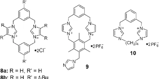
The most prevalent scaffold into which the guanidinium group has been included involves a bicyclic system, as seen in 1 (Fig. 2.1). The geometry of this structure is beneficial for the formation of discrete complexes, as the guanidinium group is constrained such that only one face contains protons available for hydrogen bonding, and thus is accessible for complexation. The synthesis of this bicyclic system was reported by McKay and Kresling in 1957 [3], and then augmented by Schmidtchen for the purpose of developing receptors [4].
Compound 2 (Fig. 2.1) consists of two bicyclic guanidiniums attached via a urethane linke. 1H-NMR binding studies with dicarboxylates and biologically relevant phosphates pointed to the utility of this compound as a ditopic binder of anionic guests [5].
The host was found to extract dicarboxylate guests from the aqueous phase into chloroform. Receptor 3, which containis naphthoyl substituents, was reported by de Mendoza and co-workers [6a]. Receptor 4 (Fig. 2.1) include a nucleotide derivative [6b].
A 1:1 complex with p-nitrobenzoate was determined for this structure using 1H-NMR titrations, as well as the extraction of the guest from aqueous to organic media. Interestingly, in this case, slight diastereomeric excesses were observed in the extraction of racemic salts, such as mandelate, naproxenate and tryptophan. This selectivity was ascribed to a three-point binding interaction between the host and guest
Fig. 2.2
Further development of the bicyclic guanidinium was performed through the attachment of a lipophilic substituent derived from Kemp’s triacid which is attached to the core through a 3,6-diaminocarbazole linker (5) (Fig. 2.2) [7]. This compound was found to complex dideoxyadenine dinucleotide.
guanidinium core involved the introduction of one naphthalene ring as well as a crown ether into the system [8]. This structure (6) (Fig. 2.2) was designed to bind amino acids guests through interactions between the ammonium group and the host crown, the guest carboxylate with the host guanidinium group and p-stacking interactions between the aromatic groups. Selectivity in the binding of amino acids with aromatic sidechains (phenylalanine and tryptophan) over those with aliphatic chains provided evidence of associations involving the aromatic moieties.
Receptor 7 (Fig. 2.2), which contains two bicyclic guanidiniums linked by a naphthalene spacer, is another example based on this system [9]. This compound was found to bind a variety of nucleotides in methanol and water.
The next advancement on this host scaffold was the introduction of a closo-borane substituent to a structure with two linked guanidinium groups (8) (Fig. 2.3) [10]. This alteration was made in an attempt to develop a bicyclic guanidinium host which had an overall neutral charge, but retained hydrophobic character.
In another example involving this scaffold with appended guanidinium groups, modified deoxycholicacid arms were appended to the core (9) (Fig. 2.3) to bind uronicacid guests [11].
Further development of the bicyclic scaffold led to its inclusion within a macrocyclic system [12].
Then, receptor 10 (Fig. 2.4) was designed and synthesized in which multiple functional groups capable of forming hydrogen bonds to anionic guest molecules are constrained within the macrocycle such that they point towards the interior.
Fig. 2.4
Finally, an example of the attachment of bicyclic guanidiniums to calix[4]arene was recently reported [13]. Here, receptor 11 (Fig. 2.4) and the corresponding monoguanidinium were found to enantioselectively extract amino acids into organic solvent. Results obtained indicated selectivity for both the binding and extraction of the L isomers, and up to 90% for phenylalanine.
Another series of compounds with preorganized guanidinium groups involves a variety of cyclic spacers linking two of these functional groups. In these cases, rigid spacers with defined geometries are generally implemented to preorganize the guanidinium moieties and thus provide the orientation for guest binding purposes. This was first exemplified in 1978 when Lehn and co-workers reported the incorporation of guanidinium groups into macrocycles 12, 13, and 14 (Fig. 2.5) [14].
Fig. 2.5
Later, Hamilton and co-workers [15] reported bisguanidiniumcontaining host 15 (Fig. 2.5) which they synthesized for the purpose of binding phosphodiester guests. The isophthaloyl spacer was used to situate the guanidinium groups in a suitable geometry to bind the trigonalbipyramidal guests.
Two tetraguanidines were developed, 16(a) and 16(b) (Fig. 2.6), for the purpose of binding DNA [16]. The multilayered structure of these compounds makes them suitable for binding into the minor grooves of double stranded DNA. Furthermore, sequence specificity for the 3’-GAA-5’ region was determined for the two structures. In addition, the incorporation of guanidinium group into RNA has recently been reported [17]. In these RNA analogs, the phosphodiester linkages in the backbone have been replaced by guanidinium groups to form polycationic strands. The goal of this work is to obtain receptors with increased affinity towards DNA and RNA by increasing electrostatic interactions upon complexation.
Another example of the appending of guanidinium groups to biological compounds is the development of guanidinoglycosides by Tor and co-workers [18].
Here, guanidinium groups were formed on saccharide scaffolds such as tobramycin, in this case generating 17 (Fig. 2.6). This was performed in an effort to increase affinity for RNA over the parent aminoglycosides through the introduction of guanidinium functionalities.
Fig. 2.6
Attachment of a guanidinium groups to xanthone led to the development of receptor 18 (Fig. 2.6) [19]. In this structure, extra interaction sites are built in: the xanthone NH hydrogen bond donor and the diisopropylbenzoate for stacking. Here, data in the analysis of certain carboxylates indicated a proton transfer from the host rather than a binding event. As a result, a series of acids with varying acidities were tested for binding affinity.
In another effort to increase the number of binding sites adjacent to a guanidinium groups, linkage to pyrrole was performed (19) (Fig. 2.6) [20]. This design was intended to utilize the pyrrole NH to facilitate the binding of N-acetyl amino acids.
Host compound 20 (Fig. 2.6) was developed which utilizes a 2,2’: 6,2” terpyridine-type ligand complexing zinc [21]. This receptor was designed to
complex.
2.2. Guanidinium groups incorporated into the triethylbenzene
scaffold.
The triethylbenzene substructure is another molecular design in which guanidinium functionalities have been exploited in the development of host molecules. The utility of this platform has been recently reviewed [22].
In 1997, Anslyn and co-workers [23] reported 21 (Fig. 2.7), based on the threefold substitution of the triethylbenzene scaffold with aminodihydroimidazolium groups. The target guest for this receptor was citrate as the geometrical orientation of the three guanidinium groups complemented the tris-carboxylate containing guest species.
Fig. 2.7
Another receptor based on the triethylbenzene scaffold is compound 22 (Fig. 2.7) [24]. This structure was designed to bind the polyanionic secondary messenger inositol triphosphate (IP3) as it contains six guanidinium groups in such a geometry as they are oriented towards the inside of the cavity.
The next receptor of this type reported (23) (Fig. 2.7) included two aminoimidazolium groups and a phenanthroline-copper complex [25]. In this case, the metal complex was introduced both as an extra binding site for the citrate guest and to allow for the signaling of binding.Introduction of the metal to the phenanthroline led to the quenching of the ligand fluorescent signal. The binding of citrate could then be observed due to the reemergence of this signal upon binding of the guest to the metal center.
A different approach to obtaining a receptor of this type involved the formation of a library of compounds based on a rationally designed core [26]. The goal of these studies was to develop a sensor for ATP. The designed core of this structure consisted of a resin-bound triethylbenzene scaffold with two guanidinium groups. A combinatorial library was then obtained by growing peptide arms off the guanidinium groups. Finally, fluorophores were added to the peptide arms and to a lysine linker for detection purposes. The resulting library was screened fluorometrically for ATP binding through inspection using UV light to obtain potential hosts. These were then sequenced and resynthesized. In the final analysis, receptor 24 (Fig. 2.8) with Ser-Tyr-Ser residues on the peptide arms showed selectivity for ATP over AMP and GTP.
Fig. 2.8
2.3. Receptors with covalently attached signalling groups
Another series of guanidinium containing receptors reported have featured the covalent attachment of this binding unit to known signalling devices. In 1996, de Silva et al. [27] reported a receptor of this type, 25 (Fig. 2.30), with azacrown, anthracene and guanidinium groups linked in series. This host was designed to complex g-aminobutyric acid (GABA) through interactions between the host guanidinium group and guest carboxylate as well as the host azacrown and the guest ammonium.
Fig 2.9
The anthracene moiety was included so that binding of the guest by the azacrown amine disrupted photoinduced electron transfer (PET) quenching of the anthracene fluorescence. As was expected, binding of the guest was detected by an increase in fluorescence. A control compound lacking the guanidinium group showed no fluorescence change in the presence of GABA, while glutamic acid, the physiological precursor to GABA also had no detectable effect.
Another example of this concept was published by Beer et al. [28], compound 26 (Fig. 2.9), which consists of a guanidinium group linked to a ferrocene unit. This host bound inorganic phosphate guest through hydrogen bonding to the guanidinium group. Upon complexation, the redox signal of the ferrocene was affected as oxidation of the group was facilitated by the introduction binding of the guest could be detected through redox analysis.
Next, compound 27 (Fig. 2.9) was established as a luminescent sensor for phosphate and phosphodiester guests [29]. Here, two neutral acylaminoimidazoline groups were linked to bipyridine, which then became one unit of a tris-bipyridine ruthenium(II) complex. Binding was detected by alteration of both the absorbance and fluorescent signals of the host, with a large fluorescence change potentially caused by rigidification of the complex.
Compound 28 (Fig. 2.9), which contains two iminoylthioureas, and the corresponding compound with 1,2,4-thiadiazoles have been reported [30]. Binding of carbonate, bicarbonate and hydrogen phosphate was achieved with these hosts. Fluorescence enhancements and spectral shifts occur.
References
[1] D.W. Christianson, W.N. Lipscomb, Acc. Chem. Res. 22 (1989) 62.
[2] F.A. Cotton, E.E. Hazen, Jr., M.J. Legg, Proc. Natl. Acad. Sci. 76 (1979) 2551. [3] A.F. McKay, M.E. Kreling, Can. J. Chem. 35 (1957) 1438.
[4] F.P. Schmidtchen, Chem. Ber. 113 (1980) 2175. [5] F.P. Schmidtchen, Tetrahedron Lett. 30 (1989) 4493.
[6] a) A. Echavarren, A. Galàn, J.-M. Lehn, J. de Mendoza, J. Am. Chem. Soc. 111 (1989) 4994. b) A. Galàn, E. Pueyo, A. Salmerʤόn, J. de Mendoza, Tetrahedron Lett. 15 (1991) 1827.
[7] A. Galàn, J. de Mendoza, C.M. Bruix, G. Deslongchamps, J. Rebek, Jr., J. Am. Chem.
Soc. 113 (1991) 9421.
[8] A. Galàn, D. Andreu, A.M. Echavarren, P. Padros, J. De Mendoza, J. Am. Chem. Soc. 114 (1992) 1511
[9] P. Schliessl, F.P. Schmidtchen, J. Org. Chem. 59 (1994) 509. [10] M. Berger, F.P. Schmidtchen, J. Am. Chem. Soc. 118 (1996) 8947.
[11] M. Segura, V. Alcázar, P. Padros, J. de Mendoza, Tetrahedron 53 (1997) 13119. [12] V. Alcázar, M. Segura, P. Prados, J. de Mendoza, Tetrahedron Lett. 39 (1998) 1033 [13] L. Fang, G.-Y. Lu, W.-J. He, Z.-S. Wang, L.-G. Zhu, Chin. J. Chem. Soc. 19 (2001) 317. [14] B. Dietrich, T.M. Flyes, J.-M. Lehn, L.G. Pease, D.L. Fyles, J. Chem. Soc. Chem.
Commun. (1978) 934.
[15] R.P. Dixon, S.J. Geib, A.D. Hamilton, J. Am. Chem. Soc. 114 (1992) 365.
[16] R. Fukutomi, A. Tanatani, H. Kakuta, N. Tomioka, A. Itai, Y. Hashimoto, K. Shudo, H. Kagechika, Tetrahedron Lett. 39 (1998) 6475.
[17] (a) D.A. Barawkar, B. Linkletter, T.C. Bruice, Bioorg. Med. Chem. Lett. 8 (1998) 1517; (b) N. Kojima, I.E. Szabo, T.C. Bruice, Tetrahedron 58 (2002) 867.
[18] N.W. Luedtke, T.J. Baker, M. Goodman, Y. Tor, J. Am. Chem. Soc. 122 (2000) 12035. [19] M. Martín, M. Almaraz, J. Hernández, A. Tejeda, M.C. Caballero, J.R. Morán,
[21] H. Aït-Haddou, S.L. Wiskur, V.M. Lynch, E.V. Anslyn, J. Am. Chem. Soc. 123 (2001) 11296.
[22] G. Hennrich, E.V. Anslyn, Chem.-Eur. J. 8 (2002) 2119.
[23] A. Metzger, V.M. Lynch, E.V. Anslyn, Angew. Chem. Int. Ed. Engl. 36 (1997) 862. [24] K. Niikura, A. Metzger, E.V. Anslyn, J. Am. Chem. Soc. 120 (1998) 8533.
[25] L.A. Cabell, M.D. Best, J.J. Lavigne, S.E. Schneider, D.M. Perreault, M.-K. Monahan, E.V. Anslyn, J. Chem. Soc. Perkin Trans. 2 (2001) 315.
[26] S.E. Schneider, S.N. O’Neil, E.V. Anslyn, J. Am. Chem. Soc. 122 (2000) 542.
[27] A.P. de Silva, H.Q.N. Gunaratne, C. McVeigh, G.E.M. Maguire, P.R.S. Maxwell, E. O’Hanlon, Chem. Commun. (1996) 2191.
[28] P.D. Beer, M.G.B. Drew, D.K. Smith, J. Organomet. Chem. 543 (1997) 259.
[29] S. Watanabe, O. Onogawa, Y. Komatsu, K. Yoshida, J. Am. Chem. Soc. 120 (1998) 229. [30] G. Hennrich, H. Sonnenschein, U. Resch-Genger, Tetrahedron Lett. 42 (2001) 2805.
3. Imidazolium receptors for the recognition of anions
Various positively charged imidazolium derivatives have been synthesized and studied as selective anion-receptors. Imidazolium group can make a strong interaction with anions through (C–H)+…X- type ionic hydrogen bond because the charge–charge electrostatic interaction dominates.[1]This type of interaction could also be envisaged in the crystal structure of imidazolium based ionic liquids [2a, 2b]and calix-[4]-pyrrole complexing with imidazolium salts.[2c]
In the following, imidazolium based receptors will be grouped according to their topological and structural classification, which includes:
1) benzene tripodal;
2) cyclophane and calix-[n]-imidazolium; 3) cavitand and calixarene;
4) fluorescent imidazolium; 5) ferrocenyl imidazolium.
3.1. Benzene tripodal systems
A benzene based tripodal receptor with three imidazolium groups 1 was reported by Sato et al.[3] . The host 1 displayed a selective binding with Cl- over Br- and I- in acetonitrile, which was confirmed via 1H NMR titration experiments.[3] Kim and coworkers clarified that this tricationic heterocycle interacts strongly with anions through (C–H)+…X- type ionic hydrogen bond between the hydrogen on the electron-deficient C(2) carbon atom of the imidazolium ring and the guest anion, and shows a selective binding for F- over other halide anions. For better anion binding affinity, the benzene based tripodal receptor was theoretically modified and experimentally realized [1] as tripodal nitro-imidazolium receptor 2.
Fig. 3.1 - Benzene tripodal systems
The homochiral tripodal imidazolium system 3 was reported by Howarth et al. [4]. This chiral tripodal system could distinguish between sodium (R)-2-aminopropionate and sodium (S)-2-(R)-2-aminopropionate From the 1H NMR experiments, it has been shown that the imidazolium salt forms a complex with the (R) enantiomer but not with the (S) enantiomer.
3.2. cyclophane and calix-[n]-imidazolium
It has been. recently reported similar benzene tripodal systems (4, 5 and 6) (Fig. 3.1) and bis imidazolium (7) [5] (Fig. 3.2)
Cyclophane and calix-imidazolium systems Alcalde et al. reported cyclophane type receptors as shown in Fig. 3.3 (8a and 8b).[6]
Fig. 3.3 - Structures of cyclophane-type imidazolium receptors 8, 9 and 10.
Interestingly, in the X-ray crystal structure of 8a.2Cl-.2H2O, the dication 8a
adopts a chair-like conformation.
On the other hand, the molecular shape of dication 8b in 8b.2Cl- 3.5H2O
assumes a cone-like conformation, but both cations 8a and 8b have similar molecular cavity dimensions: a square of 5 Å side. The deshielding effects of C(2)–H (in imidazolium rings) were reported to be in the order of H2PO4- > F->
CH3COO- > CN- > Cl-.
A tetracationic heterophane (11) (Fig. 3.4) has been reported, which forms a 1:1 inclusion complex with halides and oxoanions in DMSO-d6.[7]
From the 1H NMR titration experiments, the binding affinity of this receptor was reported to be in the order HSO4- > Br- > H2PO4- > Cl- > I- > ClO4-, which
was explained based on the size of anions.
A novel calix[4]imidazolium[2]pyridine (12) has been reported (Fig. 3.5).[8]
Fig. 3.5 - Crystal structure of calix[4]imidazolium[2]pyridine 12 complexed with
anions F
-Unlike the fluoride anion, the chloride and bromide anions of larger size do not fit in the center of the cavity of 12, and furthermore these anions favor nonspherical or surface conformations in order to keep the extra electron in a large empty space.[9]
3.3. Cavitand derivative
Recently, reported a new cavitand derivative bearing four imidazolium groups (12) as a receptor for anions (Fig. 3.6) has been reported. [10]
Fig 3.6 - Imidazolium receptors based on cavitand (12).
The binding properties towards various anions including dicarboxylates were investigated based on the 1H NMR spectroscopic experiments in acetonitrile-d3. Among the anions examined, cavitand 12 displayed a selective binding with bis(tetrabutylammonium) 1,4-phenylenediacetate.
3.4. Fluorescent imidazolium systems
A few anthracene derivatives bearing imidazolium groups as binding sites have been recently studied.[11]
Two imidazolium moieties were first immobilized on the 1,8-positions of the chemosensor (13) (Fig. 3.7), and a unique feature of the binding mode was predicted based on ab initio calculations.19a 1,8-Bis-imidazolium anthracene 13 effectively and selectively recognizes the biologically important H2PO4- ion
over other anions such as I-, Br- and Cl- in acetonitrile. It has been further demonstrated that the selectivity of these imidazolium receptors against anions can be controlled by the topology of the binding site (e.g. enhancement of rigidity). Compared to host 13, the greater rigidity in host 14 enhances the binding selectivity for H2PO4- over F
-.[19b]
Fig. 3.7 - Anthracene derivatives bearing imidazolium groups
Furthermore it has been reported a novel water-soluble imidazolium anthracene derivative (15), which not only differentiates the structurally similar compounds GTP and ATP but also acts as a potential fluorescent chemosensor for GTP in 100% aqueous solution (pH = 7.4).[19c]
A fluorescent tripodal receptor (16) bearing three benzoimidazolium and napthyl groups has been recently synthesized.[12]
This host acts as an ‘‘off–on’’ signaling chemical sensor with high selectivity for Cl- over Br- and I- through a guest-induced conformational switching process (Scheme 3.1).
-In the presence of Cl-, the tripodal system adopts a cone conformation, which leads to the excimer fluorescence by bringing the naphthalene groups into close proximity.
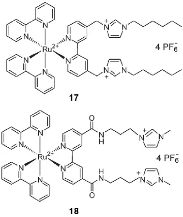
It has been also reported a new imidazolium functionalized acyclic ruthenium(II) bipyridyl receptor (17,18) (Fig. 3.8).[13]
Fig. 3.8 - Structures of fluorescent hosts 17 and 18.
Both receptors displayed a selectivity for Cl- over the other anions in acetonitrile–water (9: 1, v/v) and 22 sensed ATP in 50:50 acetonitrile–water solvent media.
It has been also reported a naphthalene derivative (23) which contains two methylene bridged bis-imidazolium rings (Fig. 3.9).[14a]
The host 19 displayed a selective affinity for I-, which was confirmed using fluorescence spectroscopy and 1HNMR. They also reported the role of aromatic (C–H)…anion interaction on 20 in addition to the imidazolium (C–H)+…anion ionic hydrogen bonding, whose strength was found to be increased with the nitro substitution on the para position.[14b]
Fig. 3.9 - Structures of hosts 19 and 20.
A fluorescent cavitand derivative bearing four imidazolium groups as well as four pyrene groups as a fluorescent receptor for GTP has been synthesized (Fig. 3.10).[15]
Since host 20 contains pyrene groups, the binding properties towards various anions were investigated based on the fluorescence experiments.
3.5. ferrocenyl imidazolium systems
Howarth et al. reported an imidazolium receptor which contains two ferrocene groups (21) (Fig. 3.11).[16]
Fig. 3.11 - Structures of ferrocenyl imidazolium receptors 21
This compound adds to a substantial number of artificial organic hosts for anions which incorporate strong hydrogen bonds (H-bonds), usually N-H···X-,[17] e.g., calixpyrrole, or C-H···X- H bonding , if the carbon atom is adjacent to a cationic center, e.g., imidazolium.[18] ring and guest anions such as Cl-, Br- and I-.
This novel type of charged hydrogen bonding is very different and intriguing in comparison with many other conventional hydrogen bonds. The role of (C2-H)+… X- hydrogen bond during the recognition process is still open to debate [19].
References
[1] H. Ihm, S. Yun, H. G. Kim, J. K. Kim and K. S. Kim, Org. Lett., 2002, 4, 2897.
[2] (a) J. D. Holbrey, W. M. Reichert, M. Nieuwenhuyzen, S. Johnston, K. R. Seddonb and R. D. Rogers, Chem. Commun., 2003, 1636; (b) J. D. Holbrey, W. M. Reichert and R. D. Rogers, Dalton Trans., 2004, 2267; (c) R. Custelcean, L. H. Delmau, B. A. Moyer, J. L. Sessler, W.-S. Cho, D. Gross, G. W. Bates, S. J. Brooks, M. E. Light and P. A. Gale,
Angew. Chem., Int. Ed., 2005, 44, 2537.
[3] K. Sato, S. Arai and T. Yamagishi, Tetrahedron Lett., 1999, 40, 5219. [4] J. Howarth and N. A. Al-Hashimy, Tetrahedron Lett., 2001, 41, 5777.
[5] Y. Bai, B.-G. Zhang, J. Xu, C.-Y. Duan, D.-B. Dang, D.-J. Liu and Q.-J. Meng, New J.
[7] K. Sato, T. Onitake, S. Arai and T. Yamagishi, Heterocycles, 2003, 60, 779.
[8] K. Chellappan, N. J. Singh, I.-C. Hwang, J. W. Lee and K. S. Kim, Angew. Chem., Int. Ed., 2005, 44, 2899.
[9] (a) N. J. Singh, A. C. Olleta, A. Kumar, M. Park, H.-B. Yi, I. Bandyopadhyay, H. M. Lee, P. Tarakeshwar and K. S. Kim, Theor. Chem. Acc., 2005, DOI: 10.1007/s00214-005-0057-1; (b) H. M. Lee, D. Kim and K. S. Kim, J. Chem. Phys., 2002, 116, 5509; (c) H.M. Lee and K. S. Kim, J. Chem. Phys., 2001, 114, 4461.
[10] S. K. Kim, B.-G. Kang, H. S. Koh, Y.-J. Yoon, S. J. Jung, B. Jeong, K.-D. Lee and J. Yoon, Org. Lett., 2004, 6, 4655.
[11] (a) S. K. Kim, N. J. Singh, S. J. Kim, H. G. Kim, J. K. Kim, J. W. Lee, K. S. Kim and J. Yoon, Org. Lett., 2003, 5, 2083; (b) J. Yoon, S. K. Kim, N. J. Singh, J. W. Lee, Y. J. Yang, K. Chellappan and K. S. Kim, J. Org. Chem., 2004, 69, 581; (c) J. Y. Kwon, N. J. Singh, H. Kim, S. K. Kim, K. S. Kim and J. Yoon, J. Am. Chem. Soc., 2004, 126, 8892. [12] Y. Bai, B.-G. Zhang, J. Xu, C.-Y. Duan, D.-B. Dang, D.-J. Liu and Q.-J. Meng, New J.
Chem., 2005, 29, 777.
[13] M. S. Vickers, K. S. Martindale and P. D. Beer, J. Mater. Chem., 2005, 15, 2784.
[14] (a) H. Kim and J. Kang, Tetrahedron Lett., 2005, 46, 5443; (b) S. In, S. J. Cho, K. H. Lee and J. Kang, Org. Lett., 2005, 7, 3993.
[15] S. K. Kim, B.-S. Moon, J. H. Park, Y. I. Seo, H. S. Koh, Y. J. Yoon, K.-D. Lee and J. Yoon, Tetrahedron Lett., 2005, 46, 6617.
[16] (a) J.-L. Thomas, J. Howarth, K. Hanlon and D. McGuirk, Tetrahedron Lett., 2000, 41, 413; (b) J.-L. Thomas, J. Howarth and A. M. Kennedy, Molecules, 2002, 7, 861.
[17] K. A. Nielsen, W.-S. Cho, J. Lyskawa, E. Levillain, V. M. Lynch, J. L. Sessler, J. O. Jeppesen, J. Am. Chem. Soc. 2006, 128, 2444 – 2451;
[18] a) K. Chellappan, N. J. Singh, I.-C. Hwang, J. W. Lee, K. S. Kim, Angew. Chem. 2005, 117, 2959 – 2963; b) K. Chellappan, N. J. Singh, I.-C. Hwang, J. W. Lee, K. S. Kim,
Angew. Chem. Int. Ed. 2005, 44, 2899 – 2903; c) N. Alhashimy, D. J. Brougham, J.
Howarth, A. Farrell, B. Quilty, K. Nolan, Tetrahedron Lett. 2007, 48, 125– 128.
[19] Zhaochao Xu, Sook Kyung Kim and Juyoung Yoon, Chem. Soc. Rev., 2010, 39, 1457– 1466
4. Amide based anion receptors
The anion coordination achieved by amine based anion receptors are here highlighted by halide-H-N amide interections . Halides are the simplest anions in terms of shape and are thus the closest analogue of metal ions. In general, they prefer a four-coordinate, tetrahedral geometry, which is understandable in terms of the four lone pairs in an sp3-hybridized electronic shell. Indeed, the most common coordination numbers are two, three, and four. However, other coordination numbers up to nine can be cited.
The coordination numbers for most examples of hydrogen-bond coordination of anions begin at two. The bromide complex 1·Br- (Fig. 4.1) was characterized by X-ray crystallography, which showed that the bromide is hydrogen bonded in a two-coordinate V-shaped geometry with a syn,syn coordination mode and the two amide hydrogen atoms at distances of 2.39 and 2.68 Ǻ from the bromide.[1] The amide groups are out of the phenyl plane, possibly as a result of the large size of the bromide ion.
Fig. 4.1
The pyridine nitrogen atom in macrocycle 2 (fig. 4.1) serves to preorganize the two adjacent amide groups for chelation, as illustrated by the two-coordinate V-shaped iodide complex [2].
Smith and co-workers extensively studied dual-host ditopic receptors (for example, receptor 3 in fig. 4.2) for both anions and cations, including halides and alkali-metal ions.[3–6]
The cavity situation in these receptors often leads to a coordination number of two for halides, and the geometry is usually V-shaped. The additional electrostatic binding component provided by an adjacent cation also assists in stabilizing the complex.
Fig. 4.2
4.1. Coordination Number 3
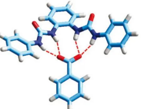
Coordination number three is often observed with pincer type chelating ligands that contain three available hydrogen bond-donor groups. Diamido-pyrroles are excellent pincer ligands for three-point hydrogen bonding.[7] For example, in the diamidopyrrole receptor 4 (Fig. 4.3) the chloride ion is bound to the ligand by three hydrogen bonds to the amide and pyrrole hydrogen atoms (4·Cl-).[8]
Fig. 4.3
Because of the geometrical placement of the hydrogen bonds imposed by the rigid ligand framework, the geometry of the anion complex probably approximates that of the splayed tines of a fork.
The related ditopic ligand 5 (Fig. 4.3) a dichloro analogue of 4 that was synthesized to enhance the hydrogen-bonding capabilities, readily holds two chloride ions in the two separate binding sites. In the solid state, each Cl- ion is bound by three NH groups: two amide groups (N···Cl = 3.27 and 3.28 Å) and a pyrrole donor (N···Cl = 3.07 Å).[9]
A related potentially tridentate ligand, the mixed imidazolium/pyrrole 6 (Fig. 4.4), was synthesized as a model for the prodigiosins.[10] Prodigiosins are natural products that contain three pyrrole units and can transport HCl across lipid membranes.[11]
Fig. 4.4
Another three-coordinate halide is found in the chloride complex of the trigonally symmetric amide-based cyclophane 7 (Fig. 4.4). [12] Two chloride ions are held by hydrogen bonding with amide hydrogen atoms of two of the three pyridine spacers. The water molecule is held by the amide groups of the third spacer, and bridges the two chloride ions.
4.2. Coordination Number 4
Probably the most common coordination number for halides is four. The chloride complex of the acyclic pyrrole based ligand 8 (Fig. 4.5) shows a rather unanticipated four-coordinate geometry, with hydrogen bonds from the two
rings.
Fig. 4.5
The acyclic thiophene-based amide ligand 9 (fig. 4.5) forms a dimeric complex with flouride (9·F-)2,[13]. Two hydrogen bonds in (9·F
-)2 form from the amide
hydrogen atoms and two more from the two CH groups of the thiophene.
Anion complexes of a number of tetraamide macrocyclic hosts were found to have four-coordinate geometries. For example, the 20-membered ring 10 (Fig. 4.6) binds one chloride ion. For most efficient binding, the ligand bends and the chloride ion sits on top of the ligand above the mean plane of the four amide nitrogen atoms,[14] hydrogen-bonded to the four host amide NH groups. The coordination mode is pyramidal and similar to what is sometimes found for transition-metal complexes with macrocyclic ligands that are too small to incorporate the metal ion.
The somewhat larger macrocycle 11 (Fig. 4.6) also binds chloride, together with a water molecule slightly above the macrocyclic cavity.[14]. Two of the coordination sites for the chloride ion are provided by the amide NH groups, a third from the neighboring water molecule, and a fourth from a water molecule from an adjacent macrocycle, which gives what could be described as a distorted tetrahedral geometry.
An expanded version of Jurczak’s tetraamide-based macrocycles is the 36-membered-ring lactam 12 (Fig. 4.7) that results from a [4+4]condensation of the methyl ester of pyridine-2,6-dicarboxylate and ethylenediamine.[15]
The macrocyclic framework has a steplike geometry, in which two opposing 2,6-pyridinedicarbamoyl groups are essentially coplanar and one of the remaining units is above and one is below that plane. Crystallographic results for the large macrocycle revealed four guests: two chloride ions
bridged by two water
molecules, all within the macrocyclic cavity (12·2 Cl-·2H2O).
The pyridine-based bicyclic receptor 13 (Fig. 4.8) and its methylated quaternary-amine form 14 (Fig. 4.9) were reported by O. Kang et al.[16] In the chloride complex H2132+·2 Cl
-·H2O, the bridgehead amines are protonated and
two of the amide loops fold in one direction while the other loop points in the opposite direction. Two chloride ions are held within the cryptand and are bridged by a water molecule.
The quaternary-amine cryptand 14 also binds chloride (14·2Cl -·2H2O).[16] In this case, all three amide loops of the ligand fold in the
same direction, thus forming a bowl-like shape. Rather than being encapsulated, one chloride ion sits at the top of the bowl and is centered between the two charged amines. A second chloride ion is located off to one side of the macrocycle and is linked by water molecules to the first chloride ion. The chloride ion directly above the host is bound to two of the amide hydrogen atoms of 14, with two additional linkages to the waters “floating” on top of the host to result in a pseudotetrahedral geometry.
Fig. 4.8 - Chloride complex H213 2+
·2 Cl-·H2O in different views
Fig. 4.9 - Chloride complex 14·2Cl-·2H2O in different views
4.3. Coordination Number 5
The common geometries of five-coordinate transition metal complexes are square pyramidal and trigonal bipyramidal. Five-coordinate halide complexes are frequently found for simple tetraamide hosts, both acyclic and macrocyclic. The acyclic host in 15·Cl-·H2O (Fig. 4.10) is deformed slightly
from planarity, and the two planar 2,6-pyridinedicarbamoyl groups are canted with respect to each other.[14]
The chloride ion acts as a template and forms four hydrogen bonds to the amide hydrogen atoms. There is an additional hydrogen bond to a water molecule on one side of the macrocycle. The resulting coordination geometry is pseudo square pyramidal.
The 18-membered-ring macrocycle 16 (Fig. 4.10) forms both fluoride and chloride complexes (16·X-·H2O). Although the 18-membered ring might be
considered an ideal size for the relatively small ion, the four amide groups undergo a slight tetrahedral distortion to accommodate the fluoride ion. With the fifth hydrogen bond to a water molecule, the resulting geometry is pseudo square pyramidal.[17]
Fig. 4.10
The hybrid tetraamide receptor 17 (Fig. 4.10) contains both 2,6-diamidopyridine and 1,3-diamidobenzene groups and also binds one chloride ion. The solid-state structure of the chloride complex (17·Cl-) shows the chloride ion held through five hydrogen bonds inside the cavity of the macrocycle.[18] The NH groups form four hydrogen bonds and the aromatic CH group forms the fifth bond with the chloride ion.
4.4. Coordination Number 6
The cyclic hexapeptide 18 (Fig. 4.11), which contains three amide groups, was found to bind halides even in aqueous solution.[19]
The crystal structure of the iodide complex revealed a 2:1 complex 192·I
in which the iodide ion is sandwiched between the two cyclopeptides. There are two crystallographically independent iodide complexes in the unit cell, both of which have virtually identical structures, indicating that a sandwich complex is the preferred coordination mode in this environment. The two cyclopeptides in both complexes are almost perfectly aligned with each other, so that there is only minor deviation from trigonal-prismatic geometry. The iodide ion is associated with the six amide hydrogen atoms, which point into the cavity.
Fig. 4.11
It has been recently reported an example of solvent fixation by the potentially ditopic receptor 19 (Fig. 4.12). The crystal structure of the complex isolated from CH2Cl2 revealed that the CH2Cl2 had undergone nucleophilic
attack by the amine lone pair with the released chloride ion being bound within the cavity to yield [19(CH2Cl)]+·Cl
-.[20] The Cl- ion is hydrogen bonded to two amide hydrogen atoms; the resulting arrangement of coordination sites is hexagonal planar.
Fig. 4.12
A bicyclic receptor 20a (Fig. 4.13) has been reported which was found to be selective for fluoride. All six NH groups are within hydrogen-bonding distances (N···F =2.84-2.89 Å) of the encapsulated fluoride ion.[21]
Fig. 4.13
The coordination geometry of the fluoride ion is approximately midway between trigonal prismatic and octahedral; the trigonal twist angle is 37°. (Fig. 4.14)
Fig. 4.14 - The fluoride complex 20a.F- and his crystal structure.
4.5. Coordination Number 9
What may be the highest coordination number found for the halides with amide-based receptors is observed with the m-xylyl analogue 20b (Fig 4.15) of the pyridine-containing cryptand 20a.[22]
Fig 4.15 - 20b.F
-The crystal structure of the fluoride complex indicates nine coordination: six bonds to the amide hydrogen atoms and an additional three interactions with the
m-xylyl CH groups (Fig. 4.16) . The 19F NMR spectra support the existence of all nine hydrogen bonds.
Fig. 4.16 - Perspective views of 20b.F-. (A) Side view. (B) End view down the
pseudo-three-fold axis.
In the following, geometries other than the spherical one will be reviewed as far as the anions coordinated by amide receptors.
4.6. Linear Anions
Examples of linear anions include azide, thiocyanate, cyanide, and bifluoride. However only one example of a crystallographically characterized linear anion complex with an amide host has been reported to date.[23]
The first inclusion complex of a linear anion was observed by Lehn for azide in an octaazacryptand.[24]
In the crystal structure of azide complex of H6216+.N3
(Fig. 4.17), the seminal example of a linear anion complex, all six of the secondary amine groups are protonated and are hydrogen bonded to the linear N3- ion in a C3
-trigonal-prismatic symmetry pattern.
The azide terminal N···N distance is very short (2.32 Ǻ) and the hydrogen-bond distances from the azide terminus to the secondary amines are 2.94 Ǻ.[24]
Although it was envisioned that bifluoride should also bind in these “linear” azacryptand receptors, a selective receptor for bifluoride was only recentlyreported, along with a crystal structure.[23]
Fig. 4.17
The structurally characterized FHF- complex of the tricycle 22 (Fig. 4.17) contains a linear FHF- ion bridging the two macrocyclic caps of the cylindrical tricycle. Although 22.HF2
contains eight amide groups as potential hydrogen-bonding sites, only four are utilized, which results in a tetrahedral four coordinate geometry based on the topology of the four bound NH groups around the anion. The other four amide hydrogen atoms are involved in three-atom hydrogen-bonding networks between the amide hydrogen atoms, and the pyridine and tertiary amine nitrogen lone pairs. The F···F distance in 22.HF2
is 2.47 Ǻ. Binding studies revealed selective binding for FHF- over other anions.
4.7. V-Shaped Anions
In addition to truly V-shaped anions such as nitrite, simple carboxylates such as acetate and benzoate will also be considered in this section.
Dual-host receptors such as 3, consisting of crown ethers covalently attached to amide containing straps, bind halides along with their associated cation. This mode of binding also works with other anion shapes, for example, nitrite. 3·Na+·NO2
(Fig. 4.18) is a relatively rare example of a crystal structure of a nitrite complex, where the nitrite ion may appropriately be considered singly coordinated [25]
The acetate complex of 3 (3Na+.AcO-) is an example of a two coordinate receptor in which the anion is also chelated by its counterion (K+), and the acetate ion lies somewhat outside the cavity. Only one of the oxygen atoms is hydrogen bonded to the amide portion of the receptor, but in this case to both
amides (N···O distances of 2.93 and 3.01 Ǻ),[25] which makes this a two-coordinate V-shaped geometry.
Fig. 4.18
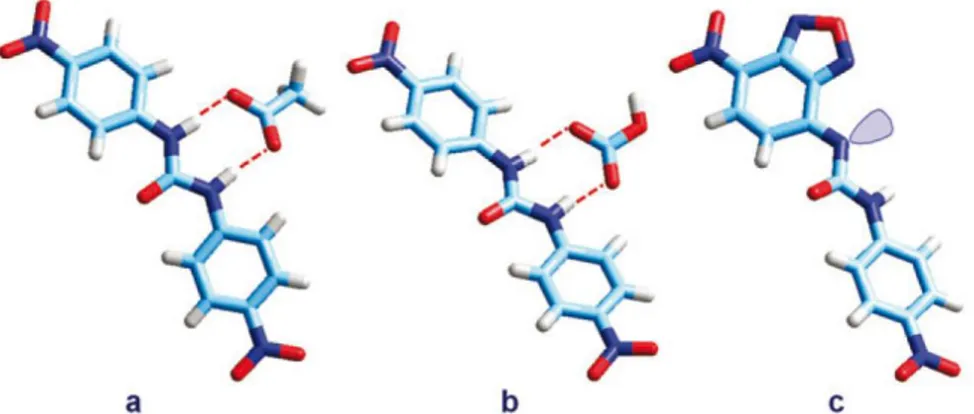
Cheng and co-workers were the first to report 23 (Fig. 4.19) as an anion receptor.[26]
Shortly thereafter, Gale and co-workers reported the crystal structures of the acetate complex of 23 [27] and the benzoate complex of 24.[28]
Both complexes exhibit three hydrogen-bonding interactions between the anions and the amide and pyrrole hydrogen atoms.
![Fig. 5.8 - Receptor 11 and the crystal structure of the complex between the protonated form (at the pyridine nitrogen atom) of receptor 11 and the chloride anion.[13]](https://thumb-eu.123doks.com/thumbv2/123dokorg/4583675.38816/71.774.99.673.224.415/receptor-crystal-structure-protonated-pyridine-nitrogen-receptor-chloride.webp)
![Fig. 5. 21 - Molecular structures of: (a) L 1 H; (b) [L 1 H···Cl] − ;(c) [L 2 H···CH 3 COO] −](https://thumb-eu.123doks.com/thumbv2/123dokorg/4583675.38816/80.774.166.616.363.658/fig-molecular-structures-l-h-cl-ch-coo.webp)