U
U
U
N
N
N
I
I
I
V
V
V
E
E
E
R
R
R
S
S
S
I
I
I
T
T
T
A
A
A
’
’
’
D
D
D
E
E
E
G
G
G
L
L
L
I
I
I
S
S
S
T
T
T
U
U
U
D
D
D
I
I
I
D
D
D
I
I
I
C
C
C
A
A
A
T
T
T
A
A
A
N
N
N
I
I
I
A
A
A
DOTTORATO INTERNAZIONALE DI RICERCA IN
NEUROBIOLOGIA
Sede amministrativa: Università di Catania
Sedi consorziate: Università di Roma “La Sapienza” e di Pavia XXV CICLO
TESI DI DOTTORATO
Dott. Gabriele Bonaventura“Tecniche analitiche di frontiera e nuove modalità di indagine genetica embrionale nello studio delle alterazioni macromolecolari responsabili delle patologie neurodegenerative” “New analytical scenarios and new approaches in the embryonic genetic investigation of the
macromolecular alterations responsible for the neurodegenerative diseases”
Coordinatore Chiar.mo Prof. Roberto Avola
Tutor Cotutor
Index
Pag. Cap. 1° MAMMALIAN STEM CELLS: EMBRYONIC AND ADULT
Pag. 13 1.1 TOTIPOTENCY, PLURIPOTENCY and MULTIPOTENCY Pag. 14 1.2 A LOOK INSIDE EMBRYONIC STEM CELLS.
Pag. 19 Cap. 2° PLURIPOTENCY CONTROLLING PATHWAYS: ROLE OF
TRANSCRIPTION FACTORS.
Pag. 24 2.1 STEM CELLS NEURAL DIFFERENTIATION
Pag. 29 CAP. 3° DIFFERENT TISSUE-DERIVED STEM CELLS: A
COMPARISON OF NEURAL TRANS-DIFFERENTIATION CAPABILITY
Pag. 33 Cap. 4° FLUORESCENCE MICROSCOPY
Pag. 33 4.1 BASIC PRINCIPLES Pag. 35 4.2 F.C.S.
Pag. 44 4.3 N&B
Cap. 5° MATERIALS & METHODS
4.1 CELL CULTURE
4.2 IMMUNOISTOCHEMISTRY EXPERIMENTS 4.3 N&B EXPERIMENTS
Cap. 6° RESULTS & DISCUSSION
Pag. 57 6.1 ADULT MESENCHIMAL STEM CELL A COMPARISON OF NEURAL TRANS-DIFFERENTIATION CAPABILITY
Pag.70 6.2 STEM CELS FROM HUMAN EMBRYO CAN IMPROVE DIFFERENTIATION TO NEURAL CELLS.
Research objective :
Since we know that a specific, sequential gene expression is determinant
in controlling long term self renewal and differentiation networks of stem
cells, the understanding of the molecular mechanisms underlined these
processes is crucial.
The principal aim of our project was to induce the differentiation process
of embryo-derived stem cells into neural cells (neurons, glial cells), to
follow during the differentiation process the changing in the expression
of characteristic “stemness” markers (OCT4, SOX2 and NANOG)
responsible for the regulatory networks involved in embryo-derived stem
cells pluripotency, whose understanding is fundamental for any potential
therapeutic application.
One of the major goals of current biological research are not only the
identification, but also the precise physico-chemical characterization of
elementary processes at level of individual proteins and nucleic acids.
These molecules are believed to be the smallest functional units in
biological systems. To address these minute quantities, very sensitive
techniques are required. Among those that allow even single molecule
measurements are atomic force microscopy (AFM) or fluorescence
spectroscopy. One outstanding feature of the latter is its noninvasiveness,
which makes it perfectly suited for measurements inside living cells.
For this reason the use of advanced spectroscopic techniques, such as
time-resolved fluorescence correlation spectroscopy (FCS) (16-21), could
allow to follow protein changes and to analyze different aspects such as
the molecular dynamics and intracellular translocation of some selected
transcription factors, tightly bound to the activation of the ESCs
differentiation processes into neural cells.
Chapter 1
Mammalian Stem Cells: Embryonic and Adult
Man has long been fascinated by the regenerative abilities of certain animals.
Regeneration is a remarkable physiological process in which remaining tissues organize to reform a missing body part. Several invertebrates, such as planarian flatworms and Hydra, regenerate tissues with speed and precision, whereas the majority of higher vertebrates are incapable of any form of whole-organ regeneration, even though they had all the necessary instructions and machinery to generate the tissue during embryonic development (1-3). Of the higher vertebrates, mammals appear to have lost the most regenerative ability, a trade-off perhaps for more proficient wound healing ability.
The most striking example of whole-organ regeneration in mammals is that of antler regeneration in elks, and in humans, liver regeneration after partial hepatectomy (4-5).
Most tissue repair events in mammals are dedifferentiation-independent events resulting from the activation of pre-existing stem cells or progenitor cells. By contrast, some vertebrates, like the salamanders, regenerate lost body parts through the dedifferentiation of specialized cells into new precursor cells. These de-differentiated cells then proliferate and later form new specialized cells of the regenerated organ (6-8).
Stem cells or progenitor cells are the common denominator for nearly all types of regeneration. They are either already pre-existing, as is the case for mammals, or created by the process of de-differentiation. Stem cells can also be found in plants in
the root and shoot meristems. Etymological origins of the term ‘stem cell’ can be traced back to early botanical monographs documenting the regenerative competence ofplant meristems.
Every day we read and listen to news reports about how stem cells promise to revolutionize medicine and change our lives with panaceas for every imaginable disease, including rhetoric that stem cell therapy will some day delay the process of ageing.
Embroiled in the hype and media frenzy are also political agendas and numerous religious and genuine ethical concerns. To further fuel the debate, embryonic stem cell research is often unjustly associated with reproductive cloning.
The hope that someday many debilitating human diseases will be treated with stem cell therapy is inspired by these remarkable examples of whole-organ and limb regeneration in animals, as well as the historical success of bone marrow transplants, which have improved the lives of many patients suffering from leukaemia and immunological and other blood disorders (9-10). Clearly, stem cell research leading to prospective therapies in reparative medicine has the potential to affect the lives of millions of people around the world for the better and there is good reason to be optimistic. However, the road towards the development of an effective cell-based therapy for widespread use is long and involves overcoming numerous technical, legislative, ethical and safety issues.
Three basic categories of cells make-up the human body: germ cells, somatic cells and stem cells. Somatic cells include the bulk of the cells that make-up the human adult and each of these cells in its differentiated state has its own copy, or copies, of the genome; the only exception being cells without nuclei, i.e. red blood cells. Germ cells are cellsthat give rise to gametes, i.e. eggs and sperm. The canonical definition of a stem cell is a cell with the ability to divide indefinitely in culture and with the potential to give rise to mature specialized cell types.When a stem cell divides, the daughter cells can eitherenter a path leading to the formation of a differentiated specialized cell or self-renew to remain a stem cell, thereby ensuring that a pool of
stem cells is constantly replenished in the adult organ (11-12). This mode of cell division characteristic of stem cells is asymmetric and is a necessary physiological mechanism for the maintenance of the cellular composition of tissues and organs in the body.
Other attributes of stem cells include the ability to differentiate into cell types beyond the tissues in which they normally reside. This is often referred to as stem cell plasticity (13-15). Stem cells are also believed to be slow cycling but highly clonogenic and generally represent a small percentage of the total cellular make-up of a particular organ.
Although there is still much to discover about the molecular mechanisms that govern stem cell-fate decisions and self-renewal, transcriptome profiling studies have highlighted several properties believed to be common to all stem cells at the molecular level. These essential attributes of ‘stemness’ are proposed to include: active Janus kinase signal transducers and activators of transcription, TGFb and Notch signalling; the capacity to sense growth factors and interaction with the extracellular matrix via integrins; engagement in the cell cycle, either arrested in G1or cycling; a high resistance to stress with upregulated DNA repair, protein folding, ubiquitination and detoxifier systems; a remodeled chromatin, acted upon by DNA helicases, DNA methylases and histone deacetylases; and translation regulated by RNA helicases of the Vasa type (16-20)
Mammalian stem cells are usually classified according to their tissue of origin. The ovary and testis contain oogonia and spermatogonia, which have been referred to as the stem cells of the gonads. In adult mammals, only the germ cells undergo meiosis to producemale and female gametes, which fuse to form the zygote that retains the ability to make a new organism thereby ensuring the continuation of the germ line. In fact, the zygote is at the top of the hierarchical stem cell tree being the most primitive and producing the first two cells by cleavage. This unique characteristic of germ cells is known as ‘developmental totipotency’. Intriguingly, Oct 4 an embryonic transcription factor critical for the maintenance of pluripotency continues to be
expressed in the germ cells but is absent in other peripheral tissues.
In mammals, the fertilized egg, zygote and the first 2, 4, 8, and 16 blastomeres resulting from cleavage of the early embryo are examples of totipotent cells.
Proof that these cells are indeed totipotent arises from the observation that identical twins are produced from splitting of the early embryo. However, the expression ‘totipotent stem cell’ is perhaps a misnomer because the fertilized egg and the ensuing blastomeres from early cleavage events cannot divide to make more of them. Although these cells have the potential to give rise to the entire organism, they do not have the capability to self-renew and, by strict definition therefore, the totipotent cells of the early embryo should not be called stem cells.
Embryonic stem (ES) cells, however, are derived from the isolated inner cell masses (ICM) of mammalian blastocysts. The continuous in vitro subculture and expansion of an isolated ICM on an embryonic fibroblast feeder layer (human or murine) leads to the development of an embryonic stem cell line. In nature, however, embryonic stem cells are ephemeral and present only in the ICM of blastocysts. The cells of the ICM are destined to differentiate into tissues of the three primordial germ layers (ectoderm, mesoderm and endoderm) and finally form the complete soma of the adult organism.
ES cells can be expanded in vitro very easily and, under optimal culture conditions, divide symmetrically to give two daughter cells. ES cell lines express the telomerase gene, the protein product of which ensures that the telomere ends of the chromosomes are retained at each cell division, preventing the cells from undergoing senescence. These cells also retain a normal karyotype after continuous passage in vitro, thus making them truly immortal. The earliest human embryonic stem cell (hESC) lines derived in the laboratory have been maintained continuously in culture for over 300 population doublings, a figure that surpasses the theoretical Hayflick limit of 50 population doublings. The establishment of hESC lines is a highly efficient procedure, with up to a 60% success rate from spare IVF blastocysts. The quality of the donated embryos appears to be an important determinant of success in
deriving hESC lines.
Nevertheless, protocols for hESC line derivation have been reproduced in many labs and are relatively easy to follow.
To qualify as a bona fide ES cell line, the following criteria must be satisfied:
(1) immortality and telomerase expression; (2) pluripotentiality and teratoma formation; (3) maintenance of stable karyotype after extended in vitro passage; (4) clonality; (5) Oct 4 and other pluripotent marker expression; and (6) ability to contribute to chimera formation through blastocyst injection. hESCs have fulfilled all criteria with the exception of chimera contribution. For obvious ethical reasons, experiments involving blastocyst injections and ectopic grafting in adult hosts cannot be performed in the human.
Primordial germ cells (PGCs) are diploid germ cell precursors that transiently exist in the embryo before they enter into close association with the somatic cells of the gonad and become irreversibly committed as germ cells. Human embryonic germ (hEG) cells, also a form of stem cell, are isolates of PGCs from the developing gonadal ridge of 5- to 9-week-old fetuses of elective abortions. Shamblott et al. reported the successful isolation and characterization of hEG cell lines. hEG cells are pluripotent and are capable of forming all three primordial germ layers.
Fetal stem cells are cell types in the fetus that eventually develop into the various organs of the body. Research with fetal stem cells has thus far been limited to only a few cell types because of the unavailability of abortuses. These include neural crest stem cells, fetal hematopoietic stem cells, fetal mesenchymal stem cells and pancreatic islet progenitors. Fetal neural stem cells are abundant in the fetal brain and have been shown to differentiate into both neurons and glial cells. Fetal blood, placenta and umbilical cord are rich sources of fetal hematopoietic stem cells. Several commercial enterprises trying to capitalize on the theoretical potential of fetal hematopoietic stem cells as a source of stem cells for cell-replacement therapy have been established in the last few years. Although working with umbilical cord blood appears to circumvent the majority of the ethical issues associated with research on
fetal material, fetal stem cell research is in many ways underdeveloped and is still in its infancy.
Adult stem cells also known as somatic stem cells can be found in diverse tissues and organs. The best-studied adult stem cell is the hematopoietic stem cell (HSC) (16-19). HSCs have been used widely in clinical settings for over 40 years and form the basis of bone marrow transplantation success. Unfortunately, HSCs—like many other adultstem cells are rare and difficult to isolate in large numbers from their in vivo niche.
For example, only approximately 1 out of 10 000 bone marrow cells is an HSC.20 Adult stem cells have also been isolated from several other organs such as the brain (neuronal stem cells), skin (epidermal stem cells), eye (retinal stem cells) and gut (intestinal crypt stem cells) (17-19). Mesenchymal stem cells (MSCs) are another well characterized population of adult stem cells. MSCs are prevalent in bone marrow at low quantities (1 out of 10 000–100 000 mononuclear cells). It is thought that they respond to local injury by dividing to produce daughter cells that differentiate into multiple mesodermal tissue types, including bone, cartilage, muscle, marrow stroma, tendon, ligament, fat and a variety of other connective tissues. The ease of culture has greatly facilitated the characterization of MSCs. In addition, recent studies have shown that the MSCs can also differentiate into neuron-like cells expressing markers typical for mature neurons, suggesting that adult MSCs might be capable of overcoming germ layer commitment (20-22). Several reports hint that MSCs can form a variety of cell types in thelaboratory, including fat cells, cartilage, bone, tendon and ligaments, muscles cells,skin cells and even nerve cells. (20-26) However, not all organ and tissues contain stem cells. The molecular marking and lineage tracing of pancreatic cells has revealed that some organs, like the islet component of the pancreas, appear not to contain any stem cells.
1.1 TOTIPOTENCY, PLURIPOTENCY AND MULTIPOTENCY
Stem cells can also be classified as totipotent, pluripotent and multipotent. Totipotency is the ability to form all cell types of the conceptus, including the entire fetus and placenta. Such cells have unlimited capability; they can basically form the whole organism. Early mammalian embryos are clusters of totipotent cells. Pluripotency is the ability to form several cell types of all three germ layers (ectoderm, mesoderm and endoderm) but not the whole organism. In theory, pluripotent stem cells have the ability to form all the 200 or so cell types in the body. There are four classes of pluripotent stem cells. These are embryonic stem cells, embryonic germ cells, embryonic carcinoma cells and recently the discovery of a fourth class of pluripotent stem cell, the multipotent adult progenitor cell from bone marrow.
It is generally assumed that the range of potential fates for hEGCs will be relatively limited compared to hESCs because hEGCs are much further along in the schema of embryonic development.
Human embryonal carcinoma (hEC) cell lines are derived from tumours of germ cell origin and have long served as the human counterpart of murine EC cells for studying human development and differentiation in vitro. hEC cell lines are capable of multilineage differentiation in vitro but, being of tumour origin, are unfortunately mostly aneuploid, which makes them unsuitable for cell-replacement therapeutics. Both hESC and hEC cell lines express similar stage-specific embryonic antigens and tumour rejection antigens on the surfaces of their cells. hEC lines also express Oct 4, grow in colonies and are morphologically similar to hESC, with individual cells displaying a high nuclear to cytoplasmic ratio. Several hEC cell lines also require the support of a feeder layer to retain pluripotent characteristics. Not all hEC cell lines
are pluripotent and some feeder-independent hEC lines have been reported to be nullipotent.
Multipotency is the ability of giving rise to a limited range of cells and tissues appropriate to their location, e.g. blood stem cells give rise to red blood cells, white blood cells and platelets, whereas skin stem cells give rise to the various types of skin cells. Some recent reports suggest that adult stem cells, such as haemopoietic stem cells, neuronal stem cells and mesenchymal stem cells, could cross boundaries and differentiate into cells of a different tissue.This phenomenon of unprecedented adult stem cell plasticity has been termed ‘transdifferentiation’ and appears to defy canonical embryological rules of strict lineage commitment during embryonic development.
1.2 A look inside the Embryonic Stem Cell
Embryonic stem cells (ESC) are pluripotent cells which give rise to all somatic cell types in the embryo. ESC can be a valuable tool for understanding the complex mechanisms involved in development of specialized cells and establishment of organ structures. Moreover, the indefinite self-renewal ability and plasticity of ESC allows for in vitro generation of an unlimited number of distinct cell types, and has opened new avenues for regenerative medicine (26-28).
The greatest therapeutic promise of human ESC (hESC) is to generate specialized cells to replace damaged tissue in patients suffering from various degenerative diseases. However, the signaling mechanisms involved in lineage restriction of ESC to adopt various cellular phenotypes are still under investigation.(27-30) Furthermore, for progression of hESC-based therapies towards clinical applications, appropriate culture conditions must be developed to generate genetically stable homogenous populations of cells, to avoid possible adverse effects following
transplantation. Other critical challenges that must be addressed for successful cell implantation include problems related to survival and functional efficacy of the grafted cells.
Following fertilization of an egg and formation of a diploid zygote, a structure referred to as a blastocyst is generated by multiple mitotic cell divisions during early embryogenesis. The blastocyst consists of an inner layer of cells called the embryoblast and an outer layer of cells called the trophoblast. The trophectoderm, also referred to as the outer cell mass, forms the extra-embryonic tissue, which eventually gives rise to the placenta, chorion, and the umbilical cord. The embryoblast, also known as the inner cell mass (ICM), develops into the embryo (31-33). Early studies of development of mouse blastocysts by Sherman et al. (1975) examined the growth and differentiation of trophoblast cells as well as the proliferation of the inner cell mass in long-term cultures. Four cell lines were obtained and maintained for more than a year. However, these lines contained cell types other than undifferentiated ESC, were not able to differentiate to all the three germ layers in vivo and eventually developed chromosomal abnormalities (34-35). Subsequently, established cultures of embryonal carcinoma stem cells were used to develop appropriate culture conditions and determine the optimal stage of isolation of pluripotent embryonic stem cells, leading to the successful derivation of the first stable mouse embryonic stem cell lines in 1981 (36-38)
The pioneering work on mouse ESC, and later advances in culturing techniques that were developed to culture nonhuman primate ESC lines (37-38) eventually led to the first successful generation of hESC lines by Thompson and coworkers (1998) and Reubinoff and coworkers (2000). These hESC were derived from human embryos that were produced by in vitro fertilization for clinical purposes. Human ESC lines described by Thompson and coworkers retained their pluripotency, were karyotypically normal when grown on mouse embryonic fibroblast (MEF) feeders, and fulfilled all the criteria for ESC including having the capability to generate large germ cell tumors that containing several different types of tissue (teratomas) when
grafted to severe combined immunodeficient (SCID) mice (39). As the SCID mouse lacks both B and T cells, these animals can be used to study the behavior of transplanted hESC in vivo without the need for immunosuppressant drugs.
To date, hundreds of hESC lines have been generated from donated embryos. Isolation of the ICM from the trophectoderm at the blastocyst stage has, for the most part, been achieved by immunosurgery or mechanical dissection. The first hESC lines were established using the immunosurgical method, which requires the use of animal-derived products including anti-human serum antibodies and guinea pig complement (39-42). Exposure to animal-derived products would prevent the later use of hESC for transplantation therapies, due to possible transfer of pathogens which would potentially initiate the patient’s innate immune mechanisms leading to an increased risk of graft rejection. Therefore, mechanical or enzymatic isolation of the ICM from the trophectoderm in a manner that avoids contact between the ICM and animal products during the derivation procedure would be advantageous for future clinical applications (43-45). In addition, laser beams have been used to derive hESC lines by creating a small opening at the zona pellucida that encapsulates the blastocyst, followed by laser-assisted isolation of the ICM (46).
Generation of hESC lines from the inner cell mass at the blastocyst stage has thus far obligated the destruction of the embryo, which has raised ethical and political concerns. In order to address this issue, much work has been devoted to isolating cells from earlier stages of embryonic development without destruction of the embryo. Initial attempts at removal of one cell at the 8-cell or morula stage resulted in variable success rates and required co-culture of isolated blastomeres with established hESC lines (47-49). Blastomere differentiation to ICM was highly inefficient because the blastomere-derived aggregates mostly gave rise to trophectoderm-like vesicles.
To circumvent this problem and increase the efficiency of hESC derivation, a modified approach using culture media supplemented with laminin was employed (50).
This strategy was almost as efficient as conventional methods used to derive hESC lines from whole blastocysts. The rationale behind this essential effect of laminin was suggested to be simulation of the natural ICM niche, which prevented polarization of the blastomeres into ICM and trophectoderm. In addition, optimization of culture conditions for this new procedure allowed successful generation of blastomere-derived hESC in feeder-free conditions, eliminating the need for co-cultures with animal-derived feeder layers or previously established hESC lines.
Indefinite self-renewal is a fundamental hallmark of successful hESC culture. When the first hESC lines were derived, MEF feeder layers were used to support the propagation of hESC in the primitive undifferentiated state (44-45). Ever since, in order to move toward xeno-free hESC culturing systems, various approaches using human-derived cell types including fibroblast feeder cells derived from fallopian tube epithelium (46), fetal foreskin, muscle (48), bone marrow (49), or amniotic epithelium (50), have been established. Alternatively, hESC can be maintained in feeder-free environments in the presence of extracellular matrices such as matrigel and fibronectin. Nevertheless, media conditioned by feeder fibroblast cells and supplementation with basic fibroblast growth factor (bFGF) were initially used to maintain hESC in an undifferentiated state in such feeder-free conditions (51-52). In feeder-free culture systems, hESC often give rise to fibroblast or stromal-like cells that may serve as supportive cells in maintaining the undifferentiated growth of hESC. Studies examining the nature of these feeder cells provided evidence that feeder cells derived from hESC can be used to support their own growth (53-54). Although these cells fulfill the growth requirements of hESCs, they are not immortal and will senesce after several passages, thereby limiting their continual use. Derivation of new feeder cells can be cumbersome and may result in highly variable culture systems.
Thus, additional efforts are required to completely eliminate the need of feeder cells and establish a defined environment for hESC growth. Studies focused on secreted factors released from MEF feeder layers, that have the capacity to maintain
self-renewal of hESC, have identified a number of factors responsible for maintenance of hESC pluripotency (55-59). In addition, high concentrations of bFGF and repression of bone morphogenetic protein (BMP) signaling by noggin have been suggested to sustain undifferentiated proliferation of hESC in serum-free media (60-63). Other multifaceted exogenous treatments of hESC with cocktails of human recombinant proteins and signaling molecules including activin A and transforming growth factor-beta 1 (TGF-β1) have also been employed for hESC culture (64-66). Although there is some evidence that maintaining hESC in feeder-free culture systems can decrease their stability and predispose them to developing genetic abnormalities (67), whether this applies to all feeder-free culture systems is unknown.
Feeder-free culture systems using medium that contains only human-sourced recombinant proteins have been developed for culture of hESC and are commercially available; however, these conditions may not be optimal for a wide range of hESC lines (68).
Therefore, even though feeder-free and serum-free defined conditions for maintenance of hESC have been developed, further investigations are needed to determine the factors responsible for maintenance of the pluripotent phenotype and stability of hESC lines in general.
Chapter 2
Pluripotency Controlling Pathways: role of Transcription Factors
Holding the capacity of self-renewal and the potential to give rise to all cell types, human embryonic stem cells (hESCs) represent a powerful system for modelling early human development and promising tools for regenerative medicine (68-69). While considerable recent progress has been made in terms of developing new techniques, allowing for the long-term culture of human stem cells, our understanding of both the intrinsic and extrinsic regulators of stem cell proliferation and of those factors controlling cell lineage determination and differentiation, is still limited.
Systematic, genome-wide interrogations have identified hundreds of genes, including several transcription factors, which have expression patterns tightly correlated with ES cell differentiation.
OCT4, SOX2 and NANOG constitute a triad of transcription factors, identified as crucial for the maintenance of ES cells self-renewal and pluripotent state. In fact ESCs lose the capacity to maintain pluripotency, upon knockdown of the expression of these factors, as confirmed by gene knock-out studies; again, they are downregulated at the onset of differentiation. Much effort has been spent in recent years to understand the molecular mechanisms underlying hESC pluripotency and differentiation, and it is now clear that both transcriptional and post-transcriptional levels of regulation have crucial roles.
Interestingly OCT4, SOX2 and NANOG form a core regulatory circuitry (70-71). The three factors co-occupy an extensive subset of their target loci, activating genes involved in the maintenance of the undifferentiated state. Moreover, in co-operation with Polycomb group proteins, the trio also repress the expression of development and differentiation genes (72-73). Finally, OCT4, SOX2 and NANOG also sustain
each other’s transcription in autoregulatory and feedforward loops (74). The maintenance of such transcriptional regulatory circuitry is crucial to preserve the pluripotency of hESCs, as even slight variations in the levels of the core factors is sufficient to trigger differentiation (75-76).
OCT-4
Oct4 (encoded by the Pou5f1 gene), belongs to the Pit-Oct-Unc (POU) family of homeodomain proteins, and is exclusively expressed within the totipotent human blastomeres, pluripotent epiblast as well as primodial germ cells (PGCs). The POU domain is a bipartite DNA-binding domain present in all POU proteins. It consists of two subdomains, called the POU-specific (POUS) and the POU homeo-domain (POUHD), which are connected by a flexible linker, variable in length. Flexibility of the linker region engendered between the two subdomains enables the POUS and the POUHD to contact the DNA-binding site independently of each other. Due to the particular configuration of the two POU subdomains, POU proteins have an intrinsic ability to adopt several binding configurations on the DNA. This results in an exceptional transactivation flexibility and interaction with different sets of coactivators [1]. In addition, POU factors possess an intriguing capability to form homo- and heterodimers that can bind to octamer motif variants. Importantly, Oct4 plays a critical role in the establishment and maintenance of pluripotency, as Pou5f1-null embryos do not form a pluripotent ICM, but rather, differentiate into trophectodermal tissue. Similarly, Oct4 is also critical for maintaining mouse ESCs (mESCs) in an undifferentiated state and has to be tightly regulated. Depletion of Oct4 mRNA by 50% is sufficient to result in the formation of trophectoderm cells, while Oct4 overexpression by 50% will promote mesodermal and endodermal differentiation. (77-78).
sequence, ATGCAAAT (79). It was later shown to be a principal factor in maintaining a stem cell state a property that generated great interest in this transcription factor’s target genes (80). POU5F1 expression may also mark adult germline compartments and certain classes of tumors (81).
SOX 2
The SOX genes belong to a large group of genes in which the DNA-binding domain is called a high mobility group (HMG) box (82). Two basic types of HMG-class proteins can be delineated. One group is characterized by proteins containing multiple HMG boxes, having a general affinity for binding DNA independent of its sequence. This group includes the HMG-1 protein, ubiquitous binding factor (UBF), and mitochondrial transcription factor 1 (MT-TF1). The second category of HMG-class proteins consists of those with a single HMG box and that bind DNA in a highly sequence-specific manner.
Sox2, which contains the high-mobility group box DNA-binding domain, is expressed within the ICM and extraembryonic ectoderm of pre-implantation blastocysts. Sox2-null mutant embryos cannot give rise to embryonic or trophectoderm lineages, indicating that Sox2 plays an essential role in early embryo precursor cells and their in vitro stem cell equivalents (83). Sox2 is expressed in other stem cells and precursor cells during development, including neural stem cells (NSC), and therefore it is likely to be involved in self-renewal and precursor differentiation. In the developing CNS, several studies have shown that all three closely related SoxB1 subfamily members, Sox1, Sox2 and Sox3, which are coexpressed in the neuroepithelium [3], function to maintain broad developmental potential and NSC identity [84-85-86]. The POU domain-containing Oct4 and the HMG domain containing Sox2 are two transcription factors that although both have independent roles in determining other cell types, at least part of their function in pluripotent cells is via a synergistic interaction between the two to drive transcription
of target genes. Currently known targets of Sox2-Oct4 synergy are Fgf4, Utf1, and Fbx15, as well as Sox2 and Pou5f1 (the gene encoding Oct4) themselves. Each of these target genes has a composite element containing an octamer and a sox binding site. Many recent characterization of a genetic link between the Sox2-Oct4 complex and Sox2 and Pou5f1 expression, as well as their in vivo binding to these genes in mouse and human ESCs, suggests that this complex is at the top of the pluripotent cell genetic regulatory network.
NANOG
Nanog, the third member of the core ESC transcription factors, was discovered through a screen for pluripotency factors that could sustain mESC self-renewal in the absence of leukemia inhibitor factor (LIF).
Nanog is a homeodomain (HD) protein that was discovered in a screen for self-renewal factors that could sustain mESCs in the absence of LIF signaling. Nanog is critical for mammalian development and is required for specification of the ICM in the preimplantation embryo (87-88). Similarly, the successful derivation of ESCs from the mouse blastocyst requires the expression of Nanog.(89) Because of the regulatory cooperation among Nanog, Oct4, and Sox2, it was believed that Nanog interacted with many other key factors in ESCs that govern pluripotency.
Human Nanog (hNanog) can be roughly divided into three regions; the N-terminus rich in Ser, Thr, and acidic residues, the HD containing the DNA-binding motif, and the C-terminus containing a potent transactivation domain (90). The Nanog HD shares highest amino acid identity (less than 50%) with the HDs of the Nk-2 family. However, Nanog neither contains the TN domain nor the NK-2-specific domain, which are highly conserved within the Nk-2 family, suggesting that Nanog is structurally and functionally distinct from members of the NK-2 family [4]. However, in spite of the biological importance of Nanog, little is known about its functional domains and molecular mechanisms. In this study, in order to identify the functional motif required for hNanog nuclear localization, we investigated its
nuclear/cytoplasmic distribution using a variety of fusion proteins constructed through deletion and site-directed mutagenesis. We found that hNanog contains two basic motifs located within the N-terminus and C-terminus of the HD and that both are required for its complete nuclear localization.
NEURAL PROGENITORS DERIVED FROM STEM CELLS
Substantial advances in pluripotent stem cell biology have fueled optimism for the development of stem cell-based procedures for brain repair. The concept of circuit reconstruction in the damaged brain through cell replacement has been pursued extensively in the many neurodegenerative disease such as in the Parkinson’s disease (PD). Clinical trials using fetal donor tissue in PD patients have infact provided proof-of-principle that new neurons, transplanted directly into the brain of the patient, can replace damaged circuitry with appropriate structural and functional features in order to significantly restore the disturbances in motor function associated with PD (90-93). Practical and ethical limitations associated with the use of fetal tissue as donor material has placed a significant emphasis on stem cells as a potentially superior cell source.
In the context of brain repair, pluripotent stem cells possess attractive features including a capacity for large-scale expansion as a cell source for neural transplantation procedures and potential for differentiation in to a range of potentially therapeutic cell types relevant for specific neurological conditions (94).
The earliest steps of embryonic neural development are orchestrated by sets of transcription factors that control at least three processes: the maintenance of proliferative, pluripotent precursors that expand the neural ectoderm; their transition to neurally committed stem cells comprising the neural plate; and the onset of differentiation of neural progenitors. The transition from one step to the next requires the sequential activation of each gene set and then its down-regulation at the correct developmental times. Identifying these proteins and determining how they interact in a gene regulatory network has been the focus of developmental genetic studies for over two decades. It is now of practical, clinical significance as well because there is a great deal of interest in determining how pluripotent stem cells differentiate into neurons in culture to provide new therapies for neurodegenerative diseases.
In vitro, neural differentiation appears to be a primary default lineage for hESC
differentiation under conditions that do not maintain pluripotency. Therefore, earliest methods for generating NSCs from hESCs, albeit with very low efficiency, were by spontaneous differentiation in the absence of conditions that promote self-renewal (44). Subsequent studies utilized the addition of specific stimuli to mimic embryonic neurogenesis to improve the yield of NSCs derived from hESCs. For murine ESCs (mESCs), retinoic acid (RA) provided reliable signaling for generating NSCs). However, RA-based signaling in hESCs appeared to be involved in a later stage of differentiation that specifies spinal cord progenitors rather than neural induction). Therefore, a reverse strategy blocking bone morphogenic protein (BMP) and/or Smad signaling pathways has been developed to efficiently generate NSCs from hESCs (80). Signaling by BMPs activated the Smad1 pathway in hESCs and promoted their differentiation into primitive endodermal cells. Inhibition of Smad signaling by noggin induced a large population of neural progenitors from hESCs that expressed early neuroectodermal markers Pax6 and nestin. The efficiency of this approach was significantly improved by dual Smad inhibition by using both noggin and a small molecule SB431524 that blocks downstream signaling of Smad 2/3 (81). However, the synchronous differentiation response of hESCs largely depends on the culture format used during the procedure; cellular response to factors in the medium is more or less uniform in monolayer cultures compared to cells grown as aggregates/multilayered colonies.
hESCs have traditionally been cultured on mouse embryonic feeder (MEF) layers. Initial studies on differentiation of hESCs involved the generation of suspended cellular aggregates called embryoid bodies (EBs) by plating detached hESC colonies in suspension culture on low adhesion plates. These EBs were capable of forming multilayered structures that could contain several cell types representing all three germ layers, recapitulating aspects of cell differentiation that occurs during early embryogenesis (82). It was suggested that this three-dimensional organization of cells as EBs was important for organized hESC differentiation (83). Neural induction of
EBs using RA or noggin resulted in a mosaic of neural progenitors at different stages of differentiation (84). These cells could eventually be dissociated and enriched by selection and purification methods. This heterogeneity in differentiation was mainly because cells of the inner layers of the EB do not have access to specific growth factors or morphogens added to the culture medium. Recent developments in hESC culture circumvent this hurdle by using reagents that allow hESC growth on feeder-free conditions using matrigel as a substrate (85). In this adherent culture system, neural induction could be directed in a synchronous fashion by noggin resulting in a homogenous population of NSCs from hESCs (81, 85, 86). In an analogous strategy, synchronous differentiation of hESCs could be achieved by co-culture with cells that produce specific factors that direct the development of a specific cell type. It is well established that mesodermal signaling is required for neural induction (87). Therefore, hESCs co-cultured with bone marrow-derived stromal cell lines promoted neural differentiation (88-90). Based on studies in mouse ESC (mESC) differentiation, this co-culture method appears to generate neural cells with superior
in vitro neuroectodermal patterning (91). However, isolation and purification of
neural cells from any co-culture system presents an added complication for clinical use.
Techniques have also been developed for the derivation of NSCs from adult stem cells. Adult human MSCs from bone marrow and umbilical cord have been shown to differentiate to putative NSCs after treatment with a combination of RA and growth factors, such as, brain-derived neurotrophic factor (BDNF), neural growth factor (NGF) and neurotrophin-3 (NT3) (92, 93). Moreover, induction of MSCs using a combination of chemicals: β-mercaptoethanol, dimethylsulfoxide and butylated hydroxyanisole, has also been reported to generate cells that express NSC markers (94). Using a similar experimental approach, adult stem cells from skin (95) and adipose tissue (96) were also demonstrated to generate putative NSCs. However, all the above cases, the differentiation potential of these putative NSCs were not completely characterized and the resulting neuronal cell types were not functionally
evaluated. Although adult stem cells could be an attractive source of autologous cells for transplantation, their potential remains to be definitively scrutinized.
hESC-derived NSCs resemble primary cultures of neuroectodermal columnar cells and form neural rosettes (90, 97). Cells forming rosettes expressed early neuroectodermal markers such as Pax6 and Sox1 (79, 90, 98). These NSCs from neural rosettes were capable of multiplying by symmetrical division over extended periods in culture. Substrates such as fibronectin promoted undifferentiated expansion of adherent cultures of NSCs in the presence of fibroblast growth factor 2 (FGF2) (85, 99-102). Epidermal growth factor (EGF) (44), laminin (103) and ascorbic acid (104) have also been used in combination with FGF2 for NSC expansion in culture. Non-adherent suspension cultures of NSCs as "neurospheres" have also been optimized with similar growth conditions but with a greater potential for expansion (105). Accumulating evidence suggests that the multipotent differentiation potential of NSCs was limited to early rosette stage cells and progressively diminished when expanded in vitro (79, 106, 107). This phenomenon mimics in vivo neural development as only neural precursors at neural plate stage exhibited broad patterning potential compared to neural precursors emerging after neural tube closure (108). Elkabetz et al. showed that neural rosettes that expressed anterior markers of the nervous system, such as Forse1, had the broadest differentiation potential (106). These cells were able to differentiate to neural cell types of anterior-posterior central and peripheral nervous system. Forse1 expression was consistently observed in early NSCs derived from EB-based or stromal cell co-culture methods (106, 109). Few other studies also corroborated that hESC-derived NSCs were unable to develop midbrain dopaminergic neurons, spinal motor neurons and oligodendrocyte progenitor cells after expanded in cultures even in the presence of growth factors (106, 110-112). Based on this observation, it can be concluded that only early NSCs were found more responsive to "caudalizing factors" such as RA (98, 110, 111). Maintenance of Forse1-expressing neural rosettes required activation of sonic hedgehog (SHH) and Notch signaling pathways for self-renewal and
maintenance (106). However, the same study showed that this maintenance was possible only when NSCs were grown at a high density, suggesting that yet unidentified autocrine factors may be required for proliferating multipotent NSCs. Therefore, future studies need to develop methods for reliable expansion of NSC without any loss of potential. This would be critical for cell therapy-based clinical use that necessitates access to a homogenous and considerably large population of NSCs.
Chapter 3
DIFFERENT TISSUE-DERIVED STEM CELLS: A COMPARISON OF NEURAL TRANS-DIFFERENTIATION CAPABILITY
The interest in stem cells has increased enormously in recent years because they can differentiate into several lineages, including adipose cells, chondrocytes, osteoblasts, endothelial cells, and they are also suitable as neuronal cell source for repair or regeneration of damaged central nervous system (CNS) structures (Fallon et al. 2000; Woodbury et al. 2000; Sugaya et al. 2001; Freed 2002; Wislet-Gendebien et al. 2005; Miller et al. 2006; Corti et al. 2007; Curtis et al. 2007; Larygin et al. 2008; Zietlow et al. 2008; Ali and Bahbahani 2010; Fathi et al. 2010; Gincberg et al. 2012; Lescaudron et al. 2012). However, cellular therapy based on CNS-derived neural stem cells have encountered many restrictions and difficulty to be used in the clinical setting, due to their limited expansion ability in culture. In fact while embryonic stem cells are totipotent, and have retained the ability to differentiate into all animal tissues, it is believed that adult stem cells have the limited ability to differentiate only into the cells of the tissue in which they reside (Alison and Sarraf 1998; Clarck et al. 2000; Brittan et al. 2002; Welm 2002; Takito and Al-Awqati 2004; Pawani et al. 2013). An increasing number of scientific discoveries seems to challenge this classical dogma, suggesting that the ability of stem cells to generate a daughter cell is not limited to mature cell types present in the tissue in which they reside, but can be surprisingly wider (Wright et al. 2001; Lemoli et al. 2005).
The first evidence for the plasticity of adult stem cells have emerged from the study on the hematopoietic system, using in vivo functional assays that use the properties of clonogenic hematopoietic immature cells: it was observed that transplanted bone marrow cells are able to give rise to "atypical" progeny and regenerate, even if at a rather low frequency, other tissues (Nye et al. 2003; Camargo et al. 2004; Lemoli et
al. 2005; Theise 2010; Covas et al. 2008).
On the other hand the adult bone marrow of several animal species (mouse, rat, human) is already known to contain immature cells as mesenchymal stem cells (MSCs) capable of generating multiple cell lines (Fuchs and Segre 2000; Fukuda 2003; Sekiya et al. 2004; Song et al. 2008; Trobridge and Kiem 2010).
For what concerning bone marrow mesenchymal stem cells (BM-MSCs), previous literature reports on in vitro studies (Orlic 2001; Brittan 2002; Jiang et al. 2002) have a high potential for expansion, good genetic stability, compatibility with tissue engineering, as well as high reparative capacity of vital organs and tissues (Muraglia et al. 2000; Hedlund et al. 2007; Darkazalli et al. 2012) they are also able to develop into other cells, such as hepatocytes, cardiomyocytes and neural cells, both neurons and glial cells (Prockop et al. 1997; Frisén 2002; Jones et al. 2002; Lee et al. 2002; Lodie et al. 2002; Mezey et al. 2003; Zhao et al. 2002; Simmons 2003; Vassilopoulos et al. 2003; Morizane et al. 2008) although, it is not currently known how the differentiation of these cells in vivo proceeds (Muraglia 2000; Zhao et al. 2002; Xian and Foster, 2006; Milanesi et al. 2012).
Also mesenchymal stem cells from perinatal tissues (cord blood and amniotic fluid) are particularly viable for our purposes. These cells have been successfully differentiated into specialized cells from the three germ layers and therefore can be described as pluripotent stem cells (Ma et al. 2005; Denner et al. 2007; Panepucci et al. 2007; McGuckin et al. 2008; Bhartiya et al. 2012). Furthermore for autologous transplantation, for foetuses and newborns, in case of genetic disorders and after banking in later stages of life, have found application.
By detailing it has been shown that cord blood mesenchymal stem cells (CB-MSCs) can differentiate into several lineages (Grontos et al. 2003; Wang et al. 2004; Anzalone et al. 2010; Bordet et al. 2010) and can be an example of multipotent or even pluripotent stem cells.
the bone marrow mesenchymal stem cells they show advantages over bone-marrow, since these later one decrease in number and differentiation potential with age (Panepucci et al. 2004; Jeong et al. 2005; Roobrouck et al. 2008).
Also the amniotic fluid has been object of our attention because it contains multiple cell types derived mainly from exfoliating surfaces of the developing foetus such as cells from the foetal skin, respiratory system, urinary and gastrointestinal tracts, along with populations of MSCs. (In’t Anker et al. 2003; Prusa et al. 2004; Schmidt et al. 2008; Jezierski et al. 2010). The uniqueness of these type of cells is their primitiveness. The characterization of this multipotent stem cell population, designated as amniotic fluid-derived stem cells (AFS), was initially described by De Coppi et al. (2007). AFS cells are characterized by high capacity for self-renewal and by their ability to differentiate towards lineages, representative of all three germ layers. Given these characteristics we explored even this source for the differentiation capability into neural like cells.
The existence of stem cells with previously unappreciated differentiation potential has been recently challenged by evidence of a novel source of mesenchymal stem cells: the human endometrium, a highly regenerative tissue undergoing monthly cycles of growth, differentiation and shedding during a woman’s reproductive years (Padikula et al. 1991; Gargett 2004; Chan et al. 2012). It has been stated that adult stem or progenitor cells are responsible for the cyclical regeneration of the endometrial functional layer each month (Padikula et al. 1991; Schwab et al. 2005; Gargett and Masuda 2010).
As human endometrial stem cells are slightly isolated, they expand rapidly, without leading to technical problems by producing a clonogenicity higher than bone marrow and cord blood mesenchymal stem cells. (Shoae-Hassani et al. 2011).
The extremely limited self-repairing capacity of adult neural tissue justifies the search for new sources of cells and the need of strategies of intervention in neurodegenerative diseases other than in the treatment of post-traumatic and
hereditary diseases.
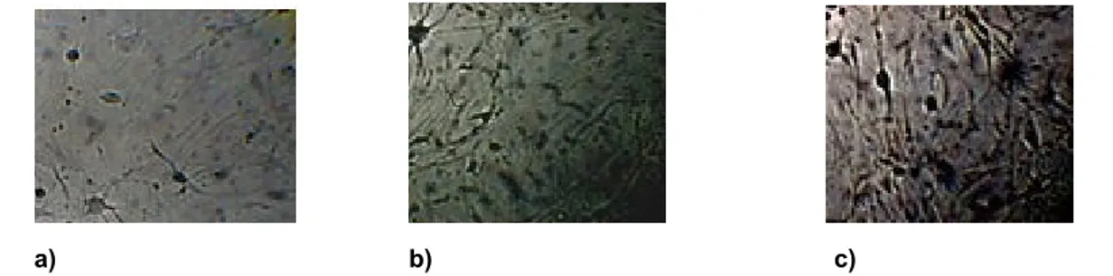
The aim of our work was to induce, by comparing, the trans-differentiation process capability of adult and perinatal stem cells in neural cells from different sources such as bone marrow, umbilical cord blood, human endometrium and amniotic fluid, by analyzing similarities and differences and by hypothesizing future therapeutic uses. We tested the expression of neural markers as GFAP, Nestin and Neurofilaments using the immunofluorescence staining assay and typical cluster of differentiation as CD34, CD90, CD105 and CD133 by using cytofluorimetric assay.
Commento [1]: <!--EndFragment-->
Chapter 4
FLUORESCENCE MICROSCOPY
Fluorescence microscopy is a powerful tool for modern cell and molecular biologists and, in particular, neurobiologists. It provides a window into the physiology of living cells at sub-cellular levels of resolution. This allows direct visualization of the inner workings of physiological processes at a systems level context in a living cell or tissue. Fluorescence microscopy enables the study of diverse processes including protein location and associations, motility, and other phenomena such as ion transport and metabolism. This versatility explains why thousands of papers describing variants of the many fluorescent microscopy techniques are published each year. When organic or inorganic specimens absorb and subsequently reradiate light, the process is typically a result of fluorescence or phosphorescence. Fluorescence emission is nearly simultaneous with the absorption of the excitation light as the time delay between photon absorption and emission is typically less than a microsecond. When the emission persists long after the excitation light is extinguished, the phenomenon is known as phosphorescence. Stokes coined the term ‘‘fluorescence’’ in the middle of the 19th century when he observed that the mineral fluorspar emitted red light when it was illuminated by ultraviolet (UV) excitation. Stokes noted that the fluorescence emission always occurred at a longer wavelength than that of the excitation light. Early investigations showed that many specimens (minerals, crystals, resins, crude drugs, butter, chlorophyll, vitamins, inorganic compounds, etc.) fluoresce when irradiated with UV light. In the 1930s, the use of fluorochromes began in biology to stain tissue components, bacteria, or other pathogens. Some of these stains were highly specific and they stimulated the development of the fluorescence microscope. Fluorescence microscopy has become an essential tool in biology as well as in materials science as it has attributes that are not readily
available in other optical microscopy techniques. The use of an array of fluorochromes has made it possible to identify cells and submicroscopic cellular components and entities with a high degree of specificity amid nonfluorescing material. The fluorescence microscope can reveal the presence of a single fluorescing molecule. In a sample, through the use of multiple staining, different probes can simultaneously identify several target molecules. Although the fluorescence microscope cannot provide spatial resolution below the diffraction limit of the respective objects, the detection of fluorescing molecules below such limits is readily achieved.
There are specimens that autofluoresce when they are irradiated and this phenomenon is exploited in the field of botany, petrology, and in the semiconductor industry. In the study of animal tissues or pathogens, autofluorescence is often either extremely faint or nonspecific. Of far greater value for such specimens are added fluorochromes (also called fluorophores), which are excited by specific wavelength irradiating light and emit light of useful intensity. Fluorochromes are stains that attach themselves to visible or subvisible structures, are often highly specific in their attachment targeting, and have significant quantum yield (the photon emission/ab- sorption ratio). The growth in the use of fluorescent mic- roscopes is closely linked to the development of hundreds of fluorochromes with known intensity curves of exciation and emission and well-understood biological structure targets.
The basic task of the fluorescence microscope is to irradiate the specimen with the desired wavelength and then to separate the much weaker emitted (fluorescent) light from the excitation light. Only the emission light should reach the eye or other detector so that the resulting fluorescing areas are contrasted against a dark background. The detection limit is largely determined by the darkness of the background. The exciting light is typically 105 or 106 times brighter than the emitted light.
When electrons go from the excited state to the ground state, there is a loss of vibrational energy. As a result, the emission spectrum is shifted to longer wavelengths
than the excitation spectrum (wavelength varies inversely to radiation energy). This phenomenon is known as Stokes’ Law or Stokes’ shift. The greater the Stokes’ shift, the easier it is to separate excitation light from emission light.
The emission intensity peak is usually lower than the excitation peak; and the emission curve is often a mirror image of the excitation curve, but shifted to longer wavelengths. To achieve maximum fluorescence intensity, the dye is usually excited at wavelengths near or at the peak of the excitation curve, and the widest possible range of emission wavelengths that include the emission peak are selected. The selection of excitation wavelengths and emission wavelengths is typically based on interference filters. In addition, the spectral response of an optical system will depend on such factors as glass transmission and detector responsivity.
Fluorescence Correlation Spectroscopy
Fluorescence correlation spectroscopy (FCS) is one of the many different modes of high-resolution spatial and temporal analysis of extremely low concentrated biomolecules. In contrast to other fluorescence techniques, the parameter of primary interest is not the emission intensity itself, but rather spontaneous intensity fluctuations caused by the minute deviations of the small system from thermal equilibrium. In general, all physical parameters that give rise to fluctuations in the fluorescence signal are accessible by FCS. It is, for example, rather straightforward to determine local concentrations, mobility coefficients or characteristic rate constants of inter or intramolecular reactions of fluorescently labeled biomolecules in nanomolar concentrations.
Fluorescence correlation spectroscopy has been developed in the early seventies as a special case of relaxation analysis. Classical relaxation methods induce certain kinds of external perturbations such as temperature or pressure jumps to a reaction system, and gain information about involved kinetic parameters from the way the system returns back to equilibrium. The novel concept of FCS with respect to these classical techniques is to take advantage of the minute spontaneous fluctuations of physical parameters that are somehow reflected by the fluorescence emission of the molecules. Such fluctuations are incessantly occurring at ambient temperatures and are generally represented as (unwanted) noise patterns of the measured signal, in our case fluorescence. The fluctuations can be quantified in their strength and duration by temporally autocorrelating the recorded intensity signal, a mathematical procedure that gave the technique its name.
Autocorrelation analysis provides a measure for the self-similarity of a time series signal and therefore describes the persistence of information carried by it. Essential information about processes governing molecular dynamics can thus be derived from the temporal pattern by which fluorescence fluctuations arise and decay.
At its first introduction by Madge, Elson and Webb in 1972, FCS was applied to measure diffusion and chemical dynamics of DNA-drug intercalation. This pioneering study was then followed by a number of other publications by many different groups describing, e.g., attempts to determine particle concentration, translational and rotational mobility in two or three dimensions, even in the cellular environment or in flow systems. Nevertheless, these early measurements suffered from poor signal-to-noise ratios, mainly because of low detection efficiency, large ensemble numbers and insufficient background suppression.
This is the basic concept of FCS: Make the number of observed molecules low enough so that each of them contributes substantially to the measured signal. Then and only then, one can truly perform analyses of spontaneous, non-coordinated fluctuations.
It is obvious that FCS can only function properly if one somehow manages to reduce the concentrations and observation volumes such that only few molecules are simultaneously detected, and at the same time increase the fluorescence photon yield per single molecule.
A major improvement could be made by using efficient fluorescent dyes to label the molecules of interest, strong and stable light sources like lasers, and ultrasensitive detectors, e.g. avalanche photodiodes with single-photon sensitivity. The final breakthrough was achieved in Stockholm by Rigler and his coworkers by combining the FCS technique with confocal detection. Here, the incoming laser light is strongly focused by a high numerical aperture objective (ideally NA > 0.9) to a diffraction limited spot. Only the few fluorophores within the illuminated region are excited. In order to limit the detection volume also in axial direction, a pinhole is introduced in the image plane, which blocks all light not coming from the focal region.
To date, most FCS measurements are performed on fluorescently labeled biomolecules diffusing in aqueous buffer solution. Because of the most elegant way of limiting the detection volume to less than one femtoliter, i.e. approximately the volume of an E.coli bacterial cell, concentrations in the nanomolar range are optimal for FCS measurements. Under these circumstances, the signal fluctuations induced by molecules diffusing into or out of the focal volume are large enough to yield good signal-to-noise ratios. During the time a particle spends in the focus, chemical or photophysical reactions or conformational changes may alter the emission characteristics of the fluorophore and give rise to additional fluctuations in the detected signal.
A substantial limitation of the single-point FCS technique is the lack of information about fluctuations occurring in the proximity of the measured point. Many processes in chemistry, physics, and biology have a spatial scale. Since the earlier days of FCS it has been well-known that diffusion processes have spatial structures that depend on the size of the volume of illumination. The larger the volume is, the longer it will take for a molecule to cross the illumination volume. The timescale of other processes such as binding to immobile locations or blinking and rotational motions is independent of the size of the illumination volume (Figure 1). This difference in the spatial extent of the fluctuation was used to distinguish among processes. However, this approach, i.e., the dependency of the timescale on the size of the illumination
volume, still uses an illumination volume that has cylindrical symmetry. With this approach it is difficult to produce illumination volumes that are large and of an arbitrary shape.
Early in the field’s development, it was understood that by moving the illumination volume in a periodic pattern in the sample at a rate such that the molecules will not move much during a period, the record of the intensity fluctuations along the path will contain spatial information about the location where the fluctuation occurred (44, 45). The analysis of the fluctuations at successive periods will contain information about the time course of the fluctuations of the points along the path (46). This approach is called scanning FCS, and it is practiced today in several labs (47–51). Conceptually, scanning FCS is different from the use of an arbitrary shape or size for the volume of illumination such as the dual-foci method (1, 52), as different volumes are excited at different times in scanning FCS. Scanning introduces a time and spatial structure to the observation that we could exploit to best match the spatial and temporal structure of the physical process we are investigating (Figure 1).
One advantage of scanning FCS is that current confocal microscopes have the capability to send the laser beam along a path (either line or circular orbit) at a very high rate. As this method is the basic element for the introduction of spatiotemporal correlations, let us examine from a conceptual point of view the various ways that the information is encoded when a laser beam performs a periodic path in the sample. Every point along the path is visited once per period. The size of the point is defined by the point-spread function (PSF).
If molecules remain in a given point for a time comparable with the period, then the intensity fluctuation at that point decays between successive periods. Because data are available at many points along the path, this experiment is equivalent to performing many single-point FCS measurements simultaneously. In this case, the time resolution of the experiment is the period, which can be a fraction of a millisecond, short enough to correlate the motion of small proteins in the cellular environment. However, if we consider two adjacent points in the path, the time difference of sampling these two points is equal to the period divided by the number