Università degli Studi di Ferrara
DOTTORATO DI RICERCA IN
"Farmacologia e Oncologia Molecolare"
CICLO XXVI
COORDINATORE Prof. Antonio Cuneo
ADENOSINE RECEPTORS IN HEALTH
AND DISEASE
Settore Scientifico Disciplinare BIO/14
Dottorando Tutore
Dott.ssa Angela Stefanelli Dott.ssa Stefania Gessi
I
TABLE OF CONTENTS
Pag
Abstract
1Adenosine receptors (ARs) in health and disease
3Adenosine 4 GPCRs 6 Adenosine Receptors 8 A1 Adenosine Receptor 10 A2A Adenosine Receptor 15 A2B Adenosine Receptor 21 A3 Adenosine Receptor 25 Conclusion 32 References 34
Downregulation of A
1and A
2BARs in human trisomy 21
mesenchymal cells from first-trimester chorionic villi
50
Introduction 51
Angiogenesis 51
Adenosine and angiogenesis in pregnancy 52
Aim of the thesis 55
Materials and methods 56
Results 61
Discussion 64
Figures Legend 67
Figures 69
References 78
A
1and A
3ARs inhibit LPS-induced hypoxia-inducible factor-1
accumulation in murine astrocytes
85
Introduction 86
Astrocyte 87
Hypoxia-inducible factor 91
HIF-Regulated genes: Neuroprotection or Inflammation 95
Effect of adenosine in astrocytes 96
Possible role of HIF, Astrocyte and Adenosine in neuroprotection 99
Aim of the thesis 100
Materials and methods 101
Results 105
Discussion 110
II Figures 118 References 132 Curriculum vitae 145 List of Publications 146 Meetings 148 Acknowledgements 149
1
Abstract
Adenosine (Ado) is an endogenous nucleoside released from almost all cell types. It exerts neuroprotective and anti-inflammatory functions by acting through four receptor subtypes A1, A2A, A2B and A3 (ARs). These receptors differ in their affinity for Ado, in the type of G
protein that they recruit and finally in the downstream signalling that are activated in target cells. The levels of Ado in the interstitial fluid are in the range of 20-200 nM, but many pathophysiological conditions such as asthma, neurodegenerative disorders, chronic inflammatory diseases and cancer are associated with changes in Ado levels. The primary aim of Ado is to reduce tissue injury and promote repair by different receptor-mediated mechanisms, including the increase of oxygen supply/demand ratio, anti-inflammatory effects and stimulation of angiogenesis. The investigation of ARs and their ligands is a rapidly growing field; there is extensive evidence for the involvement of ARs in the physiological regulation of several homeostatic processes and their implication in the ethiology of many diseases. The aim of this work was to analyse the expression of ARs and the signalling pathway, transcription factors and cytochines activated by them in different pathophysiological conditions linked to hypoxic and inflammatory conditions. At first, the role of ARs was studied in healthy and aneuploid pregnancies to understand reasons of spontaneous abortion (SA). It has been suggested that the causes of SA in aneuploidy are no different to those in euploidy, with the increased frequency in the former perhaps being ascribable to a genetically-determined imbalance in the mediators of placental perfusion and uterine contraction. So aneuploidy can be used as a model of this event. The ado transduction cascade appears to be disturbed in Trisomy 21 (TR21) through reduced expression of A2BAR and A1AR. These anomalies may be implicated in
complications such as fetal growth restriction, malformation and/or SA, well known features of aneuploid pregnancies. Therefore A1AR and A2BAR could be potential
biomarkers able to provide an early indication of SA risk and their stimulation may turn out to improve fetoplacental perfusion by increasing nitric oxide (NO) and vascular endothelial growth factor (VEGF).
Next, the effects of ARs in glial cells under inflammatory conditions have been investigated. Glial cells, astrocytes and microglia, are important contributors to inflammatory immune responses and hypoxia-inducible factor 1 (HIF-1α) is the key transcription factor that is upregulated in response to hypoxia and inflammatory stimuli. Ado, through A1AR and A3AR activation, reduces lipopolysaccharide (LPS)-stimulated
2
HIF-1α mRNA expression and protein accumulation by inhibiting LPS-triggered p42/p44 mitogen-activated protein kinase (p44/42 MAPK) and serine/threonine protein kinase (Akt) phosphorylation in normoxic and hypoxic conditions. This leads to an inhibition of genes involved in inflammation like Inducible Nitric Oxide Synthase (iNOS) and A2BARs,
that are stimulated by LPS and further increased by LPS in concert with hypoxia, whilst does not affect angiogenesis and metabolic related genes, with the exception of glucose transporter 1 (GLUT1) and hexokinase 2 (HK2) that are stimulated by LPS in normoxia and reduced by A1AR and A3AR activation. These findings add a new molecular pathway
activated by Ado in astrocytes to give a reduction of genes involved in inflammation and hypoxic injury that may cohexist in stroke, ischemia and other central nervous system (CNS) disorders.
3
Adenosine Receptors
4
Adenosine
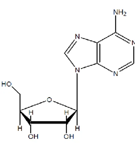
In 1929, Drury and Szent-Györgyi first reported the concept of Adenosine (Ado) acting as an extracellular signalling molecule (Drury and Szent-Gyorgyi, 1929). Ado is an endogenous nucleoside modulator released from almost all cell types (Gessi et al., 2011). Ado plays a central role as a structural element of nucleic acids and in the energy metabolism of all living organism. The physiological effects of Ado were first described in the cardiovascular system and gastrointestinal tract (Drury and Szent-Gyorgyi, 1929). It is composed of a molecule of adenine attached to a ribose sugar molecule (ribofuranose) via a β-N9-glycosidic bond (Fig. 1). Ado accumulates in the extracellular space in response to metabolic stress and cell damage, and elevations in extracellular Ado are found in conditions of ischaemia, hypoxia, trauma asthma, neurodegenerative disorders, chronic inflammatory diseases and cancer (Linden, 2001; Fredholm et al., 2001; Gessi et al., 2011; 2013). The rapid release of Ado in response to tissue-disturbing stimuli has a dual role in modulating homeostasis. First, extracellular Ado represents a pre-eminent alarm molecule that reports tissue injury in an autocrine and paracrine manner to surrounding tissue. Second, extracellular Ado generates a range of tissue responses that can be generally viewed as organ protective thereby mediating homeostasis. Ado elicits its physiological responses by binding to and activating one or more of the four transmembrane Ado receptors (ARs), denoted A1, A2A, A2B and A3. A prominent
body of evidence supports the notion that the ability of Ado, acting at its receptors, to control the immune and inflammatory systems plays a key role in the modulatory effects of Ado in both health and disease. There are many promising emerging therapeutic approaches that are focused on the modulation of Ado in the immune system (Hasko et al., 2008). Ado also has receptor-indipendent effects, because extracellular Ado can cross the cell membrane and have a role in less well-defined intracellular mechanisms, including the AMP-activated protein kinase (AMPK), adenosine kinase (AK) and S-adenosylhomocysteine hydrolase pathway (SAHase) (Antonioli et al., 2013).
Normally, it is present in body fluids in concentrations 20–200 nM, but in response to stress elevated levels of Ado, up to 300 µM are produced and released (Fredholm, 2010; Burnstock, 2008; Lopes et al., 2011).
Ado can be generated intracellularly by hydrolysis of AMP or SAHase. The former is mediated by intracellular soluble 5′ nucleotidase (5′NT) and the rate of Ado formation is largely controlled by the level of adenosine monophosphate (AMP), which provides a direct
5
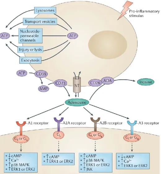
link to energy metabolism (Newby et al., 1985). Interestingly, Ado can be converted back to AMP via the action of AK and together 5′ NT and AK generate a potential futile cycle that ensures that there will always be a not insubstantial amount of intracellular Ado. The other way in which intracellular Ado is formed depend on the action of SAHase. S-Adenosyl-L-homocysteine (SAH) levels are increased with increasing rates of S-adenosylmethionine- dependent transmethylation. Interestingly, the enzyme can also catalyze the reverse reaction, i.e., formation of SAH from Ado and homocysteine and this has been used to generate an estimate of intracellular Ado concentration of around 100 nMol/L under basal conditions (Deussen et al., 1988). Indeed, whenever there is an imbalance between the rate of Adenosine triphosphate (ATP) synthesis and ATP utilization AMP tends to rise and consequently Ado formation increases. This can occur under extreme physiological conditions such as heavy exercise, increases in nerve activity, or reduced ambient oxygen, e.g., at elevated locations. It will also occur pathologically, e.g., in ischemia, local trauma, or in the interior of a solid tumor (Fredholm, 2007; Sitkovsky, 2009).
Cell membrane–embedded nucleoside trasportes, which include equilibrative nucleoside transporters (ENTs) and concentrative nucleoside transportes (CNTs), shunt extracellular Ado into the intracellular space, thereby terminating ARs signaling. ENTs, which carry nucleosides along their concentration gradient across cell membranes, include four subtypes: ENT1, ENT2, ENT3 e ENT4. CNTs, which include CNT1, CNT2 and CNT3, mediate the intracellular influx of nucleoside against their concentration gradient by using the sodium ion gradient, which occurs across the cell membrane, as a source of energy (Antonioli et al., 2013). Two enzymes play a key role in catabolizing Ado: Ado deaminase (ADA) and AK and they are responsible for an extremely short half-life of Ado in circulation (Fredholm et al., 2001; 2011; Eltzschig, 2009). AK is critically important in maintaining the physiological levels of Ado low, and also in maintaining depots of adenine nucleotides (Boison, 2006; Fredholm, 2007). However, some cells also possess concentrating transporters that drive Ado intracellularly by an ion gradient (Young et al., 2008). Extracellular Ado can be generated from extracellular degradation of adenine nucleotides via CD39 and ATP diphosphohydrolase (Deaglio and Robson, 2011) and CD73-5′nucleotidase (Colgan et al., 2006). In addition, some cells express ecto-enzymes that convert 2′, 3′,or 3′5′-cAMP to AMP for further conversion to Ado (Verrier et al., 2011). Although not all cells express these enzymes (Langer et al., 2008), they are present in most cellular microenvironments and provide a means for rapid degradation of extracellular nucleotides. Indeed, it appears that most cells can under some circumstances release nucleotides via exocytosis, specialized transporters or connexin or
6
pannexin hemichannels. There is also sometimes exocytotic release from vesicles that in addition store hormones or neurotransmitters (Lazarowsky, 2012), and perhaps even more interestingly the release of ATP can occur without the release of hormone when hormone storage granules transiently contact the cell membrane in a so called kiss-and-run encounter (MacDonald et al.,2006; Fredholm, 2013).
G protein–coupled receptors (GPCRs)
Ado is the endogenous ligand of four types of GPCRs, designated A1, A2A, A2B, and A3
(Fredholm et al., 2011).
GPCRs comprise the largest protein superfamily in mammalian genomes. They share a common seven-transmembrane (7TM) topology and mediate cellular responses to a variety of extracellular signals ranging from photons and small molecules to peptides and proteins (Lagerstrom and Schiolth, 2008). Diversity of the extracellular ligands is reflected in the structural diversity of more than 800 human GPCRs, which can be grouped into five major families and numerous subfamilies on the basis of their amino acid sequences. Subdivision on the basis of sequence homology allows the definition of rhodopsin (Class A), secretin (Class B), adhesion, glutamate (Class C) and Frizzled receptor families (Friedriksson et al., 2003). During the past few years, crystallography of GPCR has experienced exponential growth, resulting in the determination of the structures of 16 distinct receptors, 9 of them in 2012
7
alone (Katrick et al., 2013). Signal transduction by GPCRs is fundamental for most physiological processes - from vision, smell, and taste to neurological, cardiovascular, endocrine, and reproductive functions - thus making the GPCR superfamily a major target for therapeutic intervention (Overington et al., 2006). Current drug discovery efforts aim both to improve therapies for more than 50 established GPCR targets and to expand the list of targeted GPCRs (Lappano and Maggiolini, 2011; Katrick et al., 2013).
GPCRs share a common 7 TM α-helix architecture and couple to G-proteins (Lefkiowitz, 2004). The TM α-helices are connected by alternating three extracellular (EL) and cytoplasmic (CL) loops (EL1–EL3 and CL1–CL3), with the N-terminus (NT) extracellular and the C-terminus (CT) intracellular, arranged in an anticlockwise fashion as viewed from the extracellular surface. Binding of a stimulus, the so-called ‘first messenger’, to the extracellular or TM domains of a GPCR triggers conformational changes in the 7TM structure that are transmitted through the intracellular receptor domains to promote coupling between the receptor and its cognate heterotrimeric G-proteins. The receptor stimulates G-protein activation by catalysing the exchange of guanosine triphosphate (GTP) for guanosine diphosphate (GDP) on the Gα subunit and dissociation of the GTP-bound Gα subunit from the Gβγ subunit heterodimer. Once dissociated, free Gα-GTP and Gβγ subunits regulate the activity of enzymatic effectors, such as adenylate cyclases (AC), phospholipase C (PLC) isoforms and ion channels, to generate small molecules, the “second messengers”. The second messengers, in turn, control the activity of protein kinases that regulate key enzymes involved in intermediate metabolism. Hydrolysis of GTP to GDP within Gα and subsequent reassociation of Gα-GDP and Gβγ completes the G-protein cycle (Fig.2) (Oldham et al., 2008; Ding et al., 2013)
8
Figure 2: GPCR activation cycle.
ARs, like other class A GPCRs, have long been thought to exclusively occur in a monomeric state. Monomeric receptors are sufficient to induce signaling (Whorton et al., 2008). At least some studies suggest signaling via dimers occurs only at higher receptor densities (White et al., 2007). More recently, however, evidence is accumulating that ARs can form dimeric or, more generally speaking, multimeric or oligomeric structures. Through self-association, homo-oligomers (“homomers”) can be formed. Hetero-oligomerization leading to “heteromers” may be the consequence of the association between ARs and preferred partners, mostly other GPCRs, including other AR subtypes (Fredholm et al., 2011).
Adenosine receptors (ARs)
Extracellular purines (Ado, ATP, and ADP) and pyrimidines (uridine diphosphate (UDP) and uridine triphosphate (UTP) comprise a family of molecules that exert a variety of important physiological functions via the activation of cell-surface receptors termed purine receptors. Although the physiologic effects of Ado and ATP have been recognized for over 80 years, purinergic receptors were first described in 1976 and two subfamilies were identified: P1 or
9
ARs (selective for Ado), and P2 or nucleotide receptors (selective for ATP, ADP and UTP, which act as extracellular signaling molecules) (Mediero and Cronstein, 2013).
Four members of the Ado/P1 receptor family have now been cloned and characterized from a variety of species: A1, A2A, A2B, and A3, and selective agonists and antagonists have been
identified. All P1 receptors couple to G-proteins, and modulate AC activity in an inhibitory (A1, A3) or stimulatory (A2A, A2B) fashion, resulting in cyclic adenosine monophosphate
(cAMP) changes (Fig.3). P2 receptors are divided into two families: P2X and P2Y, based on molecular structure, transduction mechanisms, and pharmacological properties (Burnstock et al., 2012).
Figure 3: Ado synthesis and receptor activation in the cell. (Antonioli et al., Nature Reviews Cancer 13, 842–857;2013)
10
Each AR subtype has a different pattern of tissue expression and ligand binding properties. In cell-based systems, A1ARs have the highest affinity for Ado (Ki=10 nmol/L). The Ki values
for Ado for A2AAR A2BAR, and A3AR are 200, 2000, and 10000 nmol/L, respectively, for the
human receptors. A3ARs are also activated by the Ado metabolite inosine (Ki 2300 nmol/L)
(Rivkees and Wendler, 2012).
Many biological functions have been attributed to Ado signaling. For example, the heart rate-slowing effects of intravenous Ado that is used for patient treatment of supraventricular tachycardia are mediated through the A1AR (Delacretaz, 2006). The A2AAR is expressed on
inflammatory cells: pharmacologic studies provided critical evidence that the activation of A2AAR on neutrophils attenuates inflammatory responses (Gessi et al., 2000; Ohta and
Sitkovsky, 2001). The A2BAR is involved in hypoxia-adaptive responses (Kong et al., 2006;
Eltzschig et al., 2003; 2004), for example, during myocardial ischemia (Eckle et al., 2012), Acute Kidney Injury (AKI) (Grenz et al., 2012a, Bauerle et al., 2011) or intestinal inflammation (Grenz et al., 2012b, Frick et al., 2009) - such as that occurring during inflammatory bowel disease. A3AR has been suggested as a tumoral marker (Gessi et al.,
2004) and is involved in the inhibition of cancer growth (Fishman et al., 2004)
A
1Adenosine Receptor
The A1AR has been cloned from several animal species, including humans, and is known to
bear a close structural similarity across the species (Ravelic and Burnstock, 1998). As for signal transduction, the A1AR is coupled to members of the Gi/Go family of G proteins,
whereby it induces inhibition of AC activity (Van Calker et al., 1979). In addition, it is thought to activate PLCβ, which is known to increase inositol 1,4,5-triphosphate (IP3) and
intracellular Ca2+. A1AR is coupled to pertussis-toxin-sensitive potassium channels as well as
KATP channels, particularly in cardiac tissue and neurons. Moreover, it may inhibit Q, P and
N-type Ca2+ channels and modulate extracellular signal-regulated protein kinases (ERKs) (Fredholm et al., 2001). Furthermore, a role for the β-arrestin1/ERK mitogen-activated protein kinase (MAPK) pathway in regulating A1AR desensitization and recovery has
11
A1AR and central nervous system (CNS)
The A1AR is widely distributed throughout the CNS, featuring particularly high levels in the
brain cortex, cerebellum and hippocampus, as well as the dorsal horn of the spinal cord. It is present in both pre- and post-synaptic terminals, and modulates the activity of the nervous system at a cellular level. At the presynaptic level it mediates inhibition of neurotransmitter release, while at the postsynaptic level it induces neuron hyperpolarization. Thus, activation of A1AR via Ado is responsible for sedative, anticonvulsant, anxiolytic and
locomotor-depressant effects. Moreover, endogenous Ado levels are sufficient to tonically activate inhibitory A1AR, and caffeine, perhaps the most commonly used drug in the world, mediates
its excitatory effects by antagonising this inhibition. Ado also has a fundamental role to play in analgesia (Eltzschig et al., 2009); indeed, both spinal and systemic administration of Ado or its analogs produces anti-nociception by A1AR activation in a variety of animal models
(Boison, 2007; Gong et al., 2010; Nascimento et al., 2010; Sowa et al., 2010). These antinociceptive effects may be mediated by the inhibition of intrinsic neurons by an increase in K+ conductance and presynaptic inhibition of sensory nerve terminals, which would theoretically hinder the release of substance P and glutamate. Likewise, attenuation by N-methyl-D-aspartate (NMDA)-induced production of NO may also be involved. Furthermore, Ado has been shown to mediate opioid analgesia (Gan and Habib, 2007). In addition, it has recently been reported that allopurinol, a potent inhibitor of the enzyme xanthine oxidase used primarily in the treatment of hyperuricemia and gout, induces anti-nociception related to Ado accumulation, an effect that is completely prevented by A1AR blockade (Schmidt et al.,
2009). Compounds that are able to enhance the activity of the A1AR mediated by the
endogenous ligand within specific tissues may have potential therapeutic advantages over non-endogenous agonists, due to allosteric modulation of GPCRs. As allosteric enhancers act only on the agonist A1AR-G protein ternary complex, limiting their action to sites and times
of Ado accumulation, the use of these drugs to increase the responsiveness of the A1AR to
endogenous Ado at sites of its production is an appealing alternative to activation by exogenous agonists (Romagnoli et al., 2010), especially as the former approach minimizes side effects such as dyspnea, chest pain, atrioventricular blockage or bronchospasm.
A1AR and the respiratory system
A1ARs are responsible for many effects induced by Ado, not only in the CNS but also in
peripheral tissues (Russo et al., 2006; Baraldi et al., 2008). In particular, this signaling nucleoside has been implicated in the regulation of asthma and chronic obstructive pulmonary
12
disease (COPD) (Russo et al., 2006); Ado levels are elevated in the asthmatic lungs to an extent that can be directly correlated with the degree of inflammatory insult (Brown et al., 2008a). Unsurprisingly, therefore, A1AR expression is also increased in the epithelium and
airway smooth muscle of human asthmatics (Brown et al., 2008b). Early evidence of A1AR is
involvement in asthma was provided by studies on allergic rabbit models, where the Ado-induced acute bronchoconstrictor response was attenuated by pretreatment with A1AR
antagonists. Accordingly in human airway tissue and bronchial smooth muscle cells, activation of A1AR has been shown to produce effects that cause airway
hyper-responsiveness. In particular, activation of A1AR in human airway epithelial cells causes an
increase in the expression of the Mucin 2 (MUC 2) gene, which is responsible for mucus hypersecretion. Moreover, activation of A1AR is known to produce pro-inflammatory effects
on various types of human cells (Ponnoth et al., 2010). As a whole, these effects of A1AR in
humans suggest A1AR as an important therapeutic target in human asthma (Ethier and
Madison, 2006; Baraldi et al., 2008; Wilson, 2008). Indeed, the non-selective AR antagonists theophylline and doxofylline have been launched as bronchodilators for the treatment of various respiratory disorders (Press et al., 2007). Paradoxically, however, findings in ADA-deficient mice suggest the occurrence of anti-inflammatory actions of Ado in the lung, mediated through chronic A1AR activation of macrophages (Sun et al., 2005). Likewise, it has
been recently reported that A1AR inhibits transendothelial and transepithelial
polymorphonuclear cell migration in a murine model of lipopolysaccharide (LPS)-induced lung injury, presumably by reducing the release of chemotactic cytokines into the alveolar space. In addition, A1AR is involved in decreasing microvascular permeability and leukocyte
transmigration in endothelial cells (Ngamsri et al., 2010), suggesting also a protective and anti-inflammatory role for A1AR (Gazoni et al., 2010).
A1AR and the cardiovascular system
At a cardiovascular level, A1ARs mediate negative chronotropic, dromotropic and inotropic
effects. A1AR subtypes located on sinoatrial and atrioventricular nodes can cause bradycardia
and heart block, respectively, while their negative inotropic effects include a decrease in atrial contractility and action potential duration. Recently, it has been shown that the selective deletion of the A1AR abolishes the heart-rate slowing effects of intravascular Ado in vivo
(Koeppen et al., 2009). Stimulation of A1AR in the heart, on the other hand, exerts a
cardioprotective effects by inhibiting norepinephrine release from sympathetic nerve endings (Schutte et al., 2006). Ado also protects tissues through ischemic preconditioning (IPC), a brief period of ischemia and reperfusion that can protect the myocardium against infarction
13
from a subsequent prolonged ischemic insult. This response, which has been most widely investigated in the heart, but also occurs in other tissues (Schneyvays et al., 2005; Grenz et al., 2007), is brought about by the activation of A1AR, protein kinase C (PKC) and
mitochondrial KATP channels (Kiesman et al., 2009; Solenkova et al., 2006). A1AR agonists,
for example, tecadenoson (N6-[3(R)- tetrahydrofuranyl]adenosine), are in development for arrhythmias and atrial fibrillation; clinical studies with intravenous tecadenoson suggest that it may slow the speed of atrio ventricular nodal conduction by selectively stimulating the A1AR,
and may prevent blood pressure dropping by failing to stimulate the A2AAR (Yldiz et al.,
2007). In the kidney, on the other hand, A1AR mediates vasoconstriction, decrease in
glomerular filtration rate and the inhibition of both renin secretion and neurotransmitter release. Thus, A1AR antagonists represent a novel class of agents for potential use in the
treatment of hypertension and edema (Vallon et al., 2006). In fact, A1AR antagonists are more
effective diuretics and natriuretics than thiazides, with the added advantage of reducing the potassium wastage and reductions of renal blood flow and glomerular filtration rate seen with the latter drugs (Zhou and Kost, 2006). Furthermore, evidence from genetically altered mice indicates that transcellular NaCl transport induces the generation of Ado, which, in conjunction with angiotensin II, elicits afferent arteriolar constriction through A1AR
activation (Sun et al., 2001; Schnermann and Briggs, 2008). Moreover, clinical trials, albeit in a limited number of subjects, have demonstrated that A1AR antagonists produce natriuretic
and hypotensive effects in hypertensive patients and attenuated the furosemide-induced decline of renal hemodynamic function in heart failure patients. Hence, selective A1AR
antagonists targeting renal microcirculation are currently under development for the treatment of both chronic and acute heart failure. One of these novel pharmacological agents, rolofylline (1,3-dipropyl-8-(2-nor-1-adamantyl) xanthine, KW-3902), facilitates diuresis and preserves renal function through A1AR antagonism in patients with acute decompensated heart failure
and renal dysfunction; pilot data also suggest beneficial effects on symptoms and short-term outcomes (Slawsky and Givertz, 2009). Nevertheless, despite several studies showing improvement of renal function and/or increased diuresis with A1AR antagonists, particularly
in chronic heart failure, these findings were not confirmed in the large Phase III trials PROTECT 1 and 2 (Placebo-controlled Randomized study of the selective A1 antagonist
Rolofylline for patients hospitalized with acute heart failure and volume Overload to assess Treatment Effect on Congestion and renal funcTion) in acute heart failure patients. In fact, the pooled/meta-analysis of two studies demonstrated that treatment with rolofylline was associated with poor outcomes due to worsening renal function in patients with acute decompensated heart failure (Watherley et al., 2010; Voors et al., 2011). Nonetheless, lessons
14
can be learned from these and other studies, and there is still hope for a clinical role for A1AR
antagonists (Hocher et al., 2010; 2011).
A1AR and inflammation
Indeed, several studies have demonstrated that A1AR activation has a protective function in
vivo, inhibiting necrosis, inflammation and apoptosis. Moreover, A1AR has been implicated
as potent anti-inflammatory mediator in various kidney, heart, liver, lung and brain injury models (Gazoni et al., 2010; Pang et al., 2011). In particular A1AR activation appears to
protect against hepatic injury by upregulation and phosphorylation of heat shock protein 27, a member of a family of chaperone proteins that serves to defend against cell damage (Chen et al., 2009). A critical role for Ado in bone homeostasis via interaction with A1AR has also
been recently reported. In particular, due to the stimulatory effect played by A1AR on
osteoclast function and formation, antagonists of this receptor may be able to prevent the bone loss associated with inflammatory diseases and menopause (Kara et al., 2010a; 2010b).
Activation of A1AR inhibits lipolysis and lowers plasma concentrations of free fatty acids by
inhibiting AC and downstream cAMP formation. Unfortunately, however, the majority of full A1AR agonists are plagued by significant cardiovascular effects. Hence, selective partial
A1AR agonists have been developed (Dhalla et al., 2007a; 2007b). One such compound,
CVT-3619 (2-{6-[((1R,2R)-2-hydroxycyclopentyl) amino] purin-9-yl(4S,5S,2R,3R)-5-[(2-fluorophenylthio)- methyl]oxolane- 3,4-diol), is a partial A1AR agonist that has anti-lipolytic
effects at concentrations that do not provoke cardiovascular symptoms (Fatholahi et al., 2006; Shearer et al., 2009). A further advantage of these partial agonists is that they are accompanied by a minimal risk of ARs desensitization in response to chronic drug exposure (Vallon et al., 2006; Shearer et al., 2009). The A1AR receptor ligand candidates for novel
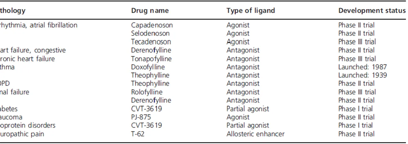
15
Table 1: Progress of A1AR ligands as novel therapeutic treatments (Gessi et al., Expert Opin. Investig.
Drugs:20;1591-1609; 2011)
A
2AAdenosine Receptor
Of the four ARs, A2AARs have taken center stage as the primary anti-inflammatory effectors
of extracellular Ado (Hasko and Pacher, 2008). The gene for the A2AAR has been cloned from
several species including dog, rat, human, guineapig and mouse, and has demonstrated a high degree of homology among human, mouse and rat (Baraldi et al., 2008). The A2AAR
stimulates AC activity through its coupling with Gs proteins; this leads to the activation of
cAMP-dependent protein kinase A (PKA), which in turn phosphorylates and activates various receptors, ion channels, phosphodiesterases and phosphoproteins such as cAMP response element-binding protein (CREB) and dopamine- and cAMP-regulated neuronal phosphoprotein (DARPP-32). PKC triggering by A2AAR activation has also been reported. In
the brain striatum, the A2A subtype stimulates Golf, another member of the Gs subfamily of G
proteins. In addition, A2AAR can interact with different types of Ca2+ channels to either
increase intracellular Ca2+ or decrease Ca2+ influx and, like the other Ado subtypes, it is involved in the modulation of ERK activity. Due to a long carboxy-terminal domain, the A2AAR possesses a greater molecular mass (45 kDa) in comparison with the other subtypes
(36-37 kDa). The A2AAR C-terminus has been defined as a crowded place where various
accessory proteins, such as D2-dopamine receptors, α-actinin, ADP-ribosylation factor nucleotide site opener, ubiquitin-specific protease 4 and translin-associated protein X, may interact. In fact, it is thought that the lack or the presence of such varied partners may explain the conflicting effects resulting from A2AAR activation, for example, neuroprotection versus
16
A2AAR structures possess common GPCR activation features (Lebon et al., 2011). GPCRs
have similar structures, consisting of 7TM helices containing well-conserved sequence motifs, indicating that they are activated by a common mechanism. Recently described structures of β-adrenoceptors highlight residues in TM region 5 (H5) that initially bind specifically to agonists rather than to antagonists, suggesting that these residues play an important role in agonist-induced activation of receptors. In this context, important information concerning the different interactions occurring between agonists and antagonists with the A2AAR has recently
come to light. In particular, agonists contain a ribose group that extends deep into the A2AAR
ligand binding pocket, where it forms polar interactions with conserved residues in H7 and non-polar interactions with residues in H3. In contrast, an inverse agonist fails to interact with any of these residues; indeed, comparison with agonist bound structures indicates that these compounds sterically prevent conformational change in H5, thereby acting as inverse agonists. Furthermore, comparison of agonist-bound structures of A2AAR with agonist-bound
structures of β-adrenoceptors has inferred that the contraction of the ligand-binding pocket caused by inward motion of helices 3, 5 and 7 may be a feature common to the activation of all GPCRs. It is evident that this detailed new structural information regarding these receptors has the potential to hugely influence rational drug design.
A2AAR and the CNS
A2AAR is found ubiquitously throughout the body, but its expression is particularly common
in the immune system and the striato-pallidal system of the brain (Fredholm et al., 2001). A2AAR localization in basal ganglia is restricted to the GABA-contained neurons of the
indirect pathway projecting from the caudate putamen to the globus pallidus, which also selectively expresses the D2 dopamine receptor and the peptide encephalin (Jenner et al.,
2009). This explains why several studies have investigated the possible involvement of A2AAR in the pathogenesis of neuronal disorders, including Huntington’s chorea and
Parkinson’s disease (Varani et al., 2007; 2010; Simola et al., 2008; LeWitt et al., 2008; Ramlackhansing et al., 2011). In fact, changes in A2AAR expression and signaling have been
reported in various experimental models of Huntington’s disease, and an aberrant amplification of A2A-stimulated AC response has been demonstrated in striatal-derived cells
engineered to express mutant Huntingtin protein. Moreover, a subsequent study also demonstrated an abnormal increase of A2AAR density in the peripheral blood cells of
Huntington’s patients, as compared with age-matched healthy subjects (Varani et al., 2007). This suggests that the aberrant A2AAR phenotype may represent a novel biomarker of
17
assessing the efficacy of novel neuroprotective approaches, not to mention providing grounds for further scientific investigation. Comparison of striatal A2AAR binding and AC activity in
one of the best-characterized animal models of Huntington’s disease, R6/2 mice, in this case of different developmental ages, showed a transient increase in A2AAR density and A2A
AR-dependent cAMP production at early presymptomatic ages with respect to age-matched wild-type animals (Varani et al., 2007). A2AAR in the CNS is also been implicated in the
modulation of motor functions. Hence, A2AAR antagonists are a useful alternative to
dopaminergic drugs in the treatment of Parkinson’s disease (Simola et al., 2008). Accordingly, the A2AAR antagonist istradefylline
(8-[2(E)-(3,4-Dimethoxyphenyl)vinyl]-1,3-diethyl- 7-methylxanthine; KW-6002) has now been pre-registered as a Parkinson’s disease treatment in North America by Kyowa Hakko Kirin (LeWitt et al., 2008). Indeed, it has been extensively demonstrated that A2AAR antagonists have the potential to reverse motor deficits
and enhance dopaminergic treatments in animal models of Parkinson’s disease. Furthermore, istradefylline, for example, in combination therapy with levodopa or dopamine agonists, has been shown to improve the symptoms of the disease in a Parkinsonian monkey model without either increasing the incidence or severity of dopaminergic related side effects or triggering or aggravating dyskinesia. In particular, an upregulation of A2AAR has been reported in
Parkinson’s patients with levodopa-induced dyskinesias (LIDs), suggesting that A2AAR
antagonists could be used, in combination with a reduction in the dosage of levodopa, to manage LIDs (Ramlackhansingh et al., 2011). The presence of an A2AAR alteration in the
postmortem putamen of Parkinson’s disease patients as compared to healthy controls has also been demonstrated, confirming that A2AAR plays a key role in this neurological pathology.
Furthermore, a selective increase of A2AAR density in the peripheral circulating cells of
patients affected by Parkinson’s disease has been observed. As a whole, these data show that A2AAR alteration is a property common to both peripheral circulating cells and the putamen
fraction in Parkinson’s disease, confirming that lymphocytes or neutrophils could represent a mirror of the CNS (Varani et al., 2010). In addition, A2AAR antagonists have been shown to
attenuate neurotoxicity induced by kainite and quinolinate (Baraldi et al., 2008).
A2AAR and the cardiovascular system
Ado has also been shown to confer important protective effects on the cardiovascular system. Regadenoson (2-[4-(Nmethylcarbamoyl)- 1H-pyrazol-4-yl]Ado), a short-acting, selective A2AAR agonist, has already been approved as an adjunctive pharmacological stress agent for
myocardial perfusion imaging studies, and was accordingly launched in the US in 2008 by Astellas Pharma. More recently, the safety and good tolerance of this drug, as well as the lack
18
of significant adverse cardiovascular events, has been demonstrated in post-heart transplant patients (Cavalcante et al., 2011). In addition, the results of a Phase III study, began in November 2009, to compare the safety and efficacy of Ado versus Apadenoson, another A2AAR agonist, in single-photon emission CT myocardial perfusion imaging in patients with
coronary artery disease is eagerly awaited in September 2011 (ClinicalTrials.gov Identifier: NCT00990327) (Kern et al., 2006; Bayes, 2007).
A2AAR, inflammation and the immune system
Activation of the A2AAR subtype on a wide range of cells, namely platelets, coronary smooth
muscle cells, endothelial cells, monocytes/macrophages and foam cells, has been shown to result in vasodilation, neo-angiogenesis, inhibition of proinflammatory cytokine production and the reduction of plaque formation (Belardinelli et al., 1998; Varani et al., 2000; Gessi et al., 2000; Blackburn et al., 2009; Bingham et al., 2010). Substantial lines of evidence have suggested that the majority of anti-inflammatory effects of endogenous Ado are mediated by A2AAR (Blackburn et al., 2009; Ohta and Sitkovsky, 2009). In particular, the dominant
mechanism involved is likely to be the suppression of cytokine and chemokine expression by immune cells through A2AAR activation. In particular, Ado regulates the production of tumor
necrosis factor (TNF-α) and macrophage inflammatory proteins (MIP)-1 α, MIP-1β, MIP-2 α and MIP-3 α, acting via neutrophil A2AAR (McColl et al., 2006). As previously mentioned,
there is a particularly strong presence of A2AAR in the immune system. Studies on
A2Aknockout (KO) models have shown that A2AAR activation inhibits interleukin (IL-2)
secretion by naive CD4+ T cells, thereby reducing their proliferation, which confirms the immunosuppressive effects of A2AAR stimulation (Naganuma et al., 2006; Sevigny et al.,
2007). Indeed, one of the mechanisms by which immunosuppression is induced is T-regulatory cell triggering of CD39 expression in order to generate Ado (Deaglio et al., 2007; Borsellino et al., 2007). Although A2AARs are generally viewed as negative regulators of
immune cells, including activated T cells, it has recently been reported that A2AAR activation
by Ado protects CD4+ T lymphocytes against activation-induced cell death. Because activation-induced cell death can be viewed as a process that terminates an immune response, the fact that it is prevented by A2AAR indicates that A2AAR activation can actually prolong
immune processes, suggesting that the role of these receptors in regulating immune responses is more complex than previously thought. It is evident, then, that further studies aimed at determining the precise role of the antiapoptotic effect of A2AAR activation in the regulation
of T-cell-mediated immune responses are required (Himer et al., 2010). It has also been demonstrated that A2AARs play an important role in the promotion of wound healing and
19
angiogenesis (Ahmad et al., 2009; Ernens et al., 2010). Moreover, A2AAR and A3AR are
responsible for the anti-inflammatory actions of methotrexate (MTX) in the treatment of inflammatory arthritis (Montesinos et al., 2006; Chan and Cronstein, 2010). In rheumatoid arthritis patients, Ado has been reported to suppress the elevated levels of pro-inflammatory cytokines, including TNF-α and IL-1β. In a recent study, an upregulation of A2AAR and
A3AR receptors was found in lymphocytes and neutrophils obtained from early rheumatoid
arthritis patients and MTX-treated patients. This alteration was associated with high levels of TNF-α and Nuclear Factor-KappaB (NF-kB) activation. Interestingly, the treatment with anti- TNF-α drugs normalized A2AAR and A3AR expression and functionality (Varani et al., 2009).
These data consolidate the involvement of A2AAR and A3AR in rheumatoid arthritis and
support the importance of these receptors in human diseases characterized by a marked inflammatory component. Activation of the A2AAR during reperfusion of various tissues has
been found to markedly reduce ischemia-reperfusion injury. In particular, in a model of ischemia-reperfusion injury in the lung, that is, A2AAR stimulation with the selective agonist
trans-4-[3-[6-amino-9-[(2R,3R,4S,5S)-5-(N-ethylcarbamoyl)- 3,4-dihydroxytetrahydrofuran-2-yl]-9H-purin- 2-yl]-2-propynyl]cyclohexanecarboxylic acid methyl ester (apadenoson) is associated with decreased inflammation and greatly protects mouse lung from injury, when administered at the time of reperfusion (Gazoni et al., 2010). It has been widely reported that hypoxia-induced accumulation of Ado may represent one of the most fundamental and immediate tissue-protection mechanisms, with A2AAR triggering signals in activated immune
cells. In these regulatory mechanisms, oxygen deprivation and extracellular Ado accumulation serve as “reporters”, while A2AAR serve as “sensors” of excessive tissue
damage (Sitkovsky et al., 2004). The hypoxia-adenosinergic tissue-protecting mechanism is provoked by inflammatory damage to blood vessels, interruption in oxygen supply, low oxygen tension (i.e., hypoxia) and by the hypoxia-driven accumulation of extracellular Ado acting via immunosuppressive, cAMP-elevating A2AARs (Sitkovsky, 2009).
A2AAR and the digestive system
Another area where A2AAR signaling has received attention as a potential therapeutic target is
the gastrointestinal tract; studies have highlighted the protective effects of A2AAR activation
in various animal models of colitis, and these protective effects can be ascribed to two major mechanisms: decreased inflammatory-cell infiltration and increased activity of regulatory T cells (Nagamuna et al., 2006; Hasko and Pacher, 2008). A2AAR stimulation was found, in
20
obtained by blocking secondary injury caused by stomach inflammation, through a reduction of myeloperoxidase and pro-inflammatory cytokines (Koizumi et al., 2009).
A2AAR and the respiratory system
Increased Ado levels have been found in the lungs of individuals with asthma or COPD, and ARs are known to be expressed on most, if not all, inflammatory and stromal cell types involved in the pathogenesis of these diseases (Polosa and Blackburn, 2009). In addition, pharmacological treatment of allergic rats with an A2AAR agonist has been shown to result in
diminished pulmonary inflammation. Moreover, a recent study in an ADA deficient model has demonstrated that genetic removal of A2AAR leads to enhanced pulmonary inflammation,
mucus production and alveolar airway destruction (Mohsenin et al., 2007). Furthermore, A2AAR induced on Invariant Natural killer T (iNKT) and NK cells can reduce pulmonary
inflammation in mice with sickle-cell anemia, improving baseline pulmonary function and preventing hypoxia-reoxygenation-induced exacerbation of pulmonary injury (Wallace and Linden, 2010). These findings further confirm the involvement of A2AAR in the
anti-inflammatory networks of the lung. In addition, a study performed in peripheral lung parenchyma has demonstrated that ARs affinity and/or density are altered in patients with COPD, as compared to smokers with normal lung function. Moreover, a significant correlation was found between the density and affinity of ARs and the forced expiratory volume in 1s:forced vital capacity ratio, a widely used index of airflow obstruction. In particular, A2AARs, as well as A3ARs, were found to be upregulated in COPD patients
(Varani et al., 2006). This alteration may represent a compensatory response mechanism and could contribute to the anti-inflammatory effects mediated by stimulation of these receptors. Given the central role of inflammation in asthma and COPD, substantial preclinical research activity with the aim of understanding the function of A2AAR in models of airway
inflammation is underway. A list of A2AAR ligands currently undergoing clinical trials as
21
Table 2: Progress of A2AAR ligands as novel therapeutic treatments (Gessi et al., Expert Opin. Investig.
Drugs:20;1591-1609; 2011).
A
2BAdenosine Receptor
A2BARs have been cloned from the rat hypothalamus, human hippocampus and mouse mast
cells. The tissue distribution of A2BAR was initially reported in peripheral organs such as the
bowel, bladder, lung and vas deferens. As for the brain, mRNA and protein have been detected in hippocampal neurons and glial cells, but not in microglial cells (Colgan et al., 2006). Following initial studies indicating selective induction of A2BAR by hypoxia, analysis
of the cloned human A2BAR promoter identified within it a functional hypoxia-responsive
region, including a functional binding site for hypoxia inducible factor 1 (HIF-1) (Kong et al., 2006, Yang et al., 2010).The same study demonstrated transcriptional coordination of A2BAR
by HIF-1α, and amplified Ado signaling during hypoxia, suggesting an important link between hypoxia and metabolic conditions related to inflammation and angiogenesis (Cohen et al., 2010). A2BAR have long been known to couple to AC activation through Gs proteins.
However, an association between A2BAR and other intracellular signaling pathways,
including Ca2+ mobilization through Gq proteins and MAPK activation, has been
demonstrated (Colgan et al., 2006). A2BAR-induced stimulation of PLC results in
mobilization of intracellular calcium and promotion of IL-8 production in human mast cells (HMC)-1 cells (Feoktikov and Biaggioni, 1995). Stimulation of A2BAR mediates the release
of IL-6 from astrocytes. Due to the neuroprotective effect of IL-6 against hypoxia and glutamate neurotoxicity, activation of A2BAR subtype provides a damage-control mechanism
22
during CNS injury (Hasko et al., 2005). Functional studies have identified A2BAR in airway
smooth muscle, fibroblasts, glial cells, gastrointestinal tract, vasculature and platelets.
A2BAR and the cardiovascular system
Vascular A2BAR may be associated with vasodilatation in both smooth muscle and the
endothelium; this subtype plays a particularly important role in the modulation of vasodilatation in certain vessels such as the mesenteric, pulmonary and coronary arteries, but not in others where the A2AAR effect predominates (Feng and Navar, 2010). In
juxtamedullary afferent arterioles, both A2AAR and A2BAR are functionally expressed, and
via the latter, the powerful vasodilatory action of Ado is exerted, counteracting A1
AR-mediated vasoconstriction (Wakeno et al., 2006). Furthermore, in mIMCD-K2 cells, a murine model system for the renal inner medullary collecting duct, Ado stimulates Cl- secretion through the cystic fibrosis transmembrane conductance regulator by activating apical A2BAR
and signaling through cAMP/PKA. This suggests that the A2BAR pathway may provide one
mechanism for enhancing urine NaCl excretion in the setting of high dietary NaCl intake (Rajagopal et al., 2010; Philipp et al., 2006). Activation of A2BAR may also prevent cardiac
remodeling after myocardial infarction (Kuno et al., 2007). Protection from infarction has been also attributed to A2BAR in ischemic post-conditioning, through a pathway involving
PKCε and Phosphoinositide 3-kinase (PI3K) (Kuno et al., 2008; Methner et al., 2010; Koda et al., 2010; Yang et al., 2010a). Furthermore, A2BAR/A3AR are known to mediate the
cardioprotective effects induced by ischemic pre-conditioning through PKCε, aldehyde dehydrogenase type-2 (ALDH2) activation and renin inhibition (Koda et al., 2010). Finally, a new role for the A2BAR has been discovered in the regulation of platelet function. In
particular, upregulated A2BAR have been found to modulate ADP receptor expression and
inhibit agonist-induced aggregation in platelets under stress in vivo (Yang et al., 2010b).
A2BAR and the digestive system
According to mRNA analysis, which has revealed large amounts of A2BAR in the cecum and
large intestine, A2BAR trigger an increase in cAMP levels in intestinal epithelial cells, which
in turn provokes Cl- secretion. This pathway results in the movement of isotonic fluid into the lumen, a process that naturally serves to hydrate the mucosal surface but, in extreme cases, produces secretory diarrhea (Strohmeier et al., 1995). Moreover, it has recently been reported that Ado increases HCO3- secretion in intact epithelium in vivo through the activation of
A2BARs expressed in the brush border membrane of duodenal villi (Ham et al., 2010). A2BAR
23
IL-6 transcription via activation of the Activating transcription factors (ATF) and CREB and CCAAT/enhancer-binding protein beta (C/EBPb) (NF-IL-6) transcription factor systems. The physiological relevance of this response lies in the fact that it provides an amplification mechanism for intestinal inflammation, as neutrophils transmigrating through the epithelial cell layer release Ado, which in turn induces the production of the neutrophil-activating IL-6. This amplification loop is further enhanced by the rapid increase in the surface expression of A2BAR that occurs after stimulation of these cells with Ado, a phenomenon made possible by
the prompt recruitment of preformed A2BAR from intracellular stores (Sitaraman et al., 2001).
Furthermore, epithelial A2B mRNA and protein have been found to be upregulated via TNF-α
in colitis, through a posttranscriptional mechanism involving microRNA (Kolachala et al., 2010). Accordingly, A2BAR gene deletion has been found to attenuate murine colitis in mice
(Kolachala et al., 2008). Conversely, however, recent studies combining pharmacological and genetic approaches have demonstrated that Ado signaling via the A2BAR dampens mucosal
inflammation and tissue injury during experimental colitis or intestinal ischemia (Frick et al., 2009). It has also been reported that A2BAR play a central regulatory role in IL-10 modulation
during the acute inflammatory phase of dextran sodium sulfate colitis, thereby implicating the A2BAR expressed on intestinal epithelial cells as an endogenously protective protein
(Eltzschig et al., 2009). The reason for these conflicting results is not clearly understood, although possible explanations may include inter-study disparity in colitis protocol, A2B
AR-deleted murine strains or animal housing conditions, the last leading to, for example, differences in the bacterial flora of the mice. Hence, additional comparison of the individual mouse strains tested is necessary if some of these apparent discrepancies are to be rectified.
A2BAR and the respiratory system
Recently, A2BARs have been implicated in the mediation of several pro-inflammatory effects
of Ado in inflammatory cells of the lung. A2BARs have been reported to mediate
degranulation and activation of canine mastocytoma and HMCs, thereby potentially playing a role in allergic and inflammatory disorders (Polosa and Blackburn, 2009). Ado constricts the airways of asthmatic patients through the release of histamine and leukotrienes from sensitized mast cells (Hasko et al., 2009); although the receptor involved seems to be the A3R
in rats, it is the A2BAR that is implicated in humans. Accordingly, A2BAR antagonists potently
inhibit the activation and degranulation of HMCs induced by Ado (Sun et al., 2006). In addition to mast cells, functional A2BARs have been found in bronchial smooth muscle cells
and lung fibroblasts. In these cells, Ado, through stimulation of the A2B subtype, increases the
24
role in the inflammatory response associated with asthma. Furthermore, it has been reported that, through A2BAR activation, Ado-differentiated dendritic cells have impaired
allostimulatory activity and express high levels of angiogenic, pro-inflammatory, immune suppressor and tolerogenic factors, including vascular endothelial growth factor (VEGF), IL-8, IL-6, IL-10, cyclooxygenase (COX-2), Transforming growth factor beta (TGF-β) and indoleamine 2,3-dioxygenase (Ben Addi et al., 2008; Novitskiy et al., 2008). Moreover, using ADA KO animals, it has been shown that Dendritic Cells (DCs) with a pro-angiogenic phenotype are highly abundant in vivo under conditions associated with elevated levels of extracellular Ado. The first evidence for the involvement of A2BAR in asthma was provided
by studies concerning the selectivity of enprofylline, a methylxanthine structurally related to theophylline (Feoktisov et al., 1998), and further support came from research demonstrating the presence of A2BAR on various type of cells involved in cytokine release in asthmatic
disease, such as smooth muscle cells, lung fibroblasts, endothelial cells, bronchial epithelium and mast cells. Expression of A2BAR has also been found in the mast cells and macrophages
of patients affected by COPD (Varani et al., 2006). In another study, activation of A2BAR in
the HMC-1 mast cell line provoked an increase in IL-8 release in vitro (Feoktistov et al., 1995).
A2BAR and inflammation
Recently, it has been reported that ADA-deficient mice treated with the selective A2BAR
antagonist 3-ethyl-1-propyl- 8-[1-[3-(trifluoromethyl)benzyl]-1H-pyrazol-4-yl]xanthine (CVT-6883) showed reduced elevations in pro-inflammatory cytokines and chemokines as well as mediators of fibrosis and airway destruction (Yang et al., 2006). Interestingly, other authors have investigated the role of A2BAR in inflammation in vivo (Schingnitz et al., 2010).
In particular, a study carried out on A2BAR KO mice, in which exon 1 of the A2BAR was
replaced by a reporter gene, consented examination of endogenous A2BAR expression in
various tissues and cell types. The results of this study show that there is abundant reporter expression in the vasculature and in macrophages. This new animal model emphasizes a role for the A2BAR not only in attenuating inflammation through the regulation of
pro-inflammatory cytokine production, but also in inhibiting leukocyte adhesion to the vasculature. Contrasting with the function of A2BAR in vasodilation, the A2BAR KO mice
have normal blood pressure (Yang et al., 2006). This apparent contradiction between pro- and anti-inflammatory effects exerted by A2BARs may be related to differences between acute and
chronic inflammation, that is, an A2BAR agonist may protect against acute
25
with a specific A2BAR agonist, 2-[6-amino-3,5-
dicyano-4-[4-(cyclopropylmethoxy)phenyl]pyridin-2-ylsulfanyl] acetamide (BAY 60-6583), have demonstrated attenuation of lung inflammation and pulmonary edema in wild type but not in A2BAR KO mice, thereby suggesting the A2BAR as a potential therapeutic target in the
treatment of endotoxin-induced forms of acute lung injury (Schingnitz et al., 2010). Furthermore, the dependence of epithelial ciliary motility and pulmonary clearance on A2BAR
activation has recently been reported (Allen-Gipson et al., 2011).
A2BAR and cancer
A2BARs play a role in cancer development by modulating both anti- and pro-tumoral effects.
In particular, A2B receptor stimulation inhibits ERK1/2 phosphorylation in breast cancer cells,
whilst it increases angiogenesis, proliferation, IL-8, VEGF and basic fibroblast growth factor in endothelial, foam and tumor cells (Gessi et al., 2010a). Recently, it has been reported that hypoxia-induced apoptosis of T cells is mediated by A2AAR and A2BAR, and that blocking the
A2AAR signaling pathways can increase the anti-apoptotic function of T cells; this appears to
suggest a new strategy for improving anti-tumor defences (Sun et al., 2010). A list of A2BAR
ligands currently undergoing clinical trials as novel therapeutic treatments is reported in Table 3.
Table 3: Progress of A2BAR ligands as novel therapeutic treatments (Gessi et al., Expert Opin. Investig.
Drugs:20;1591-1609; 2011).
A
3Adenosine Receptor
The A3AR is the only Ado subtype to be cloned before its pharmacological identification. It
was originally isolated as an orphan receptor from rat testes that possessed 40% sequence homology with canine A1 and A2A subtypes. Homologues of the rat striatal A3AR have been
cloned from sheep and humans showing, however, a large interspecies difference in A3
structure, that is, the rat A3AR possesses only 74% sequence homology with sheep and
26
by coupling with Gi proteins. In the rat mast cell line RBL-2H3 and rat brain, A3AR
stimulation activates PLC through Gq proteins. Moreover, in some cells A3AR may also
activate the MAPK signaling pathway, which is critical to the regulation of cell proliferation and differentiation (Raman et al., 2007). The A3AR is widely distributed and its mRNA is
expressed in testis, lung, kidneys, placenta, heart, brain, spleen, liver, uterus, bladder, jejunum, proximal colon, eye of rat, sheep and humans (Jacobson, 1998; Gessi et al., 2008).
A3AR and the CNS
Interestingly, a dual role of A3AR has been reported in the brain. In particular, it seems that
chronic pre-ischemic administration of the agonist 1-deoxy-1-[6-[[(3-iodophenyl)methyl] amino]-9H-purin-9-yl]-N-methyl-b-D-ribofuranuronamide (CF-101, IB-MECA) induces significant neuronal protection and reduction of the subsequent mortality, in contrast with the pronounced worsening of neuronal damage and post-ischemic mortality which accompanies acute administration of the drug. Furthermore, A3ARs also seem to play a role in a number of
CNS functions, as revealed by mice featuring functional deletions of the A3AR, including
nociception, locomotion, behavioral depression and neuroprotection. Consistent with previous reports of the neuroprotective actions of A3AR agonists, A3AR KO mice show an increase in
neurodegeneration in response to repeated episodes of hypoxia, thereby suggesting that A3AR
agonists may be useful in the treatment of ischemic and degenerative conditions of the CNS (Fedorova et al., 2003).
A3AR and the cardiovascular system
To date, several studies have provided evidence to support the theory that activation of A3AR
is crucial for cardioprotection during and following ischemia-reperfusion, and it is likely that a considerable proportion of the Ado-mediated cardioprotective effects, once largely attributed to the A1AR, may now be in partly ascribable to A3AR activation (Ge et al., 2006).
In fact, the cardioprotective effects of low levels of A3AR have been detected in transgenic
mice, which showed no adverse effects, although higher levels of A3AR expression did lead
to the development of dilated cardiomyopathy (Black et al., 2002). Similar data were observed in the case of A1AR overexpression (Funakoshi et al., 2006). The molecular
mechanism of A3AR cardioprotection has been attributed to regulation of KATP channels.
Moreover, as previously mentioned, a signaling cascade initiated by A2B/A3 subtypes that
triggers PKC-mediated ALDH2 activation in cardiac mast cells contributes to IPC-induced cardioprotection by preventing mast cell renin release and the dysfunctional consequences of local renin angiotensin system (RAS) activation. Thus, unlike classic IPC, in which cardiac
27
myocytes are the main target, cardiac mast cells are the critical site for the development of the cardioprotective anti-RAS effects of IPC (Koda et al., 2010). A role of NO in A3AR
-mediated cardioprotection has been also reported. In particular, the involvement of inducible NO synthase (iNOS) as a downstream effector of the PI3K signaling cascade after activation of A3AR at reperfusion has been demonstrated (Karjian et al., 2006; 2008; Hussain et al.,
2009). Furthermore, A3AR stimulation restores vascular reactivity after hemorrhagic shock
through a ryanodine receptor-mediated and calcium-activated potassium channel-dependent pathway (Zhou et al., 2010). Recently, it has been shown that Ado in hypoxic foam cells stimulates HIF-1α accumulation by activating all ARs. HIF-1α modulation appears to involve ERK1/2, p38 MAPK and Akt phosphorylation in the case of A1AR, A2AAR and A2BAR, while
only ERK 1/2 activation is implicated in the case of A3AR. Furthermore, Ado, through the
activation of A3AR and A2BAR, stimulates VEGF secretion in a HIF-1α-dependent way.
Finally, Ado stimulates foam cell formation, and this effect is strongly reduced by A3AR and
A2BAR blockers and by HIF-1α silencing. This study provides the first evidence that A3AR,
A2BAR mixed A3/A2B antagonists may be useful in blocking important steps in Ado-induced
atherosclerotic plaque development (Gessi et al., 2010a).
A3AR and the respiratory system
In addition to reducing injury in myocardial and vascular tissues, other beneficial anti-inflammatory actions have been attributed to the A3 subtype, with particular relevance to the
respiratory system. For example, A3ARs are expressed in human neutrophils where, together
with A2AAR, they are involved in the reduction of superoxide anion generation; they have
also been implicated in the suppression of TNF-α release induced by endotoxin from human monocytes (Gessi et al., 2002). In neutrophils, however, A3ARs also play a role in
chemotaxis, in conjunction with P2Y receptors (Chen et al., 2006, Linden, 2006). Moreover, A3AR activation seems to inhibit degranulation and superoxide anion production in human
eosinophils. Indeed, transcript levels for the A3 subtype are elevated in the lungs of asthma
and COPD patients, where expression is localized to eosinophilic infiltrates. Similar evidence has also been observed in the lungs of ADA KO mice exhibiting Ado-mediated lung disease. Treatment of ADA KO mice with 3-propyl-6-ethyl- 5-[(ethylthio)carbonyl]-2 phenyl-4-propyl-3-pyridine carboxylate (MRS 1523), a selective A3AR antagonist, prevented airway
eosinophilia and mucus production (Young et al., 2004). Nevertheless, these findings contrast sharply with the results of experiments performed in human eosinophils ex vivo, where chemotaxis was reduced by A3AR activation, suggesting that significant differences exist
28
animal (Ezeamuzie and Philips, 1999). More recently, the involvement of the A3AR in a
bleomycin model of pulmonary inflammation and fibrosis has been explored. Results demonstrated that A3AR KO mice exhibit enhanced pulmonary inflammation that involves an
increase in eosinophils. Accordingly, a selective upregulation of eosinophil-related chemokines and cytokines was seen in the lungs of A3AR KO mice exposed to bleomycin,
thereby suggesting that the A3AR performs anti-inflammatory functions in the bleomycin
model (Morschl et al., 2008). Nonetheless, the role of the A3AR in the human lung, and
indeed in asthma, still remains to be clarified. In general, receptor knockouts have provided significant new insights into Ado’s control of complex physiological (e.g., cognition) and pathological (e.g., neuroinflammation) phenomena, suggesting that further studies in these animal models would help in obtaining a clearer picture of the role of A3AR in inflammatory
lung disease. What is clear, however, is that the expression of the A3AR in asthmatic airways
is predominantly located in eosinophils (Brown et al., 2008a; Gessi et al., 2008; Wilson et al., 2008).
A3AR and cancer
A3AR ligands appear to have found very interesting applications in cancer therapies, and the
possibility that A3AR plays a role in the development of cancer has aroused considerable
interest in recent years (Merighi et al., 2003; Gessi et al., 2011). The A3 subtype has been
implicated in regulation of the cell cycle, and both pro- and antiapoptotic effects have been reported, depending on the level of receptor activation (Jacobson, 1998; Merighi et al., 2005a; Kim et al., 2010; Gessi et al., 2007; Taliani et al., 2010; Varani et al., 2011). Furthermore, the involvement of A3AR activation in inhibition of tumor growth has been demonstrated both in
vitro and in vivo, leading to clinical trials being developed to test the efficacy of A3AR
agonists in cancer treatment. The molecular mechanisms involved in the anticancer effects induced by A3AR agonists include regulation of the WNT pathway (Fishman et al., 2004),
and it has been reported that Ado upregulates HIF-1α protein expression and VEGF protein accumulation by activating the A3 subtype in tumoral cells (Merighi et al., 2005b, 2006,
2007a, 2007b). In contrast, A3AR blockade by etoposide and doxorubicin potentiates
inhibition of VEGF secretion and affects HIF-1 expression in human melanoma cancer cells. This finding appears to infer the possibility of using AR antagonists to improve the ability of chemotherapeutic drugs to block angiogenesis (Merighi et al., 2009). The reason why both agonists and antagonists appear to be useful anticancer drugs in in vitro studies is not clear. It may be that the different ligands provoke the same response as a consequence of a desensitization process undergone by the receptor after agonist stimulation, or different