See discussions, stats, and author profiles for this publication at: https://www.researchgate.net/publication/332497182
Histologic and Histomorphometric analysis of maxillary sinus augmentation
with different biomaterials. A pilot split-mouth human study.
Article in ORAL and Implantology · April 2019
CITATIONS
0
READS
82
7 authors, including:
Some of the authors of this publication are also working on these related projects:
sEMG activity of masticatory musclesView project
Analysis of the correlation between dental occlusion and body postureView project Stefano Mummolo
Università degli Studi dell'Aquila
50PUBLICATIONS 442CITATIONS
SEE PROFILE
Alessandro Nota
Università Vita-Salute San Raffaele
46PUBLICATIONS 265CITATIONS
SEE PROFILE
Enrico Marchetti
Università degli Studi dell'Aquila
45PUBLICATIONS 438CITATIONS
SEE PROFILE
Simona Tecco
Università Vita-Salute San Raffaele
123PUBLICATIONS 1,875CITATIONS
original research article
Introduction
The success of dental implants in sites defined as “favorable” is now well documented and the quantity and quality of the bone seem the deter-mining factors (1-3). Among the anatomical re-gions, the posterior maxilla (4, 5) is often con-sidered an “unfavorable” site because of the bone quality and resorption (6). To rehabilitate
this area, it is often indicated a maxillary sinus floor augmentation, a procedure to surgically el-evate the sinus membrane, creating a space for inserting a graft material (6-8). If the height of the residual alveolar bone is less than 5 mm, the implant rehabilitation procedure is performed in two surgical stages: the first one is represented by the sinus lift, followed by a waiting period of about 6 months for the maturation of the grafted biomaterial and bone, the second one consists in
H
ISTOLOGIC
AND
HISTOMORPHOMETRIC
ANALYSIS
OF
MAXILLARY
SINUS
AUGMENTA
-TION
WITH
DIFFERENT
BIOMATERIALS
. A
PI
-LOT
SPLIT
-
MOUTH
HUMAN
STUDY
S. MUMMOLO
1, A. NOTA
1,2, E. MARCHETTI
1, S. CAPUANO
1, S. TECCO
2,
G. MARZO
1, V. CAMPANELLA
31 Department of Life, Health and Environmental Sciences, University of L'Aquila, L'Aquila, Italy
2 Dental School, “Vita-Salute San Raffaele” University and “IRCCS San Raffaele”, Milan, Italy
3 Department of Clinical Sciences and Translational Medicine, University of Rome “Tor Vergata”, Rome, Italy
SUMMARY
Objectives. The aim of this pilot split-mouth controlled human study is to evaluate histologic and histomorphometric
re-sults of a highly purified xenogenic grafting material in maxillary sinus floor augmentation after six months of follow-up.
Methods. This pilot split-mouth study was conducted on 11 patients, 7 females and 4 males (mean age 53 +/- 7.9 years;
range 41-68 years), who underwent maxillary sinus floor augmentation. The following biomaterials were used: Laddec: a highly purified bovine xenograft, that was the test material; and Bio-oss a natural bone mineral, that was the control ma-terial.
Six patients (4 females, 2 males) were treated with unilateral major maxillary sinus floor augmentation with Bio-oss, and five patients (3 females, 2 males) were subjected to bilateral major maxillary sinus floor augmentation, with Bio-oss in one side and Laddec in the other side. Consequently, the test group (Laddec) included 5 samples, while the control group (Bio-oss) included 11 samples.
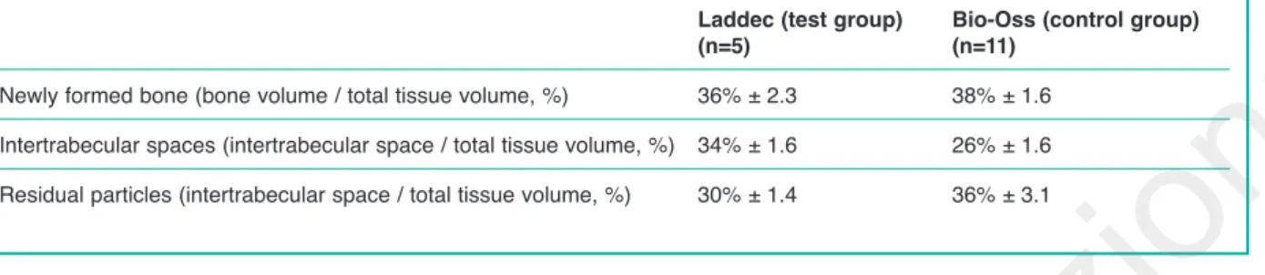
Results. For the Laddec, the newly formed bone was 36% ± 2.3; the intertrabecular spaces were 34% ± 1.6, and the
resid-ual material was 30% ± 1.4. For the Bio-Oss, the newly formed bone was 38% ± 1.6; the intertrabecular spaces were 26% ± 1.6, whereas the residual material was 36% ± 3.1.
Conclusions. Both the xenoimplants obtained a good bone regeneration with a satisfying quantity of newly formed bone
and reduced quantity of fibrous bone. The Laddec showed a better absorbability compared with Bio-Oss, whose resid-ual percentage is greater for the same elapsed time.
Key words: sinus floor augmentation, bone grafting, biomaterials, bone regeneration, maxillary sinus, histology.
©
CIC
Edizioni
original research article
the placement of the fixture/es (9-11). In max-illofacial surgery and in dentistry, the subject of controversy that still exists is the choice of the most appropriate graft material for the sinus floor augmentation. In this field, the autogenous bone seems to show the best performance, in a early phase (12). But the collection of autoge-nous bone requires an extra donor site surgery, and is associated with extra risks for morbidity and complaints of the patient. For these reasons, other graft materials are suggested (13). Sinus grafting materials may produce bone formation by osteogenesis, osteoinduction, and osteocon-duction. Whereas osteogenesis is obtained by providing osteogenic cells and matrix directly in the graft (e.g. autogenous bone), osteoinduction postulates that the grafted material is chemotac-tic to undifferentiated progenitor cells inducing them to differentiate into osteoblasts, while teoconduction permits the outgrowth of os-teogenic cells from existing bone surfaces into the graft material. Among the osteoconductive materials, there is a highly purified, xenogeneic graft biomaterial, derived by deproteinized, ster-ilized bovine bone (Laddec®) which has been
in-troduced to clinicians for its innovative features (the collagen type I matrix is preserved in asso-ciation with crystals of hydroxyapatite), and re-cently evaluated in literature (14). Consequently, the organic phase of this material (the collagen type I matrix) is not removed. This material is obtained from bovine bone after a wash with dis-tilled water and a phosphate buffer (0.4 M, pH 7.4), followed by defatting at a temperature <50 °C with ethanol/dichloromethane, and after a proteoglycan removal by urea and mercap-toethanol (International patent: PCT/WO/91/ 07194). This process of production seems able to preserve the collagen type I fibres in the matrix of this xenograft (14).
In addition, for this material are reported physi-cal characteristics very similar to human cancel-lous bone (i.e.: an average thickness of trabecu-lae of about 160 μm, an intertrabecular space of about 340 μm, and the presence of about 2 tra-beculae per mm of tissue) (14).
This material seems to stimulate osteoblastic
ac-tivity in preclinical study: it seems able to facil-itate the formation of multiple cell layers, and to increase the expression of alkaline phosphatase in mesenchymal cell cultures (15).
Subsequent clinical and histological studies, al-so conducted in humans, indicate this material as a reliable option in other oral and maxillofacial surgery procedures (as cyst enucleation or hori-zontal ridge augmentation) (15, 16).
Data about its use for sinus floor augmentation were also recently reported, but data are not con-trolled in a split-mouth design (14).
The aim of this pilot split-mouth controlled hu-man study is to evaluate histologic and histomor-phometric results of this highly purified xenogenic grafting material in maxillary sinus floor augmentation after six months of follow-up.
Methods
Subjects
This pilot split-mouth controlled study was con-ducted on 11 patients (7 females and 4 males; mean age 53 +/- 7.9 years; age range 41-68 years), who underwent maxillary sinus floor augmentation.
The subjects were selected according to the fol-lowing inclusion criteria: the presence of unilat-eral/bilateral edentulous upper jaw, residual maxillary sinus floor <5 mm. Particular attention paid to the bacterial load of the oral cavity due to the multiple possible interactions, assessed by salivary tests (17-20). Exclusion criteria were: smoking, periodontal disease, maxillary sinus pathologies, systemic con-traindications for surgical rehabilitation. All subjects signed an informed consent form be-fore being enrolled in the study. The study pro-tocol was in agreement with the Helsinki Decla-ration for studies conducted on humans and was approved by the Ethical Committee of the Uni-versity of L’Aquila.
©
CIC
Edizioni
original research article
Materials
The following biomaterials for sinus floor aug-mentation were used:
• Laddec (xenogeneic biomaterial, Ost
Devel-opment, Clermont-Ferrand, France; typology granular 600μm): highly purified bovine xenograft. This was the test material.
• Bio-oss (xenogeneic biomaterial, Geistlich,
Tiani, Italy; granular types: small 0.25-1 mm; and large 1-2 mm): natural bone mineral. This was the control material.
Six patients (4 females, 2 males) were subjected to unilateral major maxillary sinus floor aug-mentation, and were treated with Bio-oss. Five patients (3 females, 2 males) were subject-ed to bilateral major maxillary sinus floor aug-mentation, by using Bio-oss in one side and Lad-dec in the other side.
Consequently, the test group (Laddec) included 5 samples, while the control group (Bio-oss) in-cluded 11 samples.
Procedures
The following surgical protocol was applied: 1) a first surgical phase of major maxillary sinus
floor augmentation (at t0)
2) a second surgical phase after six months, at the time of implants insertion, during which were obtained the bone samples (at t1).
First surgical stage (t0)
The initial clinical situation was documented through preoperative, intra- and extra-oral pho-tos of the patient, and a panoramic X-ray exam-ination. An X-ray dental CT scan examination was also prescribed. Before surgery, the patients were subjected to antibiotic prophylaxis with penicillin, per os, 1 g every 12 hours, 3 days be-fore the surgery, and for 7 days after the surgery.
The surgical procedures were all conducted by the same operator.
After a local anaesthesia with Articaine 40 mg/ ml with adrenaline 1:100.000 vasoconstrictor (Ubis-tesin 3M ESPE), the maxillary sinus floor aug-mentation was performed using the lateral window technique (with sinus lift surgical technique) (21). A full-thickness flap was separated with a manu-al periostemanu-al elevator, and the opening of a bone window was performed on the lateral side of the sinus, using a 2 mm tungsten carbide surgical bur, with cooling by saline solution. The next step was the detachment of the Schneiderian membrane with manual periosteal elevators. The bone win-dow was then reversed, so that it formed the new sinus floor, under which the tested material (Lad-dec or Bio-oss) was grafted, following the prepa-ration procedures indicated by the manufacturers. The result was an increased bone thickness, use-ful for the subsequent implant restoration. No protective membrane was applied on the bone window. The soft tissues were thereafter reposi-tioned above the bone window, with the applica-tion of non-absorbable sutures in polimid 4/ with a 16 gauge, 3/8 circle needle. Removal of the su-tures was performed after 14 days.
Second surgical stage (t1)
At the time of the implants insertion, six months after the sinus floor augmentation, a sample of newly formed tissue was taken from the graft sites. The blocks were carefully obtained using a trephine bur with copious saline irrigation.
Preparation of the samples
Then, the samples were left in a fixative for 9 days - formaldehyde at 4% dilution - and then, after dehydration with uncatalyzed and hypo-catalyzed solutions, included in methacrylate blocks. Histologic sections of 5 microns (µm) were obtained using a microtome (22).
©
CIC
Edizioni
original research article
Some sections were stained with methylene
blue/azure II, to individuate the structural
pa-rameters, whereas the remaining sections were stained with tartrate-resistant acidic phosphatase (TRAP) to verify the presence of osteoclasts (23).
TRAP-positive cells were individuated on light microscope images of the region of interest.
Histological and
histomorphometric analysis
The histomorphometric outcomes were:
- the percentage of bone volume on total tissue volume, which expresses the quantity of new-ly formed bone
- the percentage of intertrabecular spaces on total tissue volume
- the percentage of residual material on total tissue volume.
The measurements were performed using an in-teractive software for image analysis (IAS 2000, Delta Sistemi, Rome, Italy), which automatical-ly calculates the values (24).
The differences between the two materials were evaluated with the Chi-square test, the signifi-cance was set at 0.05.
Results
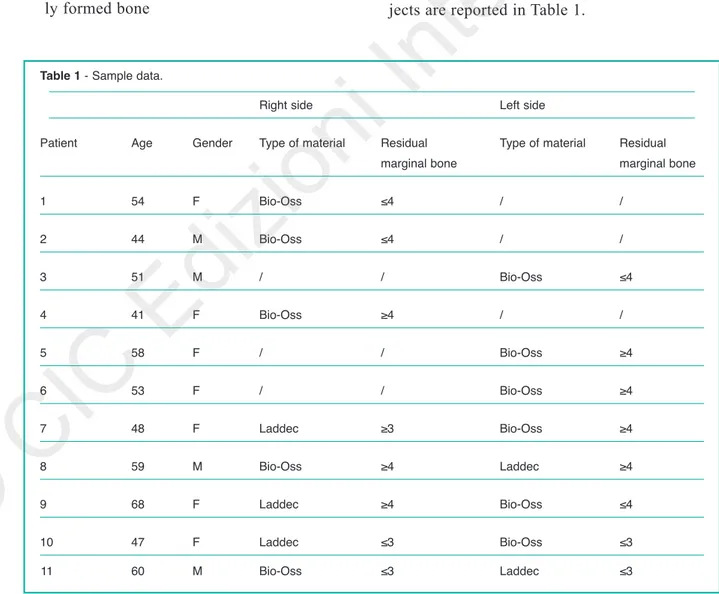
Descriptive initial data about the included sub-jects are reported in Table 1.
Table 1 - Sample data.
Right side Left side
Patient Age Gender Type of material Residual Type of material Residual
marginal bone marginal bone
1 54 F Bio-Oss ≤4 / / 2 44 M Bio-Oss ≤4 / / 3 51 M / / Bio-Oss ≤4 4 41 F Bio-Oss ≥4 / / 5 58 F / / Bio-Oss ≥4 6 53 F / / Bio-Oss ≥4 7 48 F Laddec ≥3 Bio-Oss ≥4 8 59 M Bio-Oss ≥4 Laddec ≥4 9 68 F Laddec ≥4 Bio-Oss ≤4 10 47 F Laddec ≤3 Bio-Oss ≤3 11 60 M Bio-Oss ≤3 Laddec ≤3
©
CIC
Edizioni
Internazionali
original research article
At t1, from a clinical point of view, no compli-cations were observed in any of the patients. The results of the histomorphometric analysis are re-ported in Table 2.
In general, the microscopic examination of all the processed specimens confirmed the presence newly formed bone. No acute inflammatory in-filtrate was evident.
Bio-Oss
At the histological evaluations, some sections showed the presence of osteoblastic activities, with newly formed bone directly attached to the surface of the particles of graft material, most of which appear surrounded by mature and compact bone, without bone gaps along the in-terface. The bone always results in close con-tact with the particles of the graft material. Furthermore, no inflammatory infiltrate of any kind was detected.
Laddec
At the histological evaluation, the newly formed bone tissue, which shows the characteristics of lamellar type, is not detected in the immediate bone-graft material interface. On the contrary, the presence of small capillaries, fibroblasts, and macrophages is detected, even if at a more dis-tant site. Nonetheless, in the inner part of most of the particles of the grafting material, a tissue with a typical colour of the newly formed bone
was detected, whereas on the surface of the same particles, numerous osseous gaps were identi-fied. There is, however, no necrotic reaction, no inflammatory infiltrate or foreign body reaction.
Discussion
The purpose of this histologic and histomorpho-metric evaluation is to evaluate the interactions that occur between the bone and the graft of a test and control osteoconductive xenogeneic graft materials. The clinical findings confirmed that both the xenogeneic bone substitutes, when associated with the sinus lift surgical technique occurred in recent years, allow the placement of implants in atrophic maxillary bone, regenerated with xenografts. In addition, the present findings also showed that Laddec is a suitable material for sinus floor augmentation. Despite the clinical success of xenogeneic graft materials, only few histomorphometric data are reported in the pub-lished literature about Laddec (14).
To the best of our knowledge this is the first split-mouth controlled study about it. No evi-dence of acute inflammatory infiltrate was found in any specimen in the present investigation. This also confirms that Laddec seems to not in-duce adverse immunologic response.
Specimen from the Laddec group showed newly formed bone of 36% ± 2.3, intertrabecular space of 34% ± 1.6, and residual particles of 30% ± 1.4. Bio-Oss showed 36% ± 3.1 of residual particles. The Laddec showed a better absorbability com-pared with Bio-Oss, whose residual percentage
Table 2 - Results of the hystomorphometric analysis.
Laddec (test group) Bio-Oss (control group)
(n=5) (n=11)
Newly formed bone (bone volume / total tissue volume, %) 36% ± 2.3 38% ± 1.6
Intertrabecular spaces (intertrabecular space / total tissue volume, %) 34% ± 1.6 26% ± 1.6 Residual particles (intertrabecular space / total tissue volume, %) 30% ± 1.4 36% ± 3.1
©
CIC
Edizioni
original research article
was greater for the same elapsed time. The resid-ual percentage of Bio-Oss observed in the present investigation is in accordance with literature, for the same elapsed time of 6 months (25, 26). For the Laddec, the present findings appear less encouraging respect to the pre-existing litera-ture, that reports newly formed bone of 64.72% ± 3.44, after 6 months in human specimen (in a study with fifteen subjects, not controlled) (14). The more encouraging results obtained in that sample could be associated to the use of a mem-brane that was positioned against the packed si-nus window, while no membrane was used in the present investigation.
The percentage of residual material over the to-tal tissue was 16.93%, while the present sample shows 30%. The membrane could have influ-enced the absorbability of the graft particles. In the present study, the Bio-Oss material is used as a control material, because it was consider-ably previously analyzed in literature, while lower evidence is reported for the Laddec. In light of this, from our comparative study we may say that all the biomaterials used gave good results with individual characteristics that allow preferring one to another.
For the Bio-Oss, in the present investigation, the newly formed bone resulted 38% ± 1.6; the in-tertrabecular space was 26% ± 1.6, whereas the residual material was equal to 36% ± 3.1. These data are comparable with previous literature. In a study conducted on 20 subjects analyzed af-ter 6 months from sinus augmentation, anorgan-ic bovine bone showed a newly formed bone of about 25% (25.12% ± 7.25%), and residual bio-material of about 29% (28.65% ± 9.70%) (27). In other studies on xenografts were reported da-ta of 24.63% and 29.13% of newly formed bone after 6-8 months from the sinus floor augmenta-tion (28, 29).
It was also reported a newly formed bone of 21.1% after six months in a single clinical case (30). Thus, the results obtained in the present investi-gation (38% ± 1.6 of newly formed bone) seem slightly more encouraging than the percentages of data reported by previous literature with Bio-Oss. This may depend on the different gravity of
the initial cases, and on the individual suscepti-bility.
Concerning the anatomy, we can assert that there are anatomies characterized by a greater blood supply that facilitate the incorporation of the graft, in which the new formation of bone can be found to be greater (21).
Another hypothesis could be the size of granules (in the present study was used a xenogeneic bio-material with granular type small 0.25-1 mm and large 1-2 mm), according to a recent finding that the newly bone formation appears more exten-sive in the large particle grafts compared with the small particle grafts (26.77% ± 9.63% vs 18.77% ± 4.74%, respectively) after six months in human specimen (25).
Conclusions
The xenoimplants (Laddec and Bio-Oss) both obtained a good bone regeneration with a satis-fying quantity of newly formed bone. The Lad-dec showed a better absorbability compared with Bio-Oss, whose residual percentage is greater for the same elapsed time. Further studies with Laddec are encouraged on larger samples in or-der to confirm its optimal outcome for sinus aug-mentation.
References
1. Kashi A, Saha S. Evidence-based techniques to assess
the performance of dental implants. J Oral Implantol. 2013 Dec;39(6):655-61.
2. Frascaria M, Casinelli M, Marzo G, Gatto R, Baldi M,
D’Amario M. Digital implant planning for a minimally invasive surgery approach: a case letter of a full-arch rehabilitation. J Oral Implantol. 2015 Apr;41(2):205-8.
3. Arcuri L, De Vico G, Ottria L, Condò R, Cerroni L,
Mancini M, et al. Smart fusion vs. double scan: a com-parison between two data-matching protocols for a computer guided implant planning. Clin Ter. 2016;167(3):55-62.
4. Mummolo S, Marchetti E, Albani F, Campanella V,
Pugliese F, Di Martino S, et al. Comparison between
©
CIC
Edizioni
original research article
rapid and slow palatal expansion: evaluation of se-lected periodontal indices. Head Face Med. 2014 Aug 15;10:30.
5. Andreasi Bassi M, Lopez MA, Andrisani C, Ormanier
Z, Gargari M. Full arch rehabilitation in severe maxil-lary atrophy with palatal approach implant placement: a case report. Oral Implantol (Rome). 2016;9(3):115-22.
6. Cho-Lee G-Y, Naval-Gias L, Castrejon-Castrejon S,
Capote-Moreno AL, Gonzalez-Garcia R, Sastre-Perez J, et al. A 12-year retrospective analytic study of the im-plant survival rate in 177 consecutive maxillary sinus augmentation procedures. Int J Oral Maxillofac Im-plants. 2010;25(5):1019-27.
7. Spinelli D, DE Vico G, Condò R, Ottria L, Arcuri C.
Transcrestal guided sinus lift without grafting materi-als: a 36 months clinical prospective study. Oral Im-plantol (Rome). 2016;8(2-3):74-86.
8. Di Girolamo M, Calcaterra R, Di Gianfilippo R, Arcuri
C, Baggi L. Bone level changes around platform switching and platform matching implants: a system-atic review with meta-analysis. Oral Implantol (Rome). 2016;9(1):1-10.
9. Cannizzaro G, Felice P, Minciarelli AF, Leone M,
Vi-ola P, Esposito M. Early implant loading in the at-rophic posterior maxilla: 1-stage lateral versus crestal sinus lift and 8 mm hydroxyapatite-coated implants. A 5-year randomised controlled trial. Eur J Oral Implan-tol. 2013;6(1):13-25.
10. Marchetti E, Ratta S, Mummolo S, Tecco S, Pecci R, Bedini R, et al. Evaluation of an endosseous oral im-plant system according to UNI EN ISO 14801 fatigue test protocol. Implant Dent. 2014 Dec;23(6):665-71. 11. Marrelli M, Vertucci V, Amantea M, Maletta C,
Codis-poti B, Gargari M, et al. Multiparametric evaluation of fitting accuracy for different combinations of implant-abutment coupling at marginal interface. Oral Implan-tol (Rome). 2018;11(1):77-86.
12. Handschel J, Simonowska M, Naujoks C, Depprich RA, Ommerborn MA, Meyer U, et al. A histomorpho-metric meta-analysis of sinus elevation with various grafting materials. Head Face Med. 2009 Jun 11;5:12. 13. Iorio-Siciliano V, Marzo G, Blasi A, Cafiero C,
Mignogna M, Nicolò M. Soft and hard tissue modifi-cations at immediate transmucosal implants (with Laser-Lok microtextured collar) placed into fresh ex-traction sites: a 6-month prospective study with surgi-cal reentry. Int J Periodontics Restorative Dent. 2014;34(4):541-9.
14. Guarnieri R, Belleggia F, Ippoliti S, DeVilliers P, Ste-fanelli LV, Di Carlo S, et al. Clinical, Radiographic, and Histologic Evaluation of Maxillary Sinus Lift Proce-dure Using a Highly Purified Xenogenic Graft
(Lad-dec(®)). J Oral Maxillofac Res. 2016;7(1):e3.
15. Pappalardo S, Guarnieri R. Efficacy of Platelet-Rich-Plasma (PRP) and Highly Purified Bovine Xenograft (Laddec(®)) Combination in Bone Regeneration after
Cyst Enucleation: Radiological and Histological Eval-uation. J Oral Maxillofac Res. 2013;4(3):e3. . 16. Guarnieri R, DeVilliers P, Belleggia F. GBR using
cross-linked collagen membrane and a new highly pu-rified bovine xenograft (Laddec) in horizontal ridge augmentation: Case report of clinical and histomor-phometric analysis. Quintessence Int. 2015 Sep;46 (8):717-24.
17. Mummolo S, Tieri M, Tecco S, Mattei A, Albani F, Giuca MR, et al. Clinical evaluation of salivary indices and levels of Streptococcus mutans and Lactobacillus in patients treated with Occlus-o-Guide. Eur J Paediatr Dent. 2014 Dec;15(4):367-70.
18. Mummolo S, Marchetti E, Giuca MR, Gallusi G, Tecco S, Gatto R, et al. In-office bacteria test for a microbial monitoring during the conventional and self-ligating or-thodontic treatment. Head Face Med. 2013 Feb 1;9:7. 19. Mummolo S, Nota A, Caruso S, Quinzi V, Marchetti E, Marzo G. Salivary Markers and Microbial Flora in Mouth Breathing Late Adolescents. Biomed Res Int. 2018;2018:8687608.
20. Marchetti E, Mummolo S, Di Mattia J, Casalena F, Di Martino S, Mattei A, et al. Efficacy of essential oil mouthwash with and without alcohol: a 3-day plaque accumulation model. Trials. 2011 Dec 15;12:262. 21. Romero-Millán J, Martorell-Calatayud L, Peñarrocha
M, García-Mira B. Indirect osteotome maxillary sinus floor elevation: an update. J Oral Implantol. 2012 Dec;38(6):799-804.
22. Pettinicchio M, Traini T, Murmura G, Caputi S, Degidi M, Mangano C, et al. Histologic and histomorphome-tric results of three bone graft substitutes after sinus augmentation in humans. Clin Oral Investig. 2012 Feb;16(1):45-53.
23. Schulze-Späte U, Dietrich T, Kayal RA, Hasturk H, Dobeck J, Skobe Z, et al. Analysis of bone formation after sinus augmentation using β-tricalcium phosphate. Compend Contin Educ Dent. 2012 May;33(5):364-8. 24. Martinez A, Franco J, Saiz E, Guitian F. Maxillary
si-nus floor augmentation on humans: Packing simula-tions and 8 months histomorphometric comparative study of anorganic bone matrix and β-tricalcium phos-phate particles as grafting materials. Mater Sci Eng C Mater Biol Appl. 2010 Jun 15;30(5):763-9.
25. Testori T, Wallace SS, Trisi P, Capelli M, Zuffetti F, Del Fabbro M. Effect of xenograft (ABBM) particle size on vital bone formation following maxillary sinus aug-mentation: a multicenter, randomized, controlled, clin-ical histomorphometric trial. Int J Periodontics Restora-tive Dent. 2013;33(4):467-75.
26. Iezzi G, Degidi M, Piattelli A, Mangano C, Scarano A, Shibli JA, et al. Comparative histological results of dif-ferent biomaterials used in sinus augmentation proce-dures: a human study at 6 months. Clin Oral Implants Res. 2012 Dec;23(12):1369-76.
27. Di Stefano DA, Gastaldi G, Vinci R, Cinci L, Pieri L, Gherlone E. Histomorphometric Comparison of
En-©
CIC
Edizioni
original research article
zyme-Deantigenic Equine Bone and Anorganic Bovine Bone in Sinus Augmentation: A Randomized Clinical Trial with 3-Year Follow-Up. Int J Oral Maxillofac Implants. 2015;30(5):1161-7.
28. Panagiotou D, Özkan Karaca E, Dirikan İpçi Ş, Çakar G, Olgaç V, Yılmaz S. Comparison of two different xenografts in bilateral sinus augmentation: radiographic and histologic findings. Quintessence Int. 2015;46 (7):611-9.
29. Calasans-Maia MD, Mourão CF de AB, Alves ATNN, Sartoretto SC, de Uzeda MJPG, Granjeiro JM. Maxil-lary Sinus Augmentation with a New Xenograft: A Randomized Controlled Clinical Trial. Clin Implant
Dent Relat Res. 2015 Oct;17 Suppl 2:e586-93. 30. Ohayon L. Histological and histomorphometric
evalu-ation of anorganic bovine bone used for maxillary si-nus floor augmentation: a six-month and five-year fol-low-up of one clinical case. Implant Dent. 2014 Jun;23(3):239-44.
Correspondence to: Stefano Mummolo
Department of Life, Health and Environmental Sciences University of L’Aquila, L’Aquila, Italy
E-mail: [email protected]