Lake 2016
: Conference on Conservation and Sustainable Management of Ecologically Sensitive Regions in Western Ghats[THE 10TH
BIENNIAL LAKE CONFERENCE] Date: 28-30th December 2016, http://ces.iisc.ernet.in/energy
Venue: V.S. Acharya Auditorium, Alva's Education Foundation, Sundari Ananda Alva Campus, Vidyagiri, Moodbidri, D.K. Dist., Karnataka, India – 574227
Tetrahymena thermophila: A whole cell biosensor for toxicity assessment of
Mercury
Santosh Kumar
1, 2, 3*, Daizy Bharti
1, 3, Khushboo Kashyap
1,Harpreet Kaur
1, Komal
Kamra
1,*, Antonietta La Terza
3,*1
Ciliate Biology Laboratory, SGTB Khalsa College, University of Delhi, Delhi 110 007, India.
2
Department of Biological Sciences, College of Natural Sciences, University of Ulsan, Ulsan 44610, South Korea.
3
School of Bioscience and Veterinary Medicine, University of Camerino, Via Gentile III da Varano,62032 Camerino (MC),Italy.
*Corresponding authors. Tel.: + 82 10 2895 6025; e-mail: [email protected](Kumar S.) Tel.: + 91 98 717 71417; e-mail: [email protected] (Kamra K.)
Tel.: +39 0737 403272; e-mail:[email protected](LaTerza A.)
EXTENDED ABSTRACT
Ciliated protozoans are ubiquitous, free living, and largely non-pathogenic microorganisms. Being single cell eukaryotic microorganisms they represent an essential component of all ecosystem where they are integral constituents of trophic chains and nutrient cycles (Foissner, 1987,1999, 2004; Darbyshire, 1994; Alpheiet al., 1996; Bonkowski and Schaefer, 1997; Sherr and Sherr, 2002; Cuvelieret al., 2010; Steele et al., 2011). They have been used as model organisms for the discovery of key genomic processes found across the eukaryotic tree of life, e.g., self-splicing RNAs, telomeres, and the role of RNAs in shaping germline and somatic genomes. Unlike bacteria, fungi they lack cell wall and are only separated from external environment via cell membrane, this makes them highly sensitive to any change in the environment. Considering that ciliated protozoa shows high similarity in the conserved genes (more than 800 human genes have orthologs in Tetrahymena and out of these 58 genes are associated with human diseases; Eisenet al., 2006;Fillinghamet al., 2002) between ciliates and several eukaryotes including humans, they represents a better biological tools to detect and diagnose community-level impairments in contaminated soil and water ecosystems. Thus, ciliate represents a perfect bioindicators that can be used for assessment of ecotoxicological assays for early warning deterioration of the environment. Furthermore, in response to heavy metal pollution, they express a special protein, i.e., metallothioneins (MT) rich in cysteine (cys) amino acid in which the thiol groups are able to bind heavy metals.Further, the induction of MTs by heavy metals is mainly regulated at transcription level.
In the present study, a recombinant cell line of Tetrahymena thermophilais used to assess the toxilogical impact of heavy metal on this species (La Terzaet al., 2008). The plasmid containing the gene coding forthe Green Fluorescence Protein (GFP) under the transcriptional control of an endogenousmetallothioneinepromoter was used to transfect T. thermophilacells by electroporation, according to the method described inGaertig and Gorovsky (1992).
Logarithmically growing culture of T. thermophila was washed three times in 10 mMTris pH 7.5 to remove any trace of the PPY (Proteose-Peptone-Yeast) culture mediumwhich may hinder the effect of metalused in our toxicological tests (Mercury, Hg) by chelating the metal salts. Cell count was done using a Neubauer slide and adjusted to a known dilution using 10 mMTrispH 7.5Tris. Then the suspended cells were transferred to 96-well microplates in a final volume of 100μl.Different concentrations of metal salts were prepared in 10 mMTris (pH 7.5). For metal exposures, a fixed number of cells (0.42 x 10⁵ cells/ml) were mixed with different concentrations of metal saltsin a final volume of 200 μl (100μl cell culture + 100μl metal salt solution). The exposed cells were incubated in a dark chamber at 30°C and were observed after two hours under the 20x objective on a Nikon Diaphoto TMD inverted microscope with an attached digital camera. Florescence was detected using a filter set
Lake 2016
: Conference on Conservation and Sustainable Management of Ecologically Sensitive Regions in Western Ghats[THE 10TH
BIENNIAL LAKE CONFERENCE] Date: 28-30th December 2016, http://ces.iisc.ernet.in/energy
Venue: V.S. Acharya Auditorium, Alva's Education Foundation, Sundari Ananda Alva Campus, Vidyagiri, Moodbidri, D.K. Dist., Karnataka, India – 574227
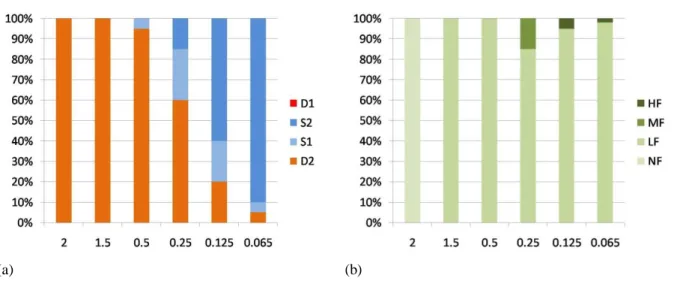
with an excitation wavelength of 470-490 nm and an emission wavelength of 520 nm. Cells showing florescence were counted on the Neubauer slide; at 2 min prior to observation, the cells were treated with dibucaine hydrochloride at a final concentration of 0.3 mM to reduce the motility without affecting viability. Florescence emission was also checked, though never detected, in the controlsamples.Based on microscopic observations, four character states (i.e., endpoints) with regard to survival weremonitored/measured, i.e., D1= death by bursting, D2= Death after formation of atypical structures,S1= Survival after formation of atypical structures, S2= Survival with normalstructure and motility. Based on the intensity of the fluorescence, four character states (endpoints) were monitored/measured – NF= no fluorescence, LF= low level of fluorescence, MF= medium level of fluorescence, HF= high level of fluorescence. In general, mercury was found to be highly toxic to the cells,; following the exposure,all cells showing atypical shapes were destined to die (Figure 1, Table 1). At metal concentrations of 2μg/ml and 1.5μg/ml, no cells survive. LC50 value (at 2 hrs) is close to 0.25μg/ml. A few cells die even at a low concentration of 0.065μg/ml. At a concentration of 2μg/ml, no cells show fluorescence. However, at 1.5μg/ml, all the cells destined to die exhibit low fluorescence. At concentrations below 0.5μg/ml some cells show medium to high fluorescence while all others show low fluorescence (Figure 2).
Metals concentrations <1.5 1.5 0.5 0.25 0.125 0.065 Mercury 100% D2 100% NF 100% D2 100% LF 95% D2 5% S1 100% LF 60% D2 25% S1 15% S2 85% LF 15% MF 20% D2 20% S1 60% S2 95% LF 5% FF 5% D2 5% S1 90% S2 98% LF 2% FF
Table 1. The effect of different concentrations of metals on the recombinant cell line of Tetrahymena
thermophile. Upper row (bold) shows different concentration used; lower row shows the effect of the
concentration; metal concentrations in μg/ml; cell concentration of 3 x 10⁵
cells/ml.
(a) (b)
Figure 1. Response of Tetrahymena thermophila to Mercury.(a) Showing cell percent viability.(b) Showing percent florescence. X axis – Mercury concentration (µg/ ml). Y axis in a) – viability, in b) fluorescence.
Lake 2016
: Conference on Conservation and Sustainable Management of Ecologically Sensitive Regions in Western Ghats[THE 10TH
BIENNIAL LAKE CONFERENCE] Date: 28-30th December 2016, http://ces.iisc.ernet.in/energy
Venue: V.S. Acharya Auditorium, Alva's Education Foundation, Sundari Ananda Alva Campus, Vidyagiri, Moodbidri, D.K. Dist., Karnataka, India – 574227
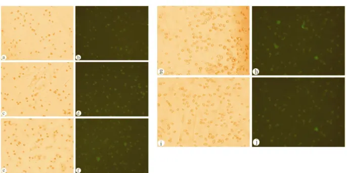
Figure 2.Photomicrographs of cells in response to different concentrations of Mercury; a, b) at concentration 1.5µg/ml; c, d) at concentration 0.5µg/ml; e, f) at concentration 0.25µg/ml; g, h) at concentration 0.125µg/ml; i, j) at concentration 0.065µg/ml.(a, c, e, g, i) cells in bright field. (b, d, f, h, j) cells showing florescence.
Acknowledgements
The study was funded by the Italian Minister of University and Research (MIUR) to ALT through the assignation of “Young Indian Research Fellowship” in the frame of a project of cooperation between Italy and India. The support from Department of Science and Technology under - Research Projects- SR/SO/AS-04/2004 and EMR/2015/001505 are acknowledged. The guidance provided by. Prof. G. R. Sapra (Retd.) and Prof. Rup Lal, University of Delhi, India, is greatly acknowledged.
References
Alphei J., Bonkowski M. & Scheu S. (1996). Protozoa, Nematoda and Lumbricidae in the rhizosphere of
Hordelymuseuropaeus (Poaceae): faunal interactions, response of microorganisms and effects on plant growth.
Oecologia. 106, 111–126.
Bonkowski M. & Schaefer M. (1997). Interactions between earthworms and soil protozoa: a trophic component in the soil food web. Soil Biol.Biochem.. 29, 499–502.
Cuvelier M.L., Allen A.E., Moniera A., McCrow J.P., Messié M, Tringe S.G. et al. (2010). Targeted metagenomics and ecology of globally important uncultured eukaryotic phytoplankton. Proc. Natl. Acad. Sci. USA. 107,14679–14684
Darbyshire J.F.(editor). (1994). Soil Protozoa. CAB International. Oxon.
Eisen J.A., Coyne R.S., Wu M., Wu D., Thiagarajan M., et al. (2006). Macronuclear genome sequence of the ciliate Tetrahymena thermophila, a model eukaryote., PloS Biol. 4, e286.
Fillingham J.S., Chilcoat N.D., Turkewitz A.P., Orias, E., Reith M. & Pearlman R.E. (2002). Analysis of expressed sequence tags (ESTs) in the ciliated protozoan Tetrahymena thermophila. J.Eukaryot.Microbiol., 49, 99–107.
Lake 2016
: Conference on Conservation and Sustainable Management of Ecologically Sensitive Regions in Western Ghats[THE 10TH
BIENNIAL LAKE CONFERENCE] Date: 28-30th December 2016, http://ces.iisc.ernet.in/energy
Venue: V.S. Acharya Auditorium, Alva's Education Foundation, Sundari Ananda Alva Campus, Vidyagiri, Moodbidri, D.K. Dist., Karnataka, India – 574227
Foissner W. (1987). Neueterrestrische und limnischeCiliaten(Protozoa, Ciliophora) ausOsterreich und Deutschland.Sitzungsberichte der osterreichischenAkademie der Wissenschaften.Wien., 195, 217–268.
Foissner W. (1999a). Soil protozoa as bioindicators: pros and cons, methods,diversity, representative examples. Agric.Ecosyst.Environ..74, 95–112.
Foissner W. (1999b). Protist diversity: estimates of the near-imponderable. Protist. 150,363–368.
Foissner W. (2004). Some new ciliates (Protozoa, Ciliophora) from an Austrian flood plain soil, including a giant, red “flagship”, Cyrtohymena (Cyrtohymenides) aspoeckinov.subgen., nov. spec. Denisia. 13, 369–382. Gaertig J. & M.A. Gorovsky. (1992). Efficient mass transformation of Tetrahymena thermophila by electroporation of conjugants. Proc. Natl. Acad. Sci. USA.89, 9196-200.
La Terza A., Barchetta S., Buonanno F., Ballarini P., Miceli C. (2008).The protozoan ciliate Tetrahymena
thermophila as biosensor of sublethal levels of toxicants in the soil.Fresenius Environmental Bulletin. 17,1144–
1150.
Sherr E.B. &SherrB.F.(2002) Significance of predation by protists in aquatic microbial food webs. Anton.Leeuw. Int. J. G. 81,293–308.
Steele J.A., Countway P.D., Xia L., Vigil P.D., Beman J.M., Kim D.Y., et al. (2011). Marine bacterial, archaeal and protistan association networks reveal ecological linkages. ISME J. 5, 1414–1425.