Oregano
feed
supplementation
affects
glycoconjugates production in swine gut
Francesca Mercati, Cecilia Dall’Aglio, Gabriele Acuti, Valerio Faeti, Federico Maria Tardella, Carolina Pirino, Elena De Felice, Paola Scocco
Material and methods
Extraction and analysis of oregano aqueous extract (OAE)
The OAE was obtained by a process of bio-liquefaction based on enzyme bio-catalysis [1]as previously
described by Franciosini et al. [2]. Briefly, the plant material was boiled in water, and treated with a specific
enzymatic preparation after cooling for four hours, and finally filtered. The OAE obtained was analyzed to
quantify
- antioxidant capacity, measured in terms of radical scavenging ability using the stable radical DPPH [3],
- total polyphenols, evaluated using the Folin-Ciocalteu reagent [4],
- reducing sugars was evaluated using the ADNS method [5].
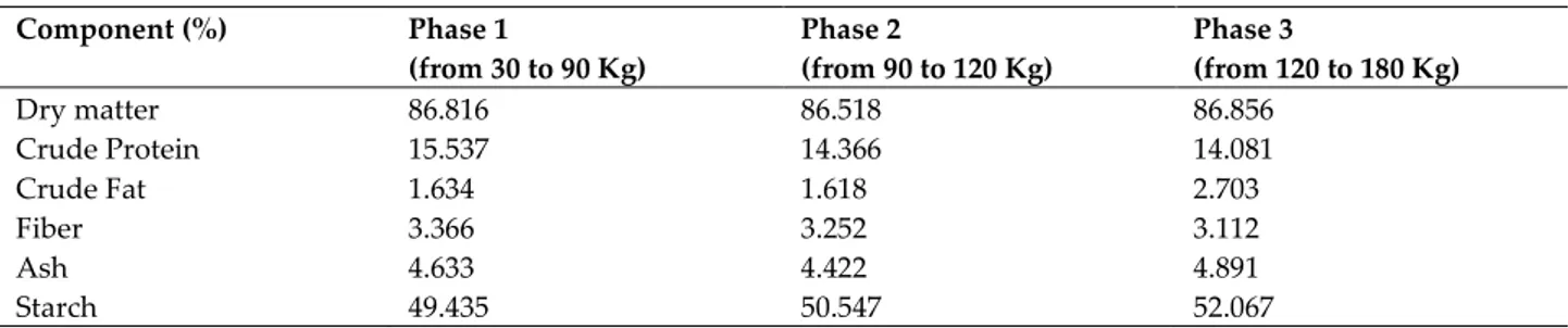
Table S1. Feed chemical analysis in the three-phase feeding program Component (%) Phase 1 (from 30 to 90 Kg) Phase 2 (from 90 to 120 Kg) Phase 3 (from 120 to 180 Kg) Dry matter 86.816 86.518 86.856 Crude Protein 15.537 14.366 14.081 Crude Fat 1.634 1.618 2.703 Fiber 3.366 3.252 3.112 Ash 4.633 4.422 4.891 Starch 49.435 50.547 52.067
Table S2 Sugar moieties visualized by performed histochemical treatments
AB= Alcian blue; Sial= sialidase; PAS= periodic acid Schiff; LID= low iron diamine; HID= high iron diamine
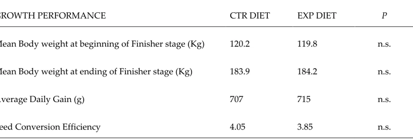
Table S3 Animal performances
GROWTH PERFORMANCE CTR DIET EXP DIET P
Mean Body weight at beginning of Finisher stage (Kg) 120.2 119.8 n.s.
Mean Body weight at ending of Finisher stage (Kg) 183.9 184.2 n.s.
Average Daily Gain (g) 707 715 n.s.
Feed Conversion Efficiency 4.05 3.85 n.s.
CTR: degermed corn-barley-soybean-based.
EXP: CTR diet supplemented (2 g/kg) with an oregano aqueous extract. P<0.05; n.s.: not significant
HISTOCHEMICAL
TREATMENTS SUGAR MOIETIES VISUALISED
AB pH2.5
Acidic groups: Sialic acid (SA), carboxylated (Hyaluronic acid, Chondroitin) and sulphated (Chondroitin-sulphates A/B/C, Heparin, Heparan sulphate) Glycosaminoglycan (GAG)-like material
Sial-AB Acidic groups without C4 not acetilated SA
KOH-Sial-AB Asialilated acidic groups (GAG-like material)
AB pH1 Sulphated GAG-like material (Chondroitin-sulphates A/B/C, Heparin, Heparan sulphate)
AB pH0.5 Highly sulphated GAG-like material (Heparin, Heparan sulphate)
PAS Vicinal hydroxyls (neutral and sialilated glycoproteins, GAG-like material)
AB/PAS Acidic groups and vicinal hydroxyls
LID Acidic groups
Table S4 Absolute P values of histochemical response difference in pig duodenum secretory structures
AB = Alcian Blue; Sial = Sialidase; PAS = Periodic Acid Shiff; LID = Low Iron Diamine; HID = High Iron Diamine
a = villus apical portion; b = villus basal portion
PIG DUODENUM P Histochemical treatment Goblet cells Duodenal glands AB pH2.5 0.01116 1 Sial-AB 0.9336 - KOH-Sial-AB 0.01066 - AB pH1 0.01471 - AB pH0.5 0.01066 - PAS a 0.8294 - b 0.1425 AB/PAS a 0.007937 1 b 0.01167 LID 0.01066 1 HID 0.01018 -
Table S5 Absolute P values of histochemical response difference in pig colon goblet cells PIG COLON P Histochemical treatments AB pH2.5 1 Sial-AB 0.01193 KOH-Sial-AB 0.01193 AB pH1 0.05701 AB pH0.5 0.0144 PAS 1 AB/PAS 0.01167 LID 1 HID 0.01471
AB = Alcian Blue; Sial = Sialidase; PAS = Periodic Acid Shiff; LID = Low Iron Diamine; HID = High Iron Diamine.
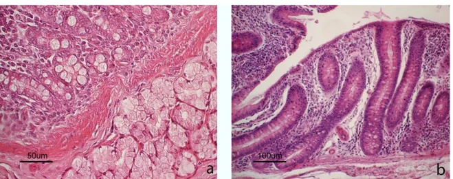
Figure S1 Light micrograph of (a) duodenum and (b) colon. In the duodenum, glandular crypts and serous duodenal glands in the tunica submucosa are showed. In the colon, deep glandular crypts are evident. Hematoxylin-eosin staining.
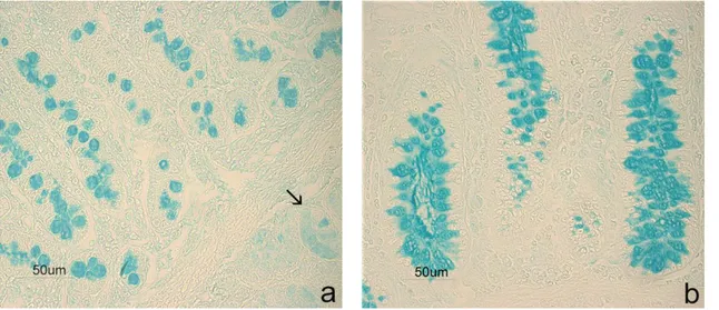
Figure S2. Controls for enzyme effectiveness. Sections incubated with enzyme-free buffer showed AB strong reactivity in swine duodenal goblet cells (a) and submucosal glands (↑), as well in colon goblet cells (b).
References
1. Setti, L.; Zanichelli, D. Bioliquefaction as a bio-refinery’s approach for the production of natural bioactive compounds for functional cosmetics. In: Morselli L, Passarini F, Vassura I, editors. Waste recovery: strategies, techniques and applications in Europe. Italy, Milano: Franco Angeli; 2009, 122–128.
2. Franciosini, M.P.; Casagrande-Proietti, P.; Forte, C.;Beghelli, D.;Acuti, G.;Zanichelli, D.; dal Bosco, A.;Castellini, C.;Trabalza-Marinucci, M.Effects of oregano (Origanumvulgare L.) and rosemary (Rosmarinus officinalis L.) aqueous extracts on broiler performance, immune function and intestinal microbial population. J. Appl. Anim. Res. 2016, 44(1), 474–479.
3. Donglin, Z.; Yasunori, H. Phenolics, ascorbic acid, carotenoids and its antioxidant activity of broccoli and their changes during conventional and microwave cooking. Food Chem. 2004, 88, 503–509.
4. Ainsworth, E.A.; Gillespie, K.M. Estimation of total phenolics content and other oxidation substrates in plant tissues using Folin-Ciocalteu reagent. Nat Protoc. 2007, 2, 875–877.
5. Bailey, M.J.; Biely, P.; Poutanen, K. Interlaboratory testing of methods for assay of xilanase activity. J. Biotechnol. 1992, 23, 257–270.