Research Doctorate Course in Bioengineering
XXV Cycle
Final dissertation
A bioengineering approach aimed at understanding
the role of hemodynamic forces acting
on human saphenous vein after
coronary artery by-pass grafting
Marco PIOLA
Advisorsprof. Monica Soncini prof. Gianfranco B. Fiore
Coordinator of the Research Doctorate Course prof. Maria Gabriella Signorini
Tutor
prof. Alberto Redaelli
3
Table of contents
Table of contents ... 3
Introduction ... 6
Rationale of the doctoral project ... 8
Outline of the thesis ... 11
Tools and procedures for ex vivo vein arterialization, preconditioning and tissue engineering ... 13
Introduction ... 14
1.1 Vessel perfusion systems ... 15
1.2 Bioreactor-based approaches for vascular tissue engineering ... 19
1.2.1 Vascular tissue engineering using cell seeding techniques ... 20
1.2.1.1 Cell seeding into bio-artificial polymer scaffolds ... 20
1.3.1.2 Cell seeding into synthetic polymer scaffolds ... 21
1.2.1.3 Re-engineering of natural vessels ... 22
1.2.2 Biomechanical mimicking in vascular tissue engineering ... 25
1.3 Conclusions ... 29
Design of a novel ex vivo vessel culture system ... 31
Introduction ... 32
2.1 Design of the EVCS ... 33
2.1.1 Design specifications ... 33
2.1.2 Architecture of the ex-vivo vessel culture system ... 33
2.1.3 SV culture chamber ... 34
2.1.4 The hydraulic circuit ... 35
2.1.4.1 Dimensioning of the fluidic circuit ... 36
2.1.5 Monitoring and control system ... 41
2.2 Overview of the EVCS ... 42
4
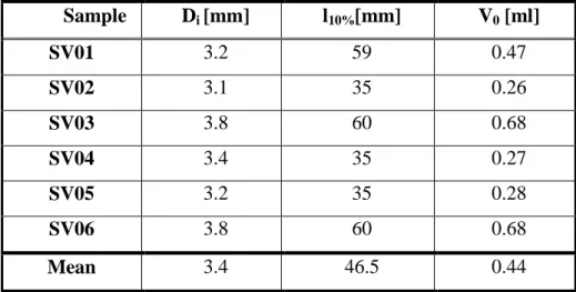
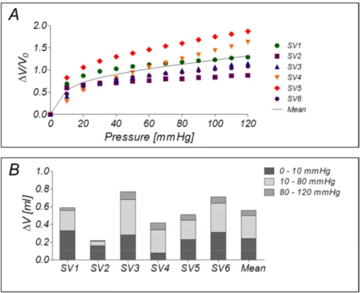
2.3.1 Pressure-volume measurements of SV segments ... 43
2.3.2 Functional assessment of the EVCS ... 45
2.4 Results of the functional experiments for testing the EVCS performance ... 45
2.4.1 Pressure-volume measurements of SV segments ... 45
2.4.2 Functional assessment of the EVCS ... 46
2.5 Conclusions ... 48
An arterialization study of human SVs in the EVCS ... 51
Introduction ... 52
3.1 Design of the conditioning experiment ... 52
3.1.1 SV samples preparation... 52
3.1.2 Mechanical conditioning of human SV within the EVCS ... 53
3.2 Methods for morphological and IF assessment of the mechanically conditioned human SV segments ... 55
3.2.1 Tissue viability evaluation ... 55
3.2.2 Histological, immunofluorescence and immunohistological analysis ... 55
3.2.3 Morphometric and proliferation measurements ... 56
3.2.4 Epigenetic profiling for histones modifications analysis ... 58
3.2.5 Protein assays ... 58
3.2.6 Statistical analysis ... 59
3.3 Results of the human SVs pressure stimulation ... 60
3.3.1 MTT and Ki67 staining reveal metabolic and proliferative activity, and a reduction of the cell density after 7 days of culture in the EVCS ... 60
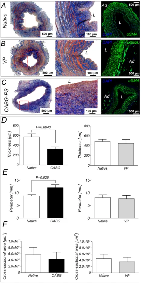
3.3.2 Morphological and immunofluorescence analyses show structural remodeling of the mechanically-conditioned human SV segments ... 62
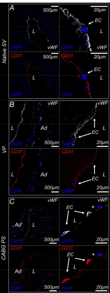
3.3.3 CABG-like pressure stimulation induced morphological remodelling and epigenetic mutations of SV vasa vasorum structures and cells... 66
3.3.4 Protein analyses confirm the remodelling of mechanically-conditioned human SV segments ... 67
3.6 Comments to the arterialization study results ... 69
5
Introduction ... 73
4.1 Evolution towards a double-compartment ex vivo vessel culture system ... 74
4.1.1 Design specification ... 74
4.1.2 Architecture of the double-compartment ex-vivo vessel culture system ... 74
4.1.3 Double-compartment SV culture chamber ... 75
4.1.4 The hydraulic circuit ... 78
4.1.4.1 Dimensioning of the de-oxygenation circuit ... 78
4.1.4.2 De-oxygenator silicone tubing dimensioning... 88
4.1.4.3 The de-oxygenator system layout and manufacturing ... 88
4.1.5 Testing of the de-oxygenator performances ... 89
4.1.6 De-oxygenator performances results... 91
4.2 Overview of the DC-EVCS ... 93
4.2.1 Functional experiments for testing the performances of the DC-EVCS ... 94
4.3 Integration of a novel left coronary artery pulse duplicator ... 95
4.3.1 Design specification ... 95
4.3.2 Architecture of the coronary artery pulse duplicator ... 96
4.3.2.1 Dimensioning, design and manufacturing of the coronary impedance subsystem ... 97
4.3.2.2 Dimensioning and manufacturing of the service impedance subsystem . 102 4.3.2.3 Dimensioning and manufacturing of the damper filter ... 109
4.4 Overview of the coronary pulse duplicator integrated with the EVCS ... 112
4.4.1 Functional experiments for testing the performance of the pulse duplicator .. 114
4.5 Conclusions ... 117
Conclusive remarks ... 120
Adapted from (Rueda et al 2008)
7 Saphenous vein (SV) graft disease represents an unresolved problem in coronary artery bypass grafting (CABG). After CABG, a progressive remodeling of the SV wall occurs, possibly leading to the lumen occlusion. This process is termed intima hyperplasia (IH). The investigation of cellular and molecular aspects of IH progression is a primary endpoint toward the generation of occlusion-free vessels that may be used as ‘life-long’ grafts. While animal transplantation and in vitro models have clarified some of the remodeling factors, the human SV pathology is far from being completely understood.
In this scenario, the aim of the present doctoral project was to explore new tools and procedures to investigate ex vivo the effects of altered mechanical load experienced by the human SV after CABG surgery. The issue of the ex vivo mimicry of the pathologic arterialization mechanism, involved in SV graft disease, was addressed by a multidisciplinary approach. Advanced bioengineering/biotechnology modelling and prototyping tools, complying with biological methods and tissue engineering/regenerative medicine requirements, were applied. Furthermore, the application of principles and methods of life-science engineering were used for providing a reliable model system, facilitating the understanding of pathogenesis of vein graft IH. Particular focus was given to the control over the environmental conditions for tightly reproducing the essential stimuli involved in the pathological SV remodelling. The integration of these methodologies led to devising a novel laboratory-oriented culture platform, that was used for conducting extensive arterialization conditioning campaigns with human SVs, under strictly controlled hemodynamic conditions.
This doctoral project is part of a wider research, involving a biological laboratory partner, aimed at discovering molecular effectors involved in vein graft disease to be targeted by pharmacologic/gene expression interfering strategies, in view of a next generation treatments and protocols to be tested in preclinical and clinical models.
This interdisciplinary project was prominently experimental, supported by computer aided design and numerical modelling. The designing and prototyping of the ex vivo platforms were performed at the Laboratory of Experimental Micro and Biofluid-dynamics (µBS Lab) of the Dipartimento di Elettronica, Informazione e Bioingegneria of the Politecnico di Milano. The preliminary biological validation with human samples and the arterialization conditioning were performed at and in strict collaboration with the
8 Laboratory of Cardiovascular Tissue Engineering of the Centro Cardiologico Monzino in Milan.
Rationale of the doctoral project
CABG using autologous vessels is a standard surgical procedure to recover myocardial perfusion in patients with coronary artery disease (de Waard et al., 2006; Parang and Arora 2009; Wallitt et al., 2007). The two most represented coronary-compatible autologous vessels to be used in CABG are the inner mammary artery (IMA) and the saphenous vein (SV). Soon after introduction of CABG, it became evident that transplanted vessels undergo a series of structural modifications, leading, within few years, to a significant reduction of vessel patency. In a number of cases, this requires re-hospitalization with stent implantation in the grafts and, ultimately, re-intervention. Compared with artery-made bypasses, SV grafts show lower long-term patency; in fact, approximately 15-30% of vein grafts fail during the first year and more than 50% patients require re-intervention within 10 years after implantation (Goldman et al., 2004; Tsui and Dashwood 2002), with high economic burden and consequences for the quality of the life of the patient. However, in most clinical cases, the use of a SV graft is unavoidable, since most patients need multiple bypass grafting procedures. In fact, the SV, due to its length and its superficial anatomical position, represents a preferred natural bypass conduit (Dashwood and Loesch 2009; Muto et al., 2010; Severyn et al., 2004; Surowiec et al., 2000).
After CABG surgery, progressive structural modifications of the SV wall, due to IH, lead to the occlusion of the graft lumen (the so called vein graft disease, VGD). The beginning of the pathology occurs at much earlier stages after grafting (one week), with the activation of biomechanical- and inflammatory-driven cascades which prime vessel remodeling (Mitra et al., 2006; Muto et al., 2010). As a first factor, the surgical procedures used during SV grafts preparation cause major insults to the integrity of the vessel endothelium and adventitia. These insults promote inflammatory responses related to a direct exposure of the intima layer to pulsatile blood flow and to a significant decrease in Nitric Oxide (NO) synthesis, with consequence for enhanced vasospasm and lower atheroprotection. The evolution of surgical techniques to harvest SV segments to be used for CABG surgery, has led to the devise of “no-touch” techniques, which preserve the
9 structure of the endothelium and the adventitia with slower progression of intima IH and enhanced vessel patency (Ahmed et al., 2004; Dashwood et al., 2005; Rueda et al., 2008; Souza et al., 2006). However, while the use of these surgical strategies lead to the maximization of vessel integrity prior to vein implantation into arterial position, an un-avoidable mechanical stimulation due to changes in pressure load and flow, appears to have prime role in VGD (Berceli et al., 2009; Hwang et al., 2011; John 2009; Tran-Son-Tay et al., 2008). In fact, immediately after grafting, SV segments are exposed to a variety of hemodynamic stimuli (vascular wall strain and stress and lumen shear stress activation) activating several cellular pathways and responses in the venous vessel wall. The new hemodynamic conditions experienced by the vein after transposition into the coronary circulation are similar to those experienced by coronary arteries. In particular, in the coronary artery circulation, the blood flow is characterized by a high pulsatile pressure, oscillating between 80 and 120 mmHg, and a pulsatile flow (with mean flow rate of 250 ml/min), which results in an elevated shear stress (1 - 7 Pa) (Bouten et al., 2011; Dummler
et al., 2011). Such forces have antagonistic effects on disease progression: elevated shear
stresses may have an atheroprotective role due to hemodynamic-related increase of NO release by endothelial cells (EC) while a non-physiologic mechanical loading of the vein wall may have a pro-pathologic effect due to mechanical ruptures in the endothelial layer and abnormal wall strain/stress of smooth muscle cells (SMCs) sheets (John 2009).
The major cause of SV graft failure is an over-proliferation of SMCs into the vessel intima layer leading to IH after few months (Lemson et al., 2000; Motwani and Topol 1998; Muto et al., 2010). SMCs proliferation begins quite shortly after CABG surgery, and precedes the later development of graft atherosclerosis that concurs to further reduce vessel patency at later times (Parang and Arora 2009; Wallitt et al., 2007). In vitro and in vivo (animal models) investigations have established that uncontrolled SMCs proliferation/IH phenomena is induced by inflammatory response, vein de-endothelialization and modified wall stress/strain (Wallitt et al., 2007). In these conditions, SMCs respond with apoptosis (Morrow et al., 2005), modified proliferation, as well as enhanced/reduced migratory activity (Qi et al., 2010). In addition, after vein implantation into arterial position, SMCs loose their typical contractile phenotype and start to proliferate and to invade the intima layer, thus reducing the vascular lumen. Furthermore, various animal models have been devised to address the pathologic evolution of the arterialized veins. These studies have
10 highlighted the relevance of different cell types and signal transduction pathways involved in the initiation of the phenomena leading to IH (Hoglund et al., 2010; Torsney et al., 2005). The parallel evolution of preclinical models mimicking human vein arterialization in mice and rabbits such as vena cava or jugular veins into carotid interposition (Hu and Xu 2002; Jiang et al., 2004), have allowed investigating the role of intrinsic and extrinsic cellular compartments and altered hemodynamics in intima thickening associated to vein arterialization. However, the establishment of IH in human arterialized veins is still far from being completely understood.
The use of bioengineering approaches is an option to study VGD. Several organ culture system were designed to recapitulate arterialization in cultured human veins for studying the physiopathology of VGD consequent to SV exposure to coronary artery-mimicking flow and pressure. As recently discussed, various devices, tailored to perform
ex-vivo culture of human SVs for a period of time spanning from 4 to 14 days and under
dynamic conditions, have been developed (Piola et al., 2012). The platforms that have been devised to this aim appeared insufficiently refined to maintain a tight control of vessels biological conditions for global molecular profiling (Piola et al., 2012). In particular, the design philosophy of the state-of-the art devices did not allow the simultaneous replication of all the main biophysical conditions acting in vivo during human vein arterialization. These lacks preclude the obtainment of reliable information on the activation of bio-mechanically stimulated molecular pathways or the possible interaction of vein-resident cells with circulating cells.
Novel ex vivo models are necessary in order to: i) tightly replicate the altered hemodynamic conditions, especially the raise in wall strain and shear stress, ii) obtain ex
vivo arterialized SV segments for investigating the mechano-biological basis of the early
events leading to IH at global molecular level, and iii) in perspective, attempt therapeutic strategies by pharmacological conditioning of the dynamically cultured vein segments.
The strategy adopted in the present doctoral project consists of devising and biologically testing a novel ex vivo model of human vein arterialization, applying bio-and tissue-engineering methods. The resulting ex vivo platform tried to overcome some of the limitations of the state-of-the-art models, focusing onto the control of the hemodynamic and biochemical microenvironments. In our view, this is crucial to obtain a global comprehension of VGD progression, and in perspective to perform comparative studies of
11 drug administration or gene expression modulation (gene therapy, siRNA and antagomir
approaches), to devise preconditioning protocols and/or regenerative medicine strategies
that reduce the clinical impact of VGD pathology.
Outline of the thesis
The dissertation is structured into 4 main chapters, that describes the various steps of the process that lead to manufacture of a novel EVCS for the in vitro mimicking of the hemodynamic forces acting on human saphenous vein after CABG surgery.
In Chapter 1, the current strategies, applied for studying the physiopathology of VGD consequent to SV exposure to coronary artery-compliant flow and pressure, are presented. The chapter is based on a published article where the state of the art of the ex
vivo arterialization models, and also the products and the related patents, is revised. The
published article is: M. Piola, M. Soncini, F. Prandi, G. Polvani, G.B. Fiore, M. Pesce.
Tools and Procedures for Ex Vivo Vein Arterialization, Preconditioning and Tissue Engineering: A Step Forward to Translation to Combat the Consequences of Vascular Graft Remodeling on Recent Patents On Cardiovascular Drug Discovery, 2012, 7 (3):
186-195.
In Chapter 2, the design of a novel ex vivo vein culture system (EVCS), aimed at replicating ex-vivo arterialization conditions experienced by SV after CABG for studying the biological mechanisms activated by SV exposure to arterial-like conditions, is presented. This version of the EVCS is conceived as pressure-driven vessel straining system for studying the early remodeling events caused by pure pulsatile arterial-like pressure. These activities were mainly carried out at the Laboratory of Experimental Micro and Biofluid Dynamics of the Dipartimento di Elettronica, Informazione e Bioingegneria of the Politecnico di Milano; preliminary functional testing of the designed device was also carried out at the Laboratory of Cardiovascular Tissue Engineering of the Centro Cardiologico Monzino. The chapter is partially based on a manuscript: “M. Piola, F. Prandi, N. Bono, M. Soncini, E. Penza, M. Agrifoglio, G. Polvani, M. Pesce, and G. B. Fiore. A compact and automated ex-vivo vessel culture system for the pulsatile pressure
conditioning of human saphenous veins. Journal of Tissue Engineering and Regenerative
12 In Chapter 3, the effects induced by different dynamic conditions on the human SV early events associated to vascular remodeling are presented. These activities were carried out at the Laboratory of Cardiovascular Tissue Engineering of the Centro Cardiologico Monzino. The chapter is partially based on a technical manuscript: “M. Piola, F. Prandi, N. Bono, M. Soncini, E. Penza, M. Agrifoglio, G. Polvani, M. Pesce, and G. B. Fiore. A
compact and automated ex-vivo vessel culture system for the pulsatile pressure conditioning of human saphenous veins. Journal of Tissue Engineering and Regenerative
Medicine, [Epub ahead of print] doi:10.1002/term.1798”.
In Chapter 4, the evolution of the bioreactor configuration from a simple pressure-driven vessel straining system to an ex vivo SV preconditioning system in the presence (or not) of controlled chemical conditions (i.e., hypoxia), arterial-like pressure, and flow patterns, is proposed. The bioreactor was upgraded to better replicate the full biomechanical stimuli involved in CABG arterialization, i.e. pulsatile wall stretch and wall shear stresses applied synchronously and with the correct phasing.
Finally in the Conclusive remarks chapter, a discussion of the overall results obtained during the development of the doctoral project is presented.
13
1
Tools and procedures for ex vivo vein
arterialization, preconditioning and tissue
engineering
This chapter is based on: M. Piola, M. Soncini, F. Prandi, G. Polvani, G.B. Fiore, M. Pesce. Tools and Procedures for Ex Vivo Vein Arterialization, Preconditioning and Tissue Engineering: A Step Forward to Translation to Combat the Consequences of Vascular Graft Remodeling. Recent Patents On Cardiovascular Drug Discovery, 2012, 7 (3): 186-195.
14
Introduction
Coronary artery bypass grafting (CABG) has been introduced in vascular surgery more than 50 years ago to combat the consequences of myocardial ischemia (Owens 2010; Parang and Arora 2009; Wallitt et al., 2007). Although the use of modern tissue engineering (TE) techniques has demonstrated the feasibility of ex vivo vessels fabrication (L'Heureux et al., 2006), the use of autologous vessels from the patients themselves is, still today, the unique option accessible to surgeons. The two most represented coronary-compatible autologous vessels to be used in CABG are the inner mammary artery (IMA) and the saphenous vein (SV). Soon after introduction of CABG, it became evident that the transplanted vessels are liable to undergo a series of structural modifications, leading to significant patency reduction, with the need for patient re-hospitalization, stent implantation or, ultimately, re-intervention (Parang and Arora 2009; Wallitt et al., 2007). The clinical impact of graft disease is different, depending on the origin of the vessels employed. It is estimated mammary artery grafts maintain an 85% patency after a 10-years while patency of SV grafts drops to 50%-60%, at a comparable time.
Basic investigations have clarified the potential contribution of vascular-resident or recruited cells (e.g., smooth muscle cells and monocytes), or of intracellular signaling activation pathways in the establishment of vein graft disease (VGD). On the other hand, a “system” level understanding of the disease progression as a whole is still unavailable. In fact, it is unclear which is the role of biomechanics on the response of vessel-resident cells, and whether changes in the global gene expression and (epi)genetic circuitries occur as a result of modified flow conditions.
In this context, the use of bioengineering approaches to reproduce arterialization in cultured human veins is likely to provide valuable tools for studying the physiopathology of VGD consequent to SV exposure to coronary artery-compliant flow and pressure. This may also help to dissect the contribution of different cellular populations in the progression of intima hyperplasia in the graft, and assess vessel responses to novel ex vivo pharmacological treatments. Finally, the conception of vessel perfusion systems may concur to establish tissue engineering (TE) protocols to obtain artificial vessels (Niklason
et al., 1999; Seliktar et al., 2003) which may be prospectively used as a novel and
15 The first section of the present chapter provides an overview of the vessel perfusion systems designed and patented to study the vein arterialization process, while the second part discusses some of the bioreactor-based strategies (and the relative patented technologies) that have been devised to produce fully engineered vessels.
1.1 Vessel perfusion systems
SV segments used for CABG undergo mechanical damages and flow/pressure loads, which are believed to activate pathways causing intima hyperplasia (IH). A crucial component contributing to VGD is the change in the pressure load and cyclic strain consequent to arterialization. In fact, in normal conditions SVs are subjected to quasi-steady flow patterns and are exposed to very low shear stresses (0.1-0.6 Pa) [7] and pressure loads (5-10 mmHg) [7]. By contrast, after CABG, SV segments are subjected to fast pulsatile flow, which is supposed to cause adaptive remodeling in the SV wall, leading to progressive bypass occlusion. The new hemodynamic stimuli are similar to those experienced by coronary arteries; namely, a mean flow rate up to 250 ml/min, a high wall shear stress in the range of 0.75-2.25 Pa [8], a systolic/diastolic pressure of approximately 120/80 mmHg with a circumferential strain of 10-15% [8].
Several attempts have been made at mimicking these conditions and characterize molecular pathways implicated in SVs arterialization. To achieve this goal, ex-vivo vessel perfusion culture systems (EVPCS) have been developed, where the survival of animal- or human-derived vessel segments is ensured by immersion into culture media, and vessel mechanical stimulation is performed by an appropriate arterial-like circulation inside the vessel lumen. This approach has been inspired by the pioneer investigation of Lindberg and Carrel (Carrel and Lindbergh 1935), proposing, for the first time, the use of ex vivo perfusion systems for studying the function of isolated organs outside the body.
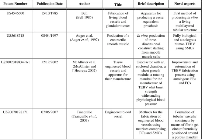
In this context, despite only one EVPCS has been patented (Clerin et al., 2010) (Table 1.1), several studies have been conducted using various devices able to expose intact vessels to steady flow perfusion.
16 Patent Number Publication Date Author Title Brief description Novel aspects
US20100331964A1 30/12/2010 Clerin et al. (Clerin et al., 2010)
Ex-vivo remodeling
of excised blood vessel for vascular
grafts
Ex vivo method and
System where excised small diameter blood vessels can be harvested and cultured to provide viable vascular grafts A modeled excised small vessel, having increased diameter, length, and wall thickness
produced by physically remodeling a small
blood vessel
Table 1.1. A relevant patent involving the use of a bioengineering approach to study vein adaptation to
arterial- like conditions: a brief description and the novel aspects of the patent content are provided.
In the attempt at studying the biology of vein arterialization, flow perfusion systems were initially designed to assess basic survival and perform pharmacological treatment of SV segments ex vivo. These systems allowed culturing SV segments for different periods of time; however, they had the limitation of not including a pressure control, which is crucial to mimic the pulsatile arterial-like flow, recognized as one of the primary cause of vessel patency reduction. Therefore, these systems were only able to elicit biological responses associated to vessel perfusion with constant flows, and, paradoxically, demonstrated the role of laminar shear stress on IH inhibition (Porter et al., 1996; Rey et
al., 2004). In a first example, 0.5 cm-length longitudinal-sliced segments of human SV
were pinned to the inside of a silicone tube and perfused for 14 days with venous (a flow rate of 70 ml/min resulting in a low shear stress of 0.1 Pa, and a pressure of 15 mmHg) and arterial (a flow rate of 500 ml/min resulting in an high shear stress of 0.9 Pa, and a pressure of 85 mmHg) flow patterns (Porter et al., 1996). The result of this study was that high-pressure constant flow inhibits IH, likely as a consequence of high shear stress. In another study by Surowiec and colleagues (Surowiec et al., 2000) a vascular perfusion culture system enabling culture of intact vessels for a period up to 96 hours was developed. This bioreactor was designed with a tightly-controlled perfusion rate in order to expose SV segments to 100 ml/min flow with a 40 mmHg intraluminal pressure and assess the effect of pharmacologic stimulation on vascular contractility. Using a similar system, Rey and colleagues (Rey et al., 2004) extended the observation time to 14 days and concluded that
ex vivo conditioning with a constant flow (resulting in low shear stresses in the range of
0.2-0.6 Pa) inhibits the formation of neointima, through a generalized cytostatic effect on all the cells of the vein.
17 A significant advancement toward the development of EVPCSs as tools to mimic SV arterialization has been the inclusion of controllers able to elevate the SV intraluminal pressure. A first example of this novel approach was reported by Miyakawa and colleagues, who developed a flow perfusion system tailored to culture human SV under venous (flow rate of 5 ml/min) or arterial conditions (flow rate of 50 ml/min and constant pressure of 80 mmHg) up to 4 days (Miyakawa et al., 2008). In this bioreactor, the effect of pressure on the IH was evident: enhanced apoptosis and vascular remodeling were observed, closely resembling early events of vein arterialization observed in animal models. In another report, Gusic and co-workers (Gusic et al., 2005a; Gusic et al., 2005b) were able to discriminate between the effects induced by flow shear stress and those elicited by mechanical wall stress, using different pressure and flow-patterns. Finally,
ex-vivo stimulation of porcine SVs was performed by adapting a previously developed system
patented by Clerin et al. ((Clerin et al., 2010; Clerin et al., 2002; Clerin et al., 2003), Tab. 1), to comply with vessel stimulation with different flow rates (5-85 ml/min), shear stress (0.26-5.6 Pa) and pressure (25-90 mmHg) amounts. Interestingly, in these reports SVs were perfused with culture medium added with Dextran in order to mimic blood viscosity (∼4 cP). The main result of these studies showed that increases in laminar shear stress neutralized intimal thickening, while ramped perfusion pressures induced IH in a dose-dependent manner.
The latest development in EVPCS design to mimic arterialization conditions has been the inclusion of perfusion systems, allowing the stimulation of vessel segments with sine-like pressure patterns at defined frequencies. In a first example, Saucy and colleagues (Saucy et al., 2010) investigated the impact of high flow pulsatile perfusion (resulting in high shear stresses in the range of 0.9-1.5 Pa) obtained by applying a flow rate of 120±15 ml/min, a pulse rate of 60 pulse/min, and a systolic/diastolic pressure of 80±10/40±10 mmHg on human SV segments for 14 days. By this, the authors were able to establish an important correlation between the expression level of urokinase and tissue-type plasminogen activators and the applied mechanical stress, thus linking expression of extracellular matrix remodeling enzymes and a biomechanical stimulus. In a second example, Voisard and colleagues developed a pulsatile system able to stimulate up to 5 SV segments in parallel and generate arterial-like conditions (a mean flow perfusion of 5000 ml/min, a pulsatile flow with a rate of 80 bpm and a systolic/diastolic pressure of 130/80
18 mmHg) (Voisard et al., 2010). Using this system, the Authors observed elevation of cellular proliferation in the vein wall as early as at 4 days after the beginning of mechanical conditioning. The most advanced perfusion system devised so far was, finally, the platform devised by Dummler and colleagues (Dummler et al., 2011). These Authors developed an EVPCS to perform pulsatile ex-vivo stimulation of SVs under venous (a flow rate of 5 ml/min and a pressure of 10 mmHg) or arterial (a flow rate of 50 ml/min and a pressure of 100 mmHg) conditions. The biological results obtained using this device showed a relatively rapid occurrence of morphological changes in the vein structure, especially in the media and in the intima layers. These morphological changes were also associated with an increase in the expression of matrix metalloprotease-2 (MMP-2).
Despite a variety of systems able to expose vein segments to arterial-like flow patterns have been devised and reported in the literature, the lack of a systematic approach at investigating the global molecular changes associated to vessel arterialization still precludes a complete understanding of the arterialization process as a pro-pathological condition in CABG. In this framework, it might be relevant to design new bioreactor-based platforms with vessel stimulation circuits containing minimized volumes, to allow the outer and the inner vein layers to be exposed to controlled amounts of small molecules and drugs while in motion. In this way, for example, the effect of novel therapeutic agents such as small molecules, gene-transferring vectors, small interfering RNAs (siRNAs) or even antagomirs (McDonald et al., 2012; Wiedemann et al., 2012) might be studied for their beneficial effects on IH inhibition directly in human samples, to be compared to those achieved in normally used animal-based methods (Thomas 2012). Another advantage of perfusion systems miniaturization might be the option to add in the inner and/or the outer stimulation circuits living cells (e.g. endothelial progenitor cells and monocytes), which are known to dynamically interact with the inner vein wall at early stages of vein arterialization, and assess their contribution to the IH prevention/progression. This may also open the way to novel tissue engineering approaches using circulating EPCs to perform grafts “pre-endothelialization” (Griese et al., 2003), or to test novel treatments aimed at minimizing homing of cells directly involved in graft inflammatory reaction and neointima accumulation.
19
1.2 Bioreactor-based approaches for vascular tissue engineering
One of the major goals in tissue engineering is the derivation of artificial tissues and organs to be used as replacements of conventionally implanted prostheses. Paradigmatic is, for example, the development of decellularization protocols of entire organs such as the heart, the lung, the windpipe, and the liver (Bader and Macchiarini 2010; Jungebluth et al., 2012; Ott et al., 2010; Ott et al., 2008; Uygun et al., 2010), followed by cellular reseeding. Given the elevated demand of CABG implantations worldwide, the engineering of vascular substitutes of IMA and SV with higher compatibility and lower patency reduction is therefore actively pursued.
In order to replace natural coronary arteries, artificial vessels for CABG require to be designed with vessel-like structure, physiology and mechanical resistance. To obtain this, an optimal combination of vascular cells (namely endothelial cells, interstitial cells and smooth muscle cells), biomaterials and mechanical stimulation must be found (Barron et
al., 2003; Dermenoudis and Missirlis 2010; Martin et al., 2004). Thus, as in the case of the ex vivo organ culture systems used for mimicking vein arterializations (see before),
bioreactors used to generate tissue-engineered blood vessels (TEBV) consist of components necessary to actuate a mechanical control of the growing vessels. These are: i) housing tools, hosting the vessel substitute, ii) sterile culture chambers and fluidic circuits for media recirculation, and iii) active systems able to stimulate the vessel substitute. In order to generate artificial vessels to be clinically used, these systems must comply with the good manufacturing practice (GMP) requirements. Therefore, they have to be customized to perform scaffold cell seeding, growth and release of artificial vessels with simple and traceable procedures (Arrigoni et al., 2008; Banes et al., 2009; Dunkelman et
al., 1998; Elizondo et al., 1999; Seifalian et al., 2002; Villalona et al., 2010). In addition to
GMP compliance, TEBV properties must finally comply with the ANSI/AAMI/ISO 7198:1998/2001 standards for tubular vascular prostheses. These regulations establish that mechanical properties, volume compliance, suture retention strength, viscoelasticity and burst pressure must be comparable to those of native vessels such as saphenous vein and internal mammary artery (IMA); e.g. a mean value of suture retention strength around 138 grams (IMA) and mean burst pressures of 1600 mmHg (Konig et al., 2009; L'Heureux et
al., 1998) and 3000 mmHg (Konig et al., 2009) (SV and IMA, respectively). The
20 should not undergo uncontrolled remodeling after implantation (Campbell and Campbell 2007; Nerem 2000; Sarkar et al., 2007; Thomas 2003).
1.2.1 Vascular tissue engineering using cell seeding techniques 1.2.1.1 Cell seeding into bio-artificial polymer scaffolds
The first attempt at generating native-like blood vessels using a simple bioreactor-assisted TE approach and a combination of endothelial cells (ECs), smooth muscle cells (SMCs), and fibroblasts (FBs) was reported in the pioneer investigation of Weinberg and Bell in 1986 (Weinberg and Bell 1986). In this study, based on a previously patented system by Bell (Bell 1985) (Table 1.2), a method for producing living blood vessels by exploiting a cell-seeded collagen gel was described for the first time. In this report, the fabrication of a multilayered blood vessel equivalent was obtained by sequential seeding different cells types into bio-artificial scaffolds. A SMC layer was first generated culturing SMCs into a collagen gel around a cylindrical mandrel. FBs were then added by adding a FB-containing cell suspension around the outer SMCs layer up to 7 days. Finally, ECs were let home to the inner layer, by perfusion-assisted seeding through the artificial vessel lumen. The final structure consisted of a vessel-like structure with ECs lining the lumen, surrounded by SMCs and FBs layers. When assessed for mechanical compliance, these bio-artificial vessels were unable to withstand physiological arterial pressures. In fact they had a burst pressure of 10 mmHg. To ameliorate the mechanical resistance of the engineered vessel, a Dacron® mesh was then used providing a final burst pressure up to 70 mmHg.
A similar method was used by McAllister and L’Heureux. These Authors attempted to produce vascular grafts using a sheet-based cell-self-assembly technique (Auger et al., 1997; L'Heureux et al., 2006; L'Heureux et al., 1993; L'Heureux et al., 1998). The same authors patented an automatic method involving the use of a rotating mandrel for the fabrication of multilayered TEBVs (McAllister and l’Heureux 2002) (Table 1.2). The resulting artificial vessels showed a structure resembling that of native arteries and a burst pressure of 2000 mmHg; however, they were not compliant as native vessels. A similar method was finally patented by Tranquillo (Tranquillo et al., 2007) (Table 1.2) for creating TEBVs including an intimal layer surrounded by a SMCs media layer constructed around a
21 tubular support. Table 1.2 resumes the patents involving the use of bioreactor-based approaches for vascular TE using sheets-based techniques.
Patent Number Publication Date Author Title Brief description Novel aspects
US4546500 15/10/1985 Bell (Bell 1985) Fabrication of living blood vessels and glandular tissues Apparatus for producing a vessel equivalent prosthesis First method of producing in vitro a living multilayered tubular structure US5618718 08/04/1997 Auger et al.
(Auger et al., 1997) Production of a contractile smooth muscle In vitro production of three-dimensional construct starting from smooth muscle cells Fully biological and autologous human TEBV using SMCs US20020188349A1 12/12/2002 McAllister et al.
(McAllister and l’Heureux 2002) Tissue engineered blood vessels and apparatus for their manufacture Bioreactor with an enclosed chamber, a sheet growth module, a rotating
mandrel for the manufacture of TEBV whit burst
strength withstanding physiological blood pressure Improvement and automation of TEBV fabrication process using autologous FBs and ECs US20070128171 07/06/2007 Tranquillo (Tranquillo et al., 2007) Engineered blood vessel
Methods for the fabrication of engineered blood vessels using matrices comprising ECs and SMCs. Formation of tubular vascular constructs by means of fibrin gel
circumferentially positioned around a porous mandrel
Table 1.2. List of relevant patents involving the use of bioreactor-based approaches for vascular tissue
engineering using sheets-based techniques: a brief description and the novel aspects of the patent content are provided.
1.3.1.2 Cell seeding into synthetic polymer scaffolds
Several biomaterials have been used for supporting and guiding cells during vascular tissue formation. Among the synthetic polymers applied in vascular engineering (Bouten et
al., 2011; Gong and Niklason 2006; Naito et al., 2011), the most widely used are
biodegradable polymers, such as polycaprolactone (PCL), polylactic acid (PLA), polyglycolic acid (PGA), and their copolymer polylactic-co-glicolic acid (PLGA) (Hoerstrup 2006; Hoerstrup et al., 2001; Wang et al., 2010). In this context, crucial are the reports of Shin’oka (Shin'oka et al., 2001; Shin'oka et al., 2005), Niklason (Niklason et al., 1999), and Hoerstrup (Hoerstrup 2006; Hoerstrup et al., 2001). While the approach of Niklason and Hoerstrup made use of biomimetic perfusion systems for scaffold
22 conditioning (see section 2.2), Shin’oka and colleagues derived TEBVs by seeding vein derived cells onto a biodegradable polymer graft composed of PCL and PLA, reinforced with PGA (1:1, w/w) with internal diameter of 10 mm, length of 20 mm, and thickness of 1 mm. After 10 days of culture, TEBVs were used to reconstruct the occluded pulmonary artery (Shin'oka et al., 2001). 42 pediatric patients were treated with this procedure; no complications occurred after a 32-months follow-up and all TEBVs were patent (Shin'oka
et al., 2005).
1.2.1.3 Re-engineering of natural vessels
An alternative strategy to the use of bio-artificial or synthetic scaffolds to produce artificial blood vessels is to “re-engineer” human- or animal-derived vessels. This strategy offers the advantage of maintaining the normal three-dimensional tissue architecture and the native extracellular matrix (ECM) composition. For this reason re-engineered vessels may have a superior mechanical performance compared with artificially fabricated vessels (Badylak et al., 2009; Fomovsky et al., 2010; Quint et al., 2011). A potential disadvantage, at least in the case of animal-derived vessels, is the possible permanence of “xeno-antigens” (e.g. α-GAL (Galili 2005)), which may preclude a full graft biological compatibility, even after recellularization.
Different strategies to perform re-engineering of allogenic or xenogenic grafts have been set up. In a first one, named “transdifferentiation”, collected donor native tissues are seeded (without prior decellularization) with recipient-specific autologous cells (such as SMCs, ECs, FBs and macrophages) followed by continuous exchange of the medium. In this way they are converted into an immunologically tolerable tissue for the recipient. This method was established and patented by Orton in 1993 (Orton 1993) and more recently, was used by Bader to transform donor-derived veins and xenogenic arteries into autologous vessels in vitro ((Bader 2012), Tab. 3). The second strategy consists in a recellularization process into a cell-free tissue obtained by means of decellularization (Brendel and Duhamel 1989). Decellularization is achieved by chemical (e.g., acids and bases, hypotonic and hypertonic solutions, ionic and non-ionic detergents, and alcohols), enzymatic (e.g., nuclease, trypsin, and collagenase) and physical (e.g. temperature, pressure and mechanical forces) methods, which can be also combined in order to disrupt
23 cells and remove cellular and nuclear material (Crapo et al., 2011; Gilbert et al., 2006; Soma and Kotturathu 2009). Different techniques are used for enhancing the efficiency of the decellularizing agents: perfusion (Montoya and McFetridge 2009; Ott et al., 2008), immersion and agitation (Pellegata et al., 2012) are the most utilized tools. After decellularization, cell removal assessment is performed using different methods, such as staining with fluorescent dyes (DAPI/ Hoechst), and standard histological staining (Hematoxylin/Eosin, Masson’s Tricrome, and Movat’s Pentachrome). Finally, in order to form a non-thrombogenic ECs barrier within the graft lumen, different strategies have been suggested. An example is the direct implantation in the patient (in situ approach) in order to to recruit circulating endothelial progenitors directly from the surrounding tissue, or to seed the decellularized vessel with autologous ECs (in vitro approach), prior to the implantation (Bischoff et al., 2004; Wolfinbarger 2003).
Decellularized natural tissues, such as human veins (Schaner et al., 2004; Soma and Kotturathu 2009; Squillace 2010; Teebken et al., 2009), human and animal arteries (Amiel
et al., 2006; Bischoff et al., 2004; Dahan et al., 2012; Kaushal et al., 2001), umbilical cord
vessels (McFetridge 2005), and small intestinal submucosa (Badylak et al., 2002; Lantz et
al., 1993) have been used as matrices, allowing repopulation with donor-derived cells by
means of in situ or in vitro approach. In table 1.3 a list of patents relevant to the use of these approaches is shown.
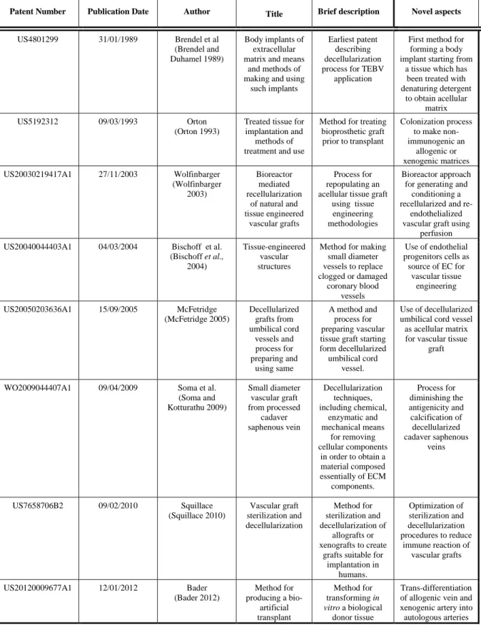
24
Table 3.1. List of patents involving the use of approaches for vascular tissue engineering using
decellularize-and-recellularize techniques of biological tissue: a brief description and the novel aspects of the patent content are provided.
Patent Number Publication Date Author Title Brief description Novel aspects
US4801299 31/01/1989 Brendel et al (Brendel and Duhamel 1989)
Body implants of extracellular matrix and means
and methods of making and using
such implants
Earliest patent describing decellularization process for TEBV
application
First method for forming a body implant starting from
a tissue which has been treated with denaturing detergent
to obtain acellular matrix US5192312 09/03/1993 Orton
(Orton 1993)
Treated tissue for implantation and
methods of treatment and use
Method for treating bioprosthetic graft prior to transplant Colonization process to make non-immunogenic an allogenic or xenogenic matrices US20030219417A1 27/11/2003 Wolfinbarger (Wolfinbarger 2003) Bioreactor mediated recellularization of natural and tissue engineered vascular grafts Process for repopulating an acellular tissue graft
using tissue engineering methodologies
Bioreactor approach for generating and
conditioning a recellularized and
re-endothelialized vascular graft using
perfusion US20040044403A1 04/03/2004 Bischoff et al.
(Bischoff et al., 2004)
Tissue-engineered vascular structures
Method for making small diameter vessels to replace clogged or damaged coronary blood vessels Use of endothelial progenitors cells as source of EC for vascular tissue engineering US20050203636A1 15/09/2005 McFetridge (McFetridge 2005) Decellularized grafts from umbilical cord vessels and process for preparing and using same A method and process for preparing vascular tissue graft starting form decellularized umbilical cord
vessel.
Use of decellularized umbilical cord vessel as acellular matrix for vascular tissue
graft WO2009044407A1 09/04/2009 Soma et al.
(Soma and Kotturathu 2009) Small diameter vascular graft from processed cadaver saphenous vein Decellularization techniques, including chemical, enzymatic and mechanical means for removing cellular components in order to obtain a material composed essentially of ECM components. Process for diminishing the antigenicity and calcification of decellularized cadaver saphenous veins US7658706B2 09/02/2010 Squillace (Squillace 2010) Vascular graft sterilization and decellularization Method for sterilization and decellularization of allografts or xenografts to create
grafts suitable for implantation in humans. Optimization of sterilization and decellularization procedures to reduce immune reaction of vascular grafts US20120009677A1 12/01/2012 Bader (Bader 2012) Method for producing a bio-artificial transplant Method for transforming in vitro a biological donor tissue Trans-differentiation of allogenic vein and xenogenic artery into autologous arteries
25
1.2.2 Biomechanical mimicking in vascular tissue engineering
Mechanical stimulation is an integral component of the cardiovascular developmental process. For example, it has been hypothesized that the beginning of pulsatile flow in the primary vascular system is a trigger of vascular and cardiac development, likely due to biomechanical effects on differentiation of cardiac and vascular progenitors. Although the global effect of the mechanical stretching in mature vessels has not been sufficiently addressed with bioreactor-assisted stimulation (see before), several studies have highlighted the role of mechanical stimulation in eliciting various responses in individual vascular cell types. For this reason, mechanical conditioning of ex vivo growing vessels is likely a crucial conditioning strategy to obtain fully functional TEBVs.
In vivo, vascular tissues are subjected to i) shear stress, acting on ECs and
influencing their orientation in the direction of flow, ii) luminal pressure, iii) stretch, mainly in the circumferential direction, and iv) longitudinal tension. Clearly, all these stimuli may have important effects on the structure of the artificially assembled vessels and may determine also their mechanical properties after the end of the preparation procedure (Niklason et al., 1999). In addition, mechanical stimuli have been shown to promote cellular responses such as the deposition of ECM components, and to cause changes in the (epi)genetic programming and the phenotype of vascular cells (Shi and Tarbell 2011).
To address this issue, several groups of researchers have developed dynamic culture systems suitable for applying, with a controlled manner, single (Dunkern et al., 1999; Hoerstrup et al., 2001; Tschoeke et al., 2008; Williams and Wick 2004) or combined biomechanical stimuli (Bilodeau et al., 2005; Dancu 2011; Fan et al., 2009; McCulloch et
al., 2004; Niklason et al., 2003; Vilender and Nickel 2008) to bio-synthetic vessels, such
as shear stress, pressure, and axial, bending and torsional loads.
In early studies, flow perfusion was used to promote the endothelialisation of polytetrafluoroethylene grafts (Dunkern et al., 1999) and to produce impermeable vascular substitutes using polyester grafts as described by Sauvage (Sauvage and Kaplan 1990). More recently, the implication of applying a 0.5-Pa shear stress (resulting from applying a steady state flow of 50 ml/min) on the properties of an engineered vascular graft was elucidated using the ElectroForce® BiodynamicTM Test Instrument (Bose Corp., Electroforce System Group, MI, USA) (Boccafoschi et al., 2012) Further Tschoeke (Tschoeke et al., 2008) developed a flow perfusion bioreactor for applying a 250 ml/min
26 fluid flow to a fibrin-based vascular graft (internal diameter of 5 mm) reinforced with a polyvinylidenefluoride mesh (with a pore size of 1-2 mm), and seeded with ovine myofibroblasts. After a stimulation period of 14 days, the graft tissue had a suture retention strength of 642 grams (similar to that of native carotid arteries, which reaches 753 grams (Cho et al., 2005)) and a burst pressure of 236 mmHg.
A biomimetic pulsatile flow is necessary to properly simulate the physiologic arterial hemodynamics in TEBV bioreactors. Pulsatile physical forces allow vascular graft performance to be enhanced by increasing ECM deposition and reinforcing mechanical resistance of PGA synthetic grafts. For example, Niklason et al. (Niklason et al., 1999), after 8 weeks of pulsatile stimulation (5% of radial distension at a rate of 165 pulse/minute), obtained an engineered vessel with a burst pressure of about 2000 mmHg, and a suture retention strenght of 90 grams, while Hoerstrup and colleagues (Hoerstrup et
al., 2001; Sodian et al., 2002) used a novel in vitro pulse duplicator system for generating a
biomimetic microenvironment supporting tissue formation. Using this apparatus, these Authors found that the mechanical properties of PGA seeded grafts increased with respect to static controls at 28 days of pulsatile culture. Analogously, Iwasaki (Iwasaki et al., 2008) developed a hemodynamic-equivalent pulsatile bioreactor consisting of a ventricular model, two synthetic polymer-made valves, a compliant silicone tube, a peripheral resistive unit, a gas exchange unit, and a housing for mounting the vessel. This complex system enabled the formation of elastic vessel grafts with mechanical properties comparable to those of native arteries after a 2-week stimulation under a constant pulse rate of 70 bpm and a gradual increase in mean flow and pressure up to 600 ml/min and 100 mmHg, respectively. Finally, Thompson and colleagues (Thompson et al., 2002) developed a bioreactor using a mechanical ventilator to induce pulsatile flow, while Narita (Narita et
al., 2004) adapted an intra-aortic-balloon pump system to develop a bioreactor able to
produce a wide range of pulsatile stimuli on a tubular PLA scaffold (flow rate spanning from 0 to 3000 ml/min, systolic and diastolic pressures ranging from 10 to 200 mmHg and from 0 to 100 mmHg, respectively).
Bioreactors can also provide very complex mechanical conditioning including axial bending and torsional stimuli. For example, Seliktar (Seliktar et al., 2000) applied cyclic strain at a frequency of 1 Hz up to 8 days to a collagen-based blood vessel construct. The results showed significant enhancement of the mechanical properties and cell organization
27 with respect to the static incubated constructs and depended on an active remodeling process. In another report (Bilodeau et al., 2005), a perfusion bioreactor equipped with a pump for applying continuous or pulsatile flow (beat rate range 50-200 bpm) and a pneumatic cylinder to stretch and twist the vessel was devised. This approach was similar to that followed in a patented method to produce a human hybrid bypass graft using a bioreactor allowing a tight control of hemodynamic forces such as pressure, flow and stretch (Dancu 2011), and that described in another patent (Niklason et al., 2003) claiming a method to perform pulsatile stretching of tubular constructs in order to align SMCs circumferentially (Table 1.4). An overview of the relevant patents regarding bioreactor-based approaches providing biomechanical stimuli to vascular TE constructs is summarized in table 1.4.
28 Patent Number Publication Date Author Title Brief description Novel aspects
US4911713 27/03/1990 Sauvage et al. (Sauvage and Kaplan 1990) Method of making vascular prosthesis by perfusion A method for fabricating an impermeable vascular prosthesis perfusing the lumen
of the textile conduit
Method for making vascular textile prosthesis impermeable, combining perfusion and chemical treatments US005792603A 08/11/1998 Dunkelman et al.
(Dunkelman et al., 1998) Apparatus and method for sterilizing, seeding, culturing, storing, shipping and testing tissue,
synthetic or native, vascular grafts Bioreactor that provide a dynamic environment for seeding, culturing and testing vascular
grafts
Dynamic environment for culturing vascular grafts of any length
and diameter applying shear and
radial stresses US005916800A 29/06/1999 Elizondo et al.
(Elizondo et al., 1999) Cardiovascular bioreactor apparatus and method Methods and apparatus for processing and shipping cardiovascular products, within sterile and aseptic
environment
New mounting system for biological
materials such as heart valves and vascular grafts
US6537567B1 25/03/2003 Niklason et al. (Niklason et al., 2003) Tissue-engineered tubular construct having circumferentially oriented smooth muscle cells
Methods for the production of organized tissue engineered constructs Use of distensible bodies to impart pulsatile stretching force to the lumens of constructs during
growth US7348175B2 25/03/2008 Vilender et al.
(Vilender and Nickel 2008) Bioreactor with plurality of chambers for conditioning intravascular tissue engineered medical products A microprocessor controlled and instrumented bioreactor for conditioning tissue engineering medical product. This system allows the control of fluid flow
and tissue displacement and allows the measurements of the material properties. Possibility of growing and conditioning vascular tissues within a controlled and strictly reproducible environment
US20090123993A1 14/05/2009 Banes et al. (Banes et al., 2009) Bioreactor for development of blood vessels Blood vessel bioreactor developed for harvest, maintain, transport and produce vascular vessels Vessel attachment/ engagement cuff connectors and clamps
US2009181448A1 16/07/2009 Fan et al. (Fan et al., 2009) Perfusion type vascular tissue bioreactor with rotary and stretching functions Multi-module bioreactor for vascular construct able to generate physiological pulsatile flow Generation of physiological pulsatile flow perfusion and
multi-mechanical stimulation; mimicking vascular compliance, inertia, and resistance US20110014597A1 20/01/2011 Frerich (Frerich 2011) Perfusable Bioreactor for the Production and/or Cultivation of a
Bioreactor with elastic walls designed for the
production of
Possibility of study the interactions between the vessel
29
Human or Animal Blood Vessel and/or a Human or Animal Tissue
human vessel tissue US7968329B2 21/06/2011 Dancu (Dancu 2011) Method of conditioning a hybrid synthetic tubular structure to yield a functional human hybrid coronary bypass graft Method for producing a human hybrid coronary
by-pass graft conditioning a synthetic tubular matrix Possibility of controlling, simultaneously and independently, hemodynamic forces in order to recapitulate in vivo vascular hemodynamic environments
Table 1.4. List of patents involving the use of bioreactor-based approaches for vascular tissue engineering
using biomechanical stimulation: a brief description and the novel aspects of the patent content are provided.
1.3 Conclusions
The use of bioreactor-assisted culture of vascular grafts and substitutes has been confined, thus far, to simple preconditioning strategies to assess basic cellular responses to arterial-like mechanical forces, or to attempt vessel engineering. In our view, the use of these systems in the context of a wider molecular comprehension of vessel response to biomechanical stimulation will lead to dissection of mechanisms governing graft failure in patients.
A first area of interest appears the application of “omics” molecular investigation to assess the biomechanics-associated changes in the vascular global gene expression pattern. This is of interest in the recent view that vein graft disease involves a possible cross-talk between various cellular components of the vessels exposed to mechanical load (Hu et al., 2004; Torsney et al., 2005), or their interaction with cells recruited in the graft by the circulation (Zhang et al., 2004). In this scenario, segments of human vessels exposed to differential mechanical loads in refined bioreactor platforms may be studied for changes in their transcriptome signature to assess the differential activation of pro- vs. anti-remodeling gene expression “modules” (Segal et al., 2003). In addition, the customization of the platforms to allow injection of defined amounts of circulating cells such as monocytes or endothelial progenitor cells (EPCs), may help dissect the molecular mechanisms underlying homing of cells participating (Hu et al., 2002), or potentially inhibiting restenosis (Brown et al., 2010; Zhu et al., 2010).
30 Another area of interest pertains to the role of recently identified vessel-resident progenitors (Ergun et al., 2011; Majesky et al., 2011; Torsney and Xu 2011) in VGD progression. These cells have been also found in human SV and have thereby named saphenous vein progenitors (SVPs) (Campagnolo et al., 2010); they are adventitial pericyte-like cells that may be activated, directly or indirectly, to differentiate into proliferating SMCs as a result of vein arterialization. This hypothesis, in line with investigations highlighting the role of adventitia cells in the progression of transplant arteriosclerosis in animal models (Hu et al., 2004; Torsney et al., 2005), may be optimally addressed using pulsatile perfusion systems where segments of human SV are exposed to arterial flow conditions, followed by immunofluorescence studies to assess SVPs mitogenic activation and differentiation. This latter approach may be finally helpful also to uncover possible changes in the epigenetic signatures associated to mechanical stress in various cell types resident in the vein, and thus to understand the involvement of chromatin and histone code (Berger 2007; Kouzarides 2007) modifications following arterialization.
In summary, while significant advancements have been made in the design and construction of bioreactor platforms to replicate arterial-like conditioning, the conception of integrated approaches to understand the biology of the vessels “as a whole” in response to alteration of mechanical conditions is only at its early stages. In our view this is crucial to obtain a global comprehension of vein graft disease progression, to perform comparative studies of drug administration or gene expression modulation (gene therapy, siRNA and antagomir approaches), to devise preconditioning protocols and/or tissue engineering strategies that reduce the clinical impact of this pathology.
31
2
Design of a novel ex vivo vessel culture system
This chapter is partially based on: “M. Piola, F. Prandi, N. Bono, M. Soncini, E. Penza, M. Agrifoglio, G. Polvani, M. Pesce, and G. B. Fiore. A compact and automated ex-vivo vessel culture system for the pulsatile pressure conditioning of human saphenous veins. Journal of Tissue Engineering and Regenerative Medicine, [Epub ahead of print] doi:10.1002/term.1798”.
32
Introduction
Saphenous vein (SV) graft disease represents an unresolved problem in coronary artery bypass grafting (CABG). After CABG, a progressive remodeling of the SV wall occurs, possibly leading to the lumen occlusion, a process termed intima hyperplasia (IH). The investigation of cellular and molecular aspects of IH progression is a primary endpoint toward the generation of occlusion-free vessels that may be used as ‘life-long’ grafts. While animal transplantation models have clarified some of the remodeling factors, the human SV pathology is far from being understood. This is also due to the lack of devices able to reproduce the altered mechanical load encountered by the SV after CABG.
In this chapter we propose the design of a compact and automated ex-vivo vessel culture system (EVCS) able to artificially produce the effects of the arterial pressure-related cyclic wall distention, one of the major biomechanical causes of IH in venous CABGs together with the pulsatile wall shear stress (Anwar et al., 2012; Berceli et al., 1990; John 2009; Muto et al., 2010; Owens 2010; Stigler et al., 2012). This aim is achieved by the development and functional assessment of a low-volume, reliable and user-friendly device, capable to replicate automatically the pulsatile pressure patterns of the physiological coronary environment. In perspective, the present EVCS could be used as tool to carry out molecular and cellular studies in order to better understand the impact of modified hemodynamic conditions on in vivo SV remodeling.
The approach applied for the design and manufacture of the device entailed a first phase of literature research, and interaction with the final user of the device; on these bases, the general and functional requirements of the bioreactor were identified, and a preliminary design of the system proposed. Subsequently, different culture chamber prototypes were designed, manufactured, and tested to define a final optimized configuration of the system. The design of the dynamic culture system made use of the 3D CAD software Pro/Engineering Wildfire 4.0 (Parametric Technology Corporation, PTC, Needham, MA), while the prototypes were manufactured using a parallel lathe (OPTI D 240x500 G-Vario, Optimum, DE), a numerical control machine (Roland MDX-40, Roland Corporation, Tokyo, JP), and a laser cutting (Versalaser VSL2.30, SK Laser, Germany), plus additional manual finishing. These activities were carried out at the Laboratory of Experimental Micro and Biofluid-Dynamics (µBS Lab) of the Dipartimento di Elettronica, Informazione e Bioingegneria of the Politecnico di Milano.
33
2.1 Design of the EVCS
2.1.1 Design specifications
The design of the EVCS took into account the general specifications of a bioreactor for tissue engineering application (Martin et al., 2004), with particular emphasis on the ease of assembly under laminar flow hood, and the safety of use in a cell culture laboratory. Specifically, the following basic requirements were addressed: i) biocompatibility of materials, ii) transparency, to ensure visual inspection for air bubble and/or medium color changes, iii) compatibility with sterilization processes, e.g., via autoclaving and/or ethylene-oxide (EtO), iv) minimization of priming volume, in order to limit the cost of soluble culture medium compounds, and v) easiness of vessel accommodation and handling during the EVCS assembly.
2.1.2 Architecture of the ex-vivo vessel culture system
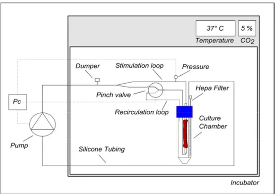
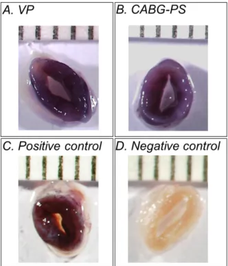
The EVCS is designed to apply a CABG-like pressure stimulation (CABG-PS), i.e. a pulsed pressure oscillating between a diastolic minimum and a systolic maximum (e.g., 80-120 mmHg), or a steady flow perfusion, i.e., a physiological venous perfusion condition (VP, e.g., 5 mmHg) within a controlled and strictly reproducible mechanical environment. A schematic representation of the system’s layout is shown in Figure 2.1. During culture, SV grafts are hosted in a culture chamber accommodated inside an incubator. The culture chamber is connected to a hydraulic circuit and actuators (pump and solenoid pinch-valve) to apply pressure stimulation to the human vessels or to allow the medium recirculate within the vessel. The hydraulic actuators are managed by a programmable monitoring and control (M/C) system, which operates via a pressure-based feedback loop.
34
Figure 2.1. Design of the EVCS. (A) Layout of the EVCS: thick lines represent the hydraulic circuit; thin
lines represent the monitoring and control (M/C) signals. In particular, the M/C system manages the hydraulic actuators (a pump and a solenoid pinch-valve) via a pressure-based signal registered by the pressure sensor.
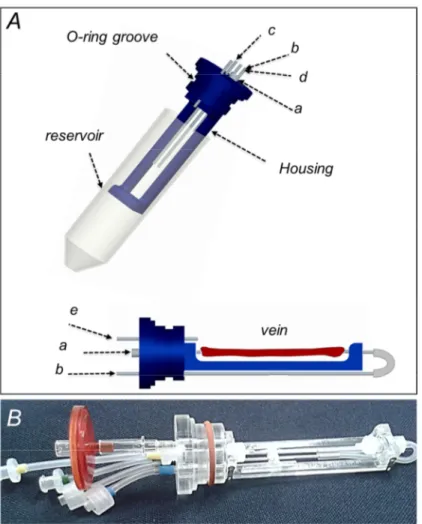
2.1.3 SV culture chamber
The culture chamber (Figure 2.2) includes a commercial reservoir and a purpose-developed vessel housing which is integrated with the reservoir cap. All the culture chamber parts built in house were designed with 3D-CAD Pro/Engineer Wildfire 4.0 (PTC, Needham, MA), and manufactured by laser cutting (Versalaser VSL2.30, SK Laser, Germany), and/or computer numerical control machining (Modela MDX-40, Japan) from polymethylmethacrylate blocks (PMMA, Plasting S.r.l., Segrate, Italy). All the utilized materials are suitable for EtO sterilization. The vessel housing allows hosting SV samples up to 5.5 cm in length. The hosted vessel segment is cannulated at both ends using polypropylene (PP) barbed fittings (Cole Parmer, IL, USA), and secured using an extensible vessel loop (Esafarma S.r.l., Italy) as an elastic tourniquet. A standard 50-ml falcon tube (International PBI S.p.A, Italy) acts as a medium reservoir. The reservoir and the housing are coupled trough a silicone O-ring.
Five ports through the cap ensure the chamber’s connection to the outside. Two ports ensure injection/removal of the culture medium to/from the vessel (Figure 2.2, port a and b). Two other ports provide connection for recirculation of the reservoir medium, i.e. the