DOTTORATO DI RICERCA IN
Scienze e Tecnologie Agrarie, Ambientali e Alimentari
Ciclo 27
Settore Concorsuale di afferenza: 07/D1 Settore Scientifico disciplinare: AGR/12
TITOLO TESI:
DETECTION AND MOLECULAR CHARECTERIZATION OF VIRUSES
INFECTING ACTINIDIA SPP.
Presentata da: Roberta Biccheri
Coordinatore Dottorato
Relatore
Giovanni Dinelli Carlo Poggi Pollini
Correlatore
Claudio Ratti
i
Acknowledgements
I would like to thank Claudio and Carlo, for providing me with the opportunity to learn so much. Thank you for all of your support and advice.
I would like to thank, Associate Professor M. N. Pearson from University of Auckland and A. G Blouin from the New Zealand Institute for Plant & Food Research for their assistance and support during in my period in New Zealand where I learned so much.
I really want to thank all my collegues of the lab in Bologna for all of their support and encouragement: Alice, Chiara, Roberta and Mattia.
I really want to thank my family and my boyfriend for all of their love and for to have supported my choices, even if it has been really hard for them.
Table of Contents
Acknowledgement i
Chapter 1: General Introduction:
1Kiwifruit origins 2
The genus Actinidia 4
Actinidia species in cultivation 7
Global kiwifruit industry 9
Production and marketing in Italy 13
Disease in Actinidia spp. 16
The aims of my study 19
References 21
Chapter 2: Virus infecting Actinidia spp.:
24Viruses of kiwifruit 25
Non-Specialist viruses 29
Alfalfa mosaic virus and Cucumber mosaic virus 29 Ribgrass mosaic virus and Turnip vein clearing virus 31
Apple stem grooving virus 33
Cucumber necrosis virus 35
Actinidia virus X 36
Kiwifruit-Adapted viruses 37
Actinidia citrivirus 37
Actinidia virus A and Actinidia virus B 39
Disease-Inducing viruses 41
Cherry leaf roll virus 41
Pelargonium zonate spot virus 42
Mechanically transmitted viruses with no known natural vectors 43
Viruses with aphid vectors 44
Viruses with presumed mealy bug vectors 44
Viruses transmitted by seed and/or pollen 45
References 47
Chapter 3:Project paper: Characterization of Cucumber mosaic
virus naturally infecting Actinida chinensis in Italy
53Abstract 54
Introduction 54
Materials and methods 57
Source plants 57
Purification of viral particles and determination of viral
protein weight 58
Random RT-PCR and sequencing 59
Circular RT-PCR 60
Construction of full-length agroinfectius clones
and leaf agroinfiltration 61
Sequence alignment and phylogenetic analysis 61
Results 62
Symptoms observed and transmissions to herbaceous indicators 62 Transmission electron microscopy (TEM)and determination of
viral protein weight 62
Sequencing 65
Sequence alignment and phylogenetic analysis 66
Detection by RT-PCR 70
Agroinfectious clones 70
Discussion 71
References 74
Chapter 4: Project paper: First report of Pelargonium zonate spot
virus infecting Actinidia
77Abstract 78
Introduction 79
Materials and methods 82
Virus isolates and host plant observation 82
Transmissions to herbaceous indicators 82
Transmission electron microscopy (TEM) 83
Purification of the viral particles 83
Random-PCR amplification and sequencing 84
Circular RT-PCR and full-length amplifications 85
Serological and molecular detection 87
Identification of the PZSV encoded suppressor of RNA silencing 87
Results 88
Symptoms on host 88
Transmissions to herbaceous indicators 88
Purification of the Viruses 90
Random-PCR amplification and sequencing 90
Circular RT-PCR and full-length amplifications 91
Serological and molecular detection 93
Evaluation of RNA silencing suppressor activity of CP and MP
proteins 94
Discussion 95
Chapter 5 Project paper: Identification and characterization of
two new viral species from Actinidia chinensis
101Abstract 102
Introduction 102
Materials and methods 104
Plants material 104
Double-stranded RNA extraction 104
Reverse transcriptase and amplification reactions 105
Sequencing data analysis 105
Terminal regions 106
Results 108
Complete genome sequence of Actinidia latent virus 108
Computational analysis of Actinidia latent virus genome 109
Phylogenetic analyses of Actinidia latent virus 115
Characterization of Kiwifruit associated totivirus 1 121
Discussion 125
References 130
Chapter 6: General discussion and conclusion
133General Discussion 134
Do viruses pose a threat to kiwifruit? 136
Mitigating virus spread 137
Conclusions 138
References 139
Appendix A : Invited Review: Viruses of kiwifruit (Actinidia species) 140
Appendix B: Materials and method 141
Appendix C: Primers and Accession numbers 154
1
Chapter 1:
2
Kiwifruit origin
The kiwifruit plant is native to eastern Asia and in 1900 it was just a plant growing in the hills and mountains of south-central China, between the Yangzi (Chang Jiang) and Pearl (Zhu Jiang) rivers (Datson & Ferguson, 2011) but palaeobiological studies estimate kiwifruit to be at least 20-26 million years old (Qian & Yu, 1991). One of the earliest descriptions of the plant and fruit (known then in China as mihoutao, monkey peach) was assigned to an author in the twelfth century Song Dynasty, who described kiwifruit as “ found in the valleys of the mountains; it is a vine with round, pubescent leaves, which grows by climbing over trees; in shape and size the fruit resembles an egg; its skin is brown; after the first frosts, it becomes sweet and edible,” as referenced by Ferguson (Ferguson, 1990b). Kiwifruit comprise more than 55 species and about 76 taxa belonging to the genus Actinidia, with a wide variability in fruit shape, size, colour and composition (Figure 1.1) (Ferguson, 1990a).
3
The original name of kiwifruit was ‘Chinese gooseberry’ and it was a name in common usage in New Zealand and elsewhere until it has been exported to the United States in 1959 . The idea to rename the fruit ‘kiwifruit’ is credited to Frieda Caplan, owner of Frieda’s Finest Produce Specialitiea, which was among the first company to import the fruit into the United States. With its brown furry skin, which resembled New Zealand’s iconic native bird the kiwi, Frieda suggested New Zealand growers to rename the fruit to get a better marketing response. Following this, the New Zealand fruit marketer Turners & Growers adopted this name and since then the name kiwifruit has achieved general acceptance across commercial, scientific and technical fields (Ferguson & Bollard, 1990). The name kiwifruit is now often used for all species within the genus
Actinidia.
Actinidia species were introduced to Europe, the U.S.A. and New Zealand in the late
19th and early 20th century (Ferguson & Bollard, 1990). New Zealand was largely responsible for the initial development and commercial growing of kiwifruit. In 1904, Isabel Fraser, returned from her travel in China, introduced the first kiwifruit seeds to New Zealand upon which the New Zealand kiwifruit industry was built. By 1910 the plants raised by a friend, Alexander Allison, produced the first fruit outside China.
Actinidia deliciosa cv. Hayward was selected around 1925 and kiwifruit production
started in New Zealand with the first commercial orchards established in 1930s and the first commercial exports of fruit of A. deliciosa started in 1953 (Ferguson & Bollard, 1990).
Until 2000 A. deliciosa cv. Hayward was the backbone of the global kiwifruit industry and almost all the international trade in kiwifruit was of this sole cultivar. When facing overproduction in the early 1990s, the New Zealand industry innovated and assessed the commercial potential of another species, Actinidia chinensis (Ferguson & Huang, 2007).
Domestication and breeding of firstly Actinidia deliciosa and more recently, A.
chinensis, from wild germplasm resulted in a lot of commercially cultivated varieties
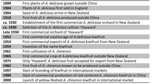
worldwide distributed. The most important steps in the development of kiwifruit as a world commercial crop are summarized in Table 1.1. Until very recently, world trade in kiwifruit had developed from the stage with one predominant green-fleshed cultivar to one commercially important fleshed cultivar with other emerging
yellow-4
fleshed cultivars and finally to the initial commercialization of red-centered yellow-fleshed cultivars. However, all this has changed with the arrival in most countries producing kiwifruit of the disease caused by Pseudomonas syringae pv actinidiae (Ferguson, 2013).
Table 1.1: Important steps in the domestication and commercialization of kiwifruit. 1899 First plants of A. deliciosa grown outside China
1904 Plants of A. deliciosa first sold in England
1904 Seed of A. deliciosa arrive in New Zealand
1910 First fruit of A. deliciosa produced outside China
ca. 1930 Establishment of the first commercial A. deliciosa orchard in New Zealand
ca. 1930 Selection of the A. deliciosa cultivar ‘Hayward’
Late 1930 First commercial orchard of ‘Hayward’
1952 First commercial coolstorage of A.deliciosa kiwifruit
1953 First commercial exports of A. deliciosa kiwifruit from New Zealand
1959 Invention of the name kiwifruit
1961 First cultivation of A. chinensis
1965 First commercial crop of A.deliciosa kiwifruit outside New Zealand
1975 Only ‘Hayward’ A. deliciosa fruit accepted for export from New Zealand
1982 First fruit of A. chinensis known to be produced outside China
1997 Start of branding of kiwifruit in international markets
1998 Start of commercial production of red-centered A. chinensis kiwifruit in China
2000 Launch of yellow-fleshed A. chinensis kiwifruit in international market
The genus Actinidia
The first specimens were collected in Nepal in 1821 by botanist NathanielWallich but the genus was established in the 1836 by Lindley that recognized the specimens as belonging to a genus that could be distinguished by its climbing habitat and the unusual radiating arrangement of the styles. Until then the taxonomy of Actinidia has remained equivocal. Lindley placed the new genus in the Dilleniaceae, giving the name
Actinidia for the stylar arrangement (from the Greek, actis, ray) and described the first
species A. callosa, but only in 1899 Van Tieghem established the family Actinidiaceae containing Actinidia and Saurauia genus. The new family designated was distinguished by the presence of raphides, the versatile anthers, the carpels being accreted into a unilocular fruit, the nature of the embryo and the structure of ovules (Ferguson, 1984). Year by year, additional species and varieties were discovered and published, including
5
Bentham in 1860. Early classification of the genus however, was extremely confusing; many Actinidia species were initially placed in different genera. Actinidia latifolia was first placed in the genus Heptaca (a doubtful genus in Tiliaceae) by Bentham in 1849, then in the genus Kadsura (Schisandraceae) by Miquel in 1861. Actinidia rufa, A.
arguta and A. polygama were first placed in the genus Trochostigma in 1843, then
transferred to Actinidia genus several years later. Actinidia kolomikta was variously placed in Prunus, Kalomikta and Trochostigma genera before finally being identified as
Actinidia by Maximowicz in 1859 (Hsieh et al., 2011). A comprehensive studied on the
taxonomy of the genus was carried out by Dunn that first revised the genus Actinidia in 1911, establishing four sections, Leiocarpae, Ampulliferae, Maculatae and Vestitae based on the degree of pubescence, shape of ovary and presence or absence of lenticels on the fruit surface. These sections may be keyed as follow:
Fruit without spots:
- Sect. Ampulliferae: Ovary bottle-shaped; - Sect. Leiocarpae: Ovary cylindric;
Fruit with spots:
- Sect. Maculatae: Leaves glabrous; - Sect. Vestiae: Leaves shaggy or woolly;
In this first revision 24 species have been recognized and almost 40 varieties or forms worldwide (Dunn, 1911). The second major revision of Actinidia genus was carried out by Li in 1952 that included the section Ampulliferae into the section Leiocarpae, in order to eliminate the ambiguous character of ovary shape because such species as A.
tetramera Maxim have the ovary intermediate shape. He further split the section Vestitae into Stellatae and Strigosae sections emphasizing the structure of leaf hairs.
These sections may be keyed as follow: - Sect. Leiocarpae: Fruit without spots; Leaves without stellate hairs:
- Sect. Maculatae: Branch and petiole glabrous; - Sect. Strigosae: Branch and petiole strigose; Leaves with stellate hairs:
- Sect. Stellatae;
In the second revision were described 36 species and over 50 varieties or forms (Li, 1952). The scheme proposed by Li was adopted also by Liang which completed a
6
revision of Chinese Actinidia in 1984. Liang described many new taxa and listed 51 species as occurring in China, but estimated that there are 54 species within the genus (Liang, 1984).
The classification of the genus Actinidia is difficult and the taxonomy of some taxa is still confusing.
In the most recent revision within the genus Actinidia was achieved by Li in 2007 describing 20 varieties and about 55 species, most of them worldwide (Li & Soejarto, 2007) and all of this have morphological features in common: the climbing growth habitat, the structure of the fruit, the characteristic radiating arrangement of styles female flowers and the dioecy (Ferguson, 2013).
The classification of some taxa still needs further study. The species of Actinidia are highly variable in their vegetative structures, as well as in their flowers and fruits, which is the main reason for the difficulty in the classification of the genus (Li, 1952). Morphologically, species of the genus Actinidia may be clearly separated into two major groups: the first group, which includes Leocarpae, has a glabrous ovary and the fruit has no spots; the second group, which includes Maculatae, Strigosae and
Stellatae, has a hairy ovary and the fruit has spots (Li et al., 2009).
All species in the genus Actinidia are seemingly dioecious, therefore there are female (fruiting) and male (pollenizer) plants (Schmid, 1978). Female plants have flowers with well-developed ovaries and styles as well as stamens. These flowers look as if they were hermaphroditc (perfect) but the pollen is not viable. After pollination, the ovules develop into seed and the ovaries into fruit: such flowers are functionally female (pistillate). Male plants have flowers with rudimentary ovaries which do not contain viable ovules; they cannot set seeds and the ovaries do not develop into fruits. Their stamens produce viable pollen and the flowers are functionally male (staminate) (Ferguson, 2013). Gender on Actinidia species appears to be controlled by an XX/XY system with the male plants having the Y chromosome. Two tightly linked genes are thought to determine gender: one stops pollen development in the female flowers and the other suppresses development of the ovary and ovules in male flowers. (McNeilage, 1997; Testolin et al. 1999). However the dioecy is not absolute, the flowers can be also bisexual able to perform the self-pollination and self-setting. Gender inconstancy has been observed in A. arguta, A. chinensis and A.deliciosa and
7
probably occurs in other Actinidia species (McNeilage, 1991). Male and female plants can differ in morphology with considerable variation in the size, shape and pubescence of leaves produced on different shoots at different times of the year even on the same plant and there can be transitional forms between taxa that overlap geographically (Dunn, 1911).
Actinidia species in cultivation
Since A. deliciosa cv. Hayward (Figure 1.2 a) has been domesticated in New Zealand around 1930, it was considered the backbone of the global kiwifruit industry and has continued to perform extraordinarily well on the global market in terms of production and sales, it remains the dominant commercial kiwifruit cultivar. The Hayward variety arises in New Zealand from a number of competing varieties to become the choice of growers, with its ability to meet all the necessary characteristic needed for a commercially successful cultivar, including taste, storage and size qualities (Ward & Courtney, 2013). Until 2000 A. deliciosa ‘Hayward’ was the cultivar of choice and almost all the international trade in kiwifruit was of this sole cultivar.
When facing overproduction in the early 1990s, the New Zealand industry innovated and assessed the commercial potential of another species, (Ferguson & Huang, 2007). The new cultivar, developed in New Zealand in 1997 by HortResearch (now Plant & Food Research), entered on the international market in 2000 under the name ZESPRI® GOLD Kiwifruit (Figure 1.2 b), reflecting the peculiar golden-yellow fruit flesh. The introduction of ZESPRI® GOLD with its different look, color and taste showed the way in bringing new customers to the kiwifruit category. The most obvious difference between A. chinensis and A. deliciosa is hairiness of the fruit, the first one has smooth skinned fruits compared with A. deliciosa, colour (A. chinensis being usually yellow compared with the green fruit A. deliciosa), fruit flavour, flower size, shoot hairiness, geographic distribution, chromosome number and leaf shape (Ferguson & Bollard, 1990). The introduction of the yellow flesh cultivar A. chinensis ‘Hort16A’ changed the industry by offering a product that complemented, rather than competed, with ‘Hayward’ resulting in an increased consumption (Belrose Inc, 2012). Since 2000, most newly planted orchards in New Zealand have been A. chinensis ‘Hort16A’ and
8
represents about 26% of the New Zealand export of kiwifruit (Belrose Inc, 2012). After introduction of ZESPRI® GOLD, a range of new cultivars were commercialized in China and Japan, some of which also internationally. A. chinensis cv. Jintaoor ENZAGoldtm, a yellow-fleshed cultivar selected in Wuhan, China, (Huang et al., 2002), is widely planted in Italy (Ferguson & Huang, 2007). Recently, A. chinensis cultivar ‘Hongyang’ (Figure 1.2 c) selected in China, with a distinctive yellow-fleshed fruit and a brilliant red around the central core, is widely cultivated for the export market particularly in Japan (Wang et al., 2003). To date, most cultivars have been selected from A. chinensis and
A. deliciosa, however A. arguta are now commercially cultivated in USA, Chile and New
Zealand (Ferguson & Huang, 2007). The fruits of A. arguta are smaller, smooth-skinned, with a rich and sweet flavour (Figure 1.2 d) (Williams et al., 2003). Despite the breeding efforts ‘Hayward’ is still the predominant fruit traded internationally, with an estimate of 90 to 95 % of all kiwifruit market (Ferguson & Seal, 2008).
Fig 1.2: Commercially produced kiwifruit: (a) A. deliciosa, green fleshed kiwifruit, (b) A.
chinensis ‘Hort16A’, gold yellow fleshed fruit, (c) A. chinensis cv ‘Hongyang’, yellow-fleshed
fruit and brilliant red around the central core, (d) A. arguta, berry sized green kiwifruit.
a b
9
Global kiwifruit industry
Actinidia species were introduced to Europe, the U.S.A and New Zealand in the late
19th and early 20th century (Ferguson & Bollard, 1990). Commercial kiwifruit growing areas have expanded rapidly and consistently since records began in 1970, with exponential growth in the 1980, static production in 1990 and steady growth over the past decade. The growth of the industry has varied significantly in short bursts and the long-term growth path has continued upward with global production doubling over the past 20 years, furthermore it is predicted that this will continue as new plantings reach full maturity in key production countries such as China and Chile (Figure 1.3) (Ward & Courtney, 2013).
Fig 1.3: Global production of kiwifruit from 1970 to 2012. Source: FAOSTAT (2014)
The international kiwifruit production is concentrated in relatively few countries. The top four countries are, China, Italy, New Zealand and Chile that collectively produce more than 80% of the world's kiwifruit crop; the top ten producing countries represent more than 96% of the world supply. Total production in 2009-2012 was 1,862,000 tonnes (Table 1.2) (Belrose Inc., 2012).
0 200 400 600 800 1000 1200 1400 1600 1970 1975 1980 1985 1990 1995 2000 2005 2010 Gl o b al ki wi fr u it p ro d u ction -to n s (000) Year
10
Table 1.2: World kiwifruit production: Top-ten producing countries 2009-2012
Rank Country Production (tons)
(Average) 1 China 480,000 2 Italy 450,000 3 New Zealand 372,833 4 Chile 230,333 5 Greece 83,167 6 France 71,851 7 Japan 33,300
8 Iran (Islamic Republic of) 31,532
9 United States of America 27,391
10 Spain 18,125
The growth in global kiwifruit production corresponded to an increase of the kiwifruit planted area. The Food & Agriculture Organisation of the United Nations statistical department (FAOSTAT, 2014) estimates in 1970 that there were < 1,000 ha of kiwifruit planted in the world outside of China. In 2010, the area of kiwifruit planted globally (including China) was estimated by O’Rourke to be over 160,000 ha (Figure 1.4) (Ward & Courtney, 2013).
Fig 1.4: Area of kiwifruit planted globally from 1970 to 2012. Source: FAOSTAT (2014)
The growth of the global kiwifruit industry has not been simply as result of using more land, but the global average yields per hectare have also increased significantly, from an estimated 5,000 in 1970 to almost 15,000 kg ha-1 in 2012 (FAOSTAT, 2014).
0 10000 20000 30000 40000 50000 60000 70000 80000 90000 100000 110000 1970 1975 1980 1985 1990 1995 2000 2005 2010 A re a o f K iwi fr u it p lan te d gl o b al ly -ha Year
11
Resulting of the increased the global volume of kiwifruit produced was also the increasing of the global exports volume of kiwifruit. According to O’Rourke (2012), around two-thirds of global kiwifruit production is exported, with Italy, New Zealand and Chile as the world’s leading exporters of kiwifruit. These countries accounted for about 90% of all exporters of kiwifruit in 2010. China is the largest producer but the production has been almost totally consumed in its domestic market with exports accounting for only 0.2 % of the global trade in kiwifruit (Table 1.3) (Ward & Courtney, 2013).
Table1.3: Major exporters: Share of global trade (volume) in fresh kiwifruit 2007-2010 (%).
Source: O’Rourke (2012) Exporter 2007 (%) 2008(%) 2009(%) 2010(%) Italy 31.1 28.6 32.7 31.0 New Zealand 32.5 35.1 30.7 30.8 Chile 14.9 14.9 15.5 15.2 Greece 3.4 3.5 4.7 6.2 France 2.7 2.4 1.9 2.2 Spain 1 0.9 1 1 United States 0.7 0.6 0.5 0.7 Portugal 0.3 0.1 0.3 0.5 China 0.3 0.2 0.1 0.2 All other 12.9 13.7 12.6 12.2 Total 100.0 100.0 100.0 100.0
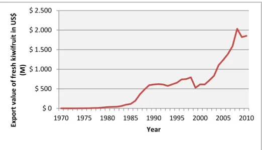
On the world kiwifruit review in 2012, the value of the global kiwifruit industry in 2012 was estimated to be around US$ 1.9 billion (O’Rourke, 2012). Until the global financial crisis in 2008, the global value of exports of fresh kiwifruit had been increasing every year since 1999, with significant growth rates between 2002 and 2008 (Figure 1.5) (FAOSTAT, 2014). During this period, consequently to this strong growth, the area of kiwifruit planted increased around 22,000 ha (FAOSTAT, 2014).
12
Fig 1.5: World value of exports of fresh kiwifruit. Source: FAOSTAT (2014)
Despite the significant growth in the global volume of kiwifruit over the past 30 years, the fruit has remained very much a niche product, accounting for less than a quarter of a percent of global fruit production. Global kiwifruit production represents about 0.22% of total production for major fruit crops, with the majority of kiwifruit consumed as fresh fruit (Table 1.4) (O’Rourke, 2012). World production has remained dominated by the traditional categories of citrus, apples, bananas and grapes. Inevitably, consumption of kiwifruit is highest in the countries that produce it. New Zealand is the largest consumer of kiwifruit with an estimated annual consumption level to be more than 5 kg of fruit per capita, while Spain, Chile, Italy and Portugal all have annual consumption level between 2 and 3 kg of kiwifruit per capita (O’Rourke, 2012).
$ 0 $ 500 $ 1.000 $ 1.500 $ 2.000 $ 2.500 1970 1975 1980 1985 1990 1995 2000 2005 2010 Exp o rt val u e o f fr e sh k iwi fr u it in US$ (M ) Year
13
Table1.4: World share of production for major fruit groups. Source: O’Rourke (2012)
Fruit category 1983-1985 (% of total) 1996-1998 (% of total) 2008-2010 (% of total) 2008-2010 versus 1996-1998 (% change) Apple 12.81 13.28 11.49 - 1.79 Other deciduous 9.68 9.66 10.87 + 1.21 Total deciduous 22.48 22.94 22.36 - 0.58 Total grapes 20.35 13.59 11.29 - 2.30 Oranges 13.84 14.70 11.48 - 3.22 Other citrus 7.60 7.25 8.96 + 1.71 Total citrus 21.45 21.95 20.44 - 1.51 Bananas 12.94 13.89 16.86 + 2.97 Other tropical 21.49 21.21 22.45 + 1.24 Total tropical 34.43 35.10 39.31 + 4.21
Other fresh fruit 0.0 5.09 5.19 + 0.10
Total berries 1.18 1.12 1.19 + 0.07
Kiwifruit 0.11 0.21 0.22 + 0.01
Total fruit 100.00 100.00 100.00 n.a.
Production and marketing in Italy
The first kiwifruit orchard were established in Italy more than 40 years ago, around 1967 and since then the commercial kiwifruit growing areas have expanded rapidly and consistently. The Italian kiwifruit industry is important both nationally and internationally. It makes up only a small part of Italian horticulture in general, but is profitable and expanding. Italy is one of two world’s biggest kiwifruit exporter together to New Zealand (Testolin & Ferguson, 2009).
Kiwifruits have been grown in Italy for more than 70 years, but for much of that time were simply as horticultural curiosities. Experimental plantings of A. deliciosa were established in 1966-67 at Lake Maggiore and over the following 2 or 3 years, small demonstration planting were established in other part of Italy using a mixture of plants sourced from the United Kingdom, including the recognized New Zealand cultivars such as ‘Hayward’. One of the first attempts at commercial-scale kiwifruit plantings in Latina in 1971, probably using plants from New Zealand, failed because the plants died from heat stress after being grown within individual plastic domes under the misapprehension that kiwifruit were tropical plants (Testolin & Ferguson, 2009). By 1973, 40-50 ha have been planted and the success of these orchards encouraged establishment of further kiwifruit orchards in Piedmont, Lazio, Veneto and,
14
subsequently, in Emilia Romagna. Five years later, in 1978, between 600 and 800 ha of kiwifruit have been planted in Italy, of which perhaps 250 ha were productive or potentially productive (Zuccherelli & Zuccherelli 1981 ). The area in kiwifruit from 1984 to 2013 increased more than 10-fold, with an initial rapid increase to 18,000 ha from 1984 to 1990, followed by a decade during which the area remained essentially unchanged or even fell slightly and then a slowly increased until 2013 (Figure 1.6). Production data follow much the same trend, an initial rapid increase from 1984 to 1990, then a plateau with wide fluctuations in production from year to year, followed by slow increase from 1999 (Figure 1.7). At least some kiwifruit plantings have been attempted in all the different regions of Italy but orchards have mainly consolidated only in four regions: Lazio, Piedmont, Emilia Romagna and Veneto (Table 1.5), with a smaller plantings in Abruzzo, Basilicata, Calabria, Campania, Lombardy and Apulia. Over the last 20 years, the biggest increases in area and production have been in south Lazio and Piedmont. Kiwifruit have become less important in regions such as Lombardy, because of frequent problems with frost and Apulia, because of damage caused by salt-laden winds and because economic conditions now favor the growing of alternative crops (Testolin & Ferguson, 2009).
Total area of kiwifruit planted in Italy from 2007 to 2012 is approximately the same but the tonnes produced fell slightly (Table 1.6)
Fig 1.6: Area of kiwifruit planted in Italy from 1984 to 2013. Source: FAOSTAT (2014) 0 5000 10000 15000 20000 25000 30000 A re a o f K iwi fr u it p lan te d in It al y -ha Year
15
Fig 1.7: Production of kiwifruit from 1984 to 2013. Source: FAOSTAT (2014)
Table 1.5: Italian regions kiwifruit production: Top-four producing regions 2013.
Source: CSO (Centro Servizi Ortofrutticoli, Ferrara) (2014)
Table 1.6: Area of kiwifruit (ha) planted and production in Italy from 2007 to 2013.
Source: CSO (Centro Servizi Ortofrutticoli, Ferrara)(2012) and FAOSTAT (2014)
2007 2008 2009 2010 2011 2012 2013 Area of kiwifruit planted (ha) 26,834 27,275 27,619 28,300 28,058 26,893 24,891 Production of kiwifruit (t) 417,151 477,100 475,790 410,522 471,929 376,327 447,560
The initial development of the Italian kiwifruit industry was based on cultivars of A.
deliciosa originating in New Zealand. ‘Abbott’, ‘ Bruno’, ‘Monty’ and ‘Hayward’ were all
planted experimentally (Ferguson & Bollard, 1990) but by 1983 over 70% of plantings were ‘Hayward’ and this reliance continued to increase for many years so that eventually it was essentially the sole fruiting cultivar grown. Only recently other cultivars of A. deliciosa and A. chinensis have started to produce commercial quantities
0 50 100 150 200 250 300 350 400 450 500 ki wi fr u it p ro d u ction in It al y -to n s (000) Year
Region % Total Area Area (ha)
Lazio 30 7,350
Piedmont 20 5,000
Emilia Romagna 16 4,000
16
of fruit (Table 1.7) but ‘Hayward’ still accounts for an overwhelming 92% of female kiwifruit plantings in Italy (Testolin & Ferguson, 2009).
Table 1.7: Tons Commercial kiwifruit cultivars in Italy from 2007 to 2013 (t)
Source: CSO (Centro Servizi Ortofrutticoli, Ferrara)(2014)
2007 2008 2009 2010 2011 2012 p2013 A. deliciosa G3 100 (t) Summer 2.700 (t) 3.400 (t) 3400 (t) 3770 (t) 3800 (t) 3.650 (t) 2.700 (t) A. chinensis HORT 16A (*) 14.200 (t) 15.000 (t) 10.000 (t) 3.600 (t) 4.000 (t) 2.500 (t) Jingold 380 (t) 2.570 (t) 4.625 (t) 5.782 (t) 5.294 (t) 6.345 (t) 5.865 (t)
* data not available
Even though Italy is one of the biggest producers of kiwifruit, the industry is still relatively small when considered in the context of the total Italian fruit production: kiwifruit account for around 2% of the total area of fruit crops and almost 3% of the total fruit production by weight (Source : FAOSTAT 2014) (Testolin & Ferguson, 2009).
Diseases in Actinidia spp.
Commercial kiwifruit growing areas have expanded rapidly and consistently since records began in 1970 and plants has been considered to be relatively diseases free until recently. Just some fungal disease was divulged previously, such as Armillaria
novae-zelandii identified in New Zealand in 1992 (Horner, 1992), Phomopsis sp. in
Greece in 2009 (Elena, 2009), Cadophora melinii identified in Italy in 2008 (Prodi et al, 2008) and verticillum wilt of gold kiwifruit in Chile (Auger et al., 2009).
Recently the phytosanitary situation of kiwifruit is changed radically with the detection of a virulent strain of Pseudomonas syringae pv. actinidiae (PSA), first in Italy and after in New Zealand (Ferrante & Scortichini, 2010; Everett et al., 2011). During spring and autumn 2008 and winter 2008–9, severe outbreaks of bacterial canker were observed on A. chinensis (yellow kiwifruit) cvs Hort 16A and Jin Tao cultivated in central Italy (Latina province). The main typical symptoms were the oozing of reddish exudates
17
along the main trunk and branches, reddening of the lenticels under the epidermis, leaf spots sometimes surrounded by a chlorotic halo, leaf wilting, twig dieback and plant wilting (Ferrante &Scortichini, 2010). This pathogen was isolated for the first time in the same area from A. deliciosa cv. Hayward in 1992 (Scortichini, 1994) but from that time until 2008 it caused only sporadic damage (i.e. leaf spotting, twig dieback), always towards A. deliciosa. Severe damage and⁄or epidemics were never observed. The epidemic affecting ‘Hort16A’ in Italy was caused by a strain that appeared to be more virulent than a strain reported in 1994 (Ferrante & Scortichini, 2010). Since 2008 PSA was spread quickly and with considerable aggression, affecting plantations in the provinces of Latina and Rome (Lazio), in Emilia-Romagna especially in the provinces of Ravenna and Forlì, in Veneto, in Piedmont and, more recently, in Calabria. The epidemic in Italy has caused severe vine losses, with removal of entire orchards as a consequence (Figure 1.8) (Scortichini et al., 2012; FAOSTAT, 2014).
Symptoms resembling those caused by PSA were first observed on A. chinensis in Te Puke, Bay of Plenty, New Zealand in November 2010 (Everett et al, 2011). Since then the disease has spread widely throughout the Bay of Plenty and also in other part of New Zealand that produce kiwifruit. The number of PSA positive orchards is now over 2,700, with 12,009 hectares (87%) on orchard where PSA has been identified (Figure 1.9) (Data from Kiwifruit Vine Health PSA Statistics Report, February 2015)
Fig 1.8: Area of kiwifruit planted in Italy and Production of kiwifruit from 2008-2013 during the
PSA epidemic. Source: FAOSTAT (2014) 21000 21500 22000 22500 23000 23500 24000 24500 25000 25500 2008 2009 2010 2011 2012 2013 Year A re a of K iw if rui t pl a nt e d in It a ly ( ha ) 0 50000 100000 150000 200000 250000 300000 350000 400000 450000 500000 k iw if rui t prod uc ti on i n It a ly -ton ne s
Area harvested (ha) Production(t)
18 Fi g 1 .9 : P SA s tat u s o f o rcha rd by regi o n in N ew Ze alan d . S ou rce: K WH Kiwif ru it Vin e Hea lth, 2 01 5 )
19
The aims of my study
In Italy, as well as in many other countries, the kiwifruit crop has been considered to be relatively disease-free and then no certification system for this species has been developed to regulate the import of propagation plant material in the European Union. The detection of a virulent strain of Pseudomonas syringae pv. actinidiae (PSA) has dictated the need to reorganize the certification system for this species in order to regulate import and exchanges of propagation plant material.
In attempting to address this issue, we also filled the lack of scientific knowledge on viruses infecting kiwifruit. Therefore, in order to study viral agents of this species, a project has been developed at the University of Bologna (Italy) in collaboration with the University of Auckland (New Zealand).
The aims of my PhD thesis were:
- to investigate and characterize the viruses that can infect kiwifruit plants in order to define the virological framework of the culture in Italy;
- to determine the best methods of investigation that can identify viral agents known or not yet known to infect Actinidia spp.;
- to characterize a strain of Cucumber mosaic virus detected in both, A. chinensis and A. deliciosa;
- to characterize a strain of Pelargonium zonate spot virus detected in A.
chinensis;
- to characterize two novel viruses, a new putative Closterovirus and a new putative Totivirus, detected by the next generation sequencing approach;
The investigations of the viruses that can infect kiwifruit plants was carried out using biological, serological and molecular techniques. The characterization of the viral isolates has been also completed with the employ of the next generation sequencing (NGS) method, which has been useful also for identification of infections caused by multiple viruses.
20
The results obtained during my PhD studies are proposed in chapters 2 to 5. Chapter 2 includes a review regarding kiwifruit viruses. The manuscript, published on the Journal of Plant Pathology is an “Invited Review” that the editors of the journal asked to our teams (New Zealand and Italy). The published paper is also included as Annex A.
Chapters 3, 4 and 5 report first identification and characterization of viral agents that infect kiwifruits in Italy. Each chapter is presented as a Project Paper then in the form that will be submitted to international scientific journals.
This format may generate some repetitions (in particular regarding Introduction, Materials and Methods and References sections) but, in my opinion, allow a better presentation of results obtained and, for sure, will speed up their publication.
21
References
Auger J., Perez I., Fullerton R.A., Esterio M., (2009). First report of Verticillium wilt of gold kiwifruit, Actinidia chinensis cv. Hort 16A, caused by Verticillium albo-atrum in Chile. Plant
Disease 93: 553.
Belrose Inc., (2012). World kiwifruit review. 2012 edition Pullman, WA, Belrose, Inc.
Datson P.M., Ferguson A.R., (2011). Actinidia. In C. Kole (Ed.), Wild Crop Relatives: Genomic and
Breeding Resources (pp. 1-20). Berlin Heidelberg: Springer.
Dunn S.T., (1911). A revision of the genus Actinidia Lindl. J. Linn. Soc. Lond. Bot. 39: 394-410. Elena K.,(2009) Occurrence of Phomopsis sp. on kiwi plantations in Nortern Greece. Hellenic
Plant Protecnion Journal 2:67-69
Everett K., Taylor R., Romberg M., Rees-George J., Fullerton R., Vanneste J., Manning M., (2011). First report of Pseudomonas syringae pv. actinidiae causing kiwifruit canker in New Zealand.
Australasian Plant Disease Notes 6: 67-71.
FAO (2014). FaoSTAT Agricolture Statistics Database. Web: .
Ferguson A.R., (1984). Kiwiftuit: a botanical review. Horticultural Reviews 6: 1-64
Ferguson A.R., (1990a). Botanical Nomenclature: Actinidia chinensis, Actinidia deliciosa and
Actinidia setosa. In: Warrington IJ, Weston GC, eds. Kiwifruit: Science and Management.
Auckland: NZHSH/Ray Richards Publisher, pp 36-57.
Ferguson A.R., (1990b). The kiwifruit in China . In: Warrington I.J., Weston G.C. (eds). Kiwifruit:
Science and Management, pp. 155-164. Auckland: Ray Richards Publisher and NZ Society for
Horticultural Science, Auckland, New Zealand.
Ferguson A.R (2013). Kiwifruit: the wild and the cultivated plants. In Boland M., Moughan P.J. (eds) Advances in Food and Nutrition Research, Nutritional Benefits of Kiwifrut 68:15-32
Ferguson A.R., Bollard E.G., (1990). Domestication of the kiwifruit. In: Warrington I.J., Weston G.C. (eds). Kiwifruit: Science and Management, pp. 165-246. Ray Richards Publisher and NZ Society for Horticultural Science, Auckland, New Zealand.
Ferguson A.R., Huang H.,( 2007). Genetic resources of kiwifruit: domestication and breeding.
Horticultural Reviews 33: 1-122.
Ferguson A.R., Seal A.G., (2008). Kiwifruit. In: Hancock J.F. (ed.). Temperate Fruit Crop Breeding, pp. 235-264. Springer, Berlin, Germany.
Ferrante P., Scortichini M., (2010). Molecular and phenotypic features of Pseudomonas syringae pv. actinidiae isolated during recent epidemics of bacterial canker on yellow kiwifruit (Actinidia
chinensis) in central Italy. Plant Pathology 59: 954-962.
Horner I.J., (1992). Epidemiology of Armillaria root-rot of kiwifruit. Acta Horticulturae 297:573-578.
22
Hsieh T.Y., Ku S.M., Chien C.T., Liou Y.T., (2011). Classifier modeling and numerical taxonomy of
Actinidia (Actinidiaceae) in Taiwan. Botanical Studies 52: 337-357.
Huang H.W., Wang S.M., Huang R.H., Jiang Z.W., Zhang Z.H., (2002). 'Jintao', a novel, hairless, yellow-fleshed kiwifruit. HortScience, 37:1135-1136.
Li H.L.,(1952). The taxonomic review of the genus Actinidia. Journal of the Arnold Arboretum, 33: 1-61.
Li J.Q., Li X.W., Soejarto D.D., ( 2007). Actinidiaceae. In: Wu C Y, Raven P, eds. Flora of China 12: 334-355. Beijing: Science Press and St. Louis: Missouri Botanical Garden Press.
Li X.W., Li J.Q., Soejarto D.D., (2009). Advances in the study of the systematics of Actinidia Lindey. Front. Biol. China, 4(1):55-61.
Liang C.F., (1984). Actinidia. In K. M. Fung (ed.), Flora Reipubliae Popularis Sinica, Science Press
49:196-324, Beijing.
McNeilage M.A., (1991). Sex expression in fruit male vines of kiwifruit. Sexual Plant
Reproduction, 4:274-278.
McNeilage M.A., (1997). Progress in breeding hermaphrodite kiwifruit cultivars and understanding the genetics of sex determination. Acta Horticulturae, 444:73-78.
Prodi A., Sandalo S., Tonti S., Nipoti P., Pisi A., (2008). Phialophora-like fungi associated with kiwifruit elephantiasis. Journal of Plant Pathology 90: 487-494.
O’Rourke D., (2012). World kiwifruit review 2012.Washington Stat: Belrose Inc. Qian Y.Q., Yu D.P., (1991). Advances in Actinidia research in China. Acta Horticulturae,
297:51-55.
Schmid R., (1978). Reproductive anatomy of Actinidia chinensis (Actinidiaceae). Bot. Jahrb. Syst.,
100:149-195.
Scortichini M., (1994). Occurrence of Pseudomonas syringae pv. actinidiae in Italy. Plant
Pathology 43:1035–8
Scortichini M., Mar celletti S., Ferrante P., Petriccione M., Firrao G., (2012). Pseudomonas
syringae pv. actinidiae: a re-emerging, multi-faceted, pandemic pathogen. Molecular Plant Pathology 13:631–40.
Testolin R., Cipriani G., Messina R., (1999) . Sex control in Actinidia is monofactorial and remains so in polyploids. In: C. C. Ainsworth,( ed). Sex determination in plants (pp 173-181). Oxford, United Kingdom, Bios Scientific Publisher.
Testolin R., Ferguson A. R., (2009). Kiwifruit (Actinidia spp.) production and marketing in Italy.
New Zealand Journal of crop and Horticultural Science, 37: 1-32.
Wang M., Li M., Meng A., (2003). Selection of a new red-fleshed kiwifruit cultivar 'Hongyang'.
23
Ward C., Courtney D., (2013). Kiwifruit: taking its Place in the Global Fruit Bowl. In Boland M., Moughan P.J. (eds) Advances in Food and Nutrition Research, Nutritional Benefits of Kiwifrut
68:1-14.
Williams M.H., Boyd L.M., McNeilage M.A., MacRae E.A., Ferguson A.R., Beatson R.A., Martin P.J., (2003). Development and commercialization of 'baby kiwi' (Actinidia arguta Planch).
Proceedings of the Fifth International Symposium on Kiwifruit, Issue 610, pp. 81-86.
24
Chapter 2:
25
Viruses of kiwifruit
The first clue of a kiwifruit infecting virus comes from New Zealand quarantine records in 1983. Gary Wood, from the Department of Industrial and Scientific Research (DSIR, New Zealand), documented local lesions observed on
Chenopodium quinoa after sap inoculation of kiwifruit imported from China and
held in quarantine. The infected kiwifruit plants were either destroyed or died during thermotherapy (G. Wood, personal comunication). In the 1980s, as Italy was becoming an important kiwifruit producer with the second greatest area planted worldwide, there were no records of viruses infecting the crop.
Caciagli and Lovisolo (1987) surveyed commercial orchards for potential viral diseases and collected samples from 100 symptomless A. deliciosa and one plant of A. deliciosa that showed chlorotic mottling. The extracts from these plants were mechanically inoculated into four herbaceous indicators (C. quinoa, C.
amaranticolor, Nicotiana glutinosa and N. clevelandii). None of the 404
inoculated indicator plants displayed symptoms. Additionally, the authors challenged young A. deliciosa plants with 17 common viruses from Italy, including Alfalfa mosaic virus (AMV) and Cucumber mosaic virus (CMV). Only three viruses, Tobacco necrosis virus (TNV), Tobacco rattle virus (TRV) and CMV, induced symptoms on the inoculated leaves of the kiwifruit and only CMV moved systemically. The authors concluded that kiwifruit may be resistant to virus infections. A few years later, during a survey in the Fujian Province in China, Lin and Gao (1995) identified one plant showing a “mosaic disease” attributed to an unidentified virus. Nitta and Ogasawara (1997) reported evidence of a graft-transmissible agent causing viruslike symptoms. Using cuttings from Actinidia
polygama plants collected in the mountains of Hiroshima Prefecture (Japan) as
rootstocks, they observed chlorotic spots and rings on the eight different A.
deliciosa varieties used as male scions. In neither case the causal agent was
identified. In 2003, Apple stem grooving virus (ASGV) was identified in a kiwifruit import from China held in New Zealand quarantine (Clover et al., 2003). This first virus identified in kiwifruit was detected by leaf symptoms, transmission electron microscopy (TEM) and mechanical transmission to herbaceous indicators and
26
identified by DAS-ELISA, RT-PCR and sequencing of amplicons. Other kiwifruit from the same consignment were subsequently studied further and new viruses were identified.
To assess the potential risk from viruses to kiwifruit, it is important to document which viruses are present in both breeding material and commercial crops and as far as possible determine where they originated from and how do they spread. In some cases viruses have been moved internationally with germplasm, while in other cases the viruses may have infected kiwifruit locally, from other plant species, following the introduction of the crop. Although China is the origin of
Actinidia until relatively recently most of the breeding and selection of
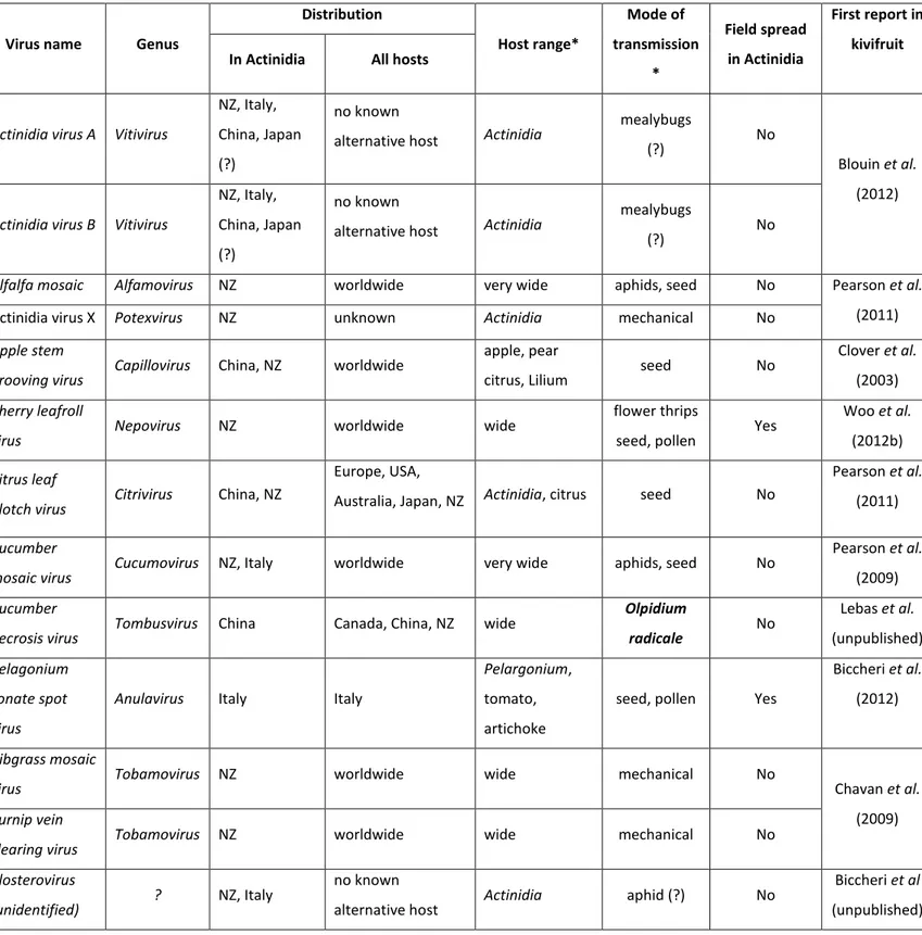
commercial cultivars was conducted in NZ involving movement of Actinidia germplasm from China to New Zealand over several decades. Subsequently there has been movement of commercial cultivars from New Zealand to several countries around the world, including Chile, Italy and back to China. In addition there has also been recent movement of germplasm and commercial varieties from China to Italy. Examination of kiwifruit germplasm and field crops in New Zealand and Italy, between 2002 and 2013 has identified a total of 13 different viruses, representing a wide taxonomic range (Table 2.1). However, many of the source plants were infected by multiple viruses which has made it difficult to attribute symptoms to individual viruses.
Recently two new detection were described outside of New Zealand and Italy, first one in China where Actinidia virus A and Actinidia virus B were detected on
A. chinensis (Zheng et al., 2014) and the second one in India with the
characterization of Apple stem grooving virus infecting A.deliciosa (Bhardwaj et
27
Table 2.1. Viruses detected naturally infecting Actinidia spp.
* data refers to the virus species in general, not specifically the Actinidia isolate (?) = presumed mode of transmission based on properties of related viruses
Virus name Genus
Distribution Host range* Mode of transmission * Field spread in Actinidia First report in kivifruit In Actinidia All hosts
Actinidia virus A Vitivirus
NZ, Italy, China, Japan (?)
no known
alternative host Actinidia
mealybugs
(?) No
Blouin et al. (2012)
Actinidia virus B Vitivirus
NZ, Italy, China, Japan (?)
no known
alternative host Actinidia
mealybugs
(?) No
Alfalfa mosaic Alfamovirus NZ worldwide very wide aphids, seed No Pearson et al.
(2011)
Actinidia virus X Potexvirus NZ unknown Actinidia mechanical No
Apple stem
grooving virus Capillovirus China, NZ worldwide
apple, pear
citrus, Lilium seed No
Clover et al. (2003)
Cherry leafroll
virus Nepovirus NZ worldwide wide
flower thrips
seed, pollen Yes
Woo et al. (2012b)
Citrus leaf
blotch virus Citrivirus China, NZ
Europe, USA,
Australia, Japan, NZ Actinidia, citrus seed No
Pearson et al. (2011)
Cucumber
mosaic virus Cucumovirus NZ, Italy worldwide very wide aphids, seed No
Pearson et al. (2009)
Cucumber
necrosis virus Tombusvirus China Canada, China, NZ wide
Olpidium radicale No Lebas et al. (unpublished) Pelagonium zonate spot virus
Anulavirus Italy Italy
Pelargonium,
tomato, artichoke
seed, pollen Yes
Biccheri et al. (2012)
Ribgrass mosaic
virus Tobamovirus NZ worldwide wide mechanical No Chavan et al.
(2009)
Turnip vein
clearing virus Tobamovirus NZ worldwide wide mechanical No
Closterovirus
(unidentified) ? NZ, Italy
no known
alternative host Actinidia aphid (?) No
Biccheri et al (unpublished)
28
To date, the viruses discovered in kiwifruit can be divided in three groups. The first group comprises AMV, ASGV, CMV, Cucumber necrosis virus (CNV), Ribgrass
mosaic virus (RMV), Turnip vein clearing virus (TVCV) and a novel potexvirus,
tentatively named Actinidia virus X (AVX). These viruses are mostly ubiquitous/ cosmopolitan and, so far, do not show a detrimental effect on commercial kiwifruit. Most of these viruses are distributed worldwide over a large host range and have been detected in alternative hosts neighboring kiwifruit orchards. The second group comprises the putatively kiwifruit specific viruses that, to date, are only known to have this single host or are likely to have a very limited host range. In this group we have identified two vitiviruses, Actinidia virus A (AcVA) and
Actinidia virus B (AcVB) and a citrivirus closely related to Citrus leaf blotch virus
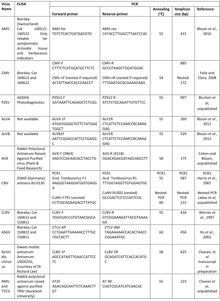
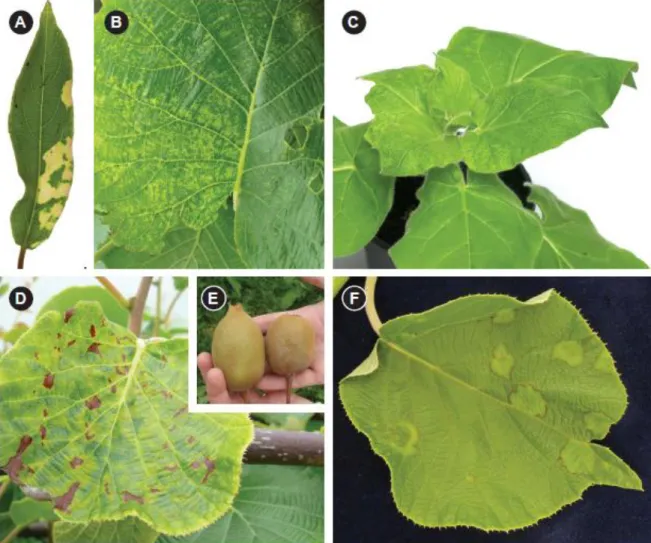
(CLBV). There is also evidence of a novel virus from the family Closteroviridae, Actinidia latent virus (AcLV). The third and most concerning group includes two viruses that have very recently been detected in kiwifruit. Cherry leaf roll virus (CLRV) in New Zealand and Pelargonium zonate spot virus (PZSV) in Italy both cause severe damage to the commercial crop. In addition, the viruses listed in Table 2.1 represent a range of different modes of transmission with varying consequences for disease spread and control. From an epidemiological perspective the known viruses can be sub-divided into those with no known natural vectors, those with aphid vectors, those with presumed mealybug vectors and those transmitted by seed and/or pollen. Almost 10 years since the first publication of kiwifruit virology, we describe now the 13 viruses detected in kiwifruit to date. This represents the first review of kiwifruit viruses, including images of symptoms (Figure. 2.1), a summary table of each virus (Table 2.1) and a summary of diagnostic tools including primer sequences and amplification conditions (Table 2.2).
29
Non-Specialist viruses
Alfalfa mosaic virus and Cucumber mosaic virus
AMV and CMV are two viruses infecting a very broad host range, with over 1200 plant host species in over 100 families for CMV (Douine et al., 1979) and 300 species in 22 plant families for AMV (Hull, 1969). The addition of Actinidia species to their host range is not unexpected. Because of the damage CMV causes on some economically important crops, it was included in the “Top 10 plant viruses” in a recent molecular pathology review (Scholthof et al., 2011). Both viruses belong to the family Bromoviridae and are efficiently vectored by a number of aphid species. They are also transmitted by seed and are easily transmissible mechanically. AMV is the type member of the genus Alfamovirus and has four bacilliform type particles (Fauquet et al., 2005).
CMV is the type member of the genus Cucumovirus and has icosahedral particles. AMV was one of the first viruses detected and identified in kiwifruit in New Zealand (Pearson et al., 2009). It was first detected in Actinidia glaucophylla, showing strong yellow mosaic patterns (Figure. 2.1A). Extracts from the chlorotic blotch easily transmitted the virus to a range of herbaceous indicator plants. In the same germplasm collection, AMV was also isolated from Actinidia guilinensis and A. fortunatii showing mottled and generally chlorotic leaves. In these hosts, the plants looked unthrifty and the virus symptoms were widespread in the block. The symptoms were observed in spring for four consecutive years. AMV and CMV have been found as a dual infection in both A. glaucophylla and A.
fortunatii and CMV was also detected in a single symptomless infection of A. glaucophylla. AMV has only been detected once in A. chinensis in New Zealand.
The plant showed a few leaves with very minor chlorosis and the symptoms could not be observed the following year. Inoculation of AMV to A. chinensis seedlings induced foliar symptoms on one or two leaves above the inoculated leaf, but newer leaves were symptomless.
30
Table 2.2. Diagnostic tools: reagents for ELISA when available and primers used and
conditions for PCR assays. Virus
Name
ELISA PCR
Forward primer Reverse-primer
Annealing (°C) Amplicon size (bp) Reference AMV Bioreba (Switzerland) Cat 140512-140522 Only reliable for symptomatic Actinidia tissue and herbaceous indicators AMV for TGTCTCACTGATGACGTG AMV rev CATACCTTGACCTTAATCCAC 55 415 Blouin et al., 2010 CMV Bioreba, Cat 160612 and 160622 CMV-F CTTTCTCATGGATGCTTCTC CMV nF (nested if required) ACTATTAACCACCCAACCT CMV-R GCCGTAAGCTGGATGGAC CMV nR (nested if required) TTTGAATGCGCGAAACAAG 54 885 Nested: 172 Felix and Clara, 2008 PZSV ADGEN Phytodiagnostics PZSV2 F GATAAATTCAGAGCTCTCGG PZSV2 R ATCTCTGCAGATTGTGTTCC 55 997 Biccheri et al, unpublished AcVA Not available AcVA 1F
ATGATGGGGTGTTCTATGGG TGGCT AcV1R CTCATTCTCCAMCCRCARAA GAG 55 269 Blouin et al., 2012 AcVB Not available AcVB1F
AATTCGGACCACTCCTGAGG C AcV1R CTCATTCTCCAMCCRCARAA GAG 55 529 Blouin et al., 2012 AVX Rabbit Polyclonal Antiserum Raised Against Purified virus (Plant & Food Research) AVX-F (3963) AAGTCCGCAACACCTACCTG AVX-R (4118) GGACAGACGATAGCAGCCTT 58 175 Cohen and Blouin, unpublished CNV DSMZ (Germany), antisera AS-0130 PCR1 Gral. Tombusvirus F1 AAGGGTAAGGATGGTGAGG A CuNV-F791 (nested) CCTCGCAGAAGACCTTATGC PCR1 Gral. Tombusvirus R1 TTTGGTAGGTTGTGGAGTGC CuNV-R1002 (nested) GCCGACTCCTCCACTCCA PCR1 55 Nested-PCR 60 PCR1 587 Nested-PCR 215 PCR1 Harris et al., 2007 Nested-PCR Lebas et al, unpublished CLRV Bioreba, Cat 160612 and 150812 CLRV-F TGGCGACCGTGTAACGGCA CLRV-R GTCGGAAAGATTACGTAAAA GG 55 416 Werner et al., 1997 ASGV Bioreba, Cat 150822 and 150812 CTLV-AP CCTGAATTGAAAACCTTTGC TGCCACTT CTLV-AM TAGAAAAACCACACTAACC CGGAAATGC 60 456 Ito et al., 2002 Actini dia citrivir us Dweet mottle antiserum Antiserum USDA253, (courtesy of Dr Richard Lee) CLBV 1F AGCCATAGTTGAACCATTCC TC CLBV 5R GCAGATCATTCACCACATG C 58 425 Chavan, et al manuscript in preparation RMV and TVCV Rabbit polyclonal antiserum raised against purified TMV (Auckland University) AT2F AGACAGCAATTCTCAAACTT GT AT 4R CGGTCGCATCATCAACAC 55 223 Chavan et al, unpublished
31
CMV has been detected in Italy on one A. chinensis plant with pale mottling of the leaves (See chapter 3). AMV and CMV can be detected by RT-PCR in Actinidia spp. (Table 2.2). DAS-ELISA can also be used for both viruses but AMV can only be detected in symptomatic tissues. Both viruses are readily transmissible to a range of herbaceous indicators including N. benthamiana, N. clevelandii, N.
glutinosa and N. occidentalis. These two viruses are similar in terms of their
abundance in the surrounding weeds and also by sharing the same vectors. Both are present worldwide and are likely to infect Actinidia spp. causing some concerns for the non-commercial species (A. glaucophylla, A. guilinensis and A.
fortunatii). Fortunately, the viruses do not appear to have a detrimental effect on
either A. chinensis or A. deliciosa. Their impact on these important crops is therefore negligible.
Ribgrass mosaic virus and Turnip vein clearing virus
RMV and TVCV are two closely related species in subgroup 3 of the genus
Tobamovirus, family Virgaviridae. Both viruses have 300 nm rod-shaped particles
with positive sense, single-stranded RNA (ssRNA) (Adams et al., 2009). RMV was first reported from Plantago (Holmes, 1941) and has been variously referred as Holmes ribgrass virus, Tobacco mosaic virus-ribgrass strain, Crucifer TMV and TMV Wasabi (Gibbs, 1999). It has been reported from at least 67 different species belonging to 15 diverse dicotyledonous and monocotyledonous families (Chavan et al., 2012). Symptoms include systemic chlorotic mottling, ring-like markings, chlorotic streaks along the veins and twisting of the petioles in
Plantago species, vein clearing in turnip (Lartey et al., 1993), necrotic mosaic in
tobacco and internal browning of tomato fruit (Oshima and Harrison, 1975). Tobamoviruses have no known natural vectors but the particles are stable and readily mechanically transmitted. They can also be carried and transmitted from the surface of seeds (Gibbs, 1977). RMV was first detected in A. deliciosa and A.
chinensis held in post-entry quarantine in New Zealand (Chavan et al., 2009) and
32
GQ401366.1) and A. deliciosa (GQ401365.1) were subsequently published (Chavan et al., 2012). RMV and TVCV were first reported in New Zealand from
Plantago spp. (Cohen et al., 2012). Subsequent studies have identified both
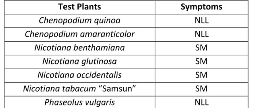
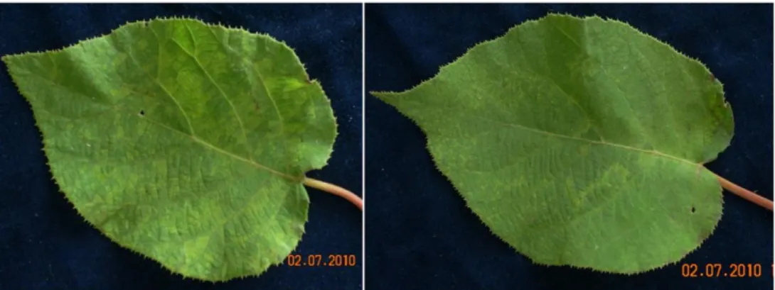
viruses in A. chinensis in New Zealand and TVCV has been identified in samples of dried leaf material of A. chinensis from both China and Italy (Cohen et al., unpublished information). Both viruses were amplified by the primers designed to detect RMV (Chavan et al., 2012) and can only be distinguished by sequencing of the amplicons. Symptoms on A. chinensis include chlorosis of leaf veins and adjacent tissue during spring and chlorotic mottles, mosaics and ringspots during summer. Symptoms on A. deliciosa include chlorotic mottling or mosaic during spring and ringspots during summer months (Chavan et al., 2009). Some of the symptoms resemble those previously described in Actinidia infected with ASGV (Clover et al., 2003) and subsequent investigation has established that most of the plants were co-infected with other viruses (Chavan et al., unpublished information). Symptoms on mechanically inoculated indicators include local chlorotic lesions in C. amaranticolor and C. quinoa, systemic mosaic and distortion in N. benthamiana, systemic necrotic ringspots and chlorotic vein banding and dark green blistering and distortion in N. clevelandii, local necrotic lesions and systemic mottle in N. glutinosa and N. occidentalis and mild systemic mottle in Phaseolus vulgaris (Chavan et al., 2009), but some of these symptoms may be caused by co-infecting viruses. For routine diagnosis, RMV and/or TVCV can be detected in Actinidia leaf samples by conventional RTPCR (Table 2.2). ELISA, using a rabbit polyclonal antiserum raised against purified TMV (M. Pearson, The University of Auckland), detected Actinidia isolates of RMV in herbaceous indicators but failed to detect the virus in infected A. chinensis and
A. deliciosa plants (Chavan et al., 2009). There are no known arthropod vectors
of tobamoviruses but they can survive in sap for prolonged periods (Oshima and Harrison, 1975). Tobamoviruses are highly infectious and readily spread by contact between infected and healthy plants or via machinery and human handling (Gibbs, 1977). Consequently, similar treatments to those recommended to prevent the spread of TMV, such as seed sterilisation using hypochlorite, should be used to prevent virus on seed coats from infecting seedlings during
33
nursery operations (Cohen et al., unpublished information). Overall, RMV and TVCV do not appear to cause significant damage to commercial kiwifruit orchards.
Apple stem grooving virus
ASGV is the type member of the genus Capillovirus, family Betaflexiviridae. Its genome consists of a positive-sense ssRNA of 6,496 nucleotides (excluding the polyA-tail) enveloped in a flexuous, filamentous particle of 620-700x12 nm. Citrus tatter leaf virus (CTLV) is regarded as an isolate of ASGV, being indistinguishable from it biologically, serologically and in genome organization. The main crop hosts are apple, European pear, Japanese pear, Japanese apricot, citrus and lilies and experimentally it infects more than 40 species in 17 plant families. It is probably found wherever apples are grown and natural spread has also been reported in citrus in China and Japan. Some Lilium ASGV strains can infect Citrus and a Pyrus isolate infects Citrus (Yoshikawa, 2000). The kiwifruit ASGV isolate from A. chinensis (AF522459) (Clover et al., 2003) has an identical genomic organization to strains from Citrus, Malus and Lilium, with a high degree of identity to Citrus (D16681), Malus (D14995) and Lilium (AB004063) isolates across the 32-terminal half (2,901 nt) of the genome. The coat protein and movement protein genes share a nucleotide identity of >95% with other strains of ASGV. The morphological, epidemiological, serological and molecular characteristics of the virus from A. chinensis are indistinguishable from those of ASGV from other hosts (Clover et al., 2003). ASGV in kiwifruit was first detected in A. chinensis budwood from Shaanxi province, (China), grafted onto healthy rootstocks of A. chinensis cv. Hort16A and grown in post-entry quarantine in New Zealand. The original source of the plants, within China, is not known. Infected plants developed interveinal mottling, chlorotic mosaics and ringspots (Clover et
al., 2003). However, these plants were subsequently found to be co-infected
with RMV and vitiviruses (R.R. Chavan, unpublished information). ASGV is often latent in commercial Malus and Citrus although it can cause graft union necrosis, tree decline and death in some apple (Yanase, 1983) and citrus (Broadbent et al., 1994) rootstock/scion combinations. It is unknown whether ASGV results in