The role of water in protein's behavior: The two dynamical crossovers
studied by NMR and FTIR techniques
Francesco Mallamace
a,c,⁎
, Carmelo Corsaro
a, Domenico Mallamace
b, Sebastiano Vasi
a,
Cirino Vasi
c, Giacomo Dugo
ba
Dipartimento di Fisica e Scienze della Terra, Università di Messina, Viale F. Stagno D'Alcontres 31, 98166 Messina, Italy
b
Dipartimento di Scienze dell'Ambiente, della Sicurezza, del Territorio, degli Alimenti edella Salute, Università di Messina, Viale F. Stagno d'Alcontres 31, 98166 Messina, Italy
c
CNR-IPCF, Istituto per i Processi Chimico-Fisici, Viale F. Stagno D'Alcontres 37, 98158 Messina, Italy
a b s t r a c t
a r t i c l e i n f o
Article history: Received 16 July 2014
Received in revised form 10 November 2014 Accepted 12 November 2014
Available online 15 November 2014
Keywords:
Protein dynamic transition Amide bending mode Lysozyme unfolding Hydration water HR-MAS
The role the solvent plays in determining the biological activity of proteins is of primary importance. Water is the solvent of life and proteins need at least a water monolayer covering their surface in order to become biologically active. We study how the properties of water and the effect of its coupling with the hydrophilic moieties of proteins govern the regime of protein activity. In particular we follow, by means of Fourier Transform Infrared spectroscopy, the thermal evolution of the amide vibrational modes of hydrated lysozyme in the temperature interval 180 Kb T b 350 K. In such a way we are able to observe the thermal limit of biological activity charac-terizing hydrated lysozyme. Finally we focus on the region of lysozyme thermal denaturation by following the evolution of the proton Nuclear Magnetic Resonance (NMR) spectra for 298 Kb T b 366 K with the High-Resolution Magic Angle Spinning probe. Our data suggest that the hydrogen bond coupling between hydration water and protein hydrophilic groups is crucial in triggering the main mechanisms that define the enzymatic ac-tivity of proteins.
© 2014 Mallamace et al. Published by Elsevier B.V. on behalf of the Research Network of Computational and Structural Biotechnology. This is an open access article under the CC BY license (http://creativecommons.org/licenses/by/3.0/).
1. Introduction
Protein activity is connected with their hydration water[1]. In fact, at least, a monolayer of water molecules called thefirst hydration shell or directly hydration water, extended over the protein surface is needed for the execution of the enzymatic activity[2,3]. The key factor of protein hydration is the H-bonding between protein surface polar groups and hydration water. Furthermore, the coupling between the hydration water and the hydrophilic moieties of the protein surface triggers the search for the correct native state (protein folding): the complex heteropolymeric amino acid sequences spontaneously fold up into organized three-dimensional structures. The spontaneity of the folding process depends on the occurrence of non-functional, or promiscuous, interactions between any suitable pair of residues that can provoke the formation of transient intermediate structures, through “non-native” interactions or frustration [4–6], that can be interpreted as roughness on the folding energy landscape [7]. The native state of a protein corresponds to a global free energy mini-mum that the protein reaches in a time range from microseconds to sec-onds[8]. If the protein shouldfind its native state just by random
searching (Levinthal paradox[9]) among the huge number of possible conformations, this search could take longer than the age of the universe.
It is noteworthy that already in 1936, Mirsky and Pauling discrimi-nated between the fundamentally different character of the native and denatured states of proteins[10]. They argued that the folding process is no more (nor less) miraculous than is the formation of a crystal from a supersaturated solution[10]. The reason lies in the cooperative nature of the denaturation and in the large magnitude of the corre-sponding enthalpy change. Indeed the native state must be nearly unique in structure, like a crystal, whereas the denatured state has a much higher entropy, reflecting the numerous disordered conforma-tions that a chain molecule could take on[10]. However one has to con-sider that the protein is afinite system and the extension of concepts such as those of nucleation and growth mechanism cannot be easily applied. From a physical point of view, proteins are hard matter at low temperature, whereas they are soft matter at high temperature depend-ing on the competition between the enthalpic end entropic contribu-tions[8]. The highly directional and polar character of the hydrogen bond seems to be the key to understand the microscopic mechanisms occurring during protein folding[10,11]. In fact, the physical and chem-ical properties of proteins depend on the characteristic of the hydrogen bonds formed within the protein residuals and with its hydration water. ⁎ Corresponding author.
E-mail address:[email protected](F. Mallamace).
http://dx.doi.org/10.1016/j.csbj.2014.11.007
2001-0370/© 2014 Mallamace et al. Published by Elsevier B.V. on behalf of the Research Network of Computational and Structural Biotechnology. This is an open access article under the CC BY license (http://creativecommons.org/licenses/by/3.0/).
Contents lists available atScienceDirect
For lysozyme in particular it has been shown how the hydrogen bond network that hydration water develops on the protein surface is stable at atmospheric pressure within a temperature interval going from≈ 225 K to ≈ 320 K[12–18]. The temperature of 225 K has been identified as the temperature of the protein glass-transition but its nature is up to now the subject of many controversies[17–22]. In fact, it was pointed out that the glass transition temperature of hydrated lysozyme depends on the hydration level, and the time scale of mea-surement[23,24]. Below 225 K, the water hydrogen bond network is highly rigid being fully developed and protein side-chains motion is hin-dered. On the contrary, above 320 K the lifetime of the hydrogen bond is too short (less than picoseconds) and does not allow to keep together the protein residuals giving rise to the unfolding process[16,25–27]. The native state of lysozyme does not evolve directly into the complete-ly unfolded (or denatured) state but passes through an intermediate state (within which the unfolding process is reversible) where rapid conformational changes occur and can provoke alteration of the folding. The alteration of the folding (or misfolding) of proteins is the source of neurodegenerative illnesses such as Alzheimer's and Parkinson's diseases[7,28].
In this paper we use two different but complementary techniques such as Fourier Transform Infrared (FTIR) and Nuclear Magnetic Resonance (NMR) spectroscopies to investigate how the coupling between the hydration water and the protein residuals evolves as a function of the temperature and determines the limits of biological ac-tivity of hydrated lysozyme. In particular, with FTIR we were able to probe the interval from 180K to 360K with a 10K step, whereas with NMR we focused on the unfolding process from 298K to 366K with a 2K step.
2. Materials and methods
Lysozyme is a small protein of 14.4 kDa; it is constituted by 129 amino acid residuals and in the native state has a globular shape. Lysozyme is easily found in animal tissues and displays anti-inflammatory and antibacterial properties. Protein samples were obtained from Fluka (L7651 three times crystallized, dialyzed, and lyophilized) and used without further purification. Samples were dried, hydrated isopiestically, and controlled by means of a precise procedure[12]. Our aim is to study thefirst monolayer of water mole-cules surrounding the protein surface and this corresponds to a hydra-tion level, h (grams of water per gram of dry protein) equals to 0.3. Fourier Transform Infrared (FTIR) absorption measurements were performed by means of a Bomem DA8 Fourier transform spectrometer, operating with a Globar source, in combination with a KBr beamsplitter and a DTGS/KBr detector. We operated in the attenuated total reflection (ATR) geometry to avoid saturation effects. Spectra were recorded with a resolution of 4 cm−1, automatically adding 200 repetitive scans in order to obtain a good signal-to-noise ratio and highly reproducible spectra; then they are normalized by taking into account the effective number of absorbers[13]. Measurements were performed at ambient pressure in the spectral region from 1300 cm−1to 1750 cm−1, in the temperature range from 180 K to 350 K. Proton NMR experiments were performed at atmospheric pressure in the temperature range 298 Kb T b 366 K by using a 700 MHz Bruker Avance spectrometer equipped with the Magic Angle Spinning (MAS) probehead. Hydrated protein samples were placed in a 50μl rotor and spun at 4000 Hz at the magic angle to increase the spectral resolution. By tilting samples of a precise angle with respect to the applied magneticfield, the hamil-tonian term corresponding to the dipolar interactions vanishes and NMR peaks become narrower. Furthermore, by spinning the rotor at the magic angle by few thousands of Hertz, line broadening effects due to susceptibility differences within the sample are removed resulting in high resolution quality spectra. The sample temperature was controlled by a cold N2flow and a heating element, calibrated by
using the frequency shift between ethylene glycol peaks. The duration
of the hard pulse was 8μs with a relative attenuation of 3 dB; the spec-tral width was 10 kHz, the acquisition time 2.9 s, the points in the time domain 64 k, the number of transient 128 and the relaxation time 2 s for a total time of about 10 min per experiment. All spectra were processed (line broadening, Fourier transform, phase correction and baseline adjustment), by using the standard routines of the Bruker software Xwinnmr version 3.5.
3. Results and discussions
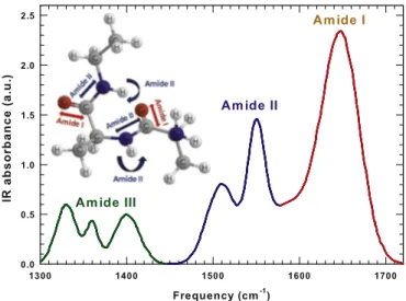
InfraRed and NMR spectroscopies are probably the most used exper-imental techniques able to study protein structure and properties[29]. In particular, FTIR spectroscopy permits a detailed analysis of the struc-ture and stability of proteins, using peptide backbone and side-chain marker bands as conformation-sensitive monitors. Specific information on the secondary structure of proteins is obtained from the analysis of the various amide bands which are indeed sensitive to the protein conformation.
In details, IR spectra of hydrated proteins provide useful structural information especially in the region of Amide I (the most intense band centered at 1600− 1700 cm−1) which is mostly a carbonyl (C_O)
stretching[30,31]. In particular, the amide I band is sensitive to hydro-gen bond pattern, dipole–dipole interaction and the geometry of the polypeptide backbone. It consists of several overlapping bands of differ-ent structural elemdiffer-ents that could be studied separately by means of a peak deconvolution[30,32].
Other intense and important Amide bands are Amide II and Amide III extending respectively from 1480 to 1580 cm−1and from 1300 to 1450 cm−1. The Amide II mode is essentially the combination of the N–H in plane bending and of the C–N stretching, while Amide III con-sists of more complex vibrational modes[31,33]. The different Amide contributions to the IR bending region are reported inFig. 1with differ-ent colors. In thefigure, the Amide I and II vibrational modes are also represented on a peptide fragment using the same color of the corre-sponding IR frequency regions.
The hydrogen bond coupling is a complex phenomenon that can be studied by analyzing the trend that IR spectra show as a function of the temperature. In particular, the behavior of the Amides peak intensity, on increasing the temperature in all the studied range, is not monotonic. In Fig. 2 we use three panels to separate the three important thermal regions within which the spectral behavior is monotonic with temperature. In each panel, for clarity we report only three significant temperatures able to describe the thermal behavior; the intermediate
Fig. 1. The different Amide contributions to the IR bending region are reported with differ-ent colors in the interval 1300− 1720 cm−1. The Amide I and II vibrational modes are also
represented on a peptide fragment using the same color of the corresponding IR frequency regions.
temperatures follow the same trend. In particular, panel A ofFig. 2shows the IR spectra in the low temperature region from 180 K to 220 K. The signal intensity decreases on increasing the temperature as indicated by the arrow. Panel B ofFig. 2shows the IR spectra in the intermediate temperature region, from 230 K to 270 K. Note that, in the intermediate temperature region, the signal intensity increases with the temperature. Panel C ofFig. 2shows the IR spectra in the high temperature region, from 280 K to 350 K. Again, in this high temperature region, the signal intensity decreases on increasing the temperature. The shoulder at about 1500 cm−1, associated with the amide II N–H residual, is well evident at low temperature and disappears upon denaturation[34].
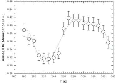
One suitable approach for the characterization of temperature-induced conformational changes in protein is to construct intensity/ temperature profiles for selected IR bands. In such a way one can deter-mine standard thermodynamic properties of the system such as transi-tion or crossover temperatures[35].Fig. 3shows the intensity of the Amide II band as a function of the temperature in the considered ther-mal range. The dotted line is a polynomial bestfit as a guide for the eye. The Amide II band, representing the N–H bending contribution, reflects directly the coupling between hydration water and protein residuals. Note that, on increasing the temperature, the intensity of the Amide II band shows a minimum at≈ 225 K. This temperature
coincides with that of the dynamical crossover observed by means of neutron scattering[12,36]and NMR spectroscopy[13,27,37]. It is note-worthy that even though these experimental techniques encompass different time scales, all lead to the same temperature range for the oc-currence of the dynamical transition. Above 225 K, when the motional amplitude (i.e., theflexibility) of hydrated lysozyme sharply increases, the intensity of the Amide II band increases with temperature up to≈ 270 K. Then, the intensity of the Amide II band starts to slowly decrease on increasing the temperature with a smooth inflection point at ≈ 320 K. This is the “magic” temperature above which water behaves as a normal liquid since the HB lifetime becomes too small (less than picoseconds) and all water molecules are essentially free
[38]. Furthermore at this temperature the refolding rate constant as-sumes the maximum value[8]. Above 320 K the intensity of the Amide II band sharply decreases on increasing the temperature (Fig. 3); water is no more a“good solvent” and lysozyme loses its glob-ular structure evolving toward a linear chain of amino acids. Thus, two temperatures appear to be relevant for the onset of different dynamical regimes (and so to the functioning) of hydrated lysozyme. The values of these temperatures agree with those already found for the same system and described in the introduction, which are 225 K and 320 K. All the described changes and their thermal borders are well described by the intensity of the Amide II band reported inFig. 3confirming the ability of the FTIR technique in detecting the structural conformational behav-ior of proteins and the corresponding role of hydration water. The spec-trum at 350 K, which is the highest measured temperature, shows (panel C) a clear peak bifurcation relatively to the Amide I band at 1650 cm−1. This is due to the onset of aggregation processes in which α -helices transform into β-sheets that tend to self-aggregate. The unfolding process is reversible in character up to≈ 346 K and then be-comes irreversible[26].
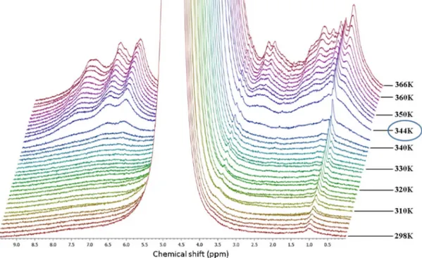
We performed a detailed NMR experiment in order to get a precise insight into the thermal denaturation of hydrated lysozyme. In fact, as shown in Fig. 3, aside from the smooth inflection point at about 320 K, the thermal trend of the intensity of the Amide II band for 280 Kb T b 330 K is quite flat. NMR spectra of hydrated proteins allow studying the different chemical groups of protein and water sep-arately. InFig. 4we present the stacked plot of NMR spectra for hydrat-ed lysozyme (h = 0.3) in the high temperature region measurhydrat-ed by means of the HR-MAS set-up. We started the measurement at 298 K and reached 366 K with steps of 2 K.
The most intense signal, which is cut off in thefigure, belongs to hy-dration water protons and is more than four orders of magnitude larger than the other contributions. Note that, except for the peak at 1 ppm (assigned to the methyl functional group), all the peaks belonging to Fig. 2. The Infrared spectra of hydrated lysozyme (h = 0.3) in the interval 1300−
1750 cm−1for 180 Kb T b 220 K (panel A), for 230 K b T b 270 K (panel B) and for 280 Kb T b 350 K (panel C). The arrows indicate the evolution of the signal intensity with temperature.
Fig. 3. The intensity of the Amide II infrared band as a function of the temperature for hy-drated lysozyme (h = 0.3). The dotted line is a polynomial bestfit as a guide for the eye.
the protons of lysozyme, begin to appear at≈ 320 K. This is a clear sign that the protons of lysozyme are immobile (protein side-chains are not flexible) on the NMR timescale at this hydration level up to ≈ 320 K. This temperature is on the border between the native state and the in-termediate region where the unfolding process starts[26,27].
The protein side-chains become more mobile on increasing the temperature, and their contribution to the free induction decay of the magnetization is indeed detectable. The temperature that marks the irreversibility of the unfolding process is≈ 344 K where the magneti-zation signal suddenly increases and all protein contributions are clearly visible (Fig. 4). The decreasing of their peak width reflects the enhanced protein mobility due to the almost complete hydrogen bonding breakage.
4. Conclusions
In this paper we have studied the coupling of water with the hydro-philic moieties of hydrated lysozyme (h = 0.3) by means of Fourier Transform Infrared and NMR spectroscopy. In particular, by looking at selected vibrations we study how the hydrogen bond interaction governs the regime of protein activity. The intensity of the amide vibra-tional modes of hydrated lysozyme in the temperature interval 180 Kb T b 350 K is able to reflect the thermal limit of biological func-tioning characterizing the studied system. Two temperatures, 225 K and 320 K, appear to be relevant for the onset of different dynamical re-gimes (and so to the functioning) of hydrated lysozyme. Their values agree with those already found for the same system and described in the introduction. Above 225 K theflexibility of hydrated lysozyme sharply increases due to the softening of the hydrogen bond network that the hydration water develops on its surface. Above 320 K water behaves as a normal liquid since the HB lifetime becomes too small (less than picoseconds). All water molecules become essentially free and water is no more a“good solvent”. The refolding rate constant as-sumes the maximum value[8]and lysozyme tends to lose its globular structure evolving toward a linear chain of amino acids.
Finally, in order to get precise insight into the thermal denaturation of hydrated lysozyme we followed the evolution of the proton NMR spectra for 298 Kb T b 366 K with the High-Resolution Magic Angle
Spinning probe. In the obtained spectra, all the peaks belonging to the protons of lysozyme, begin to appear at≈ 320 K, except that at 1 ppm (assigned to the methyl functional group). This means that at this hydration level up to≈ 320 K, the protein side-chains are not so flexible to be revealed by NMR. At higher temperatures, the protein side-chains become more mobile and their contribution is indeed well detectable. The temperature (≈344 K) at which the magnetization signal suddenly increases and all protein contributions are clearly visi-ble signs the irreversibility of the unfolding process. The corresponding decrease of the peaks width reflects the increasing protein mobility pro-voked by the almost complete hydrogen bonding breakage.
In conclusion the experimental data we have presented in this work, suggest that the main mechanisms that define the thermal limits of the enzymatic activity of proteins are given by the hydrogen bond coupling between hydration water and protein hydrophilic groups.
Acknowledgments
CC thanks the Centro Siciliano di Fisica Nucleare e Struttura della Materia (Grant no. 01/14) (Catania, Italy) for its support.
References
[1]Gregory RB. Protein solvent interaction. New York: Marcel Dekker; 1995.
[2]Rupley JA, Careri G. Protein hydration and function. Adv Protein Chem 1991;41: 37–172.
[3]Teeter MM. Water–protein interactions: theory and experiment. Annu Rev Biophys Biophys Chem 1991;20:577–600.
[4]Bryngelson JD, Wolynes PG. Spin glasses and the statistical mechanics of protein folding. Proc Natl Acad Sci U S A 1987;84:7524–8.
[5]Bowman GR, Pande VS. Protein folded states are kinetic hubs. Proc Natl Acad Sci 2010;107:10890–5.
[6]Ferreiro DU, Hegler JA, Komives EA, Wolynes PG. Localizing frustration in native pro-teins and protein assemblies. Proc Natl Acad Sci U S A 2007;104:19819–24.
[7]Chiti F, Dobson CM. Amyloid formation by globular proteins under native conditions. Nat Chem Biol 2009;5:15–22.
[8]Karplus M. Behind the folding funnel diagram. Nat Chem Biol 2011;7:401–4.
[9]Levinthal C. How to fold graciously. In: Debrunner P, Tsibris JCM, Münck E, editors. Mossbauer spectroscopy in biological systems, proceedings of a meeting held at Allerton House, Monticello, Illinois. Urbana: University of Illinois Press; 1969.
[10]Mirsky AE, Pauling L. On the structure of nature, denatured and coagulated proteins. Proc Natl Acad Sci U S A 1936;22:439–47.
Fig. 4. The stacked plot of NMR spectra for hydrated lysozyme (h = 0.3) in the high temperature region measured by means of the HR-MAS set-up. The temperature of 344 K is highlighted because it marks the irreversibility of the unfolding process.
[11]Ben-Naim A. The role of hydrogen bonds in protein folding and protein association. J Phys Chem 1991;95:1437–44.
[12]Chen S-H, Liu L, Fratini E, Baglioni P, Faraone A, Mamontov E. Observation of fragile-to-strong dynamic crossover in protein hydration water. Proc Natl Acad Sci U S A 2006;103:9016.
[13]Mallamace F, Chen SH, Broccio M, Corsaro C, Crupi V, Majolino D, et al. Role of the solvent in the dynamical transitions of proteins: the case of the lysozyme-water sys-tem. J Chem Phys 2007;127:045104.
[14]Mallamace F, Corsaro C, Broccio M, Branca C, González-Segredo N, Spooren J, et al. NMR evidence of a sharp change in a measure of local order in deeply supercooled confined water. Proc Natl Acad Sci U S A 2008;105:12725.
[15]Mallamace F, Branca C, Corsaro C, Leone N, Spooren J, Stanley HE, et al. Dynamical crossover and breakdown of the Stokes-Einstein relation in confined water and in methanol-diluted bulk water. J Phys Chem B 2010;114(5):1870–8.
[16]Zhang Y, Lagi M, Liu D, Mallamace F, Fratini E, Baglioni P, et al. Observation of high-temperature dynamic crossover in protein hydration water and its relation to re-versible denaturation of lysozyme. J Chem Phys 2009;130:135101.
[17]Lagi M, Chu X, Kim C, Mallamace F, Baglioni P, Chen SH. The low-temperature dy-namic crossover phenomenon in protein hydration water: simulations vs experi-ments. J Phys Chem B 2008;112(6):1571–5.
[18]Kumar P, Yan Z, Xu L, Mazza MG, Buldyrev SV, Chen SH, et al. Glass transition in bio-molecules and the liquid-liquid critical point of water. Phys Rev Lett 2006;97: 177802.
[19]Angell CA. Formation of glasses from liquids and biopolymers. Science 1995;267: 1924.
[20]Iben IET, Braunstein D, Doster W, Frauenfelder H, Hong MK, Johnson JB, et al. Glassy behaviour of a protein. Phys Rev Lett 1989;62:1916.
[21]Ngai KL, Capaccioli S, Shinyashiki N. The protein“glass” transition and the role of the solvent. J Phys Chem B 2008;112(12):3826–32.
[22]Ngai KL, Capaccioli S, Paciaroni A. Nature of the water specific relaxation in hydrated proteins and aqueous mixtures. Chem Phys 2013;424:37–44.
[23]Capaccioli S, Ngai KL, Ancherbak S, Paciaroni A. Evidence of coexistence of change of caged dynamics at Tgand the dynamic transition at Tdin solvated proteins. J Phys
Chem B 2012;116:1745–57.
[24]Ngai KL, Capaccioli S, Paciaroni A. Change of caged dynamics at Tgin hydrated
pro-teins: trend of mean squared displacements after correcting for the methyl-group rotation contribution. J Chem Phys 2013;138:235102.
[25]Ball P. Water as an active constituent in cell biology. Chem Rev 2008;108:74–108.
[26]Salvetti G, Tombari E, Mikheeva L, Johari GP. The endothermic effects during dena-turation of lysozyme by temperature modulated calorimetry and an intermediate reaction equilibrium. J Phys Chem B 2002;106:6081–7.
[27]Mallamace F, Corsaro C, Mallamace D, Baglioni P, Stanley HE, Chen S-H. A possible role of water in the protein folding process. J Phys Chem B 2011;115:14280–94.
[28]Selkoe DJ. Folding proteins in fatal ways. Nature 2003;426:900–4.
[29]Pelton JT, McLean LR. Spectroscopic methods for analysis of protein secondary struc-ture. Anal Biochem 2000;277:167–76.
[30]Barth A, Zscherp C. What vibrations tell us about proteins. Q Rev Biophys 2002;35: 369–430.
[31]Kong J, Yu S. Fourier transform infrared spectroscopic analysis of protein secondary structures. Acta Biochim Biophys Sin 2007;39:549–59.
[32]Mallamace D, Corsaro C, Vasi C, Vasi S, Dugo G, Mallamace F. The protein irreversible denaturation studied by means of the bending vibrational mode. Phys A Stat Mech Appl 2014;412:39–44.
[33]Banker J. Amide modes and protein conformation. Biochim Biophys Acta 1992;1120: 123–43.
[34]Mallamace F, Baglioni P, Corsaro C, Chen S-H, Mallamace D, Vasi C, et al. The influence of water on protein properties. J Chem Phys 2014;141:165104.
[35]Fabian H, Mantele W. Infrared spectroscopy of proteins. In: Chalmer Griffiths, editor. Handbook of vibrational spectroscopy, 5. Wiley; 2002. p. 3426–52.
[36]Schiro G, Natali F, Cupane A. Physical origin of anharmonic dynamics in proteins: new insights from resolution-dependent neutron scattering on homomeric polypeptides. Phys Rev Lett 2012;109:128102.
[37]Corsaro C, Mallamace D. A nuclear magnetic resonance study of the reversible denaturation of hydrated lysozyme. Phys A Stat Mech Appl 2011;390:2904–8.
[38]Mallamace F, Corsaro C, Stanley HE. A singular thermodynamically consistent temperature at the origin of the anomalous behavior of liquid water. Sci Rep 2012;2:993.