Doctoral Dissertation
Directly induced Neural Stem Cells
transplantation and prospects for stem cell-based therapy
PhD Candidate: Nunzio Vicario
UNIVERSITY OF CATANIA, Catania (Italy)
Department of Biomedical and Biotechnological Sciences
International PhD Program in Neuroscience - XXX Cycle
Section of Physiology
UNIVERSITY OF CAMBRIDGE, Cambridge (UK)
Department of Clinical Neurosciences
Wellcome Trust-Medical Research Council Stem Cell Institute
Division of Stem Cell Neurobiology
Organization
University of Catania, Catania (IT).
Department of Biomedical and Biotechnological Sciences
Section of Physiology
Ph.D. ProgramInternational Ph.D. Program
in Neuroscience
- XXX Cycle -
Other Organization(s)
University of Cambridge, Cambridge (UK).
Department of Clinical Neuroscience
Wellcome Trust-Medical Research Council
Stem Cell Institute
Division of Stem Cell Neurobiology
Document name
Doctoral dissertation
AuthorNunzio Vicario
TitleDirectly induced Neural Stem Cells transplantation and prospects for stem cell-based therapy
SummaryDespite the remarkable beneficial effects of disease-modifying agents in relapsing-remitting multiple sclerosis (MS) patients, progressive forms of (P)MS still lack effective treatments. This stark contrast is partially dependent on the difficulties researchers have found in tackling the complex pathophysiology of this phase of disease, in which chronic inflammation within the central nervous system (CNS) is coupled by ongoing neurodegeneration and demyelination.
Cell transplantation is among the most promising therapeutic approaches in regenerative medicine, combining tissue trophic and immunomodulatory effects of the graft with its intrinsic potential for cell-replacement. These are all attributes that can be harnessed to treated patients with PMS.
As such, within this thesis, I have focused my attention on investigating how cellular therapies could be used to (i) prevent neuronal damage, (ii) modulate the chronic activation of the immune system and (iii) replace the damaged myelin in PMS.
Olfactory Ensheathing Cells (OECs) are a special population of glial cells known to exert neuroprotective mechanisms and capable of promoting neuroprotection. Using in vitro models of neuron-like cells, I have demonstrated that OECs exert their neuroprotective effect by reducing Cx43-mediated cell and cell-to-extracellular environment communications. Despite this important finding, the immunomodulatory and remyelinating potential of OECs is still limited. As such, I decided to study a complementary stem cell approach that conjugates these attributes with ease in clinical applicability.
Induced Neural Stem Cells (iNSCs) are a source of autologous, stably expandable, tissue specific and easily accessible stem cells, which have the potential to differentiate into the three main neural lineages. Mouse iNSCs were characterized in vitro and in vivo and their immunomodulatory potential was initially studied. This work uncovered a novel mechanism that underpins the potential of iNSCs to interact with the chronic CNS compartmentalised activation of the innate immune system. Specifically, I found that iNSCs are able to sense extracellular metabolites, which accumulate in the chronically inflamed CNS, and to ameliorate neuroinflammation via succinate-SUCNR1-dependend mechanisms. To characterize the potential for tissue replacement and remyelination of such a promising cell line, I have also analysed how iNSCs grafts differentiate in an experimental model of focal demyelination. I found that iNSCs are able to integrate and differentiate into remyelinating oligodendrocytes (OLs) in chronic demyelinated CNS. These data suggest that iNSCs are indeed an effective source of stem cell transplantation, being able to modulate inflammation and to effectively replace lost tissue in mouse models of PMS.
Altogether the evidences gathered in this thesis are important new steps in the field of cell transplantation, which will be pivotal in the march forward for future clinical applications in chronic demyelinating CNS disorders.
Key words
Stem Cells Transplantation, induced neural stem cells (iNSCs),
Demyelination, Remyelination, Multiple Sclerosis
Language
English
Number of Pages: 122
A mio padre.
Ci manchi.
“Due to illness and therapies some children have a weak immune system and they can’t receive vaccine. Vaccination has dropped […] and this represents a life threat to her and to thousands of children in this condition. I beg you, vaccinate your children.”
Nicola Pomaro
Dad of a 5-year-old girl
In memory of Albert Bruce Sabin (Bialystok, 1906 – Washington, 1993)
Sabin developed the oral polio vaccine, which has played a key role in nearly eradicating the disease.
The Sabin’s vaccine was build off a weakened live virus. This method was arguably more effective in fully eradicating polio, instead of just preventing it. Most importantly, an oral vaccine was much easier to administer to kids than a big scary needle. For those reasons, the Sabin vaccine became the global standard for polio.
Sabin intentionally refused to patent his vaccine so that the low price would guarantee a more extensive spread of the treatment. From the development of his vaccine Sabin didn’t gain a single dollar, keeping on living with his salary as a Professor.
Cover
“Oligodendrocytes and neurons: the most spectacular and intimate cell-cell interactions in the nervous system”.
In situ hybridization for Myelin proteolipid protein (Plp) in mouse central nervous system.
“A causa della malattia e delle cure alcuni bambini hanno un sistema immunitario debole e non possono essere vaccinati. La copertura vaccinale è scesa […] e questo rappresenta un pericolo mortale per mia figlia e le migliaia di bambini nella sua condizione. Vi prego, vaccinate i vostri figli.” Nicola Pomaro,
Papà di una bimba di 5 anni
In memoria di Albert Bruce Sabin (Bialystok, 1906 – Washington, 1993)
Sabin ha sviluppato il vaccino orale per la polio che ha avuto un ruolo chiave nell’eradicare quasi totalmente questa malattia.
Il vaccino di Sabin era fatto dal virus vitale indebolito. Questo metodo era probabilmente più efficace per eradicare totalmente la polio, piuttosto che prevenirla soltanto. In aggiunta, un vaccino orale era più semplice da somministrare ai bambini piuttosto che una puntura con un grande ago spaventoso. Per queste ragioni, il vaccino di Sabin divenne lo standard mondiale contro la polio.
Sabin rifiutò intenzionalmente di brevettare il suo vaccino cosicché il basso costo potesse garantire una larga diffusione del trattamento. Dallo sviluppo del suo vaccino Sabin non guadagno un dollaro, continuando a vivere con il suo stipendio da Professore.
Copertina
“Oligodendrociti e neuroni: l’interazione cellula-cellula più spettacolare e intima del sistema nervoso”.
Ibridazione in situ per Myelin proteolipid protein (Plp) nel sistema nervoso centrale murino.
I, the undersigned Nunzio Vicario,
Place of Birth: Catania (IT)
Date of Birth: 18
thSeptember 1989
Author of the PhD thesis entitled:
“Directly induced Neural Stem Cells transplantation and prospects for stem cell-based therapy”
Authorizes the consultation of the thesis.
It is not allowed to copy or to reproduce, in whole or in part, the data and the contents of the thesis.
The whole project or part of it have already been submitted to a publisher or are in press.
Il sottoscritto Nunzio Vicario,
Nato a: Catania (IT)
Data di nascita: 18 settembre 1989;
Autore della tesi di Dottorato di Ricerca dal titolo:
“Directly induced Neural Stem Cells transplantation and prospects for stem cell-based therapy”
Autorizza la consultazione della tesi.
E’ fatto divieto di copiare o di riprodurre, in tutto o in parte, i dati e i contenuti della tesi.
L’intera ricerca o parti sono già state sottoposte a un editore o sono in attesa di pubblicazione.
Catania,
21
stAugust 2017 / 21 agosto 2017
TABLE OF CONTENTS
SUMMARY
p. 11
INTRODUCTION
p. 13
Epidemiology of Multiple Sclerosis (MS)
p. 13
Clinical forms of MS
p. 14
Pathophysiology of MS
p. 15
Cell therapies for PMS
p. 16
AIMS OF THIS THESIS
p. 19
RESULTS
p. 20
Inhibition of Cx43 mediates protective effects on
hypoxic/reoxygenated human neuroblastoma cells
p. 21
Neural stem cells respond to extracellular succinate via
SUCNR1/GPR91 to ameliorate chronic neuroinflammation
p. 39
Directly induced neural stem cells remyelinate
chronic demyelinated mouse brain
p. 91
CONCLUDING REMARKS
p. 115
REFERENCES
p. 117
SUMMARY
Despite the remarkable beneficial effects of disease-modifying agents in
relapsing-remitting multiple sclerosis (MS) patients, progressive forms of
(P)MS still lack effective treatments. This stark contrast is partially dependent
on the difficulties researchers have found in tackling the complex
pathophysiology of this phase of disease, in which chronic inflammation
within the central nervous system (CNS) is coupled by ongoing
neurodegeneration and demyelination.
Cell transplantation is among the most promising therapeutic approaches in
regenerative medicine, combining tissue trophic and immunomodulatory
effects of the graft with its intrinsic potential for cell-replacement. These are
all attributes that can be harnessed to treated patients with PMS.
As such, within this thesis, I have focused my attention on investigating how
cellular therapies could be used to (i) prevent neuronal damage, (ii) modulate
the chronic activation of the immune system and (iii) replace the damaged
myelin in PMS.
Olfactory Ensheathing Cells (OECs) are a special population of glial cells
known to exert neuroprotective mechanisms and capable of promoting
neuroprotection. Using in vitro models of neuron-like cells, I have
demonstrated that OECs exert their neuroprotective effect by reducing
Cx43-mediated cell-to-cell and cell-to-extracellular environment communications.
Despite this important finding, the immunomodulatory and remyelinating
potential of OECs is still limited. As such, I decided to study a complementary
stem cell approach that conjugates these attributes with ease in clinical
applicability.
Induced Neural Stem Cells (iNSCs) are a source of autologous, stably
expandable, tissue specific and easily accessible stem cells, which have the
potential to differentiate into the three main neural lineages. Mouse iNSCs
were characterized in vitro and in vivo and their immunomodulatory potential
was initially studied. This work uncovered a novel mechanism that underpins
the potential of iNSCs to interact with the chronic CNS compartmentalised
activation of the innate immune system. Specifically, I found that iNSCs are
able to sense extracellular metabolites, which accumulate in the chronically
inflamed CNS, and to ameliorate neuroinflammation via
succinate-SUCNR1-dependend mechanisms. To characterize the potential for tissue replacement
and remyelination of such a promising cell line, I have also analysed how
iNSCs grafts differentiate in an experimental model of focal demyelination. I
found that iNSCs are able to integrate and differentiate into remyelinating
oligodendrocytes (OLs) in chronic demyelinated CNS. These data suggest
that iNSCs are indeed an effective source of stem cell transplantation, being
able to modulate inflammation and to effectively replace lost tissue in mouse
models of PMS.
Altogether the evidences gathered in this thesis are important new steps in
the field of cell transplantation, which will be pivotal in the march forward for
future clinical applications in chronic demyelinating CNS disorders.
INTRODUCTION
Epidemiology of Multiple Sclerosis (MS)
Multiple sclerosis (MS) is an inflammatory autoimmune disorder of the central
nervous system (CNS) and one of the most common causes of neurological
disability in young adults. Most people are diagnosed between the ages of 20
and 50, although MS can occur in young children and significantly older
adults (Compston and Coles, 2008). MS is a typical disease of the female
sex, with recent longitudinal studies suggesting a significant increase in
incidence of MS in women, causing a change in the female to male ratio to
more than 3:1 (Orton et al., 2006).
The global distribution of MS shows increasing prevalence with more
distance (north or south) form the equator. However, prevalence rates may
still significantly differ among groups living in the same geographic area.
Studies indicate that immigrants who move later in life, conserve the risk of
the original geographic area, and the change in risk level may not appear
until the next generation. Instead, those who move in early childhood tend to
take on the new risk themselves.
These evidences suggest an interplay between environmental and genetic
factors in the pathogenesis of MS.
MS has a familial recurrence rate of 3% risk in first-degree relatives (siblings,
5%; parents, 2%; and children, 2%) and of 1% risk in second-degree and
third degree relatives, suggesting genetic factors in determining familiar
clustering and susceptibility.
However, many environmental factors have been reported as linked to MS
triggering. Later ages infections has been reported as helpers in trigging MS
supporting the so-called hygiene hypothesis, whereby individuals not
exposed to infections in childhood, make aberrant responses to infections in
adulthood (Martyn et al., 1993). Similarly, active smoking seems to play a role
with a 1.8 ratio for men and 1.4 ratio for women with MS versus healthy
individuals (Hedstrom et al., 2011). On the contrary, vitamin D has shown
protective effects in large epidemiologic studies. Healthy controls have higher
serum levels of 25(OH)D3 and 1,25(OH)2D3 (the active form of vitamin D)
than relapsing remitting (RR) MS patients. Moreover, RR-MS patients present
lower serum levels of vitamin D during relapses compared to the levels during
remissions. The molecular mechanisms underpinning those evidences are
still to be fully elucidated, but it has been demonstrated that 1,25(OH)2D3
supports the induction of CD4+ factor forkhead box P3 (FOXP3+) regulatory
T cells by the rendering of tolerogenic dendritic cells (Penna et al., 2005).
Clinical forms of MS
The initial phase of MS is usually characterized by a relapsing remitting
phase, in which discrete episodes of acute neurological deficits or worsening
of a given neurological function (i.e. relapse), are followed by a complete or
partial recovery (i.e. remission) (Lublin and Reingold, 1996). The
spontaneous recovery observed in the early stages of RR-MS is relieved by a
later progressive course of the disease, called secondary progressive (SP)
MS, in which failure of endogenous regenerative process occurs (Compston
and Coles, 2008). Both types can be either active or inactive and may
develop with or without a clear clinical progression (Lublin et al., 2014).
We now have several therapies to stop relapses and induce recovery in
RR-MS patients. However, still around 65% of people with RR-RR-MS will go into
SP-MS with a chronic worsening of disability within 15 years after being
diagnosed.
Of note, some patients develop a progressive course of disease from the
onset (primary progressive) while others present only a clinically isolated
syndrome (CIS). CIS, which was not included in the initial clinical
descriptions, is now recognised as the initial clinical presentation of a disease
that displays the characteristics of an inflammatory demyelinating disorder
that has yet to fulfil the MS diagnosis criteria (Miller et al., 2005a, b).
Pathophysiology of MS
The major early driver of tissue damage in MS lesions is the migration of
autoreactive lymphocytes from the periphery, which cross the blood-brain
barrier to invade the CNS. Accumulation of T and B lymphocytes, plasma
cells and activated mononuclear phagocytes triggers the secretion of
pro-inflammatory cytokines, amplifying the immune response through recruitment
of naïve microglia (Compston and Coles, 2008).
Demyelination observed in MS patients is typically the completion of a direct
insult to the oligodendrocytes (OLs) (Ferguson et al., 1997; Trapp et al.,
1998). In physiological conditions, OLs accomplish wrap segments of axons
and produce myelin. Neurons, taking advantages from this intimate contact
with OLs, are able to rapidly conduct stimuli via saltatory conduction of action
potentials that propagate throughout the so-called nodes of Ranvier. In
demyelinating conditions, the disruption of these structures impairs the
conductivity but also affects axonal tropism, leading to neurodegeneration as
a chronic condition. While axonal loss and neurodegeneration coexist with
demyelination in the progressive stage of the disease, compensatory
mechanisms are initiated to try to overcome the chronic damage. Among
these, oligodendrocyte precursors cells (OPCs) recruiting is important as
these cells can migrate into demyelinated area and differentiate into mature
myelinating OLs (Chandran et al., 2008). However, these mechanisms seem
largely inadequate, and remyelination is less successful after cycles of
demyelination and remyelination, probably because of exhausting the
capacity of tissue repair.
Cell therapies for PMS
The progressive course and failure of endogenous regenerative processes of
repair are the rationale for cell therapy-based approaches in PMS (Ben-Hur,
2011; Franklin, 2002). Cellular approaches for PMS have great advantages
compared to conventional biologics or drugs due to their intrinsic potential to
replace damaged cells, to attenuate the autoimmune response in a
non-systemic manner, and also to stimulate endogenous regenerative processes;
features summarised as therapeutic plasticity (Ben-Hur, 2011). This is a
milestone of stem cell therapy promise, and described as the combination of
abilities of cells to provide bystander effects and neurotrophic support, to
exert CNS-confined immunomodulation and to replace damaged cells.
Olfactory ensheathing cells (OECs), a special glial cell population sharing
properties with both Schwann cells (SC) and the astrocytes (Franklin and
Barnett, 1997; Wewetzer et al., 2002), are able to secrete high level of growth
factors, such as nerve growth factor (NGF), basic fibroblast growth factor
(bFGF), brain derived neurotrophic factor (BDNF) and glial derived
neurotrophic factor (GDNF), exerting remarkable neuroprotective and
neuroregenerative functions (Boruch et al., 2001; Lipson et al., 2003;
Mackay-Sim and Chuah, 2000). Several reports demonstrate that
transplantation of OECs could provide a remarkable improvement in recovery
of functions in experimental models of CNS-injury (Li et al., 1997; Li et al.,
2003; Lu et al., 2002; Nash et al., 2002). However, poor evidences support
the immunomodulatory potential of OECs and their regenerative properties
are although modest. These characteristics and the SC-like remyelinating
features of OECs grafts, prompted us to develop a stem cell-based approach
for immunomodulation, regeneration and neuroprotection (Franklin and
Barnett, 1997).
Neural Stem Cells (NSCs) are multipotent stem cells of the brain, which have
shown remarkable abilities to reduce necrosis, secondary cell loss and glial
scar formation in experimental models of CNS diseases (Keyoung et al.,
2001; Teng et al., 2002). These effects have been attributed to the generation
of a permissive environment for axonal regeneration, the secretion of
neurotrophic factors and induction of matrix metalloproteinases that allow
neurite outgrowth (Kumagai et al., 2009; Lu et al., 2003; Zhang et al., 2007).
The neurotrophins increase and the immunomodulatory functions are coupled
with the promotion of endogenous regenerative mechanisms, facilitating
endogenous oligodendrocytes precursor cells maturation into mature
remyelinating oligodendrocytes and enhancing neurogenesis and axonal
growth (Ben-Shaanan et al., 2008; Einstein et al., 2009; Pluchino et al.,
2003).
Among the crucial effects mediated by grafted cells, immunomodulation is
one of the mechanisms by which transplanted NSCs can ameliorate mice
with experimental autoimmune encephalomyelitis (EAE), a widely used
experimental model of MS. It has been demonstrated that a reduction of brain
inflammation, acute and chronic injury and demyelination leads to an overall
improvement of the clinical conditions of EAE mice (Pluchino et al., 2003;
Pluchino et al., 2005).
In this scenario, NSCs possess the potential advantages to be able to
self-renew and largely expandable, the plasticity of multipotent stem cells and the
ability to differentiate towards the main neuronal lineages, neurons,
astrocytes and oligodendrocytes, thus allowing cell replacement in the CNS
and improving endogenous spontaneous regeneration (Ben-Hur, 2011;
Keyoung et al., 2001; Nunes et al., 2003).
Recent advances in the generation of multipotent and stably expandable
induced neural stem cells (iNSCs) are exciting. In fact, the direct
reprogramming of mouse and human somatic cells into iNSCs yields
homogenous patient-specific NSCs in a relatively rapid manner. It only
requires one step that is completed within around 4 weeks in vitro and yields
iNSCs that are expandable for at least 30 passages. Also, iNSC are safer
than iPSCs, making them an outmost attractive tool for autologous cell
therapies (Meyer et al., 2015; Thier et al., 2012).
AIMS OF THIS THESIS
The general objective of this thesis was to investigate how cellular therapies
could be used to prevent neuronal damage, modulate the chronic activation
of the immune system and replace the damaged myelin in CNS disorders.
More specifically, the main aims were:
I. Characterize the mechanisms underpinning the action of OECs in
experimental in vitro models of neurodegeneration to investigate
potential candidates that mediate neuroprotective effects;
II. Study the immunomodulatory and therapeutic mechanisms of mouse
iNSCs in the EAE experimental model of MS;
III. Promote endogenous- and exogenous-mediated remyelination using
mouse iNSCs in the lysophosphatidylcholine (LPC) induced focal
demyelination model of MS.
Inhibition of Cx43 mediates protective effects on hypoxic/
reoxygenated human neuroblastoma cells
Nunzio Vicario a, Giovanna Calabrese a, Agata Zappalà a, Carmela Parenti b, Stefano Forte c, Adriana Carol Eleonora Graziano a, Luca Vanella b, Rosalia Pellitteri d, Venera Cardile a, Rosalba Parenti a, *.
a Department of Biomedical and Biotechnological Sciences, Physiology Section, University of Catania.
95125 - Catania, Italy.
b Department of Drug Sciences, University of Catania. 95125 - Catania, Italy. c IOM Ricerca, 95029 - Viagrande, Italy.
d Institute Neurological Sciences, National Research Council. 95126 - Catania, Italy.
Journal of Cellular and Molecular Medicine
Received: October 28, 2016; Accepted: February 28, 2017
Abstract
Olfactory Ensheathing Cells (OECs), a special population of glial cells, are able to
synthesise several trophic factors exerting a neuroprotective action and promoting growth
and functional recovery in both in vitro and in vivo models. In the present work, we
investigated the neuroprotective effects of OEC-conditioned medium (OEC-CM) on two
different human neuron-like cell lines, SH-SY5Y and SK-N-SH (neuroblastoma cell lines),
under normoxic and hypoxic conditions. In addition, we also focused our attention on the
role of connexins (Cxs) in the neuroprotective processes. Our results confirmed OEC-CM
mediated neuroprotection as shown by cell adherence, proliferation and cellular viability
analyses. Reduced connexin 43 (Cx43) levels in OEC-CM compared to unconditioned
cells in hypoxic conditions prompted us to investigate the role of Cx43-Gap junctions (GJs)
and Cx43- hemichannels (HCs) in hypoxic/reoxygenation injury using carbenoxolone
(non-selective GJ inhibitor), ioxynil octanoato ((non-selective Cx43-GJ inhibitor) and Gap19
(selective Cx43-HC inhibitor). We found that Cx43-GJ and Cx43-HC inhibitors are able to
protect SH-SY5Y and allow to these cultures to overcome the injury. Our findings support
the hypothesis that both OEC-CM and the inhibition of Cx43-GJs and Cx43-HCs offer a
neuroprotective effect by reducing Cx43-mediated cell-to-cell and cell-to-extracellular
environment communications.
Keywords
Olfactory glia, Growth Factors, Neuroprotection, Gap Junctions, Connexin 43
*Correspondence to: Rosalba PARENTI, Ph.D. E-mail: [email protected]
Doi: 10.1111/jcmm.13177
© 2017 The Authors.
Journal of Cellular and Molecular Medicine published by John Wiley & Sons Ltd and Foundation for Cellular and Molecular Medicine. This is an open access article under the terms of the Creative Commons Attribution License, which permits use, distribution and reproduction in any medium, provided the original work is properly cited.
Introduction
The olfactory system is a specific area of the central nervous system (CNS) capable of
supporting neurogenesis throughout the life of mammals by forming new olfactory receptor
neurons (ORNs) [1–3]. These neurons are then able to spread axons from the peripheral
nervous system of the olfactory epithelium into the CNS environment of the olfactory bulb
[4]. The capacity of ORNs to stimulate neurogenesis in the adult olfactory system may be
due to both the neural stem cells present in the olfactory epithelium and the glial cells
known as Olfactory Ensheathing Cells (OECs) [5, 6]. OECs, originally described by Golgi
and Blanes [7–9] at the end of the 19th century, are a special glial cell population sharing
properties with both Schwann cells and the astrocytes [10, 11]. Similarly to Schwann cells,
OECs express some characteristic markers such as the low affinity neurotrophin receptor
(p75NTR) and adhesion molecules such as laminin, L1 and NCAM; likewise to astrocytes
they express the S-100 protein and the glial fibrillary acidic protein (GFAP), a member of
the intermediate filament family that offers support to glial cells [12, 13]. Furthermore,
OECs are able to secrete high level of growth factors, such as nerve growth factor (NGF),
basic fibroblast growth factor (bFGF), brain derived neurotrophic factor (BDNF), glial
derived neurotrophic factor (GDNF), ciliary neurotrophic factor (CNTF), neurotrophins NT4,
NT5 and neuregulins, which exhibit important functions as neuronal supporting elements
[14–19].
Gap junctions (GJs) are specialized intercellular channels that directly connect cytoplasms
of adjacent cells, enabling direct exchanges of small molecules (less than 1200 Da). GJs
are composed by two docked hemichannels (HCs), also named connexons, one on each
cell. HCs are hexamers of homotypic or heterotypic connexins (Cxs), which are the
transmembrane proteins encoded by a multigene family of approximately 20 members in
mammals [20–25] that form respectively homotypic or heterotypic GJs.
Besides constituting ‘GJs plaques’ that allow GJ intercellular communication (GJIC), Cxs
also constitute free HCs throughout the plasma membrane, allowing exchange of a
number of autocrine and paracrine signalling molecules between the cytoplasm and the
extracellular environments [26–28]. GJs, as well as HCs, play a crucial role in a wide
range of cellular activities, including cell signalling, differentiation, growth, pro-apoptotic
signalling, either as anti-apoptotic gates or pathogenic pores depending on conditions and
cell type [29–32]. GJs and free HCs are extensively distributed in different tissues and
organs [33] in which different cell type usually expresses specific Cxs profile involved in
the selective permeability of channels formed, according to the metabolic or functional
needs [34–38]. Also in the CNS, GJs and HCs are extensively distributed among neurons
and glial cells [39–41], where, contributing to GJIC and cell-extracellular communication,
they provide specific exchange pathways under resting conditions, and also play a
context-dependent role in contradictory cell survival or cell death phenomena [42]. In
recent years, much attention has been paid to the exploitation of neuroprotective effects in
combatting the progression and chronicity of neurodegeneration. In particular, evidence
shows altered Cxs expression and functions under pathological conditions, suggesting a
central role of glial GJs and HCs in the development of various neurodegenerative
diseases [21, 43–46]. Conversely, the therapeutic potential of OECs is attracting
considerable interest owing to their exceptional ability to promote functional recovery of the
damaged CNS [3, 47–50]. However, the molecular mechanisms underlying this protective
function are not yet known. The aim of this study was firstly to investigate the
neuroprotective effects of OEC conditioned medium (OEC-CM) on two different
neuroblastoma cell lines (SH-SY5Y and SK-N-SH) exposed to hypoxic/reoxygenation
(H/R) injury. Towards this goal we explored the relationship between OEC-CM and Cxs,
HCs and GJs following H/R injury in cultures grown with and without OEC-CM. We found
that: (1) OEC-CM offers a neuroprotective effect to the SH-SY5Y and SK-N-SH cells
exposed to H/R injury; (2) injured SH-SY5Y and SK-N-SH cells show higher Cx43 levels
compared to normoxic cultures; and (3) H/R cultures grown in the presence of GJ and/or
HC chemical inhibitors, such as carbenoxolone (CBX, non-selective GJIC inhibitor) [51],
ioxynil octanoato (IO, Cx43 homotypic GJ inhibitor) [52], and Gap19 (Cx43 homotypic HC
inhibitor) [53], show higher viability compared to control cultures. Our results, showing the
neuroprotective effects of OEC-CM likely afforded by inhibition of Cx43 GJ/HC signalling
pathways, suggest potential new therapeutic tools against neurodegenerative diseases.
Materials and Methods
Primary Olfactory Ensheathing Cells cultures
Primary OECs were isolated from post-natal day 0 (P0) CD1 mice olfactory bulbs. Experiments were performed in compliance with current guidelines for animal care and in accordance with the European Community Council Directive (86/609/EEC). All efforts were made to minimize animal suffering and to use the fewest animals possible.
Ten P0 pups were decapitated, the entire brain was exposed and bulbs removed and dissected out in cold (+4 °C) Leibowitz L-15 medium (Sigma). Subsequently, collected olfactory bulbs were digested twice in fresh minimum essential medium-H (MEM-H; Sigma) containing 0.03% collagenase (Sigma) and 0.25% trypsin (Sigma) for 15 min at 37 °C. Suspension was mechanically triturated and filtrated through a 80 μm nylon filter and centrifuged at 600 g for 10 min. Cells were suspended with fresh complete Dulbecco’s modified
Eagle’s medium (DMEM, Sigma) supplemented with 10% fetal bovine serum (FBS, Sigma), 2 mM L-glutamine (Sigma), penicillin (50 U/ml, Sigma), and streptomycin (50 mg/ml, Sigma) and plated in 25 cm2 flasks. Cytosine arabinoside (Sigma) at final concentration of 1x10⁻⁵ M was added 24 hours (hrs) after plating to reduce the number of dividing fibroblasts. After 2 passages purity of OECs was verified by using immunofluorescence with p75 and S-100 (data not shown). Media were replaced twice a week for 3 passages and then cultures were used to collect the conditioned medium. OEC-CM was removed from the cultures and filtered through a membrane filter (0.22 μm pore diameter) to remove cells and debris.
Neuroblastoma cell lines cultures
The human neuroblastoma cell lines, SH-SY5Y and SK-N-SH, were purchased from ATCC (Rockville, MD, USA) and grown as previously described [56]. Briefly, cells were incubated at 37 °C in a humidified 5% CO2/95% air atmosphere and maintained in DMEM/F12 (Sigma) supplemented with 10% FBS (Sigma), 2 mM L-glutamine (Sigma), penicillin (50 U/ml, Sigma), and streptomycin (50 mg/ml, Sigma). The medium was replaced twice a week.
H/R injury and OEC-CM treatment
Cells at a density of 2.5x10⁴ cells/cm2 were seeded on appropriate supports for analysis 16 hrs before performing the experiments (time 0). The following experimental culture groups were established: untreated control groups (CTRL Normoxia) and hypoxic groups (CTRL Hypoxia) at 0, 3, 8 and 24 hrs. Each group was grown in normal culture conditions for 16 hrs, after which they were subjected to hypoxia (1% O2) for 3 hrs, followed by reoxygenation to 24 hrs. All cultures were grown with DMEM-F12 (89%), FBS (10%) and P/S (1%). For OEC-CM treatment, at time 0, media conditioning (25%, 50% and 100%) obtained from OECs cultured for 24, 48 and 72 hrs were applied in the same experimental culture groups. To verify the status of hypoxia, the expression of hypoxia-inducible factor 1-alpha (HIF1A), a specific transcription factor of the hypoxic response, was analysed by immunofluorescence (data not shown).
Immunofluorescence
Immunofluorescence analysis on SH-SY5Y cells was performed as previously described [54]. Briefly, after fixation with 4% Paraformaldehyde (PFA), cells were permeabilized in 0.2% Triton X100, blocked by incubation with 10% normal goat serum (NGS, Gibco) for 1 hr at room temperature (RT) and incubated overnight at 4 °C with the following primary antibodies: rabbit anti-p75 (1:500, Chemicon), mouse anti-S-100 (1:100; Sigma), rabbit anti-HIF1A (1:200; Sigma), mouse anti-Cx43 (1:150, Cell Signaling) and rabbit β-tubulin (1:200, Cell Signaling).
After washing, slides were incubated with the appropriate secondary antibodies: fluorescence isothiocyanate (FITC) labelled anti-rabbit antibody (1:200, Chemicon) and Cy3 labelled anti-mouse antibody (Chemicon, 1:1000) for 1 hr at RT. Nuclei were stained with DAPI (1:1000) for 5 min. Finally, slides were mounted in fluorescent mounting medium Permafluor (Thermo Scientific) and digital images were acquired using a Leica DM IRB fluorescence microscope and with Leica TCS SP8 confocal microscope. Nonspecific staining of cells was observed in control incubations in which the primary antibodies were omitted.
Western blot analysis
Cell pellets were homogenized in lysis buffer (Tris-HCl pH 7.4, 1% Triton X100, NaCl 150 mmol/L and EDTA 1 mmol/L) supplemented with a cocktail of protease inhibitors (1:100, Sigma). For Western blot quantification, 50 µg of protein were electrophoresed on 12% SDS-PAGE gels and transferred to nitrocellulose membranes. After blocking with 5% non-fat milk powder in Tris-buffered saline with 0.05%
Tween-20 (TBST), membranes were incubated overnight at 4 °C with the following primary antibodies: rabbit caspase-3 cleaved (1:1000, Cell Signaling), mouse anti-Cx47 (1:200, Invitrogen), mouse anti-Cx43 (1:1,000, Cell Signaling), rabbit anti-Cx40 (1:200, Invitrogen), mouse anti-Cx36 (1:200, Invitrogen), mouse anti-Cx32 (1:500, Novex), rabbit anti-Cx30 (1:200, Invitrogen) and rabbit β-tubulin (1:1,000, Cell Signaling). After 3 washes in TBST, the membranes were incubated with anti-mouse (1:20,000, Jackson) and anti-rabbit HRP-conjugated (1:50,000, Jackson) secondary antibodies for 1 hr at RT. Proteins bands were visualized with premixed ready-to-use chemiluminescent HRP detection reagent (Millipore) according to the manufacturer's instructions and captured with an Uvitec Cambridge Imaging System. The density of each band was quantified using ImageJ analysis software and normalized to β-tubulin levels measured in the same membranes.
Monitoring cell adherence and proliferation
Cell adherence and proliferation was monitored in real-time using the xCELLigence system E-Plate. Different cell numbers were tested, with the optimum cell density to found to be 2.5x10⁴ cells/cm2 (data not shown). Experiments were performed at different time points and with various percentages of media derived from OEC cultures mixed with fresh medium, with 100% of fresh medium employed as a control. The impedance value of each well was monitored by the xCELLigence system for a time of 24 hrs and expressed as a cell index (CI) value. Data for cell adherence were normalized at 16 hrs after plating. Normalized CI, representing a quantitative measure of cell number, was calculated by dividing CI at the time point into the CI at the normalization time point (time 0). The Rate of Cell Growth (RCG) was determined by calculating the slope of the line between starting point and ending point.
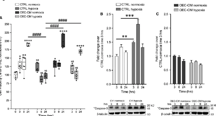
Cellular viability
Cells were trypsinized and adjusted to a concentration of 2.5x10⁴ cells/cm2 seeded in 96-well plates (Costar) and incubated for 16 hrs with basal growth medium. Then the medium was replaced with basal growth medium in control cultures and with 50% basal growth medium mixed 1:1 with OEC-CM. Normoxic cultures were placed in both normal-oxygen and in 1% O2 conditions. Cellular viability was evaluated at time 0, 3, 8 and 24 hrs after 2.5 hrs of incubation with a solution of 3-(4,5-dimethylthiazol-2-yl)-2,5-diphenyltetrazolium bromide (MTT; Sigma Aldrich, Italy) 5 mg/ml. Media were gently removed, MTT solvent (DMSO, Sigma) was added, and cells were agitated on an orbital shaker for 5 min at RT. The absorbance was measured using a Varioskan Flash spectrophotometer (Thermo Scientific) at 550 nm. Results were expressed as the percentage MTT reduction of control cells. The experiment was performed 3 times with 6 replicates per condition each time. Data are shown via standard box-and-whiskers plots in which the central-line represents the median, the upper- and lower-bounds of the boxes are min and max value and points represent all values expressed as percentage of control, assumed as 100%.
Statistical analysis
n way-Anova has been performed in order to determine the existences of interaction between n independent variables (time and treatment for two way ANOVA and time, treatment and oxygen levels for three way Anova) on a continuous dependent variable. Tukey honest significant difference (HSD) has been used as post-hoc test when ANOVA test indicated statistically significant differences to identify specific changes in time points or treatment conditions. Statistical calculation has been performed using R software (R Foundation for Statistical Computing). Values are represented in graphs as mean ± standard error of the mean (SEM). All calculation was performed using GraphPad Prism v7 software.
Results
OEC-CM exerts a protective effect in an in vitro H/R injury model
To induce hypoxia/reoxygenation (H/R) injury, 2.5x10
4cells/cm
2were plated in growth
medium and incubated in a controlled humidified atmosphere at 37 °C with constant 5%
CO
2level. Sixteen hours after plating (time 0) hypoxic cultures were exposed to 3 hrs of
hypoxia (1% O
2) and then reoxygenated to 24 hrs. In OEC-CM supplemented cell cultures
the conditioned media was added at time 0 (Fig. 1A).
To investigate the potential protective effects of OEC-CM on cell cultures after H/R injury,
we supplemented growth medium with three different percentage of conditioned medium,
25%, 50% and 100%, collected from primary OECs cultures at different conditioning times
(24, 48 and 72 hrs) (Fig. 1A). Our results showed that in normoxic conditions there was a
significant reduction of normalized cell index (CI) in all cell cultures grown in 100%
OEC-CM (Fig. 1B–D), as result of reduced cell confluence and cell number. A similar result,
although less marked than previous, was observed when the cells were cultured with 50%
OEC-CM collected after 72 hrs (Fig. 1D). This was probably linked to the lower level of
serum and nutrients compared to the fresh medium. Cell cultures grown with 25% and
50% OEC-CM, collected after 24 hrs and 48 hrs (Fig. 1A and B), did not show any
significant difference compared to cells grown in 100% growth medium (CTRL) (Fig. 1A–
C). Cells exposed to H/R injury, treated with 25%, 50% and 100% OEC-CM collected at
any time-points, showed a significant increase of normalized CI compared to CTRL (Fig.
1E–G). Our findings also indicated that OEC-CM, used at concentrations of 50% and
100%, exerted a higher protective effect on cells compared to 25% OEC-CM and CTRL
(Fig. 1E–G). To further analyse, the effects of OEC-CM on cell cultures, we evaluated the
rate of cell growth (RCG) in both normoxic and H/R injured cultures (Fig. 1H–K). Our
results demonstrated that in normoxic conditions, cell lines supplemented with 25%
OEC-CM, collected from OECs after 24, 48 and 72 hrs, did not show significant reduction of the
RCG. The same effect was observed when cells were grown in 50% OEC-CM collected
after 24 or 48 hrs. In contrast, cultures grown in 50% OEC-CM, collected after 72 hrs and
in 100% of OEC-CM (all conditioning times), showed a significant decrease of RCG (Fig.
1H and J). Interestingly, all conditioned media used at 25%, 50% and 100% on H/R injured
cultures showed a significant increase of RCG compared to control cultures (Fig. 1I and
K). Taken together, these findings suggested that, in both normoxic and H/R injured
cultures, the cells grown in 48 hrs OEC-CM mixed 1:1 in maintenance medium, showed a
higher protective effect and supported a better RCG. For this reason all further studies
have been performed by using 50% OEC-CM collected after 48 hrs. These findings
confirmed that OECs, affecting the media composition, were able to exert neuroprotective
actions and to increase cell survival and cell proliferation after injury.
OEC-CM improves cell viability in H/R cultures
In order to investigate the effect of H/R condition on cell viability, we exposed hypoxic cell
cultures to 1% O
2levels for 3 hrs and reoxygenation up to 24 hrs. Cell cultures viability
was evaluated at different time-points after injury (Fig. 2A). Our results demonstrated that
cells immediately after H/R injury and after 5 hrs of reoxygenation had significantly lower
cell viability (MTT test). Furthermore, we established that the addition of OEC-CM to the
H/R injured cultures significantly improved cell viability compared to untreated cells.
Otherwise cells cultured in presence of OEC-CM for 24 hrs in normoxic conditions did not
show any significant difference compared to control (Fig. 2A).
Further, we evaluated the levels of cleaved caspase-3, a marker of apoptosis, in normoxic
and H/R cultures with and without OEC-CM. We found that cleaved caspase-3 levels were
significantly higher in hypoxic cultures, at all reoxygenation times, compared to normoxic
cultures (Fig. 2B). Cells grown with OEC-CM, in normoxia and exposed to H/R, did not
show significant differences of cleaved caspase-3 levels compared to unconditioned
cultures (Fig. 2C).
Expression levels of different Cxs in neuroblastoma cell lines under normoxic and
hypoxic conditions
To identify whether SH-SY5Y cells, cultured in presence or in absence of OEC-CM in
normoxic and H/R conditions, expressed some specific Cxs (including Cx47, Cx43, Cx40,
Cx36, Cx32, and Cx30), we performed Western blot analyses. Our data indicated that
cells grown in all experimental conditions showed no or a very low expression of Cx47,
Cx32, Cx40 and Cx30 (Fig. 3A). The cultures, maintained under normoxic conditions,
showed basal expression levels for Cx36 and Cx43 that were reduced in presence of
OEC-CM. H/R injured cultures showed an intensely increased Cx43 levels and OEC-CM
was able to reduce this overexpression to normoxic levels (Fig. 3A). To better evaluate
levels and localization of Cx43 in SH-SY5Y cells we performed Western blots and
immunofluorescence analysis at different time-points. These experiments confirmed high
levels of this marker from the end of the injury (3 hrs) in untreated SH-SY5Y cells (Figs.
3B and 4A, B). The expression levels appeared to increase markedly after 8 hrs and then
clearly decrease at 24 hrs (Fig. 3B) in both intracellular and extracellular compartments
(Fig. 4B). However, our data showed that H/R cultures, in absence of OEC-CM, had
higher levels of Cx43 at each analysed time-point compared to the cultures in normoxia
with and without CM. On the contrary, in injured cultures, in the presence of
OEC-CM the expression levels of Cx43 seemed unaffected along the different time-points (Figs.
3A and B, 4B) compared to the cultures in normoxia with and without OEC-CM. Moreover,
H/R cultures exposed to OEC-CM exhibited an evident reduction of Cx43 expression
when compared to the unconditioned injured cultures (Figs. 3B and 4B). To further confirm
the increased Cx43 expression levels in hypoxic condition we performed Western blots
analysis on SK-N-SH at 24 hrs (Fig. 3C). These experiments confirmed that also in this
cell line the Cx43 levels were up-regulated in H/R injured cultures while did not show any
significant differences in OEC-CM cultures compared to control cultures.
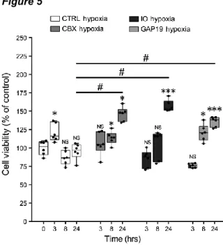
Gap junction and/or hemichannel chemical inhibitors protect SH-SY5Y cells from
H/R injury
To analyse whether the neuroprotective effect on SH-SY5Y cells exposed to H/R injury
was related to GJ and/or HC functions of Cx43 we performed MTT-viability test after
adding GJ and/or HC inhibitors. We used CBX, a non-selective GJIC inhibitor, IO to
selectively target Cx43 homotypic GJs, and Gap19 to selectively inhibit Cx43 homotypic
HCs. Results obtained from the titration of GJ and/or HC inhibitors (data not shown)
suggested that the best inhibitor concentrations were 10 μM in culture medium. Our
results demonstrated that H/R injured cultures treated with both GJ and HC chemical
inhibitors significantly increase the cell viability over time compared to the control cultures
exposed to H/R injury (Fig. 5).
Discussion
The strengthening of some endogenous neuroprotective mechanisms as a mean by which
to prevent and/or slow down the outcome of the various forms of neurodegenerative
disorders has recently emerged as one of the most popular topics in applied neurobiology.
It has been described that OECs exhibit the ability to promote regeneration in the
damaged CNS, thus they could be involved as possible mediators of repair in neurological
diseases [12, 14]. As a source of different trophic factors, OECs have attracted an
increasing interest as tool for regenerative medicine with applications that include spinal
cord injury [55, 56] or axonal growth [57, 58] with a view towards new therapeutic
approaches. It has been also demonstrated in vitro that the addition of OEC-CM to the
neuroblastoma SH-SY5Y and SK-N-SH cells exposed to the neurotoxin
6-hydroxydopamine (6-OHDA) provides neuroprotective properties [59].
In this work we showed that OEC-CM exerts a concentration and time-dependent
protective effect on SH-SY5Y and SK-N-SH cells subjected to H/R injury. Our aim was to
investigate the molecular mechanism underlying this effect. A number of evidence
revealed that, in the CNS, intercellular communication among neurons and glial cells via
GJs and/or HCs could be critical in the spread of protective and/or deleterious signals. Cxs
are dynamically expressed during injury and stress conditions and, for each condition and
context, up- or down-regulation of such proteins likely influencing gate properties of GJs
and free HCs, may influence cell survival or cell death [21, 43–46]. In particular, several
independent studies have pointed out that onset and progression of homeostatic
imbalances observed during neurodegeneration could be associated with an enhanced
HC activity in the CNS [60–66]. Here we demonstrated in vitro that SH-SY5Y and
SK-N-SH cells grown in normoxia displayed no or low expression of Cx47, Cx43, Cx40, Cx36,
Cx32 and Cx30 with or without the addition of OEC-CM. When cells are cultured under
hypoxic conditions, the Cx43 exhibited an increased expression whereas, the addition of
OEC-CM to the growth medium, restored the basal expression observed in normoxia.
These evidence suggest that, while Cx43 may be involved in hypoxic response, the
protective effect of OEC-CM may be exerted through the modulation of this specific Cxs.
Cx43 is the principal astrocytic GJ protein in the CNS where it contributes to the formation
of the functional syncytium, implicated in maintaining the homeostasis of the extracellular
milieu of neurons [67, 68]. Many studies support the potential therapeutic effects of
Cx43-GJ blockade on neuronal survival in various models of injury including stroke, epilepsy,
ischemia, optic nerve damage and spinal cord injury, with GJ communication and HC
opening leading to increased secondary damage via the inflammatory response [69–71].
To investigate the possible interplay between OEC-CM and Cx43 in protection after H/R
injury, the effects of Cx43 chemical inhibition has been assessed. When H/R stress is
induced in SH-SY5Y cells, both Cx43-GJ and Cx43-HC chemical inhibition significantly
increased the cell viability over time compared to control cultures. The functional
modulation of Cx43 provides additional support on its involvement in OEC-CM mediated
neuroprotection, likely exerted through the prevention of the spread of injury signals. One
appealing hypothesis is that OEC-CM works by influencing Cx43 expression in the
SH-SY5Y cells via paracrine factors likely involved in the physiological role of OECs within the
CNS. It is noteworthy that several studies have demonstrated a role of Cx proteins in the
regulation of tissue homeostasis occurring independently of their channel activities, in the
context of cell growth, adhesion, migration, apoptosis and signalling [72]. While further
investigations are needed to unveil the molecular details of such neuroprotection, these
data point to the possibility that the proposed model may be useful in the context of
therapeutic applications after brain injury. The involvement of Cxs in maintaining the
delicate balance of CNS cells, via GJs and/or HCs, may indeed stimulate the development
of new modulators for Cxs-based channels as novel therapeutic agents for the cure of
nervous disorders [73–76].
Acknowledgements
The authors would like to thank Dr Jayden A. Smith, (University of Cambridge, UK), for
critically review this work.
Conflicts of Interest
The authors confirm that there are no conflicts of interest.
Author contributions
N.V. and R.Pa. designed the research study; N.V., G.C. and R.Pe. performed
experiments; N.V. and S.F. collected and analysed data; C.P. and L.V. provided some
reagents and instruments; A.G., A.Z., C.P. and V.C. gave technical support and
conceptual advice; N.V., G.C. and R.Pa. wrote the manuscript.
References
1. Farbman AI. Olfactory neurogenesis: genetic or environmental controls. Trends Neurosci. 1990; 13: 362–5.
2. Graziadei PP, Graziadei GA. Neurogenesis and neuron regeneration in the olfactory system of mammals. I. Morphological aspects of differentiation and structural organization of the olfactory sensory neurons. J Neurocytol. 1979; 8: 1–18.
3. Bartolomei JC, Greer CA. Olfactory ensheathing cells: bridging the gap in spinal cord injury. Neurosurgery. 2000; 47: 1057–69.
4. Chung RS, Woodhouse A, Fung S, et al. Olfactory ensheathing cells promote neurite sprouting of injured axons in vitro by direct cellular contact and secretion of soluble factors. Cell Mol Life Sci. 2004; 61: 1238–45.
5. Doucette R. Glial influences on axonal growth in the primary olfactory system. Glia. 1990; 3: 433–49. 6. Raisman G. Specialized neuroglial arrangement may explain the capacity of vomeronasal axons to
reinnervate central neurons. Neuroscience. 1985; 14: 237–54.
7. Ramon-Cueto A, Avila J. Olfactory ensheathing glia: properties and function. Brain Res Bull. 1998; 46: 175–87.
8. Golgi C. Sulla fina anatomia dei bulbi olfattorii. Rivista Sperimentale di Freniatria. 1875; 1: 403–25. 9. Blanes T. Sobre algunos puntos dudosos de la estructura del bulbo olfatorio. Revista Trimestral
Micrografica. 1898; 3: 99–127.
10. Wewetzer K, Verdù E, Angelov DN, et al. Olfactory ensheathing glia and Schwann cells: two of a kind? Cell Tissue Res. 2002; 309: 337–45.
11. Franklin RJM, Barnett SC. Do olfactory glia have advantages over Schwann cells for CNS repair? J Neurosci Res. 1997; 50: 665–72.
12. Pellitteri R, Spatuzza M, Russo A, et al. Olfactory ensheathing cells exert a trophic effect on the hypothalamic neurons in vitro. Neurosci Lett. 2007; 417: 24–9.
13. Franceschini IA, Barnett SC. Low-affinity NGF-receptor and E-N-CAM expression define two types of olfactory nerve ensheathing cells that share a common lineage. Dev Biol. 1996; 173: 327–43.
14. Franklin RJM, Barnett SC. Olfactory ensheathing cells and CNS regeneration – the sweet smell of success? Neuron. 2000; 28: 1–4.
15. Lipson AC, Widenfalk J, Lindqvist E, et al. Neurotrophic properties of olfactory ensheathing glia. Exp Neurol. 2003; 180: 167–71.
16. Mackay-Sim A, Chuah MI. Neurotrophic factors in the primary olfactory pathway. Prog Neurobiol. 2000; 62: 527–59.
17. Woodhall E, West AK, Chuah MI. Cultured olfactory ensheathing cells express nerve growth factor, brain-derived neurotrophic factor, glia cell line-derived neurotrophic factor and their receptors. Brain Res Mol Brain Res. 2001; 88: 203–13.
18. Wewetzer K, Grothe C, Claus P. In vitro expression and regulation of ciliary neurotrophic factor and its α receptor subunit in neonatal rat olfactory ensheathing cells. Neurosci Lett. 2001; 306: 165–8.
19. Boruch AV, Conners JJ, Pipitone M, et al. Neurotrophic and migratory properties of an olfactory ensheathing cell line. Glia. 2001; 33: 225–9.
20. Simon AM, Goodenough DA. Diverse functions of vertebrate gap junctions. Trends Cell Biol. 1998; 8: 477–82.
21. Nakase T, Naus CC. Gap junctions and neurological disorders of the central nervous system. Biochim Biophys Acta. 2004; 1662: 149–58.
22. Söhl G, Willecke K. Gap junctions and the connexin protein family. Cardiovasc Res. 2004; 62: 228–32. 23. Nagy JI, Rash JE. Connexins and gap junctions of astrocytes and oligodendrocytes in the CNS. Brain
Res Brain Res Rev. 2000; 32: 29–44.
24. Mugnaini E. Cell junctions of astrocytes, ependyma, and related cells in the mammalian central nervous system, with emphasis on the hypothesis of a generalized functional syncytium of supporting cells. In: Federoff S, Vernadakis A, editors. Development, morphology, and regional specialization of astrocytes, vol. I, Orlando, FL: Academic Press; 1986. pp. 329–71.
25. Parenti R, Campisi A, Vanella A, et al. Immunocytochemical and RT-PCR analysis of connexin 36 in cultures of mammalian glial cells. Arch Ital Biol. 2002; 140: 101–8.
26. Goodenough DA, Paul DL. Beyond the gap: functions of unpaired connexon channels. Nat Rev Mol Cell Biol. 2003; 4: 285–94.
27. Evans WH, De Vuyst E, Leybaert L. The gap junction cellular internet: connexin hemichannels enter the signalling limelight. Biochem J. 2006; 397: 1–14.
28. Spray DC, Ye ZC, Ransom BR. Functional connexin “hemichannels”: a critical appraisal. Glia. 2006; 54: 758–73.
29. Plotkin LI, Manolagas SC, Bellido T. Transduction of cell survival signals by connexin-43 hemichannels. J Biol Chem. 2002; 277: 8648–57.
30. Hur KC, Shim JE, Johnson RG. A potential role for Cx43-hemichannels in staurosporin-induced apoptosis. Cell Commun Adhes. 2003; 10: 271–7.
31. Kalvelyte A, Imbrasaite A, Bukauskiene A, et al. Connexins and apoptotic transformation. Biochem Pharmacol. 2003; 66: 1661–72.
32. Krysko DV, Leybaert L, Vandenabeele P, et al. Gap junctions and the propagation of cell survival and cell death signals. Apoptosis. 2005; 10: 459–69.
33. Bruzzone R, White TW, Paul DL. Connections with connexins: the molecular basis of direct intercellular signaling. Eur J Biochem. 1996; 238: 1–27.
34. Bevans CG, Kordel M, Rhee SK, et al. Isoform composition of connexin channels determines selectivity among second messengers and uncharged molecules. J Biol Chem. 1998; 273: 2808–16. 35. Bruzzone R, White TW, Paul DL. Expression of chimeric connexins reveals new properties of the
formation and gating behavior of gap junction channels. J Cell Sci. 1994; 107: 955–67.
36. Cottrell GT, Burt JM. Functional consequences of heterogeneous gap junction channel formation and its influence in health and disease. Biochim Biophys Acta. 2005; 1711: 126-41.
37. Johnstone S, Isakson B, Locke D. Biological and biophysical properties of vascular connexin channels. Int Rev Cell Mol Biol. 2009; 278: 69–118.
38. Veenstra RD. Size and selectivity of gap junction channels formed from different connexins. J Bioenerg Biomembr. 1996; 28: 327–37.
39. Dermietzel R, Farooq M, Kessler JA, et al. Oligodendrocytes express gap junction proteins connexin32 and connexin45. Glia. 1997; 20: 101–14.
40. Kamasawa N, Sik A, Morita M, et al. Connexin-47 and connexin-32 in gap junctions of oligodendrocyte somata, myelin sheaths, paranodal loops and Schmidt-Lanterman incisures: implications for ionic homeostasis and potassium siphoning. Neuroscience. 2005; 136: 65–86.
41. Nagy JI, Rash JE. Astrocyte and oligodendrocyte connexins of the glial syncytium in relation to astrocyte anatomical domains and spatial buffering. Cell Commun Adhes. 2003; 10: 401–6.
42. Barnett SC, Riddell JS. Olfactory ensheathing cells (OECs) and the treatment of CNS injury: advantages and possible caveats. J Anat. 2004; 204: 57–67.
43. Parenti R, Cicirata F, Zappalà A, et al. Dynamic expression of Cx47 in mouse brain development and in the cuprizone model of myelin plasticity. Glia. 2010; 58(13): 1594–609.
44. Orellana JA, Avendaño BC, Montero TD. Role of connexins and pannexins in ischemic stroke. Curr Med Chem. 2014; 21: 2165–82.
45. Xie H, Cui Y, Deng F, et al. Connexin: a potential novel target for protecting the central nervous system? Neural Regen Res. 2015; 10: 659–66.
46. Li X, Zhao H, Tan X, et al. Inhibition of connexin43 improves functional recovery after ischemic brain injury in neonatal rats. Glia. 2015; 63: 1553–67.
47. Bennett MV, Contreras JE, Bukauskas FF, et al. New roles for astrocytes: gap junction hemichannels have something to communicate. Trends Neurosci. 2003; 26: 610–7.
48. Franssen EH, de Bree FM, Verhaagen J. Olfactory ensheathing glia: their contribution to primary olfactory nervous system regeneration and their regenerative potential following transplantation into the injured spinal cord. Brain Res Rev. 2007; 56: 236–58.
49. Raisman G. Olfactory ensheathing cells – anothermiracle cure for spinal cord injury? Nat Rev Neurosci. 2001; 2: 369–75.
50. Ramon-Cueto A, Corsero MI, Santos-Benito FF, et al. Functional recovery of paraplegic rats and motor axon regeneration in their spinal cords by olfactory ensheathing cells. Neuron. 2000; 25: 425– 35.
51. Rozental R, Srinivas M, Spray DC. How to close a gap junction channel. Efficacies and potencies of uncoupling agents. Methods Mol Biol. 2001; 154: 447–76.
52. Leithe E, Kjenseth A, Bruun J, et al. Inhibition of connexin 43 gap junction channels by the endocrine disruptor ioxynil. Toxicol Appl Pharmacol. 2010; 247(1): 10–7.
53. Wang N, De Vuyst E, Ponsaerts R, et al. Selective inhibition of Cx43 hemichannels by Gap19 and its impact on myocardial ischemia/reperfusion injury. Basic Res Cardiol. 2013; 108(1): 309.
54. Maugeri G, D'Amico AG, Rasà DM, et al. Expression profile of Wilms Tumor 1 (WT1) isoforms in undifferentiated and all-trans retinoic acid differentiated neuroblastoma cells. Genes Cancer. 2016; 7(1–2): 47–58.
55. Lindsay SL, Riddell JS, Barnett SC. Olfactory mucosa for transplant mediated repair: a complex tissue for a complex injury? Glia. 2010; 58: 125–34.
56. Raisman G, Li Y. Repair of neural pathways by olfactory ensheathing cells. Nat Rev Neurosci. 2007; 8: 312–9.
57. Chehrehasa F, Windus LC, Ekberg JA, et al. Olfactory glia enhance neonatal axon regeneration. Mol Cell Neurosci. 2010; 45: 277–88.
58. Su Z, He C. Olfactory ensheathing cells: biology in neural development and regeneration. Prog Neurobiol. 2010; 92: 517–32.
59. Pellitteri R, Cova L, Zaccheo D, et al. Phenotypic modulation and neuroprotective effects of olfactory ensheathing cells: a promising tool for cell therapy. Stem Cell Rev. 2016; 12: 224–34.
60. Takeuchi H, Jin S, Wang J, et al. Tumor necrosis factor-alpha induces neurotoxicity via glutamate release from hemichannels of activated microglia in an autocrine manner. J Biol Chem. 2006; 281: 21362–8.
61. Thompson RJ, Jackson MF, Olah ME, et al. Activation of pannexin-1 hemichannels augments aberrant bursting in the hippocampus. Science. 2008; 322: 1555–9.
62. Karpuk N, Burkovetskaya M, Fritz T, et al. Neuroinflammation leads to region-dependent alterations in astrocyte gap junction communication and hemichannel activity. J Neurosci. 2011; 31: 414–25.
63. Orellana JA, Froger N, Ezan P, et al. ATP and glutamate released via astroglial connexin 43 hemichannels mediate neuronal death through activation of pannexin 1 hemichannels. J Neurochem. 2011a; 118: 826–40.
64. Orellana JA, Shoji KF, Abudara V, et al. Amyloid β-induced death in neurons involves glial and neuronal hemichannels. J Neurosci. 2011b; 31: 4962–77.
65. Gulbransen BD, Bashashati M, Hirota SA, et al. Activation of neuronal P2X7 receptor-pannexin-1 mediates death of enteric neurons during colitis. Nat Med. 2012; 18: 600–4.
66. Burkovetskaya M, Karpuk N, Xiong J, et al. Evidence for aberrant astrocyte hemichannel activity in Juvenile Neuronal Ceroid Lipofuscinosis (JNCL). PLoS ONE. 2014; 9: e95023.
67. Rouach N, Koulakoff A, Giaume C. Neurons set the tone of gap junctional communication in astrocytic networks. Neurochem Int. 2004; 45(2–3): 265–72.
68. Chew SS, Johnson CS, Green CR, et al. Role of connexion 43 in central nervous system injury. Exp Neurol. 2010; 225(2): 250–61.
69. Orellana JA, Hernández DE, Ezan P, et al. Hypoxia in high glucose followed by reoxygenation in normal glucose reduces the viability of cortical astrocytes through increased permeability of connexin 43 hemichannels. Glia. 2010; 58: 329–43.
70. O'Carroll SJ, Becker DL, Davidson JO, et al. The use of connexin-based therapeutic approaches to target inflammatory diseases. Methods Mol Biol. 2013; 1037: 519–46.
71. Bennett MV, Garré JM, Orellana JA, et al. Connexin and pannexin hemichannels in inflammatory responses of glia and neurons. Brain Res. 2012; 1487: 3–15.
72. Zhou JZ, Jiang JX. Gap junction and hemichannel-independent actions of connexins on cell and tissue functions–an update. FEBS Lett. 2014; 588(8): 1186–92.
73. Yoon JJ, Green CR, O'Carroll SJ, et al. Dose-dependent protective effect of connexin43 mimetic peptide against neurodegeneration in an ex vivo model of epileptiform lesion. Epilepsy Res. 2010; 92: 153–62.
74. Schulz R, Görge PM, Görbe A, et al. Connexin 43 is an emerging therapeutic target in ischemia/reperfusion injury, cardioprotection and neuroprotection. Pharmacol Ther. 2015; 153: 90– 106.
75. Davidson JO, Green CR, Bennet L, et al. Battle of the hemichannels–Connexins and pannexins in ischemic brain injury. Int J Dev Neurosci. 2015; 45: 66–74.
76. Davidson JO, Green CR, Nicholson LF, et al. Connexin hemichannel blockade is neuroprotective after, but not during, global cerebral ischemia in near-term fetal sheep. Exp Neurol. 2013; 248: 301–8.