1
UNIVERSITÀ DEGLI STUDI DI CATANIA
Tesi di Dottorato in Farmacologia Clinica e preclinica
Ciclo XXV
NADINE LANDOLINA
INTERACTIONS AND INVASIVENESS OF MELANOMATOUS HUMAN
TUMOR CELLS WITHIN THE BLOOD BRAIN BARRIER ENDOTHELIUM.
Tesi Sperimentale di Dottorato
Coordinatore del dottorato:
Chiar. mo Prof. Renato Bernardini
Tutor:
2
Index:
1. From melanocyte to melanoma: introduction pag.2
1.1 Epidemiology pag. 4
1.2 Risk factors pag. 5
1.3 Diagnosis pag. 7
1.4 Classification and histopathology pag. 8
1.5 Biology/genetics of malignant melanoma pag.10
1.6 Treatment pag.15
1.7 Metastatic melanoma pag.17
2. The Blood Brain Barrier pag.20
2.1 Structure pag.20
2.2 Interactions of the BBB cells with other tissues pag.23
3. Aim of the study pag.24
4. Materials and methods pag.25
4.1 In vitro models for BBB pag.28
4.2 Co-culture systems pag.28
4.3 Metastatic model pag.29
5. Results pag.30
6. Conclusion pag.39
7. References pag.40
3 Introduction
1. From melanocyte to melanoma
The skin provides a vitally important protective separation between the internal and external environments. It preserves the underlying tissues from physical damage, bacterial infection, desiccation, ultraviolet irradiation and heat loss and maintain homeostasis (Presland et al.,2012). Skin consists of a stratified squamous epithelium with several cell types including Keratinocytes, Langerhans cells and melanocytes.(Eckert et al.,1989).
Keratinocytes, the most abundant cell type are specialized in the synthesis of most of the structural components of the epidermal barrier.
Langerhan’s cells are located in the suprabasal layer of the epidermis and play a pivotal role in cellular response to tumor antigens, skin graft rejection and microorganism(Williams et al., 1996). Melanocytes originate in the neural crest, a population of highly migratory embryonic cells (Fenouille et al.,2012) and are located amongst the basal layer of the epidermis, hair bulb, eyes, ears and meninges. (Williams et al., 1996).
Melanocytes are specialized exocrine cells producing melanin in specific cytoplasmic organelles called melanosomes. After the synthesis it will be transferred in the keratinocytes through PAR-2 receptor which is localized on keratinocytes surface (Bandarchi et al.,2010). Within the melanocyte , tyrosin is converted to dopa and then dopaquinone through the bifunctional enzyme tyrosinase. Dopaquinone is oxidized further to produce the pigment melanin. Each epidermal melanocyte secretes melanomes to approximately 36 adjacent keratinocytes, forming an epidermal unit. The amount of cutaneous melanin pigmentation determines the genetically programmed constitutive skin colour (Kincannon et al.,1999).
Melanoma is the most dangerous form of skin cancers, characterized by uncontrolled growth of melanocytes , the melanin-producing cells in skin (Pan et al.,2012).
The first description of melanoma as a disease entity was dated 1812 on Renè Laennec’s article “the melanoses” (Laennec, 1812). The melanocytic neoplasms whether benign or malignant are comprised of biologically different subtypes, have different progression stages and display phenotypic variation. Since then, numerous distinct categories of melanocytic neoplasms have been distinguished and multiple criteria have been defined .
4 The current WHO classification of skin tumors differentiates benign neoplasms, melanocytic nevi which can arise before or after birth, whether small or giant, involving the epidermis or the dermis, appearing in large multiplicity or solitarily (Whiteman et al. 2011).
By the way a meticulous classification of melanocytic neoplasia has had little impact on the clinical management, in particular that of metastatic melanoma. Current staging protocols do not focus on the nature of the primary melanoma and this is mostly attributable to the fact that it is among the more common causes of “metastatic cancer of unknown primary,” suggesting either a propensity to arise in unexpected sites along the neural crest migratory route, or rapid growth of poorly differentiated lesions deriving from indolent or unrecognized cutaneous primary lesions (Chin et al.,2012). Generally, once metastases have developed, melanoma undergoes a uniformly unfortunate clinical course.
Since the beginning of this century, the heterogeneous nature of malignant melanoma has been recognised and the different outcomes are strictly related to the diverse clinical appearances and different distributions by site and age (Lee and Merrill, 1970).
Given the phenotypic diversity of melanocytic neoplasia, the wide variations in age and anatomical site of primary manifestation, and the different distribution of genetic alterations in specific subsets, the question also arises whether or not cells are comprised of different subtypes of melanocytes. If so, then it needs to be determined whether these subtypes and/or their separate differentiation stages have specific susceptibilities to environmental and genetic insults, which in turn may give rise to different types of melanocytic neoplasms.
The “causal pathway” is the complex sequence of events or conditions or characteristics responsible for the transformation of a melanocyte into a neoplasm .This comprises the mutagenic factors that presumably initiate the process as the numerous host factors (be they susceptibility, protective, reparative molecular, or immunological factors) that also play a role in governing progression of melanocytic neoplasia(Whiteman et al.,2011).
Normal mammalian cellular functions are regulated by tyrosine phosphorylation and its signalling responsible for mediating normal growth, survival, differentiation, attachment and migration (Easty et al.,2011) while in oncogenesis it largely contributes to phenotypic changes described as hallmarks of cancer(Easty et al.,2011).
Malignant melanoma in particular contains more tyrosine phosphate than pigmented naevi and normal melanocytes suggesting elevated protein tyrosine kinase and/or decreased protein tyrosine phosphatase activity (Easty et al.,2011).
The Receptor tyrosine kinases (RTKs) form the largest family of oncogenes, and tyrosine kinase inhibitors have now offered remarkable therapeutic successes in some cancers. There is diverse
5 evidence for abnormalities of both tyrosine kinases and phosphatases in melanoma even if, some common RTKs driving oncogenesis and progression are still unknown(Easty et al.,2011).
Figure 1. Cutaneous melanoma; Am Fam Physician 2005;72:269-76. Copyright; © 2005 American Academy of Family Physicians
1.1 Epidemiology
The worldwide incidence of melanoma is rising at an alarming rate and in the United States it has increased by approximately 2.8% annually since 1981 (Little et al.,2012). Although malignant melanoma comprises less than 5% of malignant skin tumors; however it is responsible for almost 60% of lethal skin neoplasia. Melanoma is in general confined to economically developed countries (Fisher et al.,2012). The incidence has steadily increased over the last three decades; the annual percent increase during 1992-2002 was 2.4%. The lifetime risk of being diagnosed with melanoma in white males and white females is 2.56% and 1.73%, respectively, and in black males and black females is 0.07% and 0.09%, respectively. The estimated number of new cases of melanoma of the skin for 2008 is 62,480, 34,950 for men, and 27,530 for women. A study by Welch et al suggests that the increased melanoma incidence is primarily due to increased diagnostic surveillance(Screening for Skin Cancer: An Update of the Evidence for the U.S. Preventive Services Task Force . 2009 ).
Melanoma in children and adolescents is quite uncommon, accounting for 1–4 % of all melanomas and 3 % of pediatric cancers.SEER-based and case-matched studies suggest that the incidence of pediatric melanoma is also increasing at a rate of 1–4 % per year. When compared with melanomas
6 in adults it may be associated with thicker tumors and higher rates of lymph node metastases(Han et al.,2012).
Melanoma has long been known to occur at highest incidence in fair-skinned populations, emphasizing sunlight as the likely environmental carcinogen (McGovern, 1952). The relationship between sunlight and melanoma was first argued by Herbert Lancaster who compared melanoma mortality between populations of European extraction residing at latitudes at different proximities to the equator. This epidemiological analysis highlighted also higher melanoma mortality among the predominantly Anglo-Celtic populations residing in the tropical and subtropical states of Australia compared with those living in more temperate states. In the same way, higher melanoma mortality was observed in the lower latitudes of the North Island of New Zealand than the higher latitudes of the South Island. Further, melanoma mortality among white people residing in the lower latitudes of South Africa and the United States was systematically higher than white people residing at higher latitudes in Canada, the British Isles, and continental Europe. From these data, Lancaster hypothesized that ambient sunlight was the main environmental agent responsible for the initial trasformation of benign melanocytes into melanoma. These data which have been confirmed around the globe in the next years together with the observation that melanoma is overwhelmingly a disease of white people (Crombie, 1979a), still provide the most persuasive evidence that sunlight is the primary cause of cutaneous melanoma.
1.2 Risk factors
According to the hypothesis that sunlight was the main cause of melanoma, it was expected that sites habitually exposed to the sun would have the highest incidence. Despite the differences across populations between the anatomical sites experiencing the highest rates of melanoma, melanomas were shown to be most numerous overall on the back and limbs (particularly, the lower limb in women), the ears in men and face and neck in both sexes, followed by the head and neck, and then the shoulders, anterior trunk. As reported, Melanomas were particularly rare on sites habitually covered by clothing such as the buttocks, but also, paradoxically, on the dorsum of the hand. (Bulliard, 2000; Green et al., 1993;Bulliard et al.,1997; Elwood and Gallagher, 1998; Franceschi et al., 1996). Certain types of melanomas were observed to arise in areas that are well protected from UV radiation (such as the non-hair-bearing skin of the palms and soles and areas under the nails), and still others were recorded at sites that have no UV exposure at all (such as those on mucosal membranes).
7 On the other hand, indoor workers have an augmented risk for melanoma compared with those who work outdoors suggesting that ultraviolet radiation is in some way protective against this cancer (Rivers ,2004).
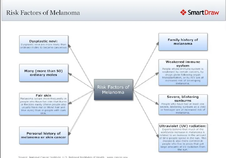
The list of risk factors includes also the presence of more than 50 acquired( common, banal) nevi, more than five dysplastic (atypical, Clark’s) nevi ,large congenital nevi, nevi larger than 6 mm, PUVA therapy, use of tanning salons, Xeroderma pigmentosum, immunosuppression, chemical exposures, scars, Marjolin’s ulcer and genetic factors (Bandarchi et al.,2011).
Individuals with fair skin, blond or red hair ,with numerous freckles and innumerable melanocytic naevi, with tendency to sunburn and tan poorly are at an increased risk for melanoma(Rivers, 2004). In particular, the red hair phenotype is in some way determined by the melanocortin-1 receptor which has a critical role in the type of melanin produced by melanocytes (eumelanin or pheomelanin). In this regard, loss-of-function mutations in the gene for the melanocortin-1 receptor protein and its variants result in red hair phenotype and may increase the risk for this cancer. In particular, Arg151Cys, Arg160Trp, and Asp294His are some of the most frequent mutations associated with the red hair and are in some way responsible for several people’s reduced ability to tan (Rivers, 2004). A second hypothesis is that individuals with variants in the melanocortin-1 receptor may not be capable of synthesizing photoprotective eumelanin resulting in a sequence of cytotoxic and mutagenic events primarily due to the increased formation of pheomelanin, free radical production and oxidative stress to the melanocyte (Rivers ,2004). Wenczel et al showed that pheomelanin or melanin intermediates (or both) are the most likely chromophores that react with ultraviolet A radiation producing reactive oxygen species which may in turn be responsible for DNA single-strand breaks and photadducts(Rivers ,2004).
More than melanoma incidence by anatomic site,‘malignant potential’ for each target cell at a given site was really the measure of interest(Whiteman et al.,2011).
8
Figure 2.Risk factors in developing melanoma; National cancer institute,U.S National institutes of health.
1.3 Diagnosis
Typical malignant melanomas are usually defined as malignant Melanoma ABCD: asymmetry, border irregularity, color variegation, diameter more then 6mm. Some dermatology experts also add E for evolution or for elevation above skin level. The ABCD checklist is a sensitive, non-specific diagnostic test (90 to 100 percent, depending on whether a positive test is defined as the presence of one, two, or three of the ABCDs).
Any Suspicious lesions should be biopsed before a real diagnosis of melanoma. Excisional biopsy with 1- to 2-mm borders is preferred. If lesion size or location makes excisional biopsy inappropriate or impractical, incisional or punch biopsies including the area appearing most suspicious may be performed. A negative incisional or punch biopsy does not necessarily rule out melanoma in a highly suspicious lesion. Shave biopsies should never be used if melanoma is suspected because lesion thickness is vitally important in determining treatment and prognosis(Rager et al.,2005).
9
1.4 Classification and histopathology
According to Wallace Clark ‘s and coworkers’ classification, there were 3 types of melanoma :the superficial spreading type(SSM), lentigo malignant type(LMM) and nodular type(NM).
Later on a fourth type of melanoma was introduced by Dr Richard Reed: acral lentiginous malignant melanoma(ALM).
Melanoma is characterized by three clinical and histomorphologically discernable steps in tumor progression:
1. Radial growth phase(RGP)related to the early phase of melanoma growth in which the tumor expands on the surface of the skin.
2. radial growth phase(RGP)-confined microinvasive which shows some malignant cells present in superficial papillary dermis.
3. vertical growth phase(VGP) which is a prominent feature for melanoma entering the tumorigenic and/or mitogenic phase.
The importance of RGP is best demonstrated by the Taran and Heenan study who monitored the development of melanoma in 1716 patients through 7 to 14 years followup. Among these level 2 melanomas(=1mm thick), only 5 evolved into metastasis.
The superficial spreading melanoma(SSM) is the most common melanoma that can occur at any
site and at any age. About 75% of SSMs arises de novo. The classic lesions delineate a well-circumbscribed polycyclic patch with variegating shades of brown, gray and black and “ pagetoid” spread of melanoma cell in epidermis. Histopathologically, the indicatory sign of SSM is the presence of enlarged melanocytes often disposed as small aggregates or nests that diffuse upward within the epidermis(pagetoid pattern).
Nodular melanoma(NM)was proposed as a separate category because it evidenced without a
significant radial growth phase and an accelerated transition to the VGP. It occurs in 15-35% of cases and could be nodular, polypoid or pedunculated.
Lentigo maligna melanoma(LMM) also called Hutchinson freckle, generally occurs on the
sun-exposed skin, face, and upper extremities of elderly patients(Bandarchi et al.,2010). It is basically in situ melanoma and the progression to metastasis, counting only 5% of lentigo maligna patients usually takes several years. In this variant melanoma cells proliferate backto-back with nest formation extensing into cutaneous adnexa. The actual treatment of lentigo maligna includes numerous methods of theraphy such as cryotherapy, superficial radiation and surgical excision with mapping and modified Mohs’surgery.( Bandarchi et al.,2010).
Acral lentiginous melanoma(ALM) commonly arises on palmar, plantar and ungula skin of black
10 arrangement of melanocytes into solitary units along the basilar epidermis does not show any evident pagetoid growth. Its lentiginous growth pattern typically dissolve in a gradual way to the adjacent non-lesional skin, making it difficult or even impossible to determine the boundary between melanoma in situ and normal skin. As LMM, ALM is prone to involve appendageal structures (such as hair follicles in LMM and sweat glands in ALM).(Whiteman et al.,2011).
(table 1)
There are additional categories of melanoma showing peculiar clinical and/or histopathological features as desmoplastic melanoma(DM), nevoid melanoma, melanoma arising from a blue nevus, melanoma arising from giant congenital nevus and melanoma in childhood. After the cutaneous form the most frequent melanoma originating sites are the mucosal epithelium(anogenital region, oropharynx and paranasal sinuses as well as the conjunctiva), uvea, encompassing intraocular structures (iris, ciliary body and choroid). (Whiteman et al.,2011).
Figure. 3 Different types of cutaneous melanoma A. Superficial spreading melanoma at the knee B. Superficial spreading
melanoma.C. Lentigo maligna on cheek. D. Nodular melanoma on shoulder.
A
B
D
C
11
Nestle FO ,Kerl H. Melanoma. In: Bolognia JL, Jorizzo JL, Rapini RP, editors. Dermatology. New York, NY: Mosby; 2003. p. 1789-815.
1.5 Biology/genetics of malignant melanoma
Like many other cancers, melanoma has a significant genetic basis. However, its genetic pathways may involve multiple genes with probable interactions with sun exposure (Bataille ,2003).
Two genes are involved in familial melanoma:CDKN2A(p16) on chromosome 9p 21 and CDK4 on chromosome 12. Penetrance of CDKN2A mutations is influenced by environmental or genetic factors. It varies with melanoma population incidence rates: it is higher in regions with higher baseline incidence (U.S.A., Australia and Sweden) than in regions with lower incidence (Europe except Sweden). Thus, the estimated penetrance of CDKN2A mutations before the age of 50 years was higher in the U.S.A., compared to Australia and Europe.(Lens et al.,2004). The first one acts as a tumour suppressor gene, regulate cell cycle and senescence. Mutations of the CDKN2A gene confer susceptibility to familial melanoma while partial or complete loss of p16 expression has also been associated with sporadic melanoma(Bandarchi et al.,2011). In particular, progressive loss of p16 has been cited in transformation of benign nevi into melanoma and metastatic melanoma (Bandarchi et al.,2011). Other alterations of cell cycle proteins(cyclin D1,pRb and p16)have been reported to contribute in transformation and progression in melanocytic tumors. Augmented expression of cyclin D1 and pRb is related with progression to melanoma cells; however both show relative decrease in thick and metastatic melanoma(Bandarchi et al.,2011).
Moreover, upregulation of PAR-1(protease-activated receptor-1 )has been identified in melanoma cell lines and tissue specimens. This is therefore responsible for mediating high levels of Cx-43
12 (gap junctional intracellular communication molecule connexion)expression which is in turn involved in tumor diapedesis and attachment to endothelial cells( Bandarchi et al.,2011). PAR-1 together with type 1 collagenase activates MMP-1(matrix metalloproteinase-1)suggested to be involved in progression of noninvasive melanoma to vertical growth phase(Bandarchi et al.,2011). Downregulation of E-cadherin and upregulation of N-cadherin may be observed in melanoma cells and this could play some role in uncontrolled proliferation, invasion and migration (Bandarchi et al.,2011).
In this regard, vascular endothelial growth factor(VEGF) and its receptors(VEGF-R1,R2 and R3) are more expressed in melanomas and advanced melanoma compared to benign nevi(Bandarchi et al.,2011).
Cortactin (a multidomain actin-binding protein important for the function of the cytoskeleton)is more expressed in melanomas than in nevi and in metastatic melanoma than in invasive primary melanoma(Bandarchi et al.,2011).MHC (major histocompatibility complex)molecule is overexpressed in primary stages of melanoma and downregulated in metastatic malignant melanoma(Bandarchi et al., 2011).PEDF(pigment epithelium-derived factor)loss is suggested to be related to the invasive phenotype and malignant progression. (Bandarchi et al.,2011).
Deregulation of microRNAs (miRNAs)using cell lines from primary or metastatic melanoma plays some role in formation and progression of melanoma.
Melanoma chondroitin sulphate proteoglycan (MCSP) support the growth, motility and invasiveness of tumor cells. Its expression is associated with augmented expression of c-Met and HGF as well as c-Met inhibition resulted in a limited growth and motility of melanoma cell lines. (Bandarchi et al.,2011).
One growth factor pathway that has been recently focused on is the RAS–RAF–MAPK–ERK signalling cascade (Ko et al.,2010). Changes in the primary sequence of the constitutively active protein RAS determined by oncogenic lesions could alter in some level the RAS signalling cascade (Ko et al.,2010). Specifically, oncogenic mutations in NRAS and BRAF occur in about 80% of the most common types of melanoma. Van’t Veer et al.(1989) and Ball et al.(1994) found a higher frequency of NRAS mutations in melanomas on sun-exposed sites(Ball et al.,1994 ;Van’t Veer et al.,1989) and later studies found these mutations in tumors arising in body sites such as the face and the head(22%),compared with the limbs(15%) or the trunck(11%). Some studies evidenced NRAS mutations associated with certain histopathological subtypes while others did not find any correlation as well as some NRAS mutations have been associated with tumor thickness and level of invasion but there was not any consistent demonstration (Whiteman et al.,2011).
13
Given the relative frequency of BRAF mutations in the most common types of melanoma, much attention has been focused on the BRAF gene and its role in this disease. This gene encodes a protein belonging to the raf family of serine/threonine protein kinases and lies downstream of RAS
(Ko et al.,2010). BRAF is highly espressed in neuronal tissue and melanocytes.
Individuals with germline BRAF mutations develop cardio-faciocutaneous syndrome, which is not associated with an increased risk of cancer or melanoma(Ko et al.,2010).
In contrast with NRAS findings ,BRAF mutations have been effectively associated with specific clinical and histopathological characteristics of melanoma. (Whiteman et al.,2011). In particular, patients with BRAF-mutated melanomas are significantly younger and have multiple melanocytic nevi on their skin than patients with melanomas without BRAF mutations (Edlundh-Rose et al.,2006; Goel et al.,2006; Liu et al.,2007; Maldonado et al.,2003; Poynter et al.,2006; Thomas et al.,2007). In Broekaert et al 2010 studies BRAF-mutant melanomas were found to be more likely to metastasize to regional lymph nodes than melanomas without BRAF mutations. In NRAS and BRAF-driven oncogenesis, the melanoma metastatization requires silencing of tumor suppressor genes including PTEN, INK4A and/or ARF. There are three RAS (HRAS, KRAS and NRAS) and three RAF(ARAF, BRAF and CRAF) genes in humans in which gain-of-function mutations in NRAS respectively (B25%) or BRAF (B40%) have been found in melanoma. NRAS mutations involve codons 12, 13 and 61 (NRASG12/G13/Q61). More than 90% of BRAF mutations T1799A point mutation in which T → A transversion converts glutamic acid for valine at the 600 position of the amino acid sequence, BRAFV600E, which results in the protein taking on a constitutive active configuration( Ko et al.,2010).
Oncogenic mutations in NRAS are responsible for the activation of both MAPK and PI3K-AKT pathways, whereas mutant BRAF activates the MAPK pathway ( Yang et al.,2012). Co-expression of NRASQ61R and BRAFV600E results in a senescent phenotype in melanoma cells. BRAFV600E cannot transformhuman melanocytes due to induction of senescence, but is capable of transforming murine melanocytes that are ARF deficient.29 RAS mutants transform melanocytes more efficiently than BRAF possibly because of the PI3K-AKT component. In the presence of PTEN suppression, BRAFactivates PI3K-AKT, and leads to full transformation to melanoma in vivo. Although several genetic events cooperating with NRAS and BRAF in melanoma have been identified, full characterization of collaborating partners has not yet been established and the role of scaffold proteins in NRAS- and BRAF-driven tumorigenesis is still unknown ( Yang et al.,2012).
GAB2 is a scaffold protein involved in the regulation of cellular processes such as proliferation, survival, migration, differentiation and morphogenesis (Yang et al.,2012). It is highly expressed in
14 metastatic melanoma as compared with benign melanocytic nevi and primary melanomas. GAB2 has oncogenic potential as demonstrated by its amplified in the acral and mucosal melanoma subset with B10% frequency(melanomas arising from melanocytes on palms, soles and mucous membranes). In cell culture systems, GAB2 promotes migration and invasion while accelerates tumorigenic potential and metastatic capability in vivo. Yang et al investigated the supporting role of GAB2 in NRAS-driven melanoma underling enhanced tumorigenesis in vivo, and facilitated angiogenic switch by upregulating HIF-1a and VEGF in tumors with mutant NRAS(Yang et al.,2012).
Mutations in KIT have also been observed in patients with melanoma arising on glabrous skin or the nail apparatus ,the mucosa, or skin with cumulative sun induced damage(CSD melanomas) and have not been detected in melanomas or skin having not this characteristic(Baedling et al.,2008;Curtin et al.,2006). KIT and BRAF mutations are somewhat of a mirror image of each other as the melanoma types in which the first are found, RAF mutations are relatively uncommon. There are several features that throw in together acral and mucosal melanomas and chronically sun damaged skin as their incidence which tends to arise with age as opposed with BRAF mutation which peak is 50yr of age, and the typical arrangement in solitary units/nests, poor circumscribed in contrast with demarcated intraepidermal component well distinguished in BRAF mutations melanoma( Viros et al.,2008).
Melanomas often show recurrent patterns of chromosomal aberrations such as losses of chromosomes 6q, 8p, 9p, and 10q along with gains of chromosomes 1q, 6p, 7, 8q, 17q and 20q compared with benign nevi (Bandarchi et al.,2011). In particular, in acral melanomas, amplifications most frequently involved chromosome 11q13, centering on the cyclin D1 locus, as well as hTERT on chromosome 5p. By contrast, cyclin D1 is infrequently amplified in mucosal melanomas, where amplifications frequently involve the CDK4 and MDM2 locus on chromosome 12q (Curtin et al., 2005).
Recently, mutations in G-proteins of the Gαq family of GTPases have been reported in certain subsets of melanocytic neoplasia. In particular, a role for the two closely related Gαq family members Gq and G11 (encoded by the genes GNAQ and GNA11, respectively) in melanocyte biology was suggested because hypermorphic mutations in both genes were found to result in skin darkening in a mutagenesis screen in mice (Van Raamsdonk et al., 2004). Recurrent mutations in GNAQ occurring early in the progression of melanocytic neoplasia in blue nevi, uveal melanomas and melanocytomas of the central nervous system were identified in subsequent studies(Küsters-Vandevelde et al., 2009). Frequent mutations of GNA11 were evidenced in the same spectrum of
15 melanocytic neoplasms in which GNAQ mutations are observed (Van Raamsdonk et al., 2010). GNAQ and GNA11 are restrictedly mutated in blue nevi and uveal melanomas, with virtually no mutations in other benign or malignant melanocytic neoplasms harboring these mutations (Van Raamsdonk et al., 2009, 2010).
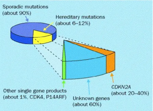
Figure 4. Prevalence of CDKN2A mutations in hereditary melanoma; Christopher B Hansen, Lisa M Wadge, Katrina Lowstuter, Kenneth Boucher, Sancy A Leachman
Clinical germline genetic testing for melanoma.
16
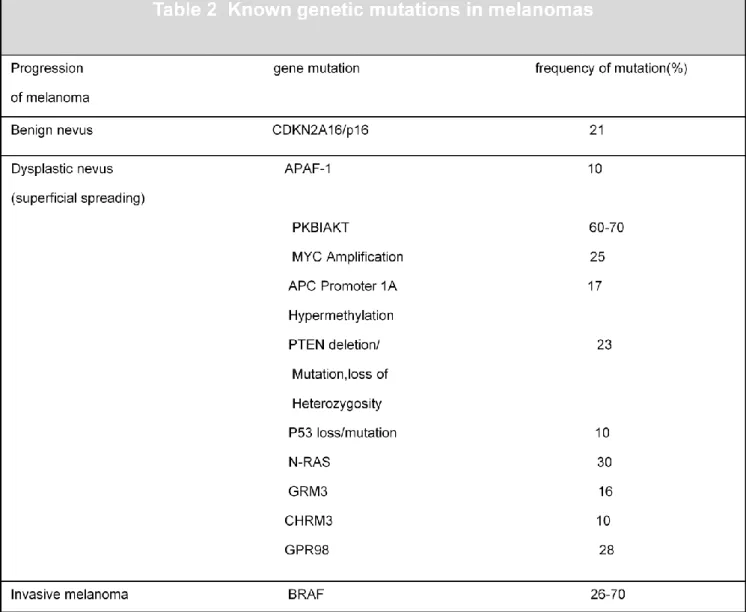
Table 2. Known genetic mutations in melanoma.
Inspired to Fonkem E, Uhlmann E.J.,Floyd S R,Mahadevan A,Kasper E,Eton O,Wong E T” Melanoma brain metastasis:overview of current management and emerging targeted therapies” Expert Rev. Neurother. 12(10), 1207–1215 (2012)
1.6 Treatment
Although high risk patients should avoid sunlight and routine checkup as a preventive measure ,the use of sunscreen is often associated with higher incidence of malignant melanoma and this is mostly attributable to modified sun-exposure behaviour (Rogers, 2000, Marks 2000). Important factors in the management of malignant melanoma are excision of primary cutaneous tumour through different protocols and sentinel lymph nodes biopsies which has had a relevant impact on managing patients of melanoma(Walsh et al.,2000). One commonly used protocol is in situ melanoma (Johnson et al.,1995, Balch 1993 ,Ringborg 1996):0.5 cm clear margin, <1 mm: 1 cm
17 clear margin 1–2mm 1–2 cm clear margin (depending on the location), 2–4mm 2 cm clear margin,
>4mm 3 cm clear margin (Green 1991, O’Rourke 1993). In transit melanoma metastasis is
characterized by in-transit cutaneous malignant melanoma deposits between the site of excision and the draining lymph nodes more than 2 cm from primary melanoma and is treated with surgical excision, interferon therapy, hypothermic isolated limb perfusion with melphalan, CO2 laser ablation, and intralesional BCG (Bandarchi et al.,2011). Satellite metastasis are defined by lesions less than 2 cm from the primary melanoma, have similar biologic behaviour and are categorized as intralymphatic metastasis as another criterion in the N category regardless of the number of lesions based on the final version of the 2009 AJCC melanoma staging and classification. Patients with positive nodes or node-negative melanomas thicker than 4 mm, ulceration, or Clark’s level IV or V may benefit of Interferon-alpha 2b which is the most commonly used FDA approved adjuvant therapy even if it does not alter the natural history of the disease for the population as a whole. In large-scale, randomized, observation-controlled trials, interferon-a2b has demonstrated a 10–20% improvement in relapse-free survival, but with no clear effect on melanoma-related mortality (Ko et al.,2010).
In stage IV melanoma, Interleukin-2 (IL-2) effects durable responses in 10–20% of patients, although is associated with severe, short-lived, toxicities (Ko et al.,2010). Among conventional cytotoxic chemotherapies, the alkylating agent dacarbazine is the only FDA approved agent determining approximately 5–10% of short-lived responses (Ko et al.,2010). Similar efficacy profiles in the metastatic context have been obtained using carmustine and temozolomide, and agents from other classes, such as taxanes and platinum-analogues(Ko et al.,2010). Two treatments have recently become available for advanced melanoma: Ipilimumab (Yervoy™), which received US FDA approval on 25 March 2011 and the FDA also approved vemurafenib (Zelboraf®) on 17 August 2011. Ipilimumab is a humanized monoclonal antibody against the cytotoxic T-lymphocyte-associated antigen 4 (CTLA-4) receptor, thereby allowing the persistent and increased activation of immune response against metastatic melanomas. Vemurafenib (Zelboraf®) is a small-molecule inhibitor of the serine threonine kinase BRAF capable of correcting the abnormal activation of the MAPK pathway. Its indication is therefore the treatment of BRAFV600E mutated malignant melanoma(Fonkem et al.,2012). In particular it has significant activity against tumor cells harboring the mutated BRAFV600E protein. Although definitive data are lacking at present, these two therapeutic strategies may offer an alternative to the management of metastatic melanoma in the brain and the rest of the CNS(Fonkem et al.,2012).
18 Routine laboratory tests such as lactate dehydrogenase (LDH) and/or imaging studies such as CT scan, MRI, and PET scan are not definitively considered to affect the longevity of patients even if patients with higher stage may benefit from these tests (Bandarchi et al.,2011 ).
The identification of signalling pathways for melanoma initiation and progression has had a pivotal role to open up exciting new areas for the investigation of novel targeted agents to be tested alone or in combination with conventional chemotherapies(Ko et al.,2010).
1.7 Metastatic melanoma
The most important parameter for the control of cancer progression is angiogenesis which is a multi-step process required for the growth of solid tumors and for the metastatic dissemination of tumor cells. Angiogenesis is the formation of new capillaries from preexisting microvessels and consists in a sequence of events, in which endothelial cells degrade their extracellular matrix, migrate into the perivascular spaces, proliferate and align to form cell–cell contacts to construct patent blood vessels. This process is mediated by the endothelial cell adhesion molecules (CAMs). They take part to the metastasis development and migration and compared to normal tissues there is a wide variation of the different CAMs between tumor types.
They are represented by four principal classes: the selectins, which are transmembrane proteins subdivided in three groups E, L and P, expressed by endothelial cells, leucocytes and platelets, respectively; the integrins, which are transmembrane glycoproteins composed of two subunits α and β; the membrane proteins immunoglobulin superfamily; and, the cadherins, mediators of cell–cell interactions( Seguin et al.,2012).
The angiogenesis particularly involves two proteins belonging to the CAMs superfamily: the integrin alpha v beta 3 (αvβ3) and the E-selectin. The integrin αvβ3 is not (or weakly) constitutively expressed in normal vascular endothelial cells, whereas it is selectively expressed on proliferating vascular endothelial cells and on vascular cells in tumours. Similarly, E-selectin is also detected at very low levels in normal adult blood vessels but is highly expressed in newly formed tumour capillaries and in activated endothelium. By their angiogenic and adhesion properties, both CAMs are involved in tumour growth and metastasis and can therefore influence cancer prognosis and/or response to antiangiogenic therapy( Seguin et al.,2012).
The melanoma cell adhesion molecule(MCAM)/CD146 has been initially identified as a melanoma progression antigen in humans(Guezguez et al.,2007). It is a 113-kDa cell adhesion glycoprotein belonging to the Ig superfamily (Guezguez et al.,2007). There are two isoforms differing by their cytoplasmic domain(MCAM long(MCAM-1)and MCAM short(MCAM-s)). Immunohistochemical
19
studies have established MCAM/CD146 as a component of the endothelial junction which is associated with actin cytoskeleton. Its interactions might be involved in the recruitment of activated T cells to the inflammation sites(Guezguez et al.,2007).
As the detachment from the primary mass occurs, followed by vasculature and colonization of distant sites, numerous cytoskeletal alterations as well as multiple changes in the tumor cell’s interactions with the environment constituted by the neighboring cells and the ECM take place ( Saladi et al.,2010). Of the phenotypic changes that occur during metastatic melanoma progression, differences in the expression of receptors for paracrine growth factors are relevant(Derkins et al.,2004). The modulating role of these factors ensures in some way, the survival of malignant cells in unusual compartments (Denkins et al.,2004).
The migratory nature of neural crest derived precursors that give rise to the melanocyte lineage has certainly an important role in the inherently high metastatic potential associated with melanoma (Saladi et al.,2010). In fact, this potential is strictly dependent on either the prometastatic genetic changes such as those involving NEDD9 amplification and epigenetic alterations that dynamically modulate the expression of genes required for each step in the process(Saladi et al.,2010). In particular, regulation of gene expression on a epigenetic level often involves changes in chromatin structure that are catalyzed by chromatin remodeling enzymes as SWI/SNF. These complexes are ATP dependent chromatin remodelling enzymes that play critical roles during organism development and increase DNA accessibility, activating or repressing gene expression (Saladi et al.,2010). The regulatory role of SWI/SNF enzymes has been recently highlighted in melanoma as promoting neural crest migration and differentiation as well as interacting with Microphthalmia – Associated Transcription Factor (MITF), a lineage survival oncogene in melanoma(Saladi et al.,2010).
Among all the forms of cancers ,melanoma has the highest propensity to metastasize in the brain. In particular, it is the third most common cause of brain metastasis behind lung(40-50%) and breast (15-25%) diagnosing approximately 10,000 patients yearly in the United States(Sloan et al.,2009). While the incidence of brain metastasis in patients with melanoma is 9.6%,melanoma metastasizes to the brain with one of the highest frequencies of any cancer capable of colonizing the CNS(Sloan et al.,2009). By the time melanoma develops, metastasis to regional and distant lymph nodes, liver, lung, and the central nervous system may well have occurred(Sloan et al.,2009). Nearly 37% of patients with stage IV melanoma eventually develop clinically apparent brain metastasis and autopsy report the prevalence of brain metastasis at 55% to 75% of patients who died of
20 melanoma(Sloan et al.,2009). The huge CNS involvement associated with malignant melanoma may be due to a “homing” influence since melanocytes and neuronal subpopulations share a common embryologic origin(Denkins et al.,2004).
Prognosis in patients with brain metastases is still poor; survival data obtained in trials of the RTOG have divided patients in three “risk classes” according to age, Karnofsky Performance Status (KPS), primary tumor control and presence of extra-CNS metastases. To this purpose, the longest median survival, reported in Class I (i.e., younger than 65, with KPS of at least 70, controlled primary tumor and absence of extra-neural metastases) patients, is 7.1 months(Salmaggi et al.,2009).
Melanoma brain metastases are multi-focal, and some can be hemorrhagic (Fidler et al.,1999). Melanoma brain metastasis often leads to neurological deterioration that progresses rapidly to death within 1 month with no treatment or 2 months with corticosteroids alone(Sloan et al.,2009). In general, patients can usually be classified at presentation into one of the three typical scenarios: patients with a solitary brain metastasis, those with oligometastatic brain tumors(typically 2 to 4) and those presenting more than 4 brain metastases classified as multiple brain metastasis(Sloan et al.,2009). The treatment of choice for single brain metastasis is surgery plus radiation, but the prognosis is poor reporting a median survival time of 4–6 months.
For multiple brain metastases, radiation and chemotherapy are employed. In this case patients may survive more than 6 months (Fidler et al.,1999). The significance of these unsatisfactory results are related to the recurrence of tumors at the site of resected lesions, as well as the growth of multiple unresected metastases in and outside the brain. Better therapy for melanoma brain metastases requires improvement in the understanding of the biology of these lesions on systemic, cellular and molecular levels(Fidler et al.,1999).
Academically, the “mestastatic cascade” is the process in which the initial spread of cells deriving from a solid tumor to a generalizing site, such as regional lymph nodes, proliferate and then spread to additional organs. In this way, even if the primary lesion has been surgically removed, the generalizing site remains intact and metastasis can occur. Generally, if brain metastasis are determined by metastasis of metastases the risk of developing fatal brain lesion may be reduced practising an aggressive, prophylactic resection of lymph node or visceral metastases (Fidler et al.,1999). On the other hand, this procedure may not result usefull in preventing brain metastasis from occurring if brain metastasis occurs by the direct spread of specialized metastatic cells from the primary lesion. Thus, the metastatic potential of cells appears fundamental to determine whether or not brain metastasis represents shedding from generalizing site metastases or the primary tumor( Fidler et al.,1999).
21 The intrinsic properties of the primary tumor as well as the specific features of the target organ certainly play a pivotal role in the different tumors tendencies to brain metastatization .
Among these features chemokines/receptors expression in tumor and brain microvessels and/or parenchyma could be relevant (Salmaggi et al.,2009). The interaction of chemokines with their receptors may act in two different phases of the metastatization process: first, in the homing of neoplastic cells from the primary site to the target organ, and secondly in local progression of the metastasis, by inhibiting apoptosis and inducing angiogenesis(Salmaggi et al.,2009).
Experimental models have recently suggested the CXCL12/CXCR4 axis as part of the cytokine/receptor system involved into metastatization. Some evidence suggests that inhibition of the receptor/ligand interaction may decrease metastasis establishment and progression also within the brain, as well as migration of tumor cells across an in vitro model of blood-brain-barrier(Salmaggi et al.,2009).
2 The Blood Brain Barrier
2.1 Structure
The nature of the blood brain barrier (BBB) was first argued by Paul Ehrlich in 1885. As it was well debated into the 20th century, the BBB represents the regulated interface between the peripheral circulation and the central nervous system(CNS) and the most critical and sensitive system in the human body(Hawkins et al.,2005). Proper neuronal function is guaranteed by narrow ranges of the ions Na+, K+ and Ca2+ which regulate the extracellular environment as well as an adequate oxygen supply satisfying the nervous tissue metabolic demands (Hawkins et al.,2005). In fact, the CNS requires for its functions approximately 20% of the oxygen consumption in humans (Hawkins et al.,2005).
It is extremely sensitive to a wide range of neurotoxic chemicals which are apparently harmless thanks to the rapid excretion by peripheral organ systems(Hawkins et al.,2005).
It is therefore essential that the interface between the CNS and the peripheral circulatory system acts as a selective barrier to potentially harmful substances, as a homeostatic regulator for the mantainence of the environmental composition and as a facilitator of nutrient transport (Hawkins et al.,2005).
Besides the BBB carrier function, its barrier function is achieved through a fourfold defence line: 1) the paracellular barrier formed by interendothelial junctions limits the free movement of
22 2) The transcellular barrier represented by the low level of endocytosis and transcytosis characteristics for brain endothelial cells and inhibits transport of substances through the cytoplasm.
3) The enzymatic barrier is made possible by a complex set of enzymes, including acetylcholinesterase, alkaline phosphatease, gamma-glutamyl transpeptidase, monoamine oxidases, and other drug metabolizing enzymes capable to degrade different chemical compounds.
4) Moreover a huge number of efflux transporters (ABC, ATP-binding cassette transporters) like ABCB1 (P-glycoprotein), ABCC1,ABCC4 and ABCG2 (BCRP) (Wilhelm et al.,2011).
The current description of the basic structure of the BBB appeared in the work of Reese, Karnovsky and Brightman in the late 1960s. Specifically, it works as a selective diffusion barrier through the presence of tight cell-cell junctions and the complete lack of fenestrations at the level of the cerebral microvascular endothelium (Hawkins et al.,2005).
Basically, the circumference of the capillary lumen is bordered by a single endothelial cell (EC) which can be distinguished from those in the periphery by increased mitochondrial content, a lack of fenestations, minimal pinocytic activity and the presence of TJ (Hawkins et al.,2005). Attached at irregular intervals to the abluminal membrane, pericytes cover approximately 22-32% of the endothelium surface and are classified into granular and filamentous subtypes(Hawkins et al.,2005). These cells, together with endothelial cells are surrounded by the basal lamina (BL) which is a membrane 30 to 40-nm thick composed of collagen type IV, Heparin sulphate proteoglycans, laminin, fibronectin and other extracellular matrix proteins( Hawkins et al.,2005). The basal lamina is contiguous with the plasma membranes of astrocyte end-feet which ensheathe cerebral capillaries( Hawkins et al.,2005).
The BBB characteristics are in some way induced by the properties of its subpopulations. As in vitro experiments showed, astrocytes have a role in the development/maintenance of BBB characteristics and in regulation of cerebral microvascular permeability in conjunction with neurons via dynamic Ca2+ signalling (Hawkins et al.,2005). Astrocytes maintain homeostasis of the brain microenvironment through the transport of various nutrients from the circulation to the neurons cooperating in neural signal transduction and buffering the ionic balance of the extracellular matrix (Fidler et al.,2010). They are sources of important regulatory factors like TGF-beta, GDNF, bFGF and IL-6 (Wilhelm et al.,2011).
23 Contractile proteins have been identified in cerebral pericytes, suggesting that they may regulate capillary blood flow (Hawkins et al.,2005).In absence of pericytes, an abnormal vasculogenesis, endothelial hyperplasia and augmented permeability take place in the brain (Wilhelm et al.,2011).
Pathological changes in cerebral blood flow and perfusion pressure interestingly accompain disruption of the BBB integrity, implying the communication between neurons and the vasculature. Whether neurons are critical in the development of the BBB phenotype has not yet been defined, but is possible that they can regulate critical aspects of BBB functions(Hawkins et al.,2005). In addition to astrocytes, pericytes and neurons, the extracellular matrix of the basal lamina also interacts with the cerebral microvascular endothelium. In particular, it works as an anchor for the endothelium via interaction of laminin and other matrix proteins with endothelial integrin receptor which are, in turn, responsible for the stimulation of numerous intracellular signalling pathways (Hawkins et al.,2005).
The interendothelial space of the cerebral microvasculature is characterized by the presence of a junctional complex formed by three different type of junctions: adherens junctions(AJ),TJ and gap junctions (Hawkins et al.,2005).Gap junctions mediate intercellular communication while a restricted permeability across the endothelium is ensured by the ubiquitous presence of both AJ and TJ (Hawkins et al.,2005).
The transmembrane components of the TJ at the BBB include junctional adhesion molecule(JAM)-1 as well as claudins, forming the primary “seal” and occludin working as additional support structure of the TJ (Hawkins et al.,2005).
In addition to these components ,there are accessory proteins identified as TJ:ZO-1,ZO-2 and ZO-3 having a critical role in the stability and function of the TJ(Hawkins et al.,2005).Other accessory proteins such as cingulin,AF-6 and 7H6 are suggested to mediate interactions between the cytoskeleton and the TJ (Hawkins et al.,2005).
TJ are dynamic structures so that possible alterations of the its proteins under physiological and pathophysiological conditions may occur (Hawkins et al.,2005). Specifically, proteins of the TJ are subject to changes in expression, subcellular localization, post-translational modification and protein-protein interactions (Hawkins et al.,2005).
Defeated protection of the BBB is a critical event in the development and progression of several diseases that affect the CNS(Hawkins et al.,2005). In some cases, several pathologies such as ischemic stroke and traumatic brain injury are responsible for increased BBB permeability whereas sometimes, the BBB disruption may be a precipitating event such as with multiple sclerosis(Hawkins et al.,2005).
24 Inflammatory mediators are known modulators of BBB permeability profoundly affecting TJ proteins(Hawkins et al.,2005).
.
Fig.5 The neurovascular unit. Shown are examples of the contribution of each of the various components of the neurovascular unit to the dynamic regulation of microvascular permeability.
Hawkins et al.,2005
2.2 Interactions of the BBB cells with other tissues
Recent studies have focused on the possibility of different populations to cross the BBB.
In particular, the emigration of sensitized leukocytes as well as macromolecules/other compartment deriving cells during inflammatory or neoplastic conditions of the CNS merits further consideration and is the topic of ongoing research (Lossinsky et al.,2004).
Tumor cells are thought to select different transendothelial routes for passage across ECs depending upon the cell type that is migrating and barrier type that is being breached (Lossinsky et al.,2004). Collectively, there is no clear understanding concerning the precise transendothelial route(s) for the various neoplastic cells in the CNS even if a transjunctional pathway may offer the best route with a path of least resistance for the cell cross (Lossinsky et al.,2004).
An accurate elucidation of how the various cell types migrate from the blood side of the vasculature to the brain parenchyma and vice-versa would be relevant for the development of specific therapies
25 that may block or reduce the attachment and migration of these cells across the BBB(Lossinsky et al.,2004).
The ability of malignant cells to penetrate such a formidable barrier depends firstly on the production /activation of enzymes responsible for the degradation of the ECM (Marchetti et al.,2003). Besides all the components released by the ECM, one type being heparan sulphate proteoglycans or HSPG is thought to have an important role in the cancer metastasis (Marchetti et al.,2003). HSPG act as a biological modulator, degrading at the level of HS chains and its catabolism is observed in inflammation, wound repair, diabetes and neoplasia, including melanoma(Marchetti et al., 2003).
Heparanase sets in brain metastasis either in local invasive processes by degrading the HS chains of HSPG and in the release of HS-bound angiogenic factors at the metastatic site(Marchetti et al.,2003).
To this purpose, the brain is an ideal environment due to its high levels of NT production(Marchetti et al.,2003). According to this hypothesis, melanoma would probably colonize brain as its preferential metastatization site for the wide range of growth factors which have been in turn released by the BBB ECM disruption (Marchetti et al.,2003).
The storage of bFGF in ECM and its release as a complex with HS fragments result in a more stable form of bFGF ,capable of binding the high-affinity plasma membrane receptors (Marchetti et al.,2003). In addition, brain metastatizing cells express relatively high levels of basement membrane hydrolytic enzymes, such as type IV collagenases, cathepsins, plasminogen activators and of course heparanase. Some of these enzymes are expressed in higher amounts by metastatic compared to nonmetastatic cells and moreover even in higher amounts by the microenvironment and by certain normal cells (microvessel endothelial cells and astrocytes, among others) (Marchetti et al.,2003). In conclusion if malignant cells receive proper paracrine signals, they can be stimulated to increase the synthesis and release of BBB-degrading enzymes(Marchetti et al.,2003). This is relevant for the invasion and together with the release of proliferating factors is the key step for brain as melanoma preferential metastatic site (Marchetti et al.,2003).
3.Aim of the study
The brain is a preferential site for metastasis thanks to its high vascularization. Tumour cells succeed in some way in crossing the blood brain barrier and produce neoplasms.
26 Firstly we focused on the different characteristics of tumour cells to release possible toxic metabolites for in vitro cerebral endothelium. To this purpose, either non metastatic and metastatic melanoma cell types were tested.
Then, the expression of molecules of invasiveness and inflammation was evaluated comparing the responses of different endothelia to three kind of tumours. The capability of tumorigenic cells to attach the brain endothelium was compared to the non cerebral endothelium.
Further, we investigate the mechanisms involved in brain melanoma metastatization using an in vitro model to understand why such a distant site as the brain is the preferential melanoma metastatic site .
4. Materials and methods
Cell cultures
A total of three tumour cell cultures were included in this study. Two types of melanoma cell cultures were tested: the metastatic melanoma COLO38 and non-metastatic ocular choroidal melanoma cell line OCM1A. Cells were grown in RPMI 1640 medium containing 2 mM glutamine, antibiotics and 10% fetal bovine serum (FCS)and cultured at 37 _C in humidified 5% CO2/95% atmosphere. All human melanoma cell lines were a generous gift of the Laboratory of Immunology, National Cancer Institute ‘‘Regina Elena’’, Rome. All materials and media were from Invitrogen Srl (San Giuliano Milanese, Italy) unless otherwise specified.
The prostatic carcinoma LnCaP was generously provided by Dr. Vera Cardile, university of Catania, section of human physiology and grew in the same conditions.; HCN-2(human neuronal culture) was grown in Dulbecco’s modified Eagle ‘s medium (DMEM,ATCC)+10%FBS and passaged up to three times.
Two types of endothelial cells were used, one an immortalized human brain endothelial cell line HCMEC/D3 I and primary human umbilical vein endothelial cells (HUVEC). HUVEC were passaged up to two times before sufficient cells were obtained for co-culture experiments in 24 well plates.
After 1 hour coating using Cultrex rat collagen I ,the HCMEC/D3 endothelial cells were grown in 1%
penicillin+streptomicin EBM2 (Lonza) +5%FBS Gold. The Endothelial basal medium(EBM) was supported with hydrocortisone(1.4uM,Sigma), ascorbic acid(5ug.ml-1,sigma), chemically defined lipid concentrate (1/100,gibco), Hepes (10mM,gibco), bFGF (1ng.ml-1).
HUVEC were obtained from human umbilical cords from healthy women who underwent
27
penicillin+streptomicin M199+20% FBS. The support medium comprised ECGS (Sigma) and
heparin(sigma).
Cell cultures of normal human astrocyte (Lonza) were included in this study. The growth medium consisted of DMEM F12(Invitrogen)+10%FBS supplemented with ascorbic acid(Sigma), bFGF
(1ng.ml-1,sigma) and insulin(2.5ng.ml). All cultures were performed in a 5% CO2 humidified
atmosphere at 37°C.
Protein extraction
In a set of experiments, either HUVEC and HCMEC were grown until reaching confluence in 100mm plastic Petri dishes. COLO38, OCM1A and LnCaP cells grown separately under starving conditions(RPMI 1% FBS). Endothelial cells were incubated for 48 hours with their medium additioned with increasing percentages of COLO 38 or OCM1A or LnCaP up to 80% conditioned medium. Membrane and cytosolic fractions of cells were prepared as described. Cells were washed with 1 ml ice-cold PBS and scraped with lysis buffer (50 mM Tris pH 7.6, 150 mM NaCl, 5 mM EDTA, 1 mM fenilmetilsulfonifluride,0.5 mg ml_1 leupeptin, 5 mg ml_1 aprotinin, 1 mg ml_1 pepstatin) and protease inhibitor. After thawing ,cells were centrifuged at a speed of 14000 rpm for 10 min at 4 _C. The cell pellets were kept at -20 and the supernatants (cytosolic proteins) were quantified using Bradford method. Fixed protein concentration (30µg) was diluted with a proper volume of SDS loading buffer(20% glicerol, 100nM Tris-HCl pH 6.8, 200nM dithiothreitol, 4% SDS, 0.1% blue bromophenol). Proteins were boiled for 5 minutes at 100°C after short speed centrifugation. Then, equal protein concentrations obtained after different treatments, according to the experiment, were separated through electrophoresis.
Western blotting
Protein samples were electrophoretically separated on different percentage acrylamide gel (in order to the molecular weight acrylamide gel from 8% to 12% were used) and transferred by semi-dry electrophoresis onto amersham hybond-ECL nitrocellulose membrane (Amersham Italia S.r.l., Milan, Italy). Membranes were blocked with 5% non-fat dried milk in TBS/Tween (blocking buffer), and incubated with the following primary antibodies overnight at 4 –C.: COX2 anti-mouse (Santa Cruz Biotechnology, Santa Cruz, CA, USA), TIMP-2(3A4) anti-mouse(Santa cruz Biotechnology, Santa Cruz, CA, USA), Sparc anti-goat (c-15)(Santa cruz Biotechnology, Santa Cruz, CA, USA), mmp1 anti-rabbit(Chemicon international), NOS2 anti-rabbit(Santa Cruz Biotechnology, Santa Cruz, CA, USA), polyclonal b-Tubulin anti-rabbit(Santa Cruz Biotechnology, Santa Cruz, CA, USA). The latter was used as a loading control. The membrane was incubated with
28 Peroxidase-labeled secondary antibodies for 1 h at room temperature. Bands were visualised using Luminata Crescendo western HRP substrate ECL(Millipore) reagents (PerBio, UK), and were developed as autoradiographs using ECL photographic film (Amersham, UK). Western blot analysis was performed on samples from two separated experiments.
Viability assay
HCMEC were seeded at 5x10⁴ cells/well into two 24-multiwell plates in EBM-2 full with 5%FBS, penicillin and streptomycin after 1hour coating. In order to obtain conditioned medium, COLO38 and OCM1A were grown in 100mm plastic dish in basal conditions until they reached confluence. Then the two dishes were incubated at 37°C for 48 hours in RPMI with 1% FBS, penicillin and streptomycin(=conditioned medium CM). Then, HCMEC were incubated for 24 hours with the following graded concentrations of COLO 38 CM (5%,10%,20%,40%,80%)in EBM-2 full. Control cells were left with their own medium(HCMEC with EBM-2 full 5%FBS). After 24 hours cells received 0.1%DMSO.The same was repeated with OCM1A and cell survival was evaluated by means of MTT test.
In a second experiment, HUVEC were seeded at 5x10⁴ cells/well into two 24-multiwell plates in 1% penicillin+streptomicin M199+20% FBS+its additives(ECGS and heparin). Identical graded
concentrations of COLO38 CM (5%,10%,20%,40%,80%) in M199 full and OCM1a CM were
prepared as previously described. The two HUVEC 24-multiwell plates were treated as previously. Control cells were incubated with equal volume of their own medium(in this case M199 with 20%FBS).
Cell proliferation was evaluated by 3-(4,5-Dimethylthiazol- 2-yl)-2,5-diphenyltetrazolium bromide (MTT, Sigma) assay. Briefly, after graded dilutions (5 through 80%)of conditioned media , MTT (10 ll) solution (5 mg/ml) was added to each well. The reaction was allowed to proceed for 3–4 h at 37 _C. The culture medium was removed and formazan crystals were dissolved by adding DMSO (200 µl). The absorbance of each well was read at 570 nm and directly correlated with the number of remaining viable cells.
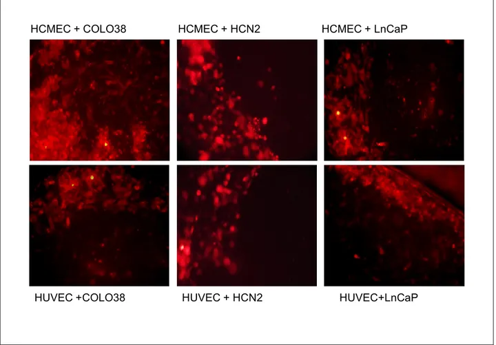
Transmigration experimental setup and fluorescence microscopy
For transmigration experiments HCMECs were gently trispinized and seeded at 1x10⁴ onto transwell permeable supports (8µm pore size polycarbonate membrane 6.5mm inserts,24 well plate, Corning) placed on 24 well plates. After reaching confluency, endothelial cells medium was replaced and astrocyte conditioned medium from the basolateral side for 24h was added. Melanoma cell lines COLO 38 and OCM1A, HCN-2 and LnCaP were grown in normal conditions.3x104 cells
29 of each type were fluorescently labelled using Cell Tracker red cmtpx 1uM (invitrogen) and plated onto the endothelial monolayer(fig.7A). Cells were left for 24 hours. Cells from the upper compartment were removed with a cotton swab(Fig.7C) and then washed with phosphate buffer 1x. The cells migrated through the endothelial monolayer and the pores of the filter were observed using fluorescence microscope and then counted.
4.1 In vitro models of BBB
The most widely used “classical” method to reproduce paracellular characteristics in vitro is based on the culture of endothelial cells on semipermeable filters. This sort of barrier define two compartments: the apical, upper compartment which can be considered as “blood-side” and the basolateral, lower compartment which is the “brain side”(Wilhelm et al.,2011).
There is significant number of publications using different types of epithelial cells or endothelial cells of non-cerebral origin for the study of different aspects of BBB functions(Wilhelm et al.,2011). Several in vitro models of the BBB used MDCK (Madin-Darby canine kidney) for its good paracellular permeability characteristics, easy culture and transfection (Wilhelm et al.,2011).
HUVECs offered a good model for the BBB study even if they were not of cerebral origins.
Primary cultures of endothelial cells of rat, mouse, pig and bovine origin have guaranteed the possibility to use transgenic animals in the case of mouse models whereas the use of human primary cells is restricted due to the lack of experimental material(Wilhelm et al.,2011).
One of the best characterized rat brain endothelial cell lines is RBE4 for retaining many BBB characteristics as well as immortalized rat cerebral endothelial cells GP8 which has been successfully used for signalling studies(Wilhelm et al.,2011).
HCMECs is probably the best characterized human cell line, expressing some junctional proteins and efflux transporters as the BBB. (Wilhelm et al.,2011).
4.2 Co-culture systems
Most of the BBB properties are induced by the presence of different subpopulations which are strictly in contact with cerebral endothelial cells.
Since the interaction of cerebral endothelial cells with astrocytes has been extensively studied, glial co-cultures are the most common models. According to the endothelial cells which are usually primary cells of rat, bovine or mouse origin; primary cells of the C6 cell line has been intensively used for glial cells. BBB-inducing medium is also used providing a sort of suitable modified BBB
30 model(Wilhelm et al.,2011). It contains 1% conditioned medium from glial-endothelial co-culture, harvested 48h after refreshing the co-culture system medium and frozen until further use(Wilhelm et al.,2011).
Pericytes are close in contact with endothelial cells therefore a co-culture of endothelial cells with pericytes or eve a triple co-culture model including also astrocytes has been also successfully employed(Wilhelm et al.,2011).
4.3 Metastatic model
In order to study the routes and mechanisms of transmigration of tumor cells through the BBB proper experimental model has been set up. The endothelial cells are cultured in large pore size filter inserts suitable for cell invasion, chemotaxis and motility studies.
Fig.6 In vitro model of the BBB.
31
Fig.7 Use of the in vitro model for brain metastasis investigations. Fluorescently labeled cancer cells are plated on confluent endothelial cultures(A). At the end of co-culture (B) cells from the upper compartment are removed(C). Fluorescence microscopy images of the tumor cells at the end of co-culture(D) and after wiping off the cells from the apical side of the filter (transmigrated cells,E)
Wilhelm et al.,In vitro models of the blood-brain barrier. Acta neurobiol Exp,71: 113-128
5. Results
To induce secretion of molecules which could affect endothelium proliferation, graded percentages of fetal bovine serum (FBS) were added to the standard medium of either the cerebral endothelium cell cultures and the non cerebral endothelium cell cultures .
32
ENDOTHELIAL CELLS TREATED WITH DIFFERENT CONCENTRATIONS OF FBS
standard composition of the medium with fixed percentage of FBS
Decreasing percentages of FBS in cell medium
P<0.05 VS CTRL
Fig.8 Treatment of endothelial cells with different concentrations of FBS.
To assess whether metastatic and non metastatic human melanoma cells could influence survival, mitogenesis and death of human endothelial blood brain barrier (HCMEC), HCMEC cells were incubated with graded concentrations of tissue culture media conditioned over 72 hours with COLO 38 metastatic human melanoma cells. HCMEC cells were incubated 48h with graded dilutions(5 through 80%) of conditioned media and then evaluated cell survival by means of the MTT test. Data indicate that COLO 38-conditioned media inhibited HCMEC cell proliferation and survival in a concentration-dependent way. Such effect started at a 5% dilution and progressively increased to reach its maximum at 80%. On the other hand , HCMEC cells incubated with corresponding dilutions of non-conditioned media responded with mitogenesis (fig. 9).
33
ENDOTHELIAL CELLS TREATED WITH COLO 38 MEDIUM
P<0.05 vs CTRL
Fig.9: Treatment of endothelial cells with medium of COLO 38.
Similar results were obtained by incubating HCMEC cells with identical dilutions of OCM1A human melanomatous cells, which bear a non metastatic phenotype. In identical experimental conditions, OCM1A–conditioned medium inhibited HCMEC cell proliferation and survival, although such effect occurred at higher concentrations compared to COLO 38-conditioned media. In fact, their effect appeared starting at a concentration of 20%. On the other hand, HCMEC cells incubated with corresponding dilutions of non-conditioned media responded with mitogenesis. (fig 10)
34
ENDOTHELIAL CELLS TREATED WITH OCM1A MEDIUM
P<0.05 vs CTRL
Fig.10 Treatment of endothelial cells with medium of OCM1A.
A first step of interaction between melanoma metastatic cell and brain endothelia is the adhesion to the endothelial cell. Molecules of the family of metalloproteinases, such as, for example, MMP1, are known to be involved in this process. Thus, we first analyzed the expression of the MMP1 protein in HCMEC cells treated with either COLO38, or OCM1A, or LnCaP cells conditioned media. To this purpose, LnCaP cell line and its conditioned medium was introduced in our experiments to compare the capability of non melanomatous tumor to interact with both the cerebral and non cerebral endothelium. The expression of MMP1 was increased in a fashion directly proportional to conditioned media concentration (fig 11). Western blot analysis was performed to evaluate the expression of molecules involved either in tumor invasiveness and in inflammation processes. Further, HCMEC incubated with increasing percentages of conditioned medium of either COLO 38, OCM1A, or LnCaP were compared to HUVEC underwent identical treatrment. As shown, the inhibitor of metalloproteinases-2 TIMP-2 complex with metalloproteinases resulting in irreversible inactivation (Fig.12). In both treated and untreated cultures, the expression of MMP1 was inversely proportional to that of TIMP-2. The latter was more expressed in cerebral endothelium compared to the non cerebral one and furthermore,