Accuracy and Reproducibility of Lymphoscintigraphy for
Sentinel Node Detection in Patients with Cutaneous Melanoma
M´onica Vidal1, Sergi Vidal-Sicart1,2, Abiguei Torrents3, Andr´es Perissinotti1, Ignacio Navales1, Pilar Paredes1,2and Francesca Pons1,2
1Nuclear Medicine Department (CDI), Hospital Cl´ınic, Barcelona, Spain;2Institut ´dInvestigaci ´o Biom ´edica Agust´ı Pi i Sunyer (IDIBAPS), Barcelona, Spain; and3Biostatistics and Data Management Platform, Hospital Cl´ınic, Barcelona, Spain
Lymphoscintigraphy is an important part of the mapping and identification of sentinel lymph nodes (SLNs). However, few studies report its reproducibility, and some concerns prevail. The aim of the study was to determine the reproducibility of lymphoscintigraphy performed by different team members fol-lowing a strict protocol to assess lymphatic drainage and the location and number of SLNs. Methods: Sixty-eight melanoma patients were included. All underwent 2 separate lymphoscintig-raphy studies, which followed the same acquisition protocol. Dis-cordance was defined as a change in localization or a failure to identify the SLN in one of the studies. Results: All patients showed lymphatic drainage, and in all cases at least 1 sentinel node was identified. In 65 of 68 patients (96%), the findings of the first lymphoscintigraphy study were similar to those of the sec-ond. This similarity was also found in the number of sentinel nodes (171 in the first study and 173 in the second). Eighty per-cent of patients showed 1–3 SLNs in both lymphoscintigraphy studies. The 2 studies differed in 3 patients (4%): 2 melanomas were located on the trunk and 1 on the head and neck. Drainage was visualized to more than 1 lymphatic basin in 19 patients (28%) in the first study versus 18 patients in the second study. Conclusion: Lymphoscintigraphy is highly reproducible in the de-tection of sentinel nodes in melanoma patients. The classic pro-tocol of radiotracer injection is reproducible and reliable enough to guarantee SLN identification, although a slight variation in iso-lated cases (especially when primary lesions are located on the trunk or the head and neck regions) is inevitable.
Key Words: sentinel lymph node; lymphoscintigraphy; melanoma
J Nucl Med 2012; 53:1193–1199 DOI: 10.2967/jnumed.112.104463
T
he technique of sentinel lymph node (SLN) biopsy with selective lymph node dissection in melanoma patients has been widely adopted by surgical oncologists as an alternative to elective lymphadenectomy for patients with clinically negativeregional lymph nodes who are at high risk for nodal metastases (1,2). The success of this technique has resulted in its use in the treatment of other cutaneous and noncutaneous malignan-cies with regional lymphatic metastatic potential (3).
Since its introduction in 1992, lymphoscintigraphy has been an essential component for preoperative SLN identi-fication in melanoma and breast cancer. Any lymph node with direct drainage from the primary tumor (i.e., the SLN) can be considered as a possible site of metastasis. The SLN is not necessarily the hottest or closest node, although that is often the case (4). For preoperative imaging, colloid particles labeled with 99mTc are currently used. Since SLN biopsy
using presurgical lymphoscintigraphy has been shown to ac-curately stage the regional lymph nodes, it has been accepted that this imaging method is a reliable and reproducible ap-proach for lymphatic mapping assessment from a melanoma to the draining SLN. The advantages of performing lympho-scintigraphy are several: identification of all draining hot nodes and all draining basins, identification of sentinel and second-tier lymph nodes, and identification of unpredictable SLNs, including in-transit lymph nodes (i.e., lymph nodes located between the primary tumor site and a drainage basin) and aberrant lymph nodes (i.e., lymph nodes located outside a standard drainage basin). Multiple hot nodes that belong to a single basin must be differentiated from multiple hot nodes that drain to separate basins and need to be identified at sur-gery and examined.
Previous studies have shown a reproducibility rate in the range of 80%–85% (5–8), with the conclusion that lymphoscin-tigraphy is not always reproducible and thus may sometimes produce false-negative SLN results. What is more, a greater variability in lymphatic drainage pathways has been reported in patients with cutaneous melanoma in the head and neck or trunk (9). In these previously mentioned works, the lympho-scintigraphic procedures were performed by the same inves-tigator so as to avoid the potential variability of several different physicians performing the 2 studies.
However, in routine clinical practice lymphoscintigraphy sometimes needs to be repeated, and different nuclear medicine physicians and technologists are often involved. This situation can introduce variables that may have an adverse effect on the reproducibility of the procedure.
Received Feb. 13, 2012; revision accepted Mar. 14, 2012.
For correspondence or reprints contact: Sergi Vidal-Sicart, Nuclear Medicine Department, Hospital Cl´ınic Barcelona, Villarroel 170, 08036 Barcelona, Spain.
E-mail: [email protected] Published online Jun. 27, 2012
COPYRIGHTª 2012 by the Society of Nuclear Medicine and Molecular Imaging, Inc.
We devised a study to assess the reproducibility of lymphoscintigraphy performed by different nuclear medi-cine team members following a strict protocol to assess lymphatic drainage and the location and number of SLNs in melanoma patients.
MATERIALS AND METHODS Patients
The study ran from December 2009 to July 2011 and involved, prospectively, 71 patients (33 women, 38 men; aged 22–82 y; mean age, 50 y) with melanoma. All patients were scheduled for lymphatic mapping and ultrasonographic study of the lymphatic draining basins on the same day.
The inclusion criteria for performing lymphatic mapping were a Breslow thickness either greater than 1 mm or—with some risk factors (ulceration, high mitotic rate, Clark level IV or V)—less than 1 mm. The primary lesion was on the lower extremity in 26 patients, on the upper extremity in 9, on the trunk in 24, and on the head and neck in 12. The mean Breslow thickness was 1.9 mm (range, 0.7–8.0 mm). The general exclusion criteria were wide local excision of a previous tumor and patients who did not accept the study. Another specific exclusion criterion was a lymph node positively ascertained by ultrasound and subsequent histology.
A first lymphoscintigraphy study was performed for lymphatic mapping (to select the lymphatic basins to be explored by ultraso-nography), part of a running protocol in our department that includes ultrasound and scintigraphy. This first study was done by one of the nuclear medicine physicians involved in the study. If ultrasound assessment of those lymph nodes did not reveal any suspicion of node involvement (in which case it would be ascertained by fine-needle aspiration cytology), a subsequent SLN biopsy procedure (with a second lymphoscintigraphy study) was performed some weeks later, the day before scheduled surgery. This second study was performed by a different physician from the nuclear medicine team involved in the study but followed the same strict injection and imaging protocol.
Informed consent was obtained from all patients, and the study protocol was approved by the Ethics Committee of Hospital Cl´ınic, Barcelona.
Lymphoscintigraphy
Injection Procedure. There is a general consensus that intra-dermal injection (raising a wheal at each injection site) is the optimal technique for injecting radiocolloid for lymphatic mapping. The number and specific site of radiocolloid aliquots to inject varies according to anatomic region, but the tracer is always injected about 0.5–1.0 cm away from the scar or the tumor margin.
During the first lymphatic mapping procedure, a photograph of the injection site with skin marks at the points of injection was taken to reduce the degree of variability when repeating the procedure. A maximum of 4 tuberculin syringes, each containing 37 MBq of 99mTc-nanocolloid (Nanocoll; GE Healthcare) in 0.1 mL were used. The total activity administered was therefore between 74 and 148 MBq in 0.4 mL. The needle was inserted as tangentially as possible to the skin surface, a few millimeters under the skin, just enough to produce a visible wheal in the skin. Care must be taken to avoid any skin contamination that could be confused with lymph node uptake. The injection site was covered with a sticking plaster to prevent leakage of tracer through the punctured skin.
Imaging Acquisition. After the radiotracer injection, an imme-diate dynamic study (1 frame/30 s for 10 min; matrix size, 128·
128) was acquired using a single-head digital g-camera (E-Cam; Siemens Healthcare). Several 5-min early static images (matrix size, 256· 256) were obtained after this dynamic study. Another set of static images (delayed) was obtained 2 h later. Body con-touring was achieved by placing a flat methacrylate source filled with 37 MBq of99mTc-pertechnetate behind the patient. The lo-cation of SLNs was defined after obtaining all acquisition views to ensure accurate marking of the site of SLNs on the overlying skin. This sequential acquisition is helpful to clarify the criteria for SLN identification on preoperative images.
The major criteria for identifying lymph nodes as SLNs are the visualization of lymphatic ducts, the time of appearance, the lymph node basin, and the intensity of lymph node uptake. The lymph node with direct lymphatic connection to the injection site was labeled the SLN, as were other nodes of delayed appearance in different lymphatic basins not previously seen. Second-echelon nodes receive drainage from an SLN, and these nodes were often visualized on lymphoscintigraphy (mainly on delayed images) but were not evaluated in the present study.
SLN biopsy was indicated when an ultrasound study without suspicion of node involvement was obtained in these cases; the second lymphoscintigraphic procedure was done in the same way as the first, by a different member of the team. Special care was taken with the site of injection (reviewing the previous photo-graphs). Lymphoscintigraphy was obtained with the same protocol as described for the first study. Both sets of images were evaluated by 2 investigators with experience in this field for similarity of the following parameters: lymphatic flow pattern, draining lymph node basin, and location and number of SLNs. After the second study, the patients underwent surgery, where the SLNs were detected with the help of the handheld g-probe and subsequently harvested for delayed histologic study.
The dissected tissue with the SLN was placed in 4% formalin solution. No frozen sections were obtained. The SLN was separated from surrounding adipose tissue, bisected, sliced, and embedded. Subsequent serial sections were made. Conventional staining with hematoxylin–eosin and immune histologic staining with S100 and human melanoma black–45 were performed.
Data Analysis and Statistics
Results were expressed as mean and SD, and percentages or as otherwise specified. The k-coefficient and the corresponding 95% confidence interval were used to assess agreement between the 2 drainage patterns. The Lin concordance coefficient and the corre-sponding 95% confidence interval were also used to evaluate con-cordance between the number of lymph nodes in each procedure. k- and Lin coefficients above 0.80 indicate excellent agreement, between 0.61–0.80 substantial agreement, between 0.41–0.60 mod-erate agreement, between 0.21–0.40 fair agreement, and lower than 0.20 poor agreement. Categoric data were compared using the Fisher exact test. The analysis was performed using SAS software, version 9.1.3 (SAS Institute Inc.), and the level of significance was estab-lished at 0.05 (2-sided).
RESULTS
In 3 of 71 patients, the ultrasound findings after the first lymphoscintigraphy study were suggestive of lymph node involvement, and subsequent fine-needle aspiration cytology showed metastases in all 3 cases (2 in the inguinal zone and the other in the axillary area). Thus, the second lymphoscintigraphy
study for SLN biopsy was not performed on these patients and only 68 patients were included in the study. Tables 1 and 2 show the principal characteristics of the primary tumor and lymphatic drainage. Five different nuclear medi-cine physicians with different levels of skill in this technique were involved in the injection procedure and lymphoscinti-graphic studies.
An excisional biopsy was previously performed on 63 of these 68 patients, and the remaining 5 had a shave biopsy with the primary melanoma left in place. The mean time that elapsed from the biopsy procedure until the first lymphoscintigraphy study was 17 d (range, 13–28 d). The time between the first and second lymphoscintigraphy studies aver-aged 19 d (range, 7–30 d). Thus, the time between the biopsy and SLN procedure averaged 32 d (range, 23–45 d). All 68 patients showed lymphatic drainage from the injection site in both studies, and at least 1 SLN was identified in all cases. In 19 patients (28%), the first lymphoscintigraphy study visual-ized drainage to more than 1 lymphatic basin. Two of these 19 patients had the primary tumor in the upper extremities, and 1 patient had the melanoma in a lower limb. Most patients in this subgroup (16/19, or 84%) presented with primary melanoma on the trunk (n5 9) or on the head and neck region (n 5 7). When the primary tumor was on the trunk, head, or neck, drainage to multiple lymphatic basins was significantly more prevalent than when the primary tumor was on the limbs (P5 0.0021, Fisher exact test). This significance was nearly the same in the second lymphoscintigraphic study (n 5 18 patients, with a similar distribution of primary locations; P5 0.0028, Fisher exact test). Lymphatic drainage paths were iden-tical for both studies in 65 of 68 patients (96%; k-coefficient, 0.98, with 95% confidence interval ranging from 0.91 to 1). This similarity was found in the number of visualized SLNs (171 in the first study and 173 in the second [Lin concordance coefficient, 0.95, with 95% confidence interval ranging from 0.92 to 0.97]) and in the location of the different SLNs (SLNs from lesions on the upper limbs were identically localized in the axilla or epicondylar area in both studies; this situation was the same for inguinal or popliteal SLNs from lower-limb mel-anomas).
In 3 patients (4% of the total study population), substantial differences were observed between the first lymphatic mapping and the second. In these patients, the
melanoma was on the trunk (2 patients) or on the head and neck (1 patient). An example of this different drainage pattern is shown in Figures 1 and 2.
Eighty percent of patients showed from 1 to 3 SLNs in both lymphoscintigraphy studies. When the primary tumor was on the trunk or the head and neck, more SLNs were depicted (#8). The number of SLNs visualized per patient in the first and the second lymphoscintigraphy studies was nearly identical (mean6 SD, 2.51 6 1.29 and 2.54 6 1.35, respectively. Some examples are depicted in Figures 3 and 4. In 5 patients (7%), in-transit SLNs were visualized in both lymphoscintigraphy studies, with no difference be-tween the two.
During surgery, a total of 177 SLNs (mean6 SD, 2.6 6 1.44) were harvested (range, 1–9). This figure implies good agreement with both sets of lymphatic mapping (Lin con-cordance coefficient, 0.78 for the first study and 0.82 for the second study). The case of 9 excised SLNs was in a patient who had a primary melanoma on the scalp with bilateral drainage to the cervical area and occipital region.
The pathologic study showed micrometastases (.0.2–2 mm) in 3 patients and macrometastases (.2 mm) in 6 patients. These figures resulted in 9 of 68 patients (13%) having SLN involvement.
DISCUSSION
Sappey’s work on lymphatic drainage of skin was ac-cepted as correct by the medical community for over 100 y. In the 1970s, new information became available based on lymphoscintigraphic studies of melanoma patients. Those studies showed that lymphatic drainage was unpredictable, especially in the trunk and in the head and neck areas. Some authors even showed that following Sappey’s guide-lines would predict drainage to the wrong lymph node field in 30% of patients (10). So, lymphoscintigraphy now plays an important role in defining the pattern of potential meta-static spread through the lymphatic system from the pri-mary lesion site and the regional nodal basins at risk (1,6). The SLN has been shown to be predictive of nodal involvement, and its pathologic status is a key prognostic factor in patients with primary melanoma. Meticulously performed lymphoscintigraphy is essential for reliable SLN biopsy and serves several purposes: to point out the
TABLE 1
Topographic Distribution of Primary Lesions and Staging Characteristics of Study Cohort
Primary site Lower extremities Upper extremities Head/neck Trunk
No. of patients 23 9 12 24
Mean Breslow thickness (mm) 2.2 1.5 2.2 1.7
TNM pathologic stage IA (n 5 2) IA (n 5 2) IB (n 5 5) IA (n 5 4)
IB (n 5 8) IB (n 5 3) IIA (n 5 3) IB (n 5 11)
IIA (n 5 4) IIA (n 5 2) IIB (n 5 3) IIA (n 5 5)
IIB (n 5 6) IIB (n 5 1) IIIC (n 5 1) IIB (n 5 1)
IIIA (n 5 2) IIIB (n 5 1) IIIA (n 5 1)
draining lymph node field at risk for metastatic disease, to indicate the number of SLNs, to help distinguish first-tier nodes from secondary nodes, to detect SLNs in unpredict-able locations, and to mark the location of the sentinel node on the skin. SLN biopsy identifies node-negative patients, for whom further treatment may not be indicated, thus reducing the number of unnecessary lymphadenectomies and avoiding complications such as lymphedema, delayed wound healing, infection, and pain (9,11). Furthermore, SLN biopsy can identify those clinically node-negative patients who actually have occult nodal metastases.
By combining preoperative lymphoscintigraphy with intraoperative detection assisted by a handheld g-probe and blue dye, one can detect the SLN in nearly all patients. With an interobserver agreement of more than 98%, cuta-neous lymphoscintigraphy is particularly useful for lym-phatic mapping of sites such as the head, neck, and trunk, in which lymphatic flow to more than a single adjacent predictable nodal group may vary from 40% to 75% (12).
At these sites, lymphoscintigraphy extends the predicted area of ambiguous lymphatic drainage from primary axial melanomas to 11 cm on either side of the midline or above and below Sappey’s line (the gently curved line drawn on the skin between a point 2 cm above the umbilicus and the level of the second lumbar vertebra on the back) instead of the usual limit of 2.5 cm from these lines when anatomic guidelines are followed (13). This ability to reliably iden-tify drainage routes enables lymphoscintigraphy to predict more than 98% of the basins that contain lymph node me-tastases (14). In the present study, lymphatic drainage was correctly identified in all patients in both lymphoscintigraphy studies.
The assessment of reproducibility of cutaneous lympho-scintigraphy for SLN detection is of great importance. A lack of reproducibility may increase false-negative rates and the risk of melanoma recurrence. A few previous studies have tackled this issue. Rettenbacher et al. (6) showed a reproduc-ibility of 84% in their study group of 100 patients by injecting TABLE 2
Lymphatic Drainage Distribution Depending on Localization of Primary Lesion of Cutaneous Melanoma
Localization of primary lesion Inguinal drainage Axillary drainage Cervical drainage Other drainage
Upper extremities U (n 5 7) Elbow and arm (n 5 1)
B (n 5 1) Lower extremities U (n 5 23) Trunk U (n 5 1) U (n 5 12) U (n 5 2) Intercostal (n 5 1) B (n 5 1) B (n 5 4) B (n 5 1) Scapular (n 5 1) Internal mammary (n 5 1) Head/neck U (n 5 2) Preauricular (n 5 5) B (n 5 3) Supraclavicular (n 5 2) U5 unilateral; B 5 bilateral.
FIGURE 1. Discordance between first (A– C) and second (D–F) lymphoscintigraphy studies of 47-y-old man with melanoma on upper back. First study showed SLNs in left supraclavicular area and left axilla. Second study, performed 4 wk later, showed similar supraclavicular drainage but absence of radiotracer uptake in axilla.
the tracer in 2 different approaches. First, they injected the tracer intracutaneously at a 2- to 5-mm distance from either the melanoma or the biopsy scar. On another day, they re-peated the investigation, injecting the radiotracer intracutane-ously exactly 10 mm from the previous injection site. In 59 of these patients, the primary tumor was on the trunk, head, or neck. This percentage was similar to what we found in the current study, in which primary melanoma was located on the trunk or the head and neck in 36 (53%) of 68 patients. In another study, Kapteijn et al. (8) identified the same lym-phatic drainage in both lymphoscintigraphy studies in 22 (88%) of 25 patients. In 3 cases, differences were observed between the 2 sets of images, with the location of the primary melanoma being the head and neck in 2 patients and an upper limb in the third. These 2 studies are different in their design, and it seems that only 1 investigator was involved in the lymphoscintigraphic procedure.
With these 2 studies in mind, the objective of the current study was to test the reproducibility of lymphoscintigraphic findings in 2 separate lymphatic mapping procedures in the
same patient, using a strict protocol performed by nuclear medicine physicians with expertise in this particular field. The radiopharmaceutical was administered to all patients around the tumor site or biopsy scar in an identical manner but by a different nuclear medicine physician each time. Two to 4 injections were given, depending on the length and location of the lesion. Intradermal injection left a small-volume (60.5 mL) skin wheal, because a larger dose tends to cause spread into deeper layers and thus to different lym-phatic ducts (15). The volume was identical in the first and second scans (although slight variations are inevitable). In our study, 96% concordance was observed between 2 studies on the same patient. In 3 of 68 patients (4%), the location of SLNs and lymphatic drainage routes could not be reproduced in the second study. Differences in drainage patterns were seen in 2 trunk melanomas and 1 head and neck melanoma. These locations are the most likely to show variability in lymphatic drainage. However, none of these patients had me-tastases in the SLNs, and so the discrepancies did not lead to any upstaging or downstaging. This high grade of
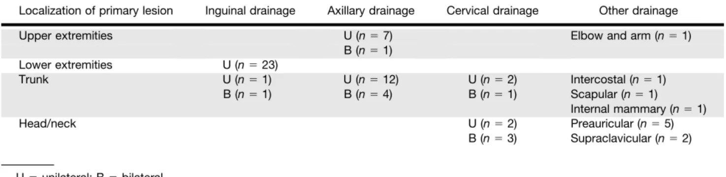
concor-FIGURE 2. Discordance between first (A– C) and second (D–F) lymphoscintigraphy studies of 53-y-old woman with melanoma on central frontoparietal area. First study showed SLNs bilaterally in cervical area. Second study, performed 2 wk later, showed similar drainage in left cervical area but absence of SLN uptake in right cervical location. Delayed images were performed (#4 h) with same results.
FIGURE 3. Concordance between first (A– C) and second (D–F) lymphoscintigraphy studies of 34-y-old woman with central mel-anoma on head. Second study was per-formed 3 wk after first study. Despite this location, clear drainage is observed in right cervical area only. Both studies show similar lymphatic pathway and number of nodes, with little difference in uptake.
dance could be influenced by the exact placement of the radiotracer injections at the same locations.
In-transit SLNs were observed in 5 patients (7% of the group) and were exactly the same in both studies. The primary tumors were on the upper limb, lower limb, and trunk (3 cases). Thus, a strong point in favor of lymphoscintigraphy is that occasionally aberrant drainage can be reliably identified (16,17). On the other hand, a hot spot does not always repre-sent a lymph node but can be caused by accidental spillage from the injection site or a drop of radiotracer, as well as the well-known occurrence of lymphatic lakes. Following the pro-cedure meticulously during lymphoscintigraphy is mandatory for best results (16,18,19).
The finding that lymphoscintigraphy is not always re-producible may be explained by small differences in injection technique or by a variation in tracer particle composition (although we always use the same radiotracer). Also, patient-related factors such as previous exertion, body hydration, and
variation in the hydrostatic pressure of blood may play a role. Finally, the time interval after excision of the melanoma may be important because granulation tissue is gradually replaced by more dense and compact fibrous tissue in the process of wound healing (5,13). Some patients had their first scinti-graphic study a few weeks after the primary lesion had been excised, and it is conceivable that such recent interventions influence to some extent the lymphatic drainage of the area concerned. In the 2 previously mentioned reproducibility stud-ies, the time interval before reinjection varied from 2 d to 4 wk, and similar or different injection site–to–tumor distances for both lymphoscintigraphy examinations were used.
Another important aspect of this study concerns the number of SLNs visualized during both studies, as well as the number of nodes surgically excised and their concordance. We found excellent concordance between the 2 mapping studies (95%), with similar numbers of visualized SLNs (171 vs. 173). A few slight differences were observed; in the second study 3 patients had a lymph node that had been missed in the first study, whereas 2 patients had a lymph node missed in their second study. The mean number of SLNs was almost identical in both studies (2.51 vs. 2.54). During surgery, 177 SLNs were harvested. Although in most patients the site and number of SLNs seen on lymphoscintigraphy studies did not vary between the 2 studies, we found that some SLNs were brighter or fainter on one study versus the other. Although more lymphatic nodes were excised during surgery than were visualized on lymphoscintigraphy, none of those extra nodes was metastatic. This final result could be in accordance with Rettenbacher et al., who found additional SLNs by extending the injection distance to the scar, but none of the extra visualized SLNs showed metastatic involvement. Thus, widening the injection distance from the site of the primary tumor may increase the ambiguous zone of drainage, cross a lymphatic watershed, or visualize additional basins not really related to drainage of the tumor site (6,20). The classic protocol for radiotracer injection is robust enough to avoid variabilities, as can be confirmed from our own results. Ap-plication of the injection far (1 cm) from the scar would be recommended only in patients with scar hypertrophy or in-flammation after excisional biopsy or in patients with no visualization after standard injection technique. On the other hand, the intraoperative use of both blue dye (or fluorescent tracers) and a g-probe is of crucial importance to compensate for the limitations of lymphoscintigraphy (21).
Finally, in our program a nuclear medicine physician in training performs more than 50 lymphoscintigraphic procedures per year for SLN biopsy in melanoma patients. The results of this study demonstrate that a well-trained learning curve in this procedure offers suitable reproducibility in such a technique. CONCLUSION
A strict and meticulous procedure during injection and imaging acquisition offers excellent reproducibility in lym-phatic mapping even when performed by different teams. However, the classic protocol of radiotracer injection is
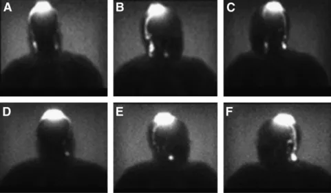
FIGURE 4. Concordance between first (A–D) and second (E–H) lymphoscintigraphy studies of 39-y-old woman with melanoma on dorsum of right hand. Second study was performed 3 wk after first study. Even with this unusual drainage to arm, reproducibility was achieved in both lymphatic pathways (in-transit nodes and axillary nodes).
reproducible and reliable enough to guarantee SLN identifi-cation, although a slight variation in isolated cases (especially when primary lesions are located on the trunk or on the head and neck regions) is inevitable.
DISCLOSURE STATEMENT
The costs of publication of this article were defrayed in part by the payment of page charges. Therefore, and solely to indicate this fact, this article is hereby marked “advertisement” in accordance with 18 USC section 1734.
ACKNOWLEDGMENTS
This work has been supported in part by RTICC RD06/ 0020/0038 and AGAUR 2009 SGR 1049 grants. No other potential conflict of interest relevant to this article was reported. REFERENCES
1. Morton DL, Wen DR, Wong JH, et al. Technical details of intraoperative lym-phatic mapping for early stage melanoma. Arch Surg. 1992;127:392–399. 2. Mariani G, Gipponi M, Moresco L, et al. Radioguided sentinel lymph node
biopsy in malignant cutaneous melanoma. J Nucl Med. 2002;43:811–827. 3. Kitajima M, Kitagawa Y. Universal applications of sentinel node technology.
Ann Surg Oncol. 2004;11(suppl):144S–146S.
4. Nieweg OE. False-negative sentinel node biopsy. Ann Surg Oncol. 2009;16:2089– 2091.
5. Uren RF, Howman-Giles R, David KV, et al. The reproducibility in routine clinical practice of sentinel lymph node identification by pre-operative lympho-scintigraphy in patients with cutaneous melanoma. Ann Surg Oncol. 2007;14: 899–905.
6. Rettenbacher L, Koller J, Ka¨ssmann H, et al. Reproducibility of lymphoscintig-raphy in cutaneous melanoma: can we accurately detect the sentinel lymph node by expanding the tracer injection distance from the tumor site? J Nucl Med. 2001;42:424–429.
7. Mudun A, Murray DR, Herda SC, et al. Early stage melanoma: lymphoscintig-raphy reproducibility of sentinel node detection, and effectiveness of the intra-operative gamma probe. Radiology. 1996;199:171–175.
8. Kapteijn BAE, Nieweg OE, Vald´es Olmos RA, et al. Reproducibility of lympho-scintigraphy for lymphatic mapping in cutaneous melanoma. J Nucl Med. 1996;37:972–975.
9. Chakera AH, Hesse B, Burak Z, et al. EANM-EORTC general recommendations for sentinel node diagnostics in melanoma. Eur J Nucl Med Mol Imaging. 2009;36:1713–1742.
10. Thompson JF, Uren RF. Lymphatic mapping in management of patients with primary cutaneous melanoma. Lancet Oncol. 2005;6:877–885.
11. Forschner A, Eigentler TK, Pflugfelder A, et al. Melanoma staging: facts and controversies. Clin Dermatol. 2010;28:275–280.
12. Uren RF, Howman-Giles RB, Shaw HM, Thompson JF, McCarthy WH. Lym-phoscintigraphy in high-risk melanoma of the trunk: predicting draining node groups, defining lymphatic channels and locating the sentinel node. J Nucl Med. 1993;34:1435–1440.
13. Vald´es Olmos RA, Nieweg OE. Reproducibility of cutaneous lymphoscintigra-phy: same or different lymphatic routes and sentinel nodes after reinjection? J Nucl Med. 2001;42:430–431.
14. Gershenwald JE, Ross MI. Sentinel-lymph-node biopsy for cutaneous mela-noma. N Engl J Med. 2011;364:1738–1745.
15. Alazraki N, Glass EC, Castronovo F, Valdes-Olmos RA, Podoloff D. Procedure guideline for lymphoscintigraphy and the use of intraoperative gamma probe for sentinel lymph node localization in melanoma of intermediate thickness. J Nucl Med. 2002;43:1414–1418.
16. Leong SP, Morita ET, Su¨dmeyer M, et al. Heterogeneous patterns of lymphatic drainage to sentinel lymph nodes by primary melanoma from different anatomic sites. Clin Nucl Med. 2005;30:150–158.
17. Ort´ın-P´erez J, Vidal-Sicart S, Dom´enech B, Rub´ı S, Lafuente S, Pons F. In-transit sentinel lymph nodes in malignant melanoma: what is their importance? Rev Esp Med Nucl. 2008;27:424–429.
18. Uren RF, Howman-Giles R, Chung D, Thompson JF. Guidelines for lymphoscin-tigraphy and F18 FDG PET scans in melanoma. J Surg Oncol. 2011;104:405– 419.
19. Willis AI, Ridge JA. Discordant lymphatic drainage patterns revealed by serial lymphoscintigraphy in cutaneous head and neck malignancies. Head Neck. 2007;29:979–985.
20. Rossi CR, De Salvo GL, Trifiro` G, et al. The impact of lymphoscintigraphy technique on the outcome of sentinel node biopsy in 1,313 patients with cuta-neous melanoma: an Italian Multicentric Study (SOLISM-IMI). J Nucl Med. 2006;47:234–241.
21. Brouwer OR, Klop WM, Buckle T, et al. Feasibility of sentinel node biopsy in head and neck melanoma using a hybrid radioactive and fluorescent tracer. Ann Surg Oncol. 2012;19:1988–1994.