Panniculitis and vitiligo occurring during BRAF
and MEK inhibitors combination in advanced
melanoma patients: Potential predictive role
of treatment efficacy
Francesca ConsoliID1*, Ausilia Maria Manganoni2, Salvatore Grisanti1, Fausto Petrelli3, Marina Venturini2, Giovanni Rangoni1, Francesco Guarneri4, Paolo Incardona5,
William Vermi6, Pier Giacomo Calzavara Pinton2, Alfredo Berruti1
1 Department of Medical and Surgical Specialties, Radiological Sciences, and Public Health, Medical Oncology, University of Brescia at ASST-Spedali Civili, Brescia, Italy, 2 Department of Dermatology, University of Brescia at ASST-Spedali Civili, Brescia, Italy, 3 Oncology Unit, ASST Bergamo Ovest, Treviglio, Italy, 4 Department of Pharmacy, ASST-Spedali Civili, Brescia, Italy, 5 Department of Laboratory Medicine, Unit of Pathology, ASST-Spedali Civili, Brescia, Italy, 6 Department of Molecular and Translational Medicine, Section of Pathology, School of Medicine, University of Brescia at ASST-Spedali Civili, Brescia, Italy
Abstract
Panniculitis and vitiligo-like lesions have been recently identified as rare cutaneous side effects of the combination of BRAF and MEK inhibitors, a standard of care in metastatic and locally advanced BRAF V600 mutated melanoma. An immune-mediated mechanism has been advocated in the pathogenesis of these skin lesions. Herein we retrospectively reviewed our institutional experience with the aim to explore the association between the occurrence of panniculitis and vitiligo-like lesions during combination therapy with dabrafe-nib (D) and trametidabrafe-nib (T) and outcome of advanced melanoma patients. Among 52 conse-cutive BRAF V600 mutated melanoma patients submitted to DT in our center, 12 (23%) developed immune related skin lesions (IRSLs): 8 panniculitis and 4 vitiligo. Patients with IRSLs diagnosis obtained a better disease response (83% versus 25%) (p = 0.001) than their counterpart and had a longer progression free survival and overall survival. The associ-ation of IRSLs and lower risk of disease progression (HR 0.19; CI 95% 0.04–0.90; p = 0.043) was confirmed after adjusting for major prognostic factors in multivariate analysis. IRSLs might represent an easy predictive surrogate marker for treatment response and favourable outcome in melanoma patients submitted to DT combination therapy.
Introduction
The combination of BRAF and MEK inhibitors, such as dabrafenib (D) and trametinib (T), actually represents the standard of care in metastatic or locally advanced BRAF V600 mutated malignant melanomas (MM) [1,2].
The results of pooled analyses of phase II and III studies revealed that DT combination leads to a long-term benefit in terms of either progression-free survival (PFS) (48% and 38% of
a1111111111 a1111111111 a1111111111 a1111111111 a1111111111 OPEN ACCESS
Citation: Consoli F, Manganoni AM, Grisanti S,
Petrelli F, Venturini M, Rangoni G, et al. (2019) Panniculitis and vitiligo occurring during BRAF and MEK inhibitors combination in advanced melanoma patients: Potential predictive role of treatment efficacy. PLoS ONE 14(4): e0214884. https://doi.org/10.1371/journal.pone.0214884
Editor: Roger Chammas, Universidade de Sao
Paulo, BRAZIL
Received: January 6, 2019 Accepted: March 21, 2019 Published: April 2, 2019
Copyright:© 2019 Consoli et al. This is an open access article distributed under the terms of the Creative Commons Attribution License, which permits unrestricted use, distribution, and reproduction in any medium, provided the original author and source are credited.
Data Availability Statement: All relevant data are
within the manuscript.
Funding: The study was supported by patients and
families and by FIRM (Fondazione Internazionale di Ricerca in Medicina), Cremona, Italy. The funders had no role in study design, data collection and analysis, decision to publish, or preparation of the manuscript.
Competing interests: The authors declared that no
patients free from progression at 1 and 2-years respectively), or overall survival (OS) (74% and 53% of patients alive after 1 and 2-years, respectively). The survival obtained by DT was supe-rior to that of single agent BRAF inhibitor D, either in the overall population or in patient sub-sets stratified according to commonly recognized prognostic characteristics [3]. Baseline serum lactate dehydrogenase (LDH) and number of metastatic sites strongly predicts OS and PFS of DT treated patients [4] and patients with normal LDH and less than three metastatic sites are expected to have a 5-years OS rate of 51% [5]. These findings notwithstanding, the prediction of prognosis of individual melanoma patients treated with DT remains difficult and new prognostic markers are needed, in addition beside BRAF mutation, no additional predic-tive parameters of DT efficacy have been up to now identified.
Dermatologic complications, such as rash, dry skin, photosensitivity, squamo-proliferative growth, hand-foot skin reactions have been frequently observed during the administration of BRAF and MEK inhibitors [6]. More recently, two additional cutaneous side effects have been described: panniculitis and vitiligo-like lesions [7,8,9]. The pathogenesis of these cutaneous reactions is not fully elucidated, although an immune-mediated mechanism is advocated [10]. Panniculitis was interpreted as a part of a systemic non-infectious inflammatory reaction, while drug-induced vitiligo as an immune-mediated destruction of melanocytes. Panniculitis during combination therapy was reportedly rare [7,11]. Vitiligo-like lesions were observed during BRAF inhibitor mono-therapy but they were rarely reported during the combination of BRAF and MEK inhibitors [8,9,12].
Preclinical and clinical evidences provide support for an immune-modulating effect of tar-get therapies, through the enhancement of a favorable tumor microenvironment. BRAF inhi-bition could enhance melanoma antigen expression and facilitate T-cell cytotoxicity [13]. Recent data are in favour of a pre-existing tumor immunity features associated to response to target therapies and the identification of specific gene signature suggests a correlation with an immune response to treatments [14,15]. Indeed, the immune system activation could account for the long-term disease control observed in a subset of patients with the combination of dab-rafenib and trametinib [2,4,5,16].
A recently published retrospective series showed a significant association between immune adverse reactions including vitiligo, erythema nodosum, uveitis and keratitis sicca and durable response to BRAF inhibitors either administered alone or in association with MEK inhibitors [12].
In this paper, we report a case series of patients who developed immune related skin lesions (IRSLs) during DT therapy with the aim to correlate these events with disease response and patient progression-free survival (PFS).
Patients and methods
From February 2015 to September 2018, we retrospectively collected data from 52 consecutive treatment naïve patients with advanced BRAF V600 mutated metastatic melanoma, who received dabrafenib (150 mg twice daily) and trametinib (2 mg daily) combination therapy, at the Medical Oncology Unit of University of Brescia at Spedali Civili in Brescia (Italy). Patients were evaluated monthly to assess safety and efficacy of treatment and a full skin examination was performed at each visit.
The diagnosis of panniculitis or vitiligo-like lesions was documented clinically and the diag-nosis was confirmed by a dermatologist, actively involved during the diagnostic workflow. A cutaneous biopsy was performed only in some cases to confirm doubtful patterns of presenta-tion. A biochemical assessment was conducted at the time of the diagnosis of panniculitis, including complete blood count, erythrocyte sedimentation rate and antistreptolysin O (ASO) titers and C-reactive protein (CRP), to exclude alternative etiologies.
This observational study was conducted in accordance with the good clinical practice guidelines and was approved by the Independent Ethic Committee of the ASST-Spedali Civili in Brescia, Italy.
We explored the associations between baseline clinical and biochemical characteristics (such as number and type of metastatic sites, level of LDH, Performance Status ECOG) and post-baseline characteristics (such as panniculitis or vilitigo-like lesions secondary to treat-ment exposure) with the Chi-square test or the Fisher test as required. To assess the impact of clinical factors in predicting overall response rate (ORR) a binary logistic regression was used both in univariate and multivariate analysis. For survival outcomes, PFS curves stratified by single covariates were generated with the Kaplan-Meier method and the difference between curves was assessed with the log-rank test. The prognostic value of clinical parameters on PFS was evaluated with the Cox proportional hazard model in uni- and multivariate backward con-ditional analysis. All tests were two-sided and the statistical significance was set at p <0.05. Hazard ratio for PFS from the present and other published studies were pooled according to fixed or random effect model according to heterogeneity. Higgins’ I2statistic values < and
>50 were defined as having low and high heterogeneity, respectively. For all other analyses the
SPSS software version 23.0 (SPSS Inc, Chicago, IL; USA) was used. The data of the present series and the published one [12] were aggregated into a meta-analysis by RevMan 5.3 software (Cochrane Collaboration, Copenhagen, Denmark) and Comprehensive Meta-analysis Soft-ware 3.
Results
Patients’ characteristics and treatments
Demography and patient characteristics were depicted inTable 1. All patients received DT combination. Twelve patients (23%) underwent immune related skin lesions (IRSLs): 7 females and 1 male had panniculitis and 3 male and 1 female had vitiligo-like dermatitis.
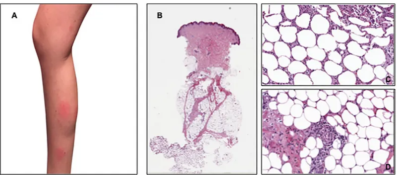
The median time to first occurrence of panniculitis was 8 months (range 1–16). This condi-tion appeared during the first month of treatment in 2 cases, while a late onset (i.e. after at least 7 months from the beginning of therapy) was observed in 6 patients. Legs were involved in all cases (Fig 1A), while in three cases, the disease also appeared on the arms. Skin lesions’ biopsies, performed in three patients, documented the presence of lobular or septal panniculi-tis without vasculipanniculi-tis (Fig 1B,Fig 1C,Fig 1D).
The eruptions had a self-limiting control and typically resolved within few days or weeks, but recurrence occurred in all cases due the continuing treatment. Dermatitis was accompa-nied by systemic symptoms, such as fever and/or arthralgia, in all cases. No history of chronic concomitant diseases was documented and no additional medications were introduced. Com-plete blood count, erythrocyte sedimentation rate were normal and ASO titers resulted nega-tive, while CRP resulted increased during the concomitance with systemic symptoms. Patients were treated symptomatically with non-steroidal anti-inflammatory drugs (NSAID), in associ-ation with topical or systemic steroids with good symptomatic clinical benefit. No treatment discontinuation was required due to IRSLs onset.
The median time to first onset of vitiligo-like lesions was 16 months (range 6–40 months). Vitiligo-like lesions had an atypical and patchy distribution of the depigmented macules, they occurred initially on the trunk in all patients. The depigmented areas progressed during time, involving head and upper limbs in two patients and also lower limbs in one of them. Confocal microscopy evaluation of vitiligo areas showed the loss of dermal papillary ring brightness and some «half-rings» structures with «scalloped border-like» features and a homogeneous reduc-tion of the normal brightly refractile papillary rings at dermo-epidermal juncreduc-tion.
Patients destined to develop IRSLs had at baseline conditions lower tumor burden (less than three metastatic sites) and normal level of LDH in comparison to their counterpart (p = 0.007 and p = 0.024 respectively, at Fisher’s two tailed test).
Relationship between the occurrence of IRSLs and progression-free
survival (PFS)
At univariate analysis, variables that were found to be significantly associated to PFS were LDH (normal versus elevated) (HR 0.16; Confidence Interval [95%CI] 0.07–0.38, p<0.0005), IRSLs (presence versus absence) (HR 0.13; CI 95% 0.03–0.56, p = 0.006), number of metastatic sites (< 3 versus � 3) (HR 0.36; CI 95% 0.17–0.78; p = 0.009) and M stage (M1a and M1b ver-sus M1c according to American Joint Committee Cancer system 7thedition) (HR 0.33; CI 95% 0.13–0.82, p = 0.017) (Table 2).
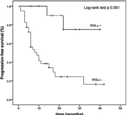
At the last follow-up examination, 2 out of 12 patients (17%) experiencing IRSLs underwent disease progression after DT therapy and all of them were alive. The corresponding propor-tions of patients who did not develop IRSLs were 27 out 40 with disease progression (67%) and 20 out of 40 alive (50%), respectively. The Kaplan Meier curves showed that the median PFS was not reached versus 9 months (95%CI 4.64–13.35 months) (p = 0.001) and the median OS was not reached versus 13 months (p = 0.002) in patients experiencing IRSLs or not, respec-tively (Figs2and3).
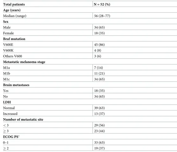
Table 1. Baseline characteristics and demographics of the entire population of fifty-two patients.
Total patients N = 52 (%) Age (years) Median (range) 56 (28–77) Sex Male 34 (65) Female 18 (35) Braf mutation V600E 45 (86) V600K 4 (8) Others V600 3 (6)
Metastatic melanoma stage
M1a 7 (14) M1b 11 (21) M1c 34 (65) Brain metastases Yes 18 (35) No 34 (65) LDH Normal 39 (63) Increased 13 (37)
Number of metastatic site
< 3 29 (56)
� 3 23 (44)
ECOG PS�
0–1 33 (63)
� 2 19 (37)
�ECOG PS = Eastern Cooperative Oncology Group Performance Status.
At multivariate analysis, normal LDH value at baseline and the occurrence of IRSLs were the only 2 variables independently associated with a lower risk of disease progression (p = 0.010 and p = 0.043, respectively) (Table 2).
Since there were no deaths in the IRSLs group, the univariate and multivariate Cox analysis for overall survival was not performed.
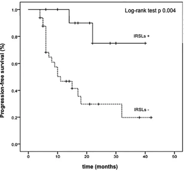
The longer PFS in the subgroup of patients experiencing IRSLs maintained the statistical significance even after introducing a landmark of 3 months to prevent the bias of early pro-gression (median PFS not reached versus 11 months in the patient subgroup with or without IRSLs, respectively) (p = 0.004) (Fig 4).
Fig 1. Clinical presentation of the skin eruptions and histopathological findings in one patient. (A) shows the clinical lesions at the left leg. (B and C) show
histological findings consistent with neutrophilic lobular panniculitis, without vasculitis (D). https://doi.org/10.1371/journal.pone.0214884.g001
Table 2. Univariate e multivariate analysis of factors associated to progression-free survival (PFS) in BRAF mutated melanoma patients treated with dabrafenib and trametinib.
Covariate Effect tested PFS (months) Univariate analysis Multivariate analysis
PFS PFS Median 95% CI p HR 95% CI P HR 95% CI P Metastatic sites < 3 32 17.61–46.38 0.005 0.36 0.17–0.78 0.009 0.74 0.32–1.70 0.482 � 3 8 3.51–12.48 M stage M1a, M1b 32 12.64–51.36 0.010 0.33 0.13–0.82 0.017 0.53 0.18–1.56 0.256 M1c 8 5–11 LDH Normal 22 8.93–35.07 <0.0005 0.16 0.07–0.38 <0.0005 0.30 0.12–0.75 0.010 Elevated 5 2.70–7.29 IRSLs IRSL+ n.r. 0.001 0.13 0.03–0.56 0.006 0.19 0.04–0.90 0.043 IRSL- 9 4.64–13.35
n.r. not reached; PFS Progression-free survival; HR hazard ratio; CI confidence interval; IRSLs+ presence of immune related skin lesions; IRSLs- absence of immune related skin lesions
Fig 2. Kaplan Meier estimates of progression-free survival (PFS) in patients with (IRSLs+) or without immune relate skin lesions (IRSLs-).
https://doi.org/10.1371/journal.pone.0214884.g002
Fig 3. Kaplan Meier estimates of overall survival (OS) in patients with (IRSLs+) or without immune relate skin lesions (IRSLs-).
Relationship between the occurrence of IRSLs and disease response to therapy
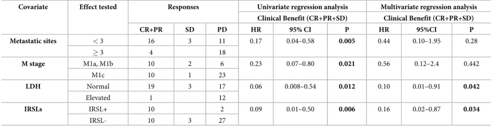
In the present series DT combination confirmed to be an active scheme since, according to RECIST criteria version 1.1, 8 patients (15%) obtained a complete response (CR), 12 patients (23%) a partial response (PR), 3 patients (6%) disease stabilization while 29 patients (56%) pro-gressed. The overall response rate (CR+PR) was 38%. Ten out of 12 patients (83%) developing IRSLs attained a disease response to DT treatment; the corresponding response rate in patients without IRSLs was 25% (10 out 40 patients). This difference in response rate attained the statis-tical significance (Chi-square test, p = 0.001). No patient destined to develop IRSLs had a pri-marily resistant disease to DT. Two patients experienced disease progression after an initial response: one with panniculitis discontinued treatment after complete remission and pro-gressed after 9 months; the second, with vitiligo-like lesions, had cerebral disease progression after a partial response lasting 17 months. Both of them were crossed to an anti-PD1 therapy and were alive at the last follow-up. The multivariate logistic regression analysis revealed that LDH level and IRSLs occurrence were the only significant independent predictors of clinical response (Table 3).Fig 4. Landmark analysis of progression-free survival (PFS) at 3 months in patients with (IRSL+) or without immune related skin lesions (IRSL-).
Pooled analysis of immune-related adverse events (irAEs) during BRAF
target therapies and melanoma patients’ outcome
Our data and those published by Ben-Betzalel et al [12] were evaluated in a pooled analysis. As depicted inFig 5, the pooled HR of the 2 studies (HR 0.53, CI 95% 0.29–0.98, p = 0.04) was sig-nificantly in favour of the association of immune related adverse events (irAEs) onset with a lower risk of progression.
Discussion
The use of combination regimens with BRAF and MEK inhibitors has contributed to modify the prognosis of patients with BRAF V600 mutated melanomas and preclinical and clinical evidences actually available, suggest that the long term efficacy of these therapies could be attributed in part to an immune-modulating effect [16]. Prognostic parameters assessed at baseline conditions, such as tumor burden, levels of LDH and Performance Status have been repeatedly found to correlate with the disease response and patient prognosis [3,4,5].
In this report, we performed a retrospective analysis on 52 consecutive patients who were homogeneously treated with BRAF and MEK inhibitors, as current standard treatment in BRAF mutant metastatic disease. We provided explorative information on whether immune related skin lesions, occurring during combined target therapy, could be associated with a durable response of melanoma lesions and improve the patient outcome. Interestingly, patients experiencing IRSLs obtained a consistently higher response rate than patients who did
Table 3. Univariate and multivariate regression analysis of factors associated to response to target therapy.
Covariate Effect tested Responses Univariate regression analysis Multivariate regression analysis Clinical Benefit (CR+PR+SD) Clinical Benefit (CR+PR+SD)
CR+PR SD PD HR 95% CI P HR 95%CI P Metastatic sites < 3 16 3 11 0.17 0.04–0.58 0.005 0.44 0.10–1.95 0.28 � 3 4 18 M stage M1a, M1b 10 2 6 0.23 0.07–0.80 0.021 0.56 0.12–2.4 0.442 M1c 10 1 23 LDH Normal 19 3 17 0.06 0.008–0.54 0.012 0.10 0.01–0.91 0.042 Elevated 1 12 IRSLs IRSL+ 10 2 0.09 0.01–0.50 0.006 0.16 0.02–0.87 0.034 IRSL- 10 3 27
CR complete response; PR partial response; SD stable disease; PD progressive disease; HR hazard ratio; CI confidence interval; IRSLs+ presence of immune related skin lesions; IRSL- absence of immune related skin lesions
https://doi.org/10.1371/journal.pone.0214884.t003
Fig 5. Meta-analysis of effect of immune related adverse effect during target therapy on progression-free survival (PFS).�Calculated Hazard Ratio and Confidence interval (CI) from the survival curves provided in the original report of Ben-Betzalel et al [12]. irAEs, immune related adverse events.
not (83% versus 25%). Moreover PFS significantly favored patients with IRSLs, after adjusting for major prognostic factors (HR 0.19, p = 0.043).
Our data are consistent to those recently published by Ben-Betzalel et al [12] who explored the role of immune related adverse events (irAEs), as clinical predictor of response to treat-ment in 78 patients submitted to BRAF targeting therapies. They defined as immune related events vitiligo, erythema nodosum, uveitis and keratitis sicca and the median time to occur-rence of irAEs from the therapy start was 6.3 months. The results of this retrospective analysis revealed an association between irAEs and PFS after adjustment for LDH and number of met-astatic sites. The pooled analysis of our paper to that of Ben-Betzalel et al confirmed the PFS advantage associated with the occurrence of IRSLs.
The patient population of the Ben-Betzalel et al study, however, was different from that pre-sented in this paper, since it consisted mainly of patients treated with BRAF inhibitor mono-therapy, that was adopted in the past when the combination treatment was not the standard of care. Moreover, the frequency of erythema-nodosum like lesions in this paper (defined as red tender cutaneous nodules), that are compatible with the diagnosis of panniculitis, was signifi-cantly lower than that observed in our patients. In addition, the reported ocular side effects were not observed in our population. Panniculits is notoriously a short lived dermatitis, so the diagnosis may escape in the absence of repeated careful skin examinations by expert dermatol-ogists during therapy. The greater proportion of panniculitis in our patients is probably due to the strict integration between oncologists and dermatologists both being involved at every time visit in each DT treated patients. Moreover, we noticed a rapid involution of individual painful inflammatory nodules and plaque without scarring or identifying the presence of a contusion appearance.
The correlation between immune-related side effects and both disease response and patient prognosis suggests a synergic interplay between the cytotoxic effect of BRAF and MEK inhibi-tors combination and immune system activation. Molecular target therapy could counteract the immunosuppressive effect of oncogenic BRAF signaling, leading to beneficial local and sys-temic immune-modulating effects [17]. Along this line, irAEs or IRSLs could be considered an
in-vivo manifestation of immune system activation and the late onset of these side effects
underlined the latency of immune system activation. Noteworthy, in this study panniculitis was detected earlier than vitiligo and this underlines the importance of the careful diagnosis this skin abnormality.
The retrospective nature, the limited number of patients included and the relatively short follow-up are the most important limitations of this study. Our data need confirmation in a larger patient series recruited prospectively.
In conclusions, the identification of surrogate parameters of efficacy of modern therapies in advanced melanoma is of paramount importance. The immune related skin lesions, panniculi-tis and vitiligo, occurring during treatment with BRAF and MEK inhibitors, are easily detect-able and contribute to identify a patient subset destined to obtain a long-term clinical benefit from the therapy. The surrogacy role of these parameters deserves validation.
Acknowledgments
The study was supported by patients and families, by FIRM (Fondazione Internazionale di Ricerca in Medicina), Cremona, Italy; by ‘‘gli Amici di Carlo” in memory of Carlo Ridon and ‘‘gli Amici di Andrea” in memory of Andrea Gadeschi.
Author Contributions
Data curation: Francesca Consoli, Salvatore Grisanti, Fausto Petrelli, Marina Venturini, Paolo Incardona, William Vermi, Alfredo Berruti.
Formal analysis: Francesca Consoli, Salvatore Grisanti, Fausto Petrelli, Alfredo Berruti. Investigation: Marina Venturini, Giovanni Rangoni, Paolo Incardona, William Vermi. Methodology: Francesca Consoli, Salvatore Grisanti, Fausto Petrelli, Francesco Guarneri,
Wil-liam Vermi, Alfredo Berruti.
Project administration: Francesca Consoli, Giovanni Rangoni. Software: Francesca Consoli, Fausto Petrelli.
Supervision: Francesca Consoli, Ausilia Maria Manganoni, Salvatore Grisanti, Paolo Incar-dona, Pier Giacomo Calzavara Pinton, Alfredo Berruti.
Validation: Francesca Consoli, Ausilia Maria Manganoni, Paolo Incardona, William Vermi, Pier Giacomo Calzavara Pinton, Alfredo Berruti.
Visualization: Francesca Consoli, Alfredo Berruti.
Writing – original draft: Francesca Consoli, Ausilia Maria Manganoni, Salvatore Grisanti, Alfredo Berruti.
Writing – review & editing: Ausilia Maria Manganoni, Salvatore Grisanti, Pier Giacomo Cal-zavara Pinton, Alfredo Berruti.
References
1. Long GV, Weber JS, Infante JR, Kim KB, Daun A, Gonzales R et al. Overall survival and durable responses in patients with BRAF V600-mutant metastatic melanoma receiving dabrafenib combined with trametinib. J Clin Oncol 2016; 34(8): 871–878.https://doi.org/10.1200/JCO.2015.62.9345PMID: 26811525
2. Long GV, Flaherty KT, Stroyakovskiy K, Gogas H, Levchenko E, de Braud F et al. Dabrafenib plus tra-metinib versus dabrafenib monotherapy in patients with metastatic BRAF V600E/ K-mutant melanoma: long-term survival and safety analysis of a phase 3 study. Annals Oncol 2017; 28: 1631–39
3. Long GV, Grob JJ, Nathan P, Ribas A, Robert C, Schadendorf D et al. Factors predictive of response, disease progression, and overall survival after dabrafenib and trametinib combination treatment: a pooled analysis of individual patient data from randomised trials. Lancet Oncol 2016; 17: 1743–54 https://doi.org/10.1016/S1470-2045(16)30578-2PMID:27864013
4. Schadendorf D, Long GV, Stroyakovskiy K, Karaszewska B, Hauschild A, Levchenko E et al. Three year pooled analysis of factors associated with clinical outcomes across dabrafenib and trametinib com-bination therapy phase 3 randomised trials. Eur J Cancer 2017; 82: 45–55https://doi.org/10.1016/j. ejca.2017.05.033PMID:28648698
5. Long GV, Eroglu Z, Infante J, Patel S, Daud A, Johnson DB, et al. Long-Term Outcomes in Patients With BRAF V600–Mutant Metastatic Melanoma Who Received Dabrafenib Combined With Trametinib JCO 2018; 36 (7): 667–673
6. De Golian E, Kwong BY, Swetter SM, Pugliese SB. Cutaneous Complications of Targeted Melanoma Therapy. Curr. Treat. Options in Oncol. 2016; 17 (11): 57
7. Mossner R, Zimmer L, Berking C, Hoeller C, Loquai C, Richtig E et al. Erythema nodosum-like lesions during BRAF inhibitor therapy: Report on 16 new cases and review of the literature. JEADV 2015; 29: 1797–1806https://doi.org/10.1111/jdv.13039PMID:25752368
8. Ruiz-Villaverde R, Sa´nchez-Cano D. Can Vemurafenib Induce Vitiligo in Metastatic Melanoma Patients? Balkan Med J 2016, 33(1): 115–116
9. Nasca MR, Lacarrubba F, Ferrau` F, Micali G. Vitiligo of the Face in a Patient Treated With Vemurafenib for Metastatic Melanoma. J Drugs Dermatol. 2016 Jun 1; 15 (6): 766–8. PMID:27272087
10. Manganoni AM, Farfaglia R, Sereni E, Farisoglio C, Pavoni L, Calzavara-Pinton PG et al. Melanoma of unknown primary with nodal metastases, presenting with vitiligo-like depigmentation. G Ital Dermatol Venereol. 2012; 147(2):210–211. PMID:22481586
11. Galliker NA, Murer C, Kamarashev J, Dummer R, Goldinger SM. Clinical observation of panniculitis in two patients with BRAF-mutated metastatic melanoma treated with a combination of a BRAF inhibitor and a MEK inhibitor. Eur J Dermatol 2015; 25(2): 177–80https://doi.org/10.1684/ejd.2014.2512PMID: 25788221
12. Ben-Betzalel G, Baruch EN, Boursi B, Steinberg-Silman Y, Asher N, Shapira-Frommer R et al. Possible immune adverse events as predictors of durable response to BRAF inhibitors in patients with BRAF V600-mutant metastatic melanoma. Eur J Cancer. 2018 Sep; 101:229–235https://doi.org/10.1016/j. ejca.2018.06.030PMID:30096703
13. Frederick DT, Piris A, Cogdill AP, Cooper ZA, Lezcano C, Ferrone CR et al BRAF inhibition is associ-ated with enhanced melanoma antigen expression and a more favorable tumor microenvironment in patients with metastatic melanoma. Clin Cancer Res 2013; 19: 1225–1231https://doi.org/10.1158/ 1078-0432.CCR-12-1630PMID:23307859
14. Yan Y, Robert C, Larkin J, Ascierto PA, Dreno B, Maio M et al. Genomic features of complete respond-ers (CR) vrespond-ersus fast progressors (PD) in patients with BRAFV600-mutated metastatic melanoma treated with cobimetinib + vemurafenib or vemurafenib alone. Annals of Oncology 2016; 27(Suppl 6): 1111O
15. Hauschild A, Larkin J, Ribas A, Dreno B, Flaherty KT, Ascierto PA et al. Modeled prognostic subgroups for survival and treatment outcome in BRAF V600-mutated metastatic melanoma pooled analysis of 4 randomized clinical trials. Jama Oncol 2018; 4 (10): 1382–88https://doi.org/10.1001/jamaoncol.2018. 2668PMID:30073321
16. Ascierto PA, Dummer R. Immunological effects of BRAF + MEK inhibition. Oncoimmunology 2018; 7 (9): e1468955https://doi.org/10.1080/2162402X.2018.1468955PMID:30228935
17. Renner K, Singer K, Koehl GE, Geissler EK, Peter K, Siska PJ et al. Metabolic hallmarks of tumor and immune cell in the tumor microenvironment. Front Immunol. 2017; 8: 248.https://doi.org/10.3389/ fimmu.2017.00248PMID:28337200