UNIVERSITÀ DEGLI STUDI DI SALERNO
Dipartimento di Farmacia
Dottorato di ricerca
in
Biologia dei Sistemi
Ciclo XII — Anno di discussione 2014
Coordinatore: Chiar.mo Prof. Antonietta Leone
Neuroimmune regulation of JCV by
Immune mediators in glial cells
settore scientifico disciplinare di afferenza
:
bio/11
Dottorando Tutore
Dott. Francesca Isabella De Simone Chiar.mo Prof. Maria Caterina
Turco
Co-tutore
It would not have been possible to write this doctoral thesis without the
help and support of the kind people around me, to only some of whom it is
possible to give particular mention here
.
I would like to express my
sincere gratitude to my PI Dr. Ilker Kudret Sariyer, my mentor at
Neuroscience Department at Temple University, Philadelphia, USA. He
has been for me not only a guide in my studies and research, but also a
friend who pushed me many times so I could improve as a scientist and as
a woman.
I would also like to thank Prof. Maria Caterina Turco, Nunziatina De
Tommasi and Vanessa Nicolin for always being there for me, whenever
times got rough, or even just for their encouragement and insightful
comments.
My sincere thanks also go to Dr. Kamel Khalili, Laura H. Carnell Professor
and Chair Department of Neuroscience for all his support, encouragement
and valuable advice.
I would like to thank my lovely family: my father and brother, for all their
constant love and support, and for never making me feel alone even if so
far away from home. Last but not least, the biggest thank you goes to my
dearest friends, old and new ones… people who have always been there for
me, and always will be…for those who have been my guardian angels and
took good care of me no matter what, and the ones who stayed up all
night helping me in my final push… home is where the heart is…
TABLE OF CONTENTS
ABSTRACT ... 1 INTRODUCTION ... 2 1.1 Polyomaviruses ... 2 1.2 JCV ... 6 1.3 Regulatory Proteins ... 9 1.4 Late Proteins ... 161.5 Transcription of JCV Genes ... 22
1.6 JCV Lifecycle ... 25
1.7 Infection and Latency of the JC virus ... 30
JCV Associated Demyelinating Diseases ... 33
2.1 Progressive Multifocal Leukoencephalopathy ... 33
2.2 Immune response against infectious agents: innate and adaptive ... 37
2.3 Inflammation ... 39
2.4 Immune system within the CNS ... 40
Material and Methods ... 42
3.1 Cell lines and culture ... 42
3.2 Plasmid constructs and viral strains ... 43
3.3 Western blots ... 43
3.4 PBMC cultures ... 44
3.5 Luciferase Reporter Assay ... 44
3.6 JCV infection ... 45
3.7 Quantitative-‐PCR (Q-‐PCR) analyses of JCV copy numbers in growth media ... 45
3.8 DpnI assay and detection of replicated-‐viral DNA by Southern blotting ... 46
3.9 Cytokine array ... 47
3.10 Autophagic imaging and flux assays ... 47
3.11 RT-‐PCR ... 48
RESULTS ... 49
4.1 Conditioned-‐media from induced PBMCs inhibits JCV infection on glial cells. ... 49
4.2 Soluble immune mediators secreted by activated PBMCs inhibit JCV early and late gene transcription in glial cells. ... 52
4.3 Conditioned media from PBMCs inhibits viral replication induced by T-‐Ag. ... 55
4.4 Conditioned-‐media from PBMCs induced by PMA and Ionomycin down-‐regulates LT-‐ Ag in a glioblastoma cell line. ... 60
4.5 Cytokine array analysis of conditioned media. ... 62
4.7 IL-‐2 and RANTES down regulate JCV Early and Late transcripts in a dose-‐dependent
manner in glial cells. ... 66
4.8 Time dependent effect of IL2 on LT-‐Ag regulation ... 69
4.9 IL2 down-‐regulates Large-‐T expression levels through autophagy ... 71
CONCLUSION ... 75
ABSTRACT
The human polyomavirus JC (JCV) is a small DNA virus responsible for the initiation of progressive multifocal leukoencephalopathy (PML), an often lethal disease of the brain characterized by lytic infection of oligodendrocytes in the central nervous system (CNS). Patients undergoing immune modulatory therapies for the treatment of autoimmune diseases such as multiple sclerosis, and individuals with an impaired-immune system, most notably AIDS patients, are in the high risk group of developing PML. Previous studies suggested that soluble immune mediators secreted from PBMCs inhibited viral genomic replication. However little is known regarding the molecular mechanism of this regulation. Here we investigated the impact of conditioned media (CM) from activated PBMCs on viral replication and gene expression by molecular virology techniques. Our data showed that viral gene expression as well as viral replication was suppressed by the CM. Further studies revealed that soluble immune mediators from PBMCs possessed a dual control on T-antigen expression at transcription and post-transcription level. These observations demonstrate a novel role of immune mediators in regulation of JCV gene expression, and provide a new avenue of research to understand molecular mechanism of viral reactivation in patients who are at risk of developing PML.
CHAPTER 1
INTRODUCTION
1.1 Polyomaviruses
Polyomaviruses were originally classified as Papovaviridae, which included both polyomaviruses and papillomaviruses. In 2000, the International Committee on the Taxonomy of Viruses split the family into Polyomaviridae and Papillomaviridae. Polyoma stems from the Greek words "poly," meaning many, and "oma," meaning tumors, as many of these viruses have been found to cause tumors in non-native host species.
There are 15 known polyomaviruses. They can infect a diverse array of species from humans to non-human primates, murinae, bovinae, and aves (Table 1). The most commonly studied polyomaviruses are the simian virus 40 (SV40), mouse polyoma, and human polyomaviruses JCV and BKV. Over the past few years, new human polyomaviruses have been discovered after the screening of infected human respiratory secretions and tumor tissues. The first among these new human polyomaviruses was discovered by sequencing DNA in respiratory secretions and analyzing its sequences using GenBank. The analysis results suggested that the discovered DNA molecule presented a circular structure homologous to polyomaviruses. Furthermore, its
genes were found to be closely related to the early genes of polyomaviruses, such as JCV, BKV and SV40, while the late genes were found to be quite divergent.
This first new human polyomavirus has been named "KIPyV" (or "KI") after its discovery at the Karolinska Institute. Its infectivity and effect on humans has not been characterized.
Simultaneous to the KI discovery, another polyomavirus was identified in patients with respiratory disorders. This virus was isolated using patient nasal secretions and it was named “WU” because it was discovered in a research lab at Washington University. WU viral genes were found to be similar to those of SV40, BKV and JCV, although a low homology, ranging between 30% and 40%, to these already known polyomaviruses was found. However, when WU was compared to the newly discovered KI, investigators found a much higher homology with it, about 65%. Interestingly, the WU virus is ubiquitous across human races and populations. In fact it has been isolated in most continents and in patients whose ages range between 3 and 53 years old. Similar to the KI virus, WU does not seem to be contagious or capable of replicating in respiratory cells. In some studies, it has been hypothesized that WU and KI viruses belong to a separate branch of the human polyomavirus family and that they share common characteristics with murine or simian families.
The third newly discovered human polyomavirus has been called Merkel Cell Polyomavirus (MCP), because it was isolated from the analysis of Merkel Cell Carcinoma (MCC). In addition, MCP has been hypothesized to be the cause of the tumorigenesis seen in MCC. MCC is an aggressive form of skin cancer, commonly found in elderly people and immunodepressed patients. As this cancer’s phenotype was similar to that of Kaposis’ sarcoma (tumors caused by herpesviruses); MCC viral components were investigated. MCC samples were
analyzed using a digital transcriptome subtraction approach, which is a technique developed to identify foreign transcripts using cDNA-sequencing data. When this technique was used on MCC, it was found that MCC had a genome with homology to other polyomaviruses and that its genome was integrated into the tumor DNA. Further, there were sequences resembling large-T, the viral capsid proteins and the viral origin.
H o st Virus Characteristics
Human JC virus (JCV)
•
Infects kidney epithelium in healthy patients•
Infects oligodendrocytes in immuno- compromised patients, causing PML BK virus (BKV)•
Infects kidney epithelium in healthy patients•
Causes PVAN in transplant recipientsWU
•
Found in patients with respiratory syndromesK I Py V
•
Found in respiratory secretions Merkel Cell polyomavirus•
Found in Merkel cell tumorsMonkey simian virus 40 (SV40)
•
Naturally occurring in kidneys of macaques•
Causes PML-like illness in immuno- compromised macaquessimian agent 12 (SA12)
•
Found naturally in baboonslymphotrophicpapovavirus (LPV)
•
Found in lymphoblasts of African green monkeysCattle bovine polyoma virus
•
Common in cattle Rabbit rabbit kidney vacuolating virus•
Found in wild rabbitsMouse mousepolyoma virus
•
Naturally occurring in kidneys of mice K virus•
Naturally occurring in lung epithelium of mice Hamster hamsterpapovavirus•
Found to produce tumors in hamstersRat ratpolyomavirus
•
Found in parotid gland of athymic rat Parakeet Budgerigar Fledgling Disease virus•
Causes fatal illness in avian species(BFDV)
Table 1:Polyomaviridae. All 15 known polyomavirus family members and their
associated diseases.(Adapted from Fields Virology, Fifth Edition Knipe and Howley, 2007) Legend. PML: Progressive Multifocal Leukoencephalopathy
PVAN: Polyomavirus Associated Nephropathy
1.2 JCV
JC virus (JCV) is a member of Polyiomaviridae family, characterized by icosahedral capsids, circular and double-stranded DNA. Polyomaviruses (PyVs) are renowned for their ability to infect a very broad spectrum of species, including humans, other primates, rabbits, rodents and birds. (M.J. Imperiale 2001). BL Padgett discovered JCV in brain tissue of a patient (John Cunningham) while treating his Hodgkin’s lymphoma. Eventually the patient died of progressive multifocal leukoencephalopathy (PML), a lytic infection of the myelin-producing oligodendrocytes in the Central Nervous System (CNS) (Padgett et al., 1971).
JC viral genome is characterized by a specific bipartite organization that is composed of two regions, called early and late transcription units, which, despite being similar in size, are transcribed in opposite directions. A common hypervariable non-coding control region (NCCR), also called a Regulatory Region (RR), divides them. This region contains the origin of replication (ORI) as well as promoter and enhancer elements, as shown in Figure1. (Frisque et al., 1984).
The early region starts transcription before DNA replication begins. This early transcription unit is able to encode for early regulatory proteins, such as small-t, Large-T, T’135 and t’165. These are produced by alternative splicing of the viral early mRNA. (Frisque et al., 1984; Saribas et al., 2010).
The LT-Ag is the JCV main regulatory protein and is necessary for the viral genome replication, for the genome late promoter transactivation and the autoregulation of its own early promoter as well. (Saribas. et al., 2010). Although the small-t-Ag function not being completely clear, it appears to be responsible for regulating the viral cycle replication and, together with Large-T, it pushes the cell into S-phase of cell cycle, where all DNA viruses replicate their DNA
(Sariyer IK et al., 2008; TK et al., 2008; Frisque RJ 2001). Large-T antigen and their variants are multifunctional, interacting with both host and viral proteins and DNA (Ferenczy et al., 2012).
The late side of the viral genome is transcribed concomitant with DNA replication and it encodes for all structural capsidic proteins, VP1, VP2 and VP3, and a small regulatory protein Agno, which accumulates mostly around the perinuclear region of the infected cells, but is also found in the nucleus in a lesser extent (25-30%) (Ferenczy et al., 2012). They all result from alternatively spliced late pre-mRNA (Saribas et al., 2010).
The viral DNA is packaged with histones H2A, H2B, H3, and H4 and creates a mini-chromosome structure that is almost indistinguishable from the host's chromatin.
1.3 Regulatory Proteins
All polyomavirus T-antigens are characterized by four conserved domains: the J domain, origin-binding domain (OBD), zinc (Zn)-binding domain, and ATPase domain (Figure 2).
Figure 2: Domain structure and biological activities of SV40 large T antigen and cellular binding partners. (a) SV40
large T antigen consists of four well-folded domains [ J domain, origin-binding domain (OBD), zinc (Zn)-binding domain, and AAA+ ATPase domain; represented by blue ovals] and two large variable disordered regions (shown by curves). Boundaries of each domain are indicated by the amino acid residue numbers. The J domain binds to Hsc70 and functions as its co-chaperone. The J domain also interacts with DNA polymerase (Pol) α primase. The N-terminal disordered region immediately downstream of the J domain harbors the LXCXE motif (diamond ). This motif is critical for the interaction between the T antigen and the pRb proteins. Additional cellular targets of this region include Bub1 and Cul7. The OBD binds to the SV40 replication origin, as well as to two host proteins, replication protein A (RPA) and Nijmegen breakage syndrome 1 (Nbs1). The Zn-binding domain mediates oligomerization of T antigen. The AAA+ ATPase domain binds to and hydrolyzes ATP, which is essential for SV40 T antigen helicase to unwind its template DNA during viral DNA replication. This domain also interacts with two cellular proteins, p53 and topoisomerase I (Topo I). The C-terminal disordered region contains the host range (HR) activity and the adenovirus-helper function. The thick brown curve highlights the region critical for the HR activity. SV40 T antigen also binds the Fbw7 ubiquitin ligase through a phosphodegron motif within the HR region. The boxes next to pRb, RPA, and p53 show the crystal structures of T antigen in complex with the corresponding cellular targets. Protein Data Bank identifiers (PDB ID) are indicated.. Reprinted with permission. ( Ping An et al., 2012)
The N' terminal region of all T antigens is also known as a J domain, due to its homology to bacterial DnaJ chaperone (Srinivasan et al. 1997). Mutations in the J domain are defective for viral DNA replication in cell culture (Peden and Pipas 1992). However, the J domain is dispensable for DNA replication in vitro (Collins BS, Pipas JM. 1995).
This could be explained by considering that the chaperone activity of T antigen is required to remove an inhibitor of replication which is already present in cells but that has been eliminated from cell-free replication systems.
The J domain of SV40 also binds and stimulates Hsc70, assisting in the release of bound cell cycle regulators. Hsc70 only interacts with cell cycle regulators when it is bound by SV40 Large-T. It is known that the binding of Hsc70 to Large-T is a critical step for the viral lifecycle. In fact, when this interaction does not occur, Large-T is unable to enhance replication of viral DNA (Borowiec et al., 1990).
Moreover, the J domain allows E2F transcription factors to dissociate from retinoblastoma (Rb) proteins. The released E2F is capable of binding to DNA, stimulating the transcription of its products, and causing cell cycle progression (Wu et al. 2004). Large-T (LT) is able to interact with retinoblastoma (Rb) family members through its LXCXE motif and with p53 in its C' terminal ATPase domain (Wessel et al., 1992).
Interactions of LT with p53 and Rb allow for cell cycle progression. This is a key step in the lifecycle of polyomaviruses without this step the genome is not replicated and the capsid proteins are not produced. The dissociation of E2F requires the binding of Rb and the LT J domain activity to work in cis (Srinivasan et al., 1997).
The OBD is a sequence-specific DNA-binding domain that recognizes the sequence GAGGC. The viral ORI is centered by four of these elements and this interaction is essential for
Introduction
the initiation of viral DNA replication. Another essential mechanism for replication is the association of OBD with replication protein A (RPA), as shown in Figure 2 and Figure 3.
Figure 3: Simian virus 40 (SV40) large T antigen is the master molecule directing viral DNA replication. (a) Simplified
schematic of the initiation process of SV40 viral DNA replication. T antigen double hexamer helicase (two sets of six ovals) initiates the distortion and melting of SV40 viral origin and subsequently unwinds the double-stranded DNA (dsDNA) template bidirectionally (represented by two parallel gray lines). The unwound single-stranded DNA (ssDNA) is shown by disordered gray curves. In addition to T antigen, nine cellular factors are required to reconstitute SV40 DNA replication in vitro. The cellular proteins that interact with T antigen at this stage are RPA, DNA polymerase α primase, and topoisomerase I (Topo I). Replication protein A (RPA) is a ssDNA-binding protein necessary for unwinding the double-stranded template DNA, whose C-terminal domain is required for interaction with T antigen. DNA polymerase α primase synthesizes RNA primers (short red curves) about 11 nucleotides in size, which serve as a starting point of DNA synthesis. Topo I and II function to resolve topological problems caused by unwinding and to establish and maintain the double helical configuration of daughter dsDNA. (b) Replication elongation of SV40 DNA. SV40 T antigen helicases continue to unwind template DNA and recruit RPA, α primase, and Topo I through specific interactions. More cellular replicative factors are involved in the elongation process. Replication factor C (RFC) and proliferating cell nuclear antigen (PCNA) facilitate the switch from α primase to DNA polymerase (Pol) δ, which then extends the nascent ssDNA (blue curves) from the primer. For synthesis of the lagging strand, the α primase has to produce primers repeatedly. (c) During the termination stage of viral DNA replication, RNase H and maturation factor 1 (MF1), a 5′ to 3′ nuclease, are required to remove the primer. Finally, DNA ligase covalently closes the gaps of the newly synthesized strands and completes the replication. Reprinted with permission. ( Ping An et al., 2012)
The last two domains are characterized by Zn-binding and ATPase domains which together constitute the enzymatic core for Large-T’s DNA helicase activity. The former domain is responsible for T antigen hexamer formation, which represents the active helicase form, whereas
lead to changes in relative positioning of residues between neighboring T antigen monomers (trans-effect) rather than within the same monomer (cis-effect). The trans-effect in the context of the hexamer results in the twisting/untwisting between the two layers of the Zn-binding domains and the AAA+ ATPase domains, as well as the expansion and constriction of the hexameric chan-nel (Figure 2g), implying that these two conformational changes are coupled to melting of the
origin and continuous unwinding of helicase activity.
In Vitro Replication of SV40 Viral DNA
Replication of the SV40 DNA has been reconstituted successfully in vitro, providing important insights into our understanding of the mechanisms of eukaryotic DNA replication. Multiple cellu-lar proteins are needed to complete replication of SV40 DNA (101). The cellu-large T antigen interacts with RPA, DNA polymerase α primase, and Topo I during replication (Figure 3). The domains and motifs important for mediating these interactions have been mapped through mutagenesis and biochemical assays (4, 46, 47, 54). Recent NMR studies have provided more information on hRPA C-terminal-mediated assembly of the SV40 replisome (4) and on the docking site for αprimase on the large T antigen helicase domain (46). Protein-protein interactions between T antigen and the cellular replication factors are critical for orchestrating the multiple steps involved in synthesizing progeny viral DNA, although the underlying mechanistic details have not been completely elucidated. a Replication initiation 5' 3' 3' 5' RPA Topo I RPA RPA αprimase RPA Helicase dsDNA ssDNA αprimase αprimase αprimase Topo I RNA primer 5' 3' 3' 5' dsDNA 5' Nascent ssDNA RNase H MF1 × × × × × DNA ligase c Termination stage b Replication elongation αprimase Forward Lagging Pol δ RPA RFC PCNA Topo I Topo I Pol δ RPA Pol δ 5' 3' 3' 5' dsDNA ssDNA 5' RNA primer Nascent ssDNA αprimase Helicase Helicase Figure 3
Simian virus 40 (SV40) large T antigen is the master molecule directing viral DNA replication. (a) Simplified schematic of the initiation process of SV40 viral DNA replication. T antigen double hexamer helicase (two sets of six ovals) initiates the distortion and melting of SV40 viral origin and subsequently unwinds the double-stranded DNA (dsDNA) template bidirectionally (represented by two parallel gray
lines). The unwound single-stranded DNA (ssDNA) is shown by disordered gray curves. In addition to T antigen, nine cellular factors
are required to reconstitute SV40 DNA replication in vitro. The cellular proteins that interact with T antigen at this stage are RPA, DNA polymerase α primase, and topoisomerase I (Topo I). Replication protein A (RPA) is a ssDNA-binding protein necessary for unwinding the double-stranded template DNA, whose C-terminal domain is required for interaction with T antigen. DNA polymerase αprimase synthesizes RNA primers (short red curves) about 11 nucleotides in size, which serve as a starting point of DNA synthesis. Topo I and II function to resolve topological problems caused by unwinding and to establish and maintain the double helical configuration of daughter dsDNA. (b) Replication elongation of SV40 DNA. SV40 T antigen helicases continue to unwind template DNA and recruit RPA, α primase, and Topo I through specific interactions. More cellular replicative factors are involved in the elongation process. Replication factor C (RFC) and proliferating cell nuclear antigen (PCNA) facilitate the switch from α primase to DNA polymerase (Pol) δ, which then extends the nascent ssDNA (blue curves) from the primer. For synthesis of the lagging strand, the αprimase has to produce primers repeatedly. (c) During the termination stage of viral DNA replication, RNase H and maturation factor 1 (MF1), a 5′to 3′nuclease, are required to remove the primer. Finally, DNA ligase covalently closes the gaps of the newly
synthesized strands and completes the replication.
www.annualreviews.org• SV40 Large T Antigen 223
Annu. Rev. Microbiol. 2012.66:213-236. Downloaded from www.annualreviews.org
achieve proper helicase functionality, the synergic interaction of OBD, Zn-binding and ATPase is required.
As a consequence of these domains’ interaction with ORI, nucleotide-binding and hydrolysis conformational changes occur. Understanding such conformational changes is fundamental to shed light on the mechanisms that regulate the functions of multi protein machines. Computational studies suggest that all polyomaviruses have a partially unstructured region between J domain and OBD, where several binding motifs for cellular proteins and nuclear localization signals can be found. This region is also the target for Rb proteins.
The specific capacity of J domain alone to adopt different conformations due to its flexible nature, predicts that the J domain–Hsc70 chaperone function can be positioned to act on different T antigen–cell protein complexes. The role played by small t antigen in JCV infections has not been studied in detail yet.
It has been shown that SV40 small t is able to interact with the protein phosphatase 2A (PP2A), which is a cellular phosphatase which plays important roles in both cell growth and transformation (Valle et al., 2006).
SV40 small t is therefore capable of interacting with PP2A and inhibiting its activity.
Such inhibition has a stimulating effect on extracellular signal-regulated kinase (ERK) and mitogen-activated protein kinases (MAPK) pathways, leading to up-regulation of AP-1 transcriptional activity (Frost et al., 1994).
Protein–protein interaction studies have demonstrated that PP2A associates with agnoprotein, a JCV late viral protein highly involved in proper capsid maturation process. PP2A association to Agno causes its dephosphorylation at PKC-specific sites. Therefore Sm t-Ag by interacting with PP2A, inhibits the dephosphorylation of agnoprotein (Sariyer et al., 2008).
Finally, T' proteins were discovered in 1995 and originally thought to be degradation products of large T (Trowbridge and Frisque 1995).
All of these have the N’ terminal J domain, but their sequences are different at the C’ terminus region. Such difference is thought to change the phosphorylation status and therefore influence their interactions with the Rb family members p107 and p130 (Bollag et al., 2000 ).
It is hypothesized that T'135, T'136, and T'165 all play a very important role controlling changes in the cell cycle needed for viral replication and transcription (Bollag et al., 2006;.Prins, and Frisque 2001 ).
The T-antigens result from alternative splicing of a common pre-cursor pre-mRNA. They are classified as large T, small t, and sliced variants such as T'135, T'136, and T'165. Large-T antigen is
the major key regulatory protein, and plays a key role in deregulation of cell cycle and also in viral DNA replication. To promote all of these actions, Large-T protein is structurally composed of a variety of domains capable of interacting with cellular factors. Besides Large-T antigen, Small t and the T' proteins also have a regulatory function, but their roles have not been as fully characterized. The N' terminal region of all T antigens has been described as a J domain, due to its homology to bacterial DnaJ chaperone. This domain has been shown to stimulate the ATPase activity of Hsp70 (DnaK) and is able to functionally substitute for the bacterial DnaJ.
Additionally, human DnaJ homologues can substitute for the SV40 J domain. The J domain of SV40 binds and stimulates Hsc70, assisting in the release of bound cell cycle regulators. Hsc70 only interacts with cell cycle regulators when it is bound by SV40 Large-T. The binding of Hsc70 to Large-T is critical step for the viral lifecycle. In fact, when this interaction does not occur, Large-T is unable to enhance replication of viral DNA.
Large-T (LT) is able to interact with retinoblastoma (Rb) family members through its LXCXE motif and with p53 in its C' terminal ATPase domain (Figure 3). These physical interactions have been demonstrated by immunoprecipitation assays of virally infected cells. In addition, these contacts are required for the transforming ability of large T, demonstrated by soft agar assays. Interactions of LT with p53 and Rb allow for cell cycle progression. Rb negatively regulates the E2F transcription factor; large T breaks this association. The released E2F is competent to bind to DNA, stimulate the transcription of its products, and cause cell cycle progression. This is a key step in the lifecycle of polyomaviruses: without cell cycle progression, the genome is not replicated and the capsid proteins are not produced. The dissociation of E2F requires the binding of Rb and the LT J domain activity to work in cis.
Recently, microRNAs (miRNAs) were found during the late phase of the JCV lifecycle. PolyomavirusmiRNAs were first discovered in SV40, using an algorithm aimed at recognizing pre-miRNA in small genomes. A pre-miRNA was identified in the SV40 genome that produced a hairpin capable of being processed by RNA-induced silencing complex (RISC). This hairpin was able to produce two miRNAs targeting the early mRNA of SV40. Similar analysis has since been performed for JCV. This analysis showed JCV also contains a homologous miRNA that targets the early mRNAs. This miRNA is unique in that both cleavage products target the same early transcript. The miRNA for JCV down-regulates Large-T antigen late during the viral lifecycle. SV40 was still infectious in the absence of the pre-miRNA in vitro. However, it is hypothesized the viral miRNAs are important for downregulating Large-T to evade immune response in vivo.
The role small t plays in JCV infection has not been extensively studied. We can gain insights into its role through its known functions in SV40 infection. SV40 small t has been
shown to interact with the protein phosphatase 2A (PP2A). PP2A is a cellular phosphatase with roles in both cell growth and transformation. SV40 small t interacts with PP2A and blocks its inhibition of protein kinase C. This release of inhibition stimulates extracellular signal-regulated kinase (ERK) and mitogen-activated protein kinases (MAPK) pathways, leading to increases in NF1B gene expression.
Recently, small t in JCV has also been shown to interact with PP2A. However these studies showed this interaction blocked the effect of PP2A on the late viral protein Agno. The authors suggest this regulation of Agno is critical for proper capsid maturation. The T' proteins were discovered in 1995 and originally thought to be degradation products of Large-T. They all share the N' terminal J domain, but their sequences diverge at their C' terminus. This difference is thought to change their phosphorylation status and influence their interactions with the Rb family members p107 and p130.
All three are hypothesized to be important to tightly control the changes in the cell cycle needed for viral replication and transcription.
1.4 Late Proteins
The late region contains agno, Vp1, 2, and 3. The JC virus capsid is composed of V antigens (VAg). Specifically, the capsid consists of 360 molecules of the major coating protein Vp1 arranged in 72 pentameters, which create an icosahedral shape (Yan et al., 1996). Either one of the two minor coating protein, Vp2 or Vp3, lies in the center of each pentameter (Figure 3) (Chen et al.,1998). The pentameters are linked together through N’ terminal regions of Vp1 that invade the next protein, while the C’ terminal Vp1 tails bind to the adjacent pentameters, tying the viron together.
Vp1 presents a barrel structure with three large exterior loops and it constitutes the outer region of the capsid. Vp1 is capable of interacting with cellular receptors (see Figure 4).
Figure 4: Vp1 pentamer and virus capsid. (A) JCV Vp1 pentamer created using SwissModel software. The
structure was created using known SV40 Vp1 structure as a model. The molecule has a #-barrel structure with the outer, receptor interacting loops shown in yellow, purple, and red. (B) Vp1 molecules form a pentamer, each color indicates a unique monomer. The N' and C' terminal tails from each molecule are used to tie the virion together. The N' terminal tails interact within the pentamer, the C' terminal tails tie adjacent pentamers together. Stehle et al, Structure 1996, used with written permission from Cell Press. (C) Arrangement of the pentamers on the viral capsid surface. The virion has 5- and 6-fold symmetry. Stehle et al, Structure 1996, used with written permission from Cell Press. (D) Cross-section of the virion. Inside each pentamer there is a minor protein, Vp2 or Vp3. The minor proteins also interact with the viral DNA, which is complexed with histones and act as a bridge for the entire structure. Reprinted with permission Kate Manley.
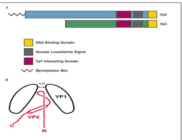
Vp3 and the terminal two-thirds of Vp2 are identical. This shared domain is composed of the Nuclear Localization Signal (NLS), the DNA binding domain and the Vp1 interacting domain (Barouch and Harrison 1994; Clever et al., 1993; Clever and Kasamatsu 1991; Gharakhanian and Kasamatsu 1990).
Vp2 N’ terminus can also be modified by a myristoylation moiety (Figure5). This is a process by which a fatty acid is co-translationally added to a protein. Myrostoyl proteins can either be cytoplasmatic or membrane-associated. After the methionine is removed and an N’ terminal glycine residue is recognized, the myristoyl group is transfected by the enzyme N-myristoyltansferase (NMT). In order to anchor a protein within a membrane, the myristoylation process needs a basic region adjacent to the insertion point or an additional anchor such as a palmitylation. A series of modifications can also modulate the above-mentioned membrane association. For instance, if a membrane-associated protein becomes phosphorylated, the negative charge will repel it out of the membrane. Conformational changes can also influence the myristoylation site exposure, for example, it can be captured in a hydrophobic pocket until a stimulus exposes it. Viral proteins are fundamental for viral uncoating and for viral release from membrane-bound compartments.
In a previous study (Gharakhanian et al., 2003), the role of minor coat proteins in SV40 was assessed and it was found that Vp2 was unessential while Vp3 was necessary for infection. These studies also suggested that the importance of Vp3 lay in its ability to activate poly (ADP-Ribose) polymerase (PARP). It is because of this over-activation of PARP that intracellular ATP seems to be depleted. This, in turn, causes cellular necrosis, releasing the virus (Gordon-Shaag et al., 2003).
In recent work, the importance of both these minor proteins has been demonstrated for SV40 infection. In addition, these proteins’ ability to lyse bacteria could represent a further tool that the virus could benefit from in order to release itself from the cell (Daniels et al., 2006).
Recently, a new minor protein Vp4 has been identified. Vp4 is not present in the virion, but it is found in cellular lysates during late time points of infection. Some investigators have
hypothesized that Vp4 is a lytic factor produced to complete the viral lifecycle (Daniels et al., 2007).
Figure 5: Minor protein domains. (A) Vp3 is identical to two-thirds of Vp2. This shared region is comprised of the DNA
binding domains, the nuclear localization signal, and the Vp1 interacting domain. Vp2 is modified N' terminally with a myristoylation moiety. (B) Possible orientation of Vp2/3 within the Vp1 pentamer as determined from X-ray crystallography. Chen et al, EMBO 1998, used with written permission from Nature publishing Group.
Related work on minor proteins has been carried out on mouse polyoma (mPy), with Vp3 and Vp2 myristoylation mutants being produced. In this study, it was proven that Vp2 and Vp3 were fundamental for both early and late events in the viral lifecycle. Specifically, the myristoylation site was switched with alanine, glutamate, glutamine and histidine using
site-directed mutagenesis. Although the alanine change showed a delay in early kinetics, with Vp1 production occurring later than in normal circumstances, this mutant did not show any virion stability defects. In a single round of infection, both the changes to glutamate and glutamine did not show any significant delayed kinetics. However, in the long term, these changes displayed a reduced re-infection capability, which was likely due to structure interactions with the host cells. Moreover, the glutamate substitution also generated a virion morphology modification. Finally, the histidine change generated an inability to either enter or release from the cells, similar to what happens with Vp2 and Vp3 mutants (Krauzewicz et al., 1990;Mannova et al.,2002;Sahli et al., 1993).
JCV requires the presence of both Vp2 and Vp3 minor proteins for its viral replication. In addition, it needs the myristoylation site on Vp2, as large groups are not able to prevent the loss of the myristoylation site (Gasparovic et al., 2006).
Large T Vp1 Vp2 Vp3 Agno
SV40 72% 78% 79% 75% 62%
BKV 82% 75% 72% 66% 79%
Table 2: Sequence similarity to JCV. Comparison of the sequence similarity between SV40 and BKV to JCV early and late
proteins.
During the late viral lifecycle phase, Agno-protein is produced, even though it is not packaged within virions. This phenomenon makes understanding Agno-protein’s role in JCV infection difficult. Despite recent studies having begun to shed some light on the properties of Agno-protein in JCV infection, much of what is known of this protein derives from studies carried out on Sv40.
All the late transcripts are produced in a polycistronic manner and its reading frame is the leader sequence for all the late gene products (Hay et al., 1982). However, for a long time, the fact that the leader sequence actually produced a protein product remained unknown.
The SV40 Agno-protein was discovered in the early 1980s. It is a basic small protein (~ 61 a.a.), with very short half-time life (~ 2 hours), which suggests that it is a regulatory protein (Jay et al., 1981). SV40’s basic properties give it a special affinity for DNA binding. Agno has often been associated with both replicating DNA and partially assembled virions (Jackson and Chalkley, 1981).
Interestingly, JCV exhibits growth defects if Agno-protein is removed. On the other hand, the removal of this protein does not affect the early gene production, DNA replication, or late gene transcription or translation. Virionsare still produced, but at a lower rate and they are releasedin smaller amounts (Resnick and Shenk. 1986).
Another interesting property of the Agno-protein is that it has been often characterized as localized to the cytoplasm and perinuclear space by indirect immunofluorescence (Nomura et al.,1983;Safakand Khalili 2001).
Furthermore, Agno-protein has multiple potential phosphorylation sites that induce a reduction in viral growth. It has been found to be a substrate for PKC and it is thought that variations in Agno phosphorylation also change its cellular localization (Sariyer et al., 2006).
To this point, recent studies have demonstrated the role of Agno as substrate for PP2A and that small-t proteins’ interaction with PP2A seems to regulate the Agno dephosphorylation (Sariyer et al., 2008.). Considering the highly basic nature of Agno, it has been hypothesized that changes in the phosphorylation status of Agno-protein control its DNA binding characteristic (Safak et al., 2001)
1.5 Transcription of JCV Genes
Once the viral genome is delivered to the newly infected cells, it acquires the histone H1 and resembles cellular chromatin (Major and Imperiale 2007). Once in the host nucleus, the JC virus genome serves as a template for RNA polymerase II (pol II) transcriptional machinery. The regulation of such machinery depends on the sequence of the NCCR, but also from the availability of host transcription factors (Ferenczy et al., 2012).
The NCCR is thought to be the key of cell type specificity and it is composed of well conserved regions surrounding the transcription start sites of both early and late coding regions. Additionally, it is also composed of a central region which contains many transcription factor binding sites. The NCCR early –proximal side contains pre-origin and origin of replication (ORI).
The original viral sequence isolated from a PML patient is known as Mad-1, since it was isolated at the University of Wisconsin-Madison (Ferenczy et al., 2012).
The Mad-1 NCCR is composed of two 98-bp tandem repeats, each one composed of a TATA box which can position mRNA start site (Ghosh et al., 1981) and multiple transcription factor binding sites (Frisque RJ. 1983). The NCCR Mad-1 tandem repeat is known as the “prototype” sequence and is composed of three sets of sequences, “a” (where the TATA box is), “c” and “e” respectively.
It is acknowledge that TATA boxes contained in the 98-bp tandem repeat are essentials for the transcription of the early and late viral genes (Daniel and Frisque 1993). Although the Mad-1 variant was the first variant isolated, it has been shown that many JCV isolates from PML
patients are actually missing the second TATA box which may not be required for JC replication (Martin et al., 1985).
The JC virus promoter contains multiple binding sites for transcription factors and transcriptional repressors.
The Nuclear factor for activated T-cells (NFAT) is a transcription factor required for JCV transcription. NFAT4 is activated by calcium release, presumably triggered by virus-receptor interactions. Once activated, it moves to the nucleus where it is able to interact with the JCV genome and drive transcription (Manley K, et al. 2006). Additionally, JCV has binding sites for NF1-X (Monaco MC, et al., 2001), NFkB (Ranganathan and Khalili, 1993), SP-1 ( Henson et al., 1992) and many others, who bind certain variants of the NCCR activating transcription of early genes.
On the other hand, NF1-A (Ravichandran and Major 2008), c-jun (Ravichandran et al., 2006;Kim J, et al. 2003), c-fos ( Kim J, et al. 2003) SF2/ASF (Sariyer and Khalili 2011) and others have been shown to repress early transcription levels.
The NF1 family on cellular DNA binding proteins is critical to JCV transcription and replication. Three NFI binding sites have been identified in the NCCR of JCV ( Amemiya et al., 1989).
Dimerization, DNA binding, and DNA replication domains of NF1proteins are found in the N terminus and are separable from the transcriptional activating domain (Gronostajski 2000). All NF1 genes (-A, -B, -C, -X) share homology on the N-terminus portion but differ at the C-terminus, which is responsible for transactivation and the repressive activity (Gronostajski 2000). It is known that all NF1 proteins are able to homo- and heterodimerize and are able to compete for the same binding site influencing transcription levels. This could explain why overexpression
of the NF1-X gene supports increased viral activity whereas NF1-a reduces the ability of permissive cell types to support JC virus infection (Ferenczy et al 2012).
Members of the activating protein 1 ( AP-1) family play a key role regulating the activation of JCV transcription (Amemiya et al., 1992).
NF1 binding to and activation of JCV are reduced by the presence of c-jun (Amemiya et al., 1992). This is thought to be due to the overlapping AP-1 and NF1 binding sites in the NCCR of JCV, suggesting that c-jun is able to physically block NF1-induced activation (Ferenczy et al 2012).
Interestingly both NF1 and AP-1 family members interact with Large-T antigen but in an antagonistic manner. NF1 appears to increase Large T –dependent early and late gene expression (Amemiya et al., 1989), and therefore contribute to enhanced viral replication (Ravichandran et al., 2006.)
AP-1 members c-jun and c-fos, instead, have been shown to interact with Large T and suppress its activation and, consequently, viral DNA replication (Kim J, et al. 2003).
It is well established that Large T antigen is able to facilitate binding of YB-1 to the viral lytic control element (LCE), and that YB-1 together with Large T increases the displacement of Pur-α from the viral promoter, and therefore stimulate late gene expression (Chen et al., 1995.; Chen and Khalili K 1995).
T antigen therefore promote late transcription by interacting with components of the basal transcriptional machinery, including TATA binding protein (TBP), TBP-associated factors (TAFs), and transcription factors, including Sp1 (Kim et al., 2000), but they can also function directly as a TAF (Damania et al., 1998).
1.6 JCV Lifecycle
Virus Entry, Trafficking, and Uncoating
In order to infect glial cells, JCV has to bind to specific cell surfacereceptors, penetrate the plasma membrane, then target its double-stranded DNA genome to the nucleus (Pho et al.,2000). JCV binding and entry into the cell requires both an N' linked glycoprotein with an∀ (2-6)- or∀2-3)-linked sialic acid (Liu et al.,1998) and the serotonin receptor 5-HT2A (Elphick et al., 2004; Dugan et al., 2008)
It has still not been established whether the sialic acid is on the serotonin receptor itself. Following binding to the cell surface receptors, the virus is internalized by the ligand inducible clathrin-dependent pathway (Querbes et al., 2004).
The virus is initially trafficked to early endosomes and uses a Rab-5- dependent pathway to access the caveosome, from which it traffics to the endoplasmic reticulum (ER) (Figures 6 and 7) (Querbes et al., 2006).
Figure 6: Sequential events during JCV life cycle(1) Adsorption of virus to the cell surface receptors; (2) entry by
clathrin-mediated endocytosis; (3) uncoating of virions and nuclear transport (uncoating takes place either in the either in endoplasmic reticulum or in the nucleus); (4) transcription of early coding region; (5) translation to produce early regulatory proteins, LT-Ag, Sm t-Ag and T’ proteins (T’135, T’136 and T’165); (6) import of LT-Ag into nucleus to initiate viral DNA replication and late gene activation; (7) replication of viral genome; (8) transcription of viral late genome; (9) translation of viral late transcript to produce agnoprotein and capsids (VP1, VP2 and VP3); (10) Nuclear import of capsid proteins; (11) assembly of viral progeny in the nucleus; (12) release of virions from infected cells. Agno: Agnoprotein; JCV: JC virus; LT-Ag: Large T antigen; Sm t-Ag: Small t antigen Reprinted with permission. ( Saribas et al., 2010 )
Virus trafficking is pH dependent, as demonstrated by an increase in endosomal pH causing a reduction in virus infection (Ashok and Atwood. 2003). Many viruses require low pH for one of three main reasons: viral membrane fusion, protease activation and vesicular trafficking.
Influenza has three proteins in its envelope, hemagglutinin (HA), neuraminidase (NA) and M2, the proton channel. Upon entering the endosome, the acidic pH causes HA to undergo conformational rearrangement, exposing a membrane penetrating form of the HA protein
Fig. 3. Sequential events during JCV life cycle
(1) Adsorption of virus to the cell surface receptors; (2) entry by clathrin-mediated endocytosis; (3) uncoating of virions and nuclear transport (uncoating takes place either in the either in endoplasmic reticulum or in the nucleus); (4) transcription of early coding region; (5) translation to produce early regulatory proteins, LT-Ag, Sm t-Ag and T’ proteins (T’135, T’136 and T’165); (6) import of LT-Ag into nucleus to initiate viral DNA replication and late gene activation; (7) replication of viral genome; (8) transcription of viral late genome; (9) translation of viral late transcript to produce agnoprotein and capsids (VP1, VP2 and VP3); (10) Nuclear import of capsid proteins; (11) assembly of viral progeny in the nucleus; (12) release of virions from infected cells.
Agno: Agnoprotein; JCV: JC virus; LT-Ag: Large T antigen; Sm t-Ag: Small t antigen
NIH-PA Author Manuscript
NIH-PA Author Manuscript
(Bullough et al., 1994). In the endosome, M2 will pump protons into the viral particle, thereby releasing viral-genome complexes from the envelope (Pinto et al., 1992). Ebola virus also requires acidification of the endosomes, but low pH does not allow for membrane fusion. Low pH activates cathepsins, which are endosomal cysteine proteases.
Figure 7: JC virus lifecycle. JC virus binds to cells using an ∀(2-6)- or ∀(2-3)-linked sialic acid and the serotonin receptor
5-HT2AR (1). After binding, JCV is internalized using clathrin-dependent endocytosis where it traffics to early endosomes (2 and
3). JCV requires pH at early times during infection to complete its trafficking to the caveosome and the ER (4 and 5). Uncoating is hypothesized to occur in the ER. The virus is then delivered to the cytoplasm where it can import into the nucleus using nuclear pores (6). Once inside the nucleus, the virus transcribes its early genes, replicates its genomes, and transcribes late genes. Virus assembly also takes place in the nucleus (7).
Ebola requires both cathepsin B and L to create a viral peptide which is then able to induce endosomal membrane fusion (Chandran et al., 2005; Schornberg et al., 2006). Reoviruses also require low pH and cathepsin B and L for efficient disassembly and membrane penetration. This
is confirmed by observation that when reoviruses are digested prior to infection to generate their infectious subvirion particle (ISVP), they are able to overcome the requirement for low pH and cathepsins (Baer et al., 1999; Ebert et al., 2002). One of the rate-limiting steps in the viral lifecycle is the uncoating of the viral genome and its delivery to the nucleus. Recently, a role for ER chaperones has been discovered for both SV40 and mouse polyoma. For mouse polyoma, interactions with ERp29, a protein disulfide isomerase (PDI) family member, cause conformational changes within the viral capsid (Magnuson et al., 2005).These conformational changes allow the virus to interact with lipid membranes so it can deliver its genome to the cytoplasm.
Furthermore, in vitro studies of mouse polyoma reveal Vp2 is capable of binding to and penetrating into the lipid membrane of the ER (Rainey-Barger et al., 2007). After the genome has reached the cytoplasm, it is able to import into the nucleus using the traditional nuclear pore pathway.
SV40 localizes and exposes its minor proteins in the ER (Norkin et al., 2002). It is hypothesized that upon delivery of the SV40 genome to the ER, chaperones uncoat the virus, where it becomes a candidate for the ER-associated degradation (ERAD) pathway (Schelhaas et al., 2007). The ERAD pathway then pulls the partially assembled virus into the cytoplasm. SV40 is unable to undergo this retrotranslocation when the proteasome and membrane protein Derlin-1 are inhibited, which further support the role of an ERAD pathway in SV40 infection. JCV makes its way through the cell through a series of filamentous networks. Treating cells with nocodazole, cytochalsin D and acrylamide disrupts these networks and renders JCV no longer infectious. This indicates that JCV infection requires microtubules, microfilaments and intermediate filaments during its lifecycle (Ashok and Atwood. 2003). The current model suggests actin is important
during early points of infection, either by directly interacting with virus-containing vesicles or indirectly affecting clazthrin-dependent endocytosis. Microfilaments and microtubules are used for subsequent steps in the lifecycle as the virus continues to be transported in vesicles to the caveosome and then the ER.
1.7 Infection and Latency of the JC virus
Seroepidemiological studies have indicated that more than 70% of the human population have been exposed to JCV during their childhood but exhibit no symptoms of clinical disease (Walker and Padgett, 1983). However, inimmunocompromised patients suffering from lymphoproliferative diseases, in AIDS patients or patients undergoing immunosuppressive therapy, JCV reactivates and leads to development of PML (Chang et al., 1996), a rare disease characterized by a lytic infection of oligodendrocytes in the central nervous system (CNS) that generally affects adults but rarely children (Brew B.J. et al., 2010).
Since its development in patients who were known to be seropositive long before clinical manifestation is not associated with the increase in JCV-specific IgM antibody titer, it’s possible that the establishment of PML is consequent to a reactivation of JCV from a latent state (Major E.O. et al., 1992). How the viral infections occur remains unclear, however it is known that primary infection occurs most likely in stromal or immune cells of the upper respiratory system (Berger et al. 2006). The virus then appears to be transported by infected lymphocytes to kidneys and bone marrow where it remains latent (Ferenczy et al., 2012). It is known that viral reactivation occurs outside the CNS, and that, once the reactivation is completed, it crosses the
blood-brain barrier transported by B cells and enters the brain where it replicates vigorously in oligodendrocytes, leading todemyelination (Brew B.J. et al., 2010; Saribas et al., 2010).
Several observations indicate that a vital role in controlling the virus is played by the cellular immune response which requires the collaboration of both innate and adaptive immunity: NK-cells destroy virus infected NK-cells. As for adaptive immunity, B-lymphocytes produce antibodies able to neutralize free virus in fluids, whereas T-lymphocytes can kill infected cells before the viral maturation and therefore its release, preventing cell-to-cell transmission (Koralnik I.J. 2002). Therefore, a reduction of CD4+ T cells can cause a lack of immune control of JCV and thus increase the likelihood of JCV reactivation and PML development (Bayliss J. et al., 2013). It is likely that, in healthy individuals, the immune system retains the virus in a latent state. Therefore, alterations in immune system function could promote reactivation of viral gene expression and start the lytic phase of infection (Chang et al., 1996).
Figure 8: initial JCV infection is thought to occur in tonsillar tissue after inhalation. Lymphocytes infected with JCV
carry virions to the kidney and bone marrow, which are thought to be the primary sites of viral latency. Following reactivation of JCV, the virus is thought to cross the blood–brain barrier within B cells and infect oligodendroglia. The change in JCV color from red to green indicates genetic rearrangement. Abbreviations: JCV, JC virus; PML, progressive multifocal leukoencephalopathy. Reprinted with permission. (Brew et al., 2010)
CHAPTER 2
JCV ASSOCIATED DEMYELINATING
DISEASES
2.1 Progressive Multifocal Leukoencephalopathy
Progressive Multifocal Leukoencelopathy (PML) is a fatal disease that develops from a lytic infection of the myelin-producing oligodendrocytes in the central nervous system (CNS). JC virus (JCV) has been identified to be the causative agent of PML. JCV has been shown to have a limited tropism, in fact its effects are limited to oligodendrocytes, astrocytes, B-lymphocytes, tonsils and kidney epithelial cells. It is very common in humans, studies estimate that about 70% of the human population is seropositive for JCV. It is not completely understood how most humans get infected, but it is hypothesized that the initial infection is subclinical and contracted during childhood. JCV is mostly latent, but can be reactivated in case of immunosuppression and lytically infect oligodendrocytes and cause PMC .
PML was first identified in the 1950s, but it took a decade to discover the viral origin of this disease. Considering that many of the early patients also had lymphatic leukemias or Hodgkin’s Disease, PML was thought to be related to complications of lymphoproliferative diseases. Over time, the pathology became more studied and it was found in patients with quite different conditions. It was soon discovered that all the patients had in common the fact that their immune system was impaired, thus researchers started attributing a viral nature to PML (Karl and Astrom 2001). In the 1960s, using electron micrographs of brain tissues collected from PML patients, for the first time viruses resembling papilloma were seen (Zu Rhein, 1965). At the time, human polyomaviruses had not yet been characterized. Then when better staining techniques were available, these brain tissue virions were identified as polyoma and not papilloma (Zu Rhein, 1965). Furthermore, they were found to be present in every brain tissue sample harvested from patients with PML. It was during the same period that cell culture techniques were introduced and these allowed researchers to grow and investigate Simian Virus 40, that is another polyomavirus. Simultaneously, the first primary human fetal glial cell cultures (PHFG) were introduced. In 1970s, for the first time the JC polyoma virus was identified in the brain biopsy of a patient whose name was John Cunningham. In 1971, using Cunningham’s brain sections, that were larger and with more virions than usual, these virions were isolated from brain matter and cultured in PHFG cells. This is why the virus was named after John Cunningham’s initials (JC).
PML was once thought to be a rare disease, but it has recently become more widespread because of the Acquired Immune Deficiency Syndrome (AIDS). It has been estimated that 4-6% of AIDS patients will develop PML (Major et al., 1992 ), but this is not the only group at risk of contracting PML. Others at risk are patients undergoing chemotherapy, transplant recipients and patients with Multiple Sclerosis (MS) or Crohn’s Disease who are treated with natalizumab.
Although PML can affect all the above-mentioned patient groups, 85% of cases are reported in Human immunodeficiency Virus (HIV)- positive patients.
In more detail, PML is known to cause multiple large demyelination areas, large bizarre astrocytes and nuclear inclusions in oligodendrocytes (See Figure 9). The common PML diagnostic tools are Magnetic Resonance Imaging or Cerebrospinal Fluid (CSF) Polymerase Chain Reaction (PCR). PML is also commonly known to cause lesions that are diffused and subcortical. Interestingly, lesions caused by PML are quite different from those caused by MS. These lesions are characterized by having edges that are not neat, they are not regularly shaped and tend to grow asymmetrically.
Figure 9: Histological features of PML. (A) Gross examination of JCV- induced lesions occurring at the subcortical white
matter. A coronal section of the frontal lobe of the brain from a PML patient is shown. (B) Apparent myelin loss, as result of JCV infection of oligodendrocytes, is made detectable by Luxol blue staining (40×). Demyelinated areas are visibly distinguishable as white plaque areas. (C) Hematoxilin and Eosin staining of the brain sections from a PML patient. Infected oligodendrocytes are indicated with a round dark staining of the eosinophilic inclusion bodies (arrow head). An arrow points to an infected astrocyte (400×). Reproduced with permission from (Saribas et al., 2010).
Fig. 1. Histological features of PML
(A) Gross examination of JCV- induced lesions occurring at the subcortical white matter. A coronal section of the frontal lobe of the brain from a PML patient is shown. (B) Apparent myelin loss, as result of JCV infection of oligodendrocytes, is made detectable by Luxol blue staining (40×). Demyelinated areas are visibly distinguishable as white plaque areas. (C) Hematoxilin and Eosin staining of the brain sections from a PML patient. Infected oligodendrocytes are indicated with a round dark staining of the eosinophilic inclusion bodies (arrow head). An arrow points to an infected astrocyte (400×). Reproduced with permission from (111).
NIH-PA Author Manuscript
NIH-PA Author Manuscript
PML might cause different symptoms, such as limb weakness and ataxia at first and as the disease progresses cognitive, speech and visual impairments may occur. Patients rarely survive more than one year since disease onset.
As of today, no cure has been found for Progressive Multifocal Leukoencephalopathy. All the clinical trials that have been implemented, using different drugs such as cytosincearabinoside, topotecam, cidofovir and high dose of azodothymidine (AZT), have proven not effective. They have failed to improve the symptoms and they were even toxic in some cases. The current gold standard treatment is Highly Active Anti-Retroviral Therapy (HAART) in HIV infected patients, whose goal is to alleviate the underlying immunosuppression to slow down the disease progression.
In fact, recent studies have showed how the use of HAART with a high central nervous system penetration effective score might be associated with prolonged survival of patients with HIV-related PML (Yoganathan et al., 2012).
2.2 Immune response against infectious agents: innate and adaptive
A healthy immune response against infectious organisms requires the collaboration of both innate and adaptive immunity.The first line of defense against pathogens is called the innatenon-antigen specific immunity, and is carried out by macrophages, neutrophils, the competent system and NK (natural killer) cells. Among all of these, only the activated NK cells are actually able to destroy the virus-infected cells.
As for the adaptive, or antigen-specific immunity, is mainly carried out by B and T lymphocytes where B cells produce antibodies to neutralize free virus, and T cells can kill infected cells prior viral maturation, and by doing that, limiting viral transmission (Koralnik 2002).
T cells are subdivided into CD4+ helper cells, and CD8+ cytotoxic T lymphocytes (CTLs). CD4+ cells, whose roll is mainly to stimulate macrophages, and CD8+ cells (TH1 response) or B cells (TH2 response) by producing specific cytokines are able to recognize viral epitopes presented on MHC class II molecules. Stimulated CD4+ cells will proliferate and then produce cytokines such as interferon (IFN), granulocyte-macrophage colony stimulating factor (GM-CSF), and tumor necrosis factor (TNF) in case of TH1 cells, or interleukin (IL)-4, IL-5 and IL-10 in TH2 cells.
CD8+ T cellswho are responsible for the destruction of viral-infested cells, are able to recognize viral epitopes bound to MHC class I molecules. This happens when a newly synthesized viral protein gets tagged for destruction in the cytoplasm of an infected cell and is degraded into peptides by the proteasome system (Koralnik 2002).
2.3 Inflammation
Inflammation is the cellular response to pathogen invasion that results from vascular dilation and an increased movement of immune cells into the affected area resulting in clinical appearance of swelling and reddening. Many diseases originate from or are exacerbated by the inflammatory response such as chronic inflammation resulting in asthma, infections in the limbs of patients with diabetes and advancement of certain cancers. Thymoquinone may have a therapeutic role in inflammation, cancer and diabetes. The immune response is often manifested by the release of pro-inflammatory cytokines and chemokines from cells in the innate immune system. Cytokines and chemokines can work to activate immune cells or elicit a chemoattractive response in other cells in the vicinity. Cytokines such as IL-1 and TNF-α are markers of inflammation. Chemokines and adhesion molecules including MIP-1 and sICAM-1 are also primary indicators of inflammation. Th1- and Th2-dependent immune responses result in the production of various cytokines. When Th1 cells are activated, they produce pro-inflammatory cytokines such as IL-1, IL-2, IL-12, IFN-γ, and TNF-α that stimulate macrophages, Natural Killer cells, cytotoxic T cells and movement of other cells into the affected area. Whereas when the Th2 cells are activated, they stimulate B cell proliferation and antibody production. In response to Th-2 mediated or humoral immunity, anti-inflammatory cytokines IL-4, IL-5, IL-10 and IL-13 are released. The ability of a substance to control the balance between Th1- and 3 Th2-associated releases of cytokines has been associated with both pro- and anti-inflammatory properties.
2.4 Immune system within the CNS
A range of mechanisms exists to limit immune responses in the CNS; in fact, the CNS is considered to be an immune-privileged site. The idea of an “immune privilege” comes from the presence of the blood-brain barrier (BBB) and the blood–cerebrospinal fluid barrier (BCSFB), that play a role in delaying the immune response to non-tumor foreign tissue in the CNS (Galea et al., 2007).
This delay is related to several factors. The CNS lacks conventional lymphoid drainage (Weller et al., 2010) and CNS-derived antigen may be transported to cervical lymphnodes in the fluid phase (Weller et al., 1996) or associated with dendritic cells (DC) (Karman et al., 2004). Since it contains few APCs, and neurons only express MHC under exceptional conditions, the parenchyma of the normal brain and spinal cord has poor capacity for antigen processing and presentation (Neumann et al., 1995). Moreover, lymphocytes have to be activated before they can cross the BBB (Wekerle et al., 1986; Prendergast et al. 1998), and once they arrive in the CNS the environment remains hostile to activated lymphocytes expressing FAS.
In fact, FAS ligands (FASLor CD95L) are a type-II transmembrane protein belonging to the tumor necrosis factor (TNF) family. Its binding with its receptor results in death by apoptosis (Bechmann et al., 1999; Flugel et al., 2000). Moreover, Fas ligand/receptor interactions play an important role in the regulation of the immune system and the progression of cancer.
Microglia, the innate immune cells of the CNS, further respond to inflammation by up-regulation of immune-regulatory molecules including B7-H1 (Magnus et al., 2005) and IDO (Kwidzinski et al., 2005), while neurons protect themselves by secreting TGF- Β upon contact with activated lymphocytes (Liu et al., 2006).