74
EVALUATION OF SYMPTOMS AND PREVENTION OF CANCER IN
MENOPAUSE: THE VALUE OF VULVAR EXAM
AR Palumbo
1, C Fasolino
1, G Santoro
1, V Gargano
3, M Rinaldi
3, B Arduino
3, M Belli
2, M Guida
1 1. University of Medicine and Surgery, Unit Obstetrics and Gynecology, Salerno, Italy2. LILT (Lega Italiana per la Lotta contro i Tumori), Avellino, Italy
3. Department of Obstetrics and Gynecology, A.U.O."San Giovanni di Dio e Ruggi d'Aragona", via S. Leonardo, Salerno, Italy
Corresponding Author: Maurizio Guida (e-mail: [email protected])
Abstract - Vulvar and vaginal atrophy (VVA), is a chronic medical condition experienced by
post-menopausal women, with prevalence estimated ranging from 10% to 50% [1]. VVA is characterized by a
constellation of symptoms, that may affect daily activities, sexuality, relationships, and quality of life [3].
Early recognition and effective treatment of VVA may enhance sexual health and the quality of life of
wom-en and their partners. Some vulvar conditions such as lichwom-en sclerosus are more prevalwom-ent in the postmwom-eno-
postmeno-pausal years. Lichen sclerosus has been suggested as a precursor of Vulvar squamous cell carcinoma. The
vulvar exam in post-menopausal women plays an essential role in prevention of cancer because it allows to
identify women who should undergo vulvar skin biopsy in order to early detect pre-neoplastic lesions for
early diagnosis of cancer of the vulva.
Keywords: vulvar cancer, cancer prevention, vulvar examination
I. INTRODUCTION
Vulvar and vaginal atrophy (VVA), is a chronic medical condition experienced by post-menopausal women, with prevalence estimated ranging from 10% to 50% [1]. VVA, resulting from the loss of estrogen stimulation, is characterized by thinning of the epithelial lining of the vagina and lower genitourinary tract, by loss of vaginal elasticity, by vaginal dryness and by an increase of vagi-nal Ph [2], determining a significant impact on sexual health and quality of life. VVA is characterized by a con-stellation of symptoms, such as vaginal dryness, burning and irritation, lack of lubrication, dyspareunia, dysuria, and urinary urgency that may affect daily activities, sexu-ality, relationships, and quality of life [3]. Unlike hot flushes that usually resolve over time, VVA has a chronic progressive nature throughout the menopausal transition and beyond. The presence and severity of symptoms are variable, from mild discomfort to great impairment, de-pending also on age, time and type of menopause, parity and vaginal delivery, frequency of coital activity, ciga-rette smoking and certain medical conditions [4]. The REVIVE survey was an online evaluation of post-menopausal women in the US. The recently published REVIVE (REal Women’s VIews of Treatment Options for Menopausal Vaginal ChangEs) survey, offers many insights into the impact of VVA symptoms on women’s lives. The most commonly reported VVA symptoms
among post-menopausal women were vaginal dryness (55%), dyspareunia (44%), and local irritation (37%) [5]. While the clinical picture of VVA and other conditions subsequent to menopause have been thoroughly de-scribed, there has been little quantitative research on the association of VVA with other disorders, such as the structural modifications of the vulvar skin.
Vulvar itching is the main symptom of Lichen sclerosus of the vulva, a chronic dermatologic condition char-acterized by pruritus, inflammation and tissue scar-ring, most often affecting post-menopausal women. [6] Additionally, women may report cracking or bleeding of the vulvar skin and perianal area.
Patients with lichen sclerosus especially those with squamous cell hyperplasia have an increased risk of vul-var malignancy. The increased risk of developing squa-mous cell carcinoma is approximately 5 percent in pa-tients with lichen sclerosus [7]. Accurate diagnosis is es-sential for effective treatment. The risk of progression to malignancy associated with some of these diseases dic-tates long-term surveillance [8].
This paper discusses implication of VVA symptoms on women’s lives and stresses value of vulvar exam in early identification of pre-neoplastic lesions in post-menopausal women.
II. METHODOLOGY Our study population included 44 sexually active post-menopausal women 50 to 75 years old, recruited from LILT ambulatory in Avellino, Italy. We administered the
Vulvovaginal Symptoms Questionnaire (VSQ), a previ-ous validated instrument to post-menopausal women. The first six questions of the questionnaire comprise the symptom subscale (itching, burning, dryness, dyspareunia, urinary frequency, urinary urgency). Wom-en who answered "Yes" to any of the first six symptom questions were considered to have vulvovaginal symp-toms.
Eligible participants were all Italian women in post-menopause, which is defined as the presence of amenor-rhea for at least 12 months. These women consulted an outpatient gynecological service for a routine gynecolog-ical examination. Among eligible women, only those who signed an informed consent were enrolled into the study. All women were subjected to a medical interview through which demographic information, personal and family medical history and comorbidities were recorded. Subjec-tive symptoms considered were: vaginal dryness, itching, burning, dysuria and dyspareunia.
Objective signs taken into consideration were: thinning of vaginal rugae, bleeding at slightest touch, bleeding at in-tense touch and presence of petechiae.
III. RESULTS
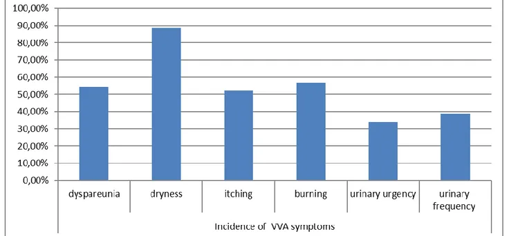
Post-menopausal women included in Our study reported at least one vulvovaginal symptom. The most common symptom reported is dryness 88.63% (n/N= 39/44), whereas the symptoms recorded less frequently are uri-nary frequency 38.63% (n/N= 17/44) and uriuri-nary urgency 34.09% (n/N=15/44). Vulvovaginal symptoms have im-portant repercussions on the sexual sphere, in fact the symptom of dyspareunia was recorded in 54.54% (n/N= 24/44) of sexually active post-menopausal women. Size-able proportions of women with vulvovaginal symptoms report itching in 52.27% (n/N=23/44) and burning 56.81% (n/N=25/44). (Graphic 1: Incidence of VVA
symptoms)
Graphic 1: Incidence of VVA symptoms
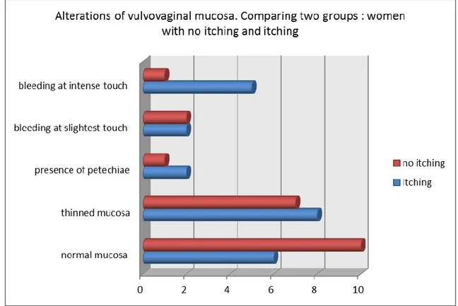
Objective examination of the external genitalia allowed to record several alterations of the vulvar skin. Alterations of the vulvovaginal mucosa were classified into five cat-egories: normal mucosa, thinned mucosa, bleeding at the slightest touch, bleeding at intense touch and presence of petechiae. (Graphic 2: Alterations of vulvovaginal
muco-sa. Comparing two groups: women with no itching and itching)
In the subgroup of post-menopausal women presenting itching 52.27% (n/N=23/44), only 26,08% (n/N=6/23) of sexually active post-menopausal women have normal mucosa, whereas 73,91% (n/N= 17/23) show alterations of the vulvar skin. (Graphic 3: Alterations of
76 Graphic 2: Alterations of vulvovaginal mucosa. Comparing two groups: women with no itching and itching
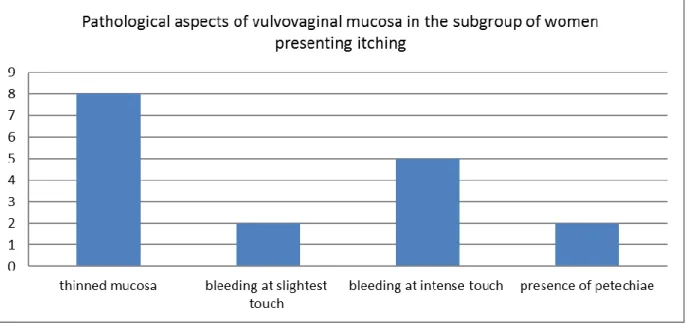
Graphic 3: Alterations of vulvovaginal mucosa in the subgroup of women presenting itching
The results of the evaluation of the external genitalia show the presence of thinned mucosa in 47,05% (n/N= 8/17), bleeding at slightest touch in 11,75% (n/N= 2/17), bleeding at intensive touch in 29,41% (n/N= 5/17) and
presence of petechiae in 11,75% (n/N= 2/17). (Graphic 4:
Pathological aspects of vulvovaginal mucosa in the
Graphic 4: Pathological aspects of vulvovaginal mucosa in the subgroup of women presenting itching
IV. DISCUSSION
Pathogenesis of VVA symptoms is reflected in the re-duction in the level of estrogenic hormones, typical phe-nomenon which occurs in the period of menopause. Estrogen receptors, alpha and beta, are expressed throughout the squamous epithelium, connective tissue and smooth muscle of the vulva, vagina, urethra, and bladder trigone and are critical for mediating numerous biochemical and physiologic functions during a woman’s reproductive years [9]. With loss of estrogen stimulation, profound changes occur within the vulvovaginal and uro-genital mucosa. In the dermal layer, collagen fibers fuse and undergo hyalinization, whereas the elastin fibers fragment. The result of these changes is an overall loss of mucosal elasticity and the vaginal epithelium thinning [10].
Estrogen-induced parakeratosis of the vulvar squamous epithelium progressively decreases after menopause and is rarely seen in older women. The vagina loses its rugae, and there is a shortening and narrowing of the vagina. The mucosa of the vagina, introitus, and labia minora be-come thin and pale. Surrounding vascularity, highly es-trogen dependent, also decreases. [11]
Vulvar disease in the post-menopausal age group is rela-tively common. Some vulvar conditions such as lichen sclerosus are more prevalent in the post-menopausal years. Often more than one condition is present at the same time [8]. Lichen sclerosus of the vulva is a chronic dermatologic condition characterized by pruri-tus, inflammation and tissue scarring [6]. The distribu-tion of skin changes may involve the perianal skin, and scarring can cause fusion of the labia minora to the labia majora and flattening of the clitoral hood, which may result in immobility [12].
Physical examination is important for differentiating lichen sclerosus from other causes of vulvar pruritus, such as lichen planus, lichen simplex chronicus, atrophic vaginitis, irritant contact dermatitis, eczema, psoriasis, vulvovaginal candidiasis and skin cancers (e.g., vulvar intraepithelial neoplasia and extra-mammary Paget disease) [13].
Patients with lichen sclerosus, especially those with squamous cell hyperplasia, have an increased risk of vul-var malignancy and should be monitored accordingly. The increased risk of developing squamous cell carcino-ma is approxicarcino-mately 5 percent in patients with lichen sclerosus. [7].
Recognition and treatment of these vulvar conditions is important for symptom relief, sexual function, prevention of progressive vulvar scarring, and to provide surveil-lance for associated vulvar cancer [14].
Vulvar cancer is a relatively rare gynecologic malignancy with an annual incidence in developed countries of ap-proximately 2 per 100,000 women. Vulvar squamous cell carcinoma (VSCC) has two etiological pathways: a high risk human papillomavirus (HPV)-dependent route, which has usual vulvar intraepithelial neoplasia (uVIN) as a precursor lesion, and an HPV-independent route, which is associated with differentiated VIN (dVIN), li-chen sclerosus, and genetic alterations, such as TP53 mu-tations [15].
Most VSCCs originate in intraepithelial lesions, named vulvar intraepithelial neoplasia (VIN), that precede the development of VSCC by a variable period of time. Epi-thelial disorders are found adjacent to VSCC in 50–70% of patients. Most of these intraepithelial changes appear to have some degree of dysplasia [16].
Lichen sclerosus (LS) has also been suggested as a pre-cursor of HPV-independent VSCC, but the mechanism of
78 carcinogenic progression from LS has not been fully
de-lineated. The association between the two entities has been established mainly because the majority of vulvar cancers will have lichen sclerosus, squamous cell hyper-plasia or differentiated VIN in the adjacent epidermis [17] and because some longitudinal cohort studies have shown that women with LS have a significantly higher risk of developing VSCC.[18]
It is clear the value of the vulvar exam to identify the presence of skin changes associated with the presence of a preneoplastic lesions.
Cancer Research UK publishes the symptoms of vulvar cancer [19]
a lasting itch pain or soreness
thickened, raised, red, white or dark patches on the skin of the vulva
an open sore or growth visible on the skin burning pain when you pass urine vaginal discharge or bleeding
a mole on the vulva that changes shape or col-our
a lump or swelling in the vulva
The presence of these alterations should be an alarm bell and must lead the gynecologist to perform a vulvar skin biopsy. Vulvar biopsy is the only method to get a histo-pathological diagnosis when there is a vulvar lesion of uncertain significance, or in the presence of persisting symptoms, such as vulvar irritation or itching [20].
V. CONCLUSION
VVA symptoms negatively affect the quality of life of menopausal women, complicating interpersonal relation-ships and married life. Early recognition and effective treatment of VVA may enhance sexual health and the quality of life of women and their partners.
Lichen sclerosus has been suggested as a precursor of HPV-independent VSCC. The etiology of lichen scle-rosus is multifactorial; therefore, patient education, be-havior modification, and regular follow-up with an expe-rienced clinician are essential to ensure effective control of patient symptoms and management of the skin condi-tion. The vulvar exam in post-menopausal women plays an essential role in the early detection of pre-neoplastic lesions. In presence of injuries such as itch, pain, raised patches and bleeding, the gynecologist must start a thera-peutic treatment. If during the follow-up of skin lesions there is a lack of response to treatment or a progression of lesions, the gynecologist must proceed with the execution of a biopsy of the vulvar skin. The recognition of vulvar lesions is important for the prevention and early diagnosis of cancer of the vulva.
REFERENCES
[1] Dennerstein L, Dudley EC, Hopper JL, Guthrie JR, Burger HG, A prospective population-based study of menopausal symptoms. Obstet Gynecol 2000 Sep; 96(3);351-8
[2] B. Mac, M.B. ride, D.J. Rhodes, L.T. Shuster, Vulvo-vaginal atrophy, Mayo Clin. Proc. 85 (2010) 87–94. [3] North American Menopause Society, The role of local vaginal estrogen fortreatment of vaginal atrophy in post-menopausal women: 2007 positionstatement of The North American Menopause Society, Menopause 14 (2007)355.
[4] Nappi RE, Lello S, Melis GB, Albani F, Polatti F, Genazzani AR. LEI (Lack of tEstosterone Impact) survey in a clinical sample with surgical menopause. Climacteric 2009; 12:533–40
[5] Susan Wysocki, Sheryl Kingsberg and Michael Krychman, Management of Vaginal Atrophy: Implica-tions from the REVIVE Survey. Clinical Medicine In-sights: Reproductive Health 2014:8
[6] Goldstein AT, Marinoff SC, Christopher K, Srodon M. Prevalence of vulvar lichen sclerosus in a general gy-necology practice. J Reprod Med. 2005;50(7):477-480 [7] Thomas RH, Ridley CM, McGibbon DH, Black MM. Anogenital lichen sclerosus in women. J R Soc Med. 1996;89(12):694-698.
[8] Olsson A, et al. Postmenopausal vulval dis-ease.Review article, Menopause Int. 2008
[9] Pettersson K, Gustafsson JA. Role of estrogen recep-tor beta in estrogen action. Annu Rev Physiol 2001: 63: 165–192.
[10] Castelo-Branco C, Cancelo MJ, Villero J, Nohales F, Julia MD. Management of post-menopausal vaginal atro-phy and atrophic vaginitis. Maturitas 2005: 52 (Suppl 1): S46–S52;
[11] Forsberg JG. A morphologist’s approach to the vagina – age-related changes and estrogen sensitivity. Maturitas 1995: 22 (Suppl): S7–S15.
[12] Oyama N, Chan I, Neill SM, et al. Development of antigen-specific ELISA for circulating autoantibodies to extracellular matrix protein 1 in lichen sclerosus. J Clin Invest. 2004;113(11):1550-1559.
[13] Neill SM. Vulvar lichen sclerosus. In: Black MM, ed. Obstetric and Gynecologic Dermatology. 2nd ed. London, UK: Mosby; 2002:137-142.
[14] Thorstensen KA, et al. Recognition and management of vulvar dermatologic conditions: lichen sclerosus, li-chen planus, and lili-chen simplex chronicus. J Midwifery Womens Health. 2012 May-Jun.
[15] Trietsch MD, et al Genetic and epigenetic changes in vulvar squamous cell carcinoma and its precursor lesions: a review of the current literature. Gynecol Oncol. 2015 Jan;136(1):143-57
[16] Marta del Pino, Leonardo Rodriguez-Carunchio & Jaume Ordi Pathways of vulvar intraepithelial neoplasia and squamous cell carcinoma. Histopathology 2013, 62, 161–175.
[17] Sturgeon SR, Brinton LA, Devesa SS, Kurman RJ. In situ and invasive vulvar cancer incidence trends (1973
to 1987). Am J Obstet Gynecol 1992;166(May (5)):1482– 5.
[18] Al-Ghamdi A, Freedman D, Miller D, et al. Vulvar squamous cell carcinoma in young women: a clinicopath-ologic study of 21 cases. Gynecol Oncol 2002;84(January (1)):94–101
[19] Allan B. Maclean Vulval cancer: prevention and screening. Best Practice & Research Clinical Obstetrics and Gynaecology Vol. 20, No. 2, pp. 379–395, 2006 [20] H.P. Van de Nieuwenhof, I.A.M Van der Avoort, J.A. de Hullu Review of squamous premalignant vulvar lesions. Critical Reviewers in Oncology/Hematology. Volume 68, Issue 2, November 2008, Pages 131-156.