Hohaus et al. Blood Cancer Journal (2018) 8:54
DOI 10.1038/s41408-018-0096-1
Blood Cancer Journal
C O R R E S P O N D E N C E
O p e n A c c e s s
Risk factors for venous thromboembolism
in patients with lymphoma requiring
hospitalization
Stefan Hohaus
1, Maria Chiara Tisi
1,2, Francesca Bartolomei
1, Annarosa Cuccaro
1, Elena Maiolo
1, Eleonora Alma
1,
Francesco D
’Alò
1, Silvia Bellesi
1, Elena Rossi
1and Valerio De Stefano
1Lymphoma is among the malignancies at high risk of
venous thromboembolism (VTE)1. The VTE risk is the
highest upfront during the first month after lymphoma
diagnosis and decreases over time2. This upfront risk may
be related to tumor burden and start of chemotherapy as contributing factors.
Routine assessment of thrombosis risk is recommended
for patients with newly diagnosed neoplastic diseases3.
Khorana et al developed a risk model for predicting chemotherapy-associated VTE based on baseline clinical and laboratory variables; however, only a minority of
patients (12.6%) in the study cohort had lymphomas1.
Several studies indicate a higher VTE risk in patients with
aggressive non-Hodgkin lymphomas (NHL)2,4–8,
advanced stage disease (III/IV)2, localization in the central
nervous system (CNS)9, and use of anthracyclines2. The
incidence of VTE during chemotherapy in NHL patients was investigated analyzing the databases of 12 Italian clinical trials, identifying DLBCL histology and Khorana
score as risk factors8. A recent monocentric study
iden-tified mediastinal involvement, BMI > 30 kg/m2
, reduced mobility, extranodal localization, development of neu-tropenia and hemoglobin level < 100 g/L as VTE risk factors in patients who had received at least one
che-motherapy cycle10.
However, the inclusion criteria adopted in some of the aforementioned studies can produce an underestimation of the risk. Deriving risk scores from clinical trials and/or out-patient populations could produce results not tailored for lymphoma patients more prone to VTE because of a
poorer clinical condition as suggested by the need for hospitalization. Hospitalization, in turn, is a risk factor for
VTE in general and in cancer patients in particular11,12.
The aim of our study was to determine the incidence of VTE and to identify lymphoma-specific risk factors. We chose to investigate those patients with at least one hos-pital stay during the period of initial staging or subsequent therapy, in order to address this issue in a population with a reduced risk dilution.
Our study is a monocentric retrospective analysis of 857 adult patients with newly diagnosed lymphomas con-secutively admitted and treated in our center from 2004 to 2015, and having had at least one hospital stay. The study was approved by our institutional review board. All patients provided written consent.
Diagnoses included the following: Diffuse large B cell lymphoma (DLBCL), n = 438; Hodgkin lymphoma (HL), n = 192; Follicular lymphoma (FL), n = 80; Peripheral T-cell lymphoma (PTCL), n = 61; Mantle T-cell lymphoma (MCL), n = 53; Primary CNS lymphoma (PCNSL), n = 33. DLBCL, PTCL, and MCL were labeled as aggressive
lymphomas. Median age was 57 years (range 18–90).
Other patient characteristics are shown in Table1.
We recorded all first objectively diagnosed deep vein
thromboses (DVT) and/or pulmonary embolisms (PE).
Thrombosis of superficial veins (n = 2) and of arteries (n
= 8) was not considered an event of interest. Diagnosis of
VTE was accepted only if it was confirmed by objective
methods. VTE was classified as symptomatic when DVT
and/or PE were associated with clinical signs or symp-toms, and as incidental when routine imaging for disease evaluation revealed clinically asymptomatic events.
VTE was registered as heralding when was present at diagnosis before the start of treatment, and as
treatment-© The Author(s) 2018
Open Access This article is licensed under a Creative Commons Attribution 4.0 International License, which permits use, sharing, adaptation, distribution and reproduction in any medium or format, as long as you give appropriate credit to the original author(s) and the source, provide a link to the Creative Commons license, and indicate if changes were made. The images or other third party material in this article are included in the article’s Creative Commons license, unless indicated otherwise in a credit line to the material. If material is not included in the article’s Creative Commons license and your intended use is not permitted by statutory regulation or exceeds the permitted use, you will need to obtain permission directly from the copyright holder. To view a copy of this license, visithttp://creativecommons.org/licenses/by/4.0/.
Correspondence: Valerio Stefano ([email protected])
1
Institute of Hematology, IRCCS Policlinico Gemelli Foundation, Catholic University of the Sacred Heart, Rome, Italy
2Division of Hematology, San Bortolo Hospital, Vincenza, Italy
Blood Cancer Journal
1234567890() :,; 1234567890( ):,; 1234567890() :,; 1234567890( ):,;
related, when occurred during the first-line therapy in a
time interval up to 9 months from thefirst cycle (i.e., in a
time frame in which the first-line regimens consisting of
6–8 cycles of chemotherapy are usually completed). Patients were followed from the time of diagnosis until the development of VTE, death, or loss to follow-up,
whichever camefirst. The cumulative incidence of VTE
was calculated from diagnosis according to the
Kaplan–Meier method. VTE present at diagnosis were
recorded as time 0. Risk factors for VTE were analyzed by univariate and multivariate analysis; a ROC analysis
identified optimal cutoff points for continuous variables.
In the competing risk analysis, we censored patients at the time of death. Competing-risk regression was based on Fine and Gray’s proportional subdistribution hazards
model13. All tests were two-sided and P values < 0.05 were
considered as significant. Statistical analyses were per-formed using the STATA 12 software (STATACORP, College Station, TX, USA).
Seventy-five patients did not complete the 9 months period and were censored at the time of last observation. During the entire observation period of 12,093 months (median 14 months per patient, range 6-15 months), 95 VTE events were observed. This corresponds to an overall rate of 11.1% (95/857); 18 patients had PE, that was
isolated in 10 cases, and 11 had splanchnic venous thrombosis. In 54 patients, VTE was present at diagnosis
or occurred until the first cycle of therapy, while in 41
VTE occurred during the first-line therapy. VTE was
symptomatic in 57/95 patients (60%); heralding and treatment-related VTE were symptomatic in 35/54 (64.8%) and 22/41 (53.6%) patients, respectively (p = 0.29).
VTE rate differed with histology. PCNSL had a VTE peak incidence in PCNSL (9/33, 27.2%); further, the VTE
rate was significantly higher in aggressive lymphomas
(DLBCL= 12.6%, 55/438; PTCL 13.1%, 8/61; MCL 11.3%,
6/53; overall 13.3%, 78/585) than in HL (6.8%, 13/192), or FL (5%, 4/80) (overall 6.2%, 17/272) (p = 0.01). In the
univariate analysis, age > 60 years, ECOG≥ 2, aggressive
histology, PCNSL, bulky disease > 10 cm, albumin levels≤
4.0 g/dL, and elevated LDH levels resulted significantly
associated with VTE (Table1).
The multivariate logistic regression analysis included only the factors with significance at the univariate
analy-sis; ECOG≥ 2, bulky disease > 10 cm, and PCNSL retained
their significance as VTE risk factors (Table1).
All characteristics that we identified as VTE risk factors
with the exception of age were confirmed as risk factors
for symptomatic VTE (Table 1). Most importantly, the
Table 1 Patient characteristics and VTE risk
All VTE Symptomatic VTE
Parameter Number of
patients
Univariate analysis Multivariate analysis Univariate Analysis Multivariate Analysis
OR 95% CI P value OR 95% CI P value OR 95% CI P value OR 95% CI P value Age > 60 years (n= 857) 384 (44.8%) 1.61 1.05–2.49 0.03 1.12 0.70–1.90 0.7 1.71 0.99–2.95 0.06 Male gender (n= 857) 431 (50.3%) 1.05 0.69–1.61 0.8 1.19 0.70–2.04 0.5 Aggressive histologya (n= 857) 585 (68.3%) 2.17 1.23–3.72 0.007 0.74 0.53–1.03 0.08 2.61 1.26–5.40 0.01 1.10 0.47–2.60 0.8 PCNSL (n= 857) 33 (3.9%) 3.22 1.45–7.15 0.004 3.70 1.41–9.69 0.008 5.06 2.17–11.2 < 0.001 6.32 2.21–18.2 Stage, III–IV (n = 849) 537 (63.2%) 1.34 0.84–2.13 0.2 0.89 0.51–1.55 0.7
Bulk > 10 cm (n= 849) 208 (24.5%) 2.67 1.71–4.15 0.001 3.23 1.85–5.63 0.0001 2.04 1.17–3.58 0.01 2.84 1.42–5.72 0.003 ECOG≥ 2 (n = 849) 270 (31.8 %) 3.28 2.10–5.10 0.001 1.80 1.03–3.13 0.04 4.26 2.39–7.60 < 0.001 2.56 1.28–5.11 0.008 WBC > 11 × 109/l (n= 831) 179 (21.5%) 1.31 0.80–2.15 0.3 1.70 0.93–3.08 0.08 Plt > 350 × 109/l (n= 832) 233 (28%) 0.83 0.50–1.36 0.5 0.48 0.23–1.0 0.05 Hb < 10 g/dl (n= 839) 147 (17.5%) 1.06 0.61–1.85 0.8 1.34 0.70–2.61 0.4 Albumin < 4 g/dl (n= 736) 417 (56.7%) 2.5 1.45–4.30 0.001 1.74 0.95–3.19 0.07 2.27 1.16–4.46 0.02 1.31 0.62–2.79 0.4 LDH elevated > UNV (n= 835) 314 (37.6%) 2.30 1.49–3.58 0.001 1.07 0.62–1.86 0.8 2.03 1.42–3.51 0.01 1.34 0.66–2.71 0.5
P values < 0.05 are shown in bold
Multivariate logistic regression analysis included 717 patients
OR odds ratio, PCNSL primary central nervous system lymphoma, WBC white blood cell count, Plt platelet count, Hb hemoglobin, UNV upper normal value
a
Aggressive histology includes diffuse large B cell lymphoma, mantle cell lymphoma and peripheral T-cell lymphoma; follicular lymphoma and Hodgkin lymphoma were considered non-aggressive histology
Hohaus et al. Blood Cancer Journal (2018) 8:54 Page 2 of 4
multivariate analysis identified the same three risk factors
(ECOG≥ 2, bulky disease > 10 cm, and PCNSL) as risk
factors for symptomatic VTE (Table1).
Only a minority of patients (16%), in particular, those > 60 years and with a reduced performance status did not receive anthracyclines. Even after adjustment for such covariates, no association between use of doxorubicin and VTE was observed (data not shown).
We next grouped patients to the presence of risk fac-tors. PCNSL patients had the highest VTE rate (27.2%).
Patients with reduced performance status (ECOG≥ 2) or
bulky disease (>10 cm diameter) had a very similar VTE risk (53/270, 19.6% and 41/208, 19.7%, respectively), and
were therefore grouped together in a group termed“bulk
and/or ECOG” (69/377, 18.3%). The VTE rate in patients
without these risk factors (n = 447) was 3.8%. The rate of symptomatic VTE was similar in the three risk groups (24.2% in PCNSL, 11.1% in the bulk and/or ECOG group,
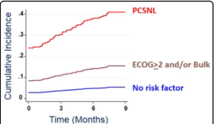
and 1.6% in the low-risk group). Figure1 shows the risk
over time in a competing-risk model. The curves suggest that the VTE risk factors are valid both for VTE events at diagnosis and during therapy.
An analysis focusing on patients with score 0 did not identify a risk factor possibly discerning patients at lowest risk (data not shown).
In conclusion, we identified VTE risk factors in patients
with lymphoma that are well different from VTE risk
factors described for solid tumors. We identified three
main clinical risk factors: CNS localization, tumor bulk > 10 cm and reduced performance status. We confirm aggressive lymphoma histology as VTE risk factor in univariate, but not in multivariate analysis.
Bulk and reduced ECOG probably contribute to slowing
down the bloodflow in the venous bed. In fact, analyzing
for associations between localization of VTE and risk factors, we found an association between the site of the bulk and thrombosis (data not shown). A reduction of the performance status to the ECOG grade 2 or greater that
defines a reduction of the daily activity with variation
from partial to complete immobilization was associated with a higher VTE localization to the lower extremities.
Analyzing for laboratory abnormalities, we found lower albumin and elevated LDH levels at diagnosis to be associated with VTE. LDH is a proliferation marker typically elevated in aggressive lymphomas. Low albumin levels may be due to an altered nutritional status or an inflammatory environment reducing albumin production and may lead to endothelial cell activation increasing the
thrombotic risk14. Other laboratory abnormalities, in
particular, the CBC counts that are an essential part of the
Khorana score, did not prove to be significant in
lym-phoma patients.
Comparison with other studies8,10is difficult because of
differences in patient selection. Our study cohort is a real-life case series including both HL and NHL, including also VTE that was present before the start of therapy. This is well justified as the recent analysis of a Swedish registry of 40,354 NHL patients showed that the incidence of
thrombosis started to increase about five months before
diagnosis, and reached its peak a month before
diag-nosis15. We included only patients with at least one
hos-pital stay. Patients as those with low-grade histology who did not require a hospital stay were not retrieved. As expected, this may explain the higher incidence rate of
VTE with respect to other reports1,2,8,10. Therefore, our
study tackles the issue of VTE frequency in a population at increased risk, in whom special care is required and
specific information are needed.
In summary, we identified lymphoma-specific VTE risk
factors that could be useful tools to plan tailored antith-rombotic prophylaxis.
Conflict of interest
S.H. has received consulting and lecture fees from Novartis, Takeda, and institutional research grants from Roche; V.D.S. has received consulting and lecture fees from Amgen, Bayer, Celgene, Novartis, and institutional research grants from Novartis. The remaining authors declar no conflict of interest. Publisher's note
Springer Nature remains neutral with regard to jurisdictional claims in published maps and institutional affiliations.
Fig. 1 Cumulative incidence rates of VTE. The graph shows the results of a competing risk regression analysis. Death was counted as a competitive event. VTE before the start of systemic therapy were considered heralding events and were counted as events at time 0, while VTE during thefirst nine months from treatment start was considered treatment-related events. Subdistribution hazard risk was 5.2-fold (95% CI, 3.1–8.8) for patients with the risk factors reduced performance status (ECOG > 2) and/or bulky disease (>10 cm), and 8.0-fold (95% C.I., 3.7–17.5) for patients with PCNSL.Analysis of the treatment-related VTE in 803 patients, after exclusion of the 54 ones who presented with VTE before the start of therapy, confirmed a VTE risk gradient similar to that of the overall cohort (see text) (5/29, 17.2% in PCNSL, 25/333, 7.5% in the bulk and/or ECOG group, and 11/441, 2.5% in the low-risk group)
Hohaus et al. Blood Cancer Journal (2018) 8:54 Page 3 of 4
Received: 25 March 2018 Revised: 27 April 2018 Accepted: 8 May 2018
References
1. Khorana, A. A., Kuderer, N. M., Culakova, E., Lyman, G. H. & Francis, C. W. Development and validation of a predictive model for chemotherapy-associated thrombosis. Blood 111, 4902–4907 (2008).
2. Sanfilippo, K. M. et al. Incidence of venous thromboembolism in patients with non-Hodgkin lymphoma. Thromb. Res. 143, 86–90 (2016).
3. Lyman, G. H. et al. Venous thromboembolism prophylaxis and treatment in patients with cancer: American Society of Clinical Oncology clinical practice guideline update. J. Clin. Oncol. 31, 2189–2204 (2013).
4. Caruso, V. et al. Thrombotic complications in adult patients with lymphoma: a meta-analysis of 29 independent cohorts including 18 018 patients and 1149 events. Blood 115, 5322–5328 (2010).
5. Mohren, M. et al. Increased risk of thromboembolism in patients with malignant lymphoma: a single-centre analysis. Br. J. Cancer 92, 1349–1351 (2005).
6. Park, L. C. et al. Incidence, risk factors and clinical features of venous throm-boembolism in newly diagnosed lymphoma patients: results from a pro-spective cohort study with Asian population. Thromb. Res. 130, e6–e12 (2012). 7. Lund, J. L., Østgård, L. S., Prandoni, P., Sørensen, H. T. & de Nully Brown, P. Incidence, determinants and the transient impact of cancer treatments on venous thromboembolism risk among lymphoma patients in Denmark. Thromb. Res. 136, 917–923 (2015).
8. Santi, R. M. et al. Khorana score and histotype predicts incidence of early venous thromboembolism in non-Hodgkin lymphomas. A pooled-data ana-lysis of 12 clinical trials of Fondazione Italiana Linfomi (FIL). Thromb. Haemost. 117, 1615–1621 (2017).
9. Goldschmidt, N., Linetsky, E., Shalom, E., Varon, D. & Siegal, T. High incidence of thromboembolism in patients with central nervous system lymphoma. Cancer 98, 1239–1242 (2003).
10. Antic, D. et al. Development and validation of multivariable predictive model for thromboembolic events in lymphoma patients. Am. J. Hematol. 91, 1014–1019 (2016).
11. Heit, J. A. et al. Relative impact of risk factors for deep vein thrombosis and pulmonary embolism: a population-based study. Arch. Intern. Med. 162, 1245–1248 (2002).
12. Prandoni, P. & Samama, M. M. Risk stratification and venous thrombopro-phylaxis in hospitalized medical and cancer patients. Br. J. Haematol. 141, 587–597 (2008).
13. Fine, J. P. & Gray, R. J. A proportional hazards model for the subdistribution of a competing risk. J. Am. Stat. Assoc. 94, 496–509 (1999).
14. Falanga, A., Marchetti, M. & Russo, L. The mechanisms of cancer-associated thrombosis. Thromb. Res. 135(Suppl 1), S8–S11 (2015).
15. Birgisdóttir, A. M., Sverrisdóttir, I. S., Landgren, O., Björkholm, M. & Kristinsson, S. Y. Risk of thrombosis in patients with non-Hodgkin’s lymphoma: a population-based cohort study. Haematologica 102(s1), 1602017 (2017).
Hohaus et al. Blood Cancer Journal (2018) 8:54 Page 4 of 4