INTRODUCTION
Williams syndrome, also known as Williams-Beuren syndrome (WBS), is a multi-system, congenital and rare disorder involving the cardiovascular system, connecti-ve tissue, and the central nervous system (1). It is a genetic disorder caused by a hemizygous microdeletion of chromosome 7 (7q11.23). It was first reported inde-pendently by Williams et al. and Beuren et al., who described children with a number of developmental and physical abnormalities (2, 3). In addition, children with WBS have a distinct cognitive profile associated with mental retardation, with relatively preserved lan-guage abilities but generally poor visuo-spatial skills (4).
Physical features include characteristic facial features with full prominent cheeks, wide mouth, long philtrum, small nose with depressed nasal bridge, heavy orbital ridges, medial eyebrow flare, dental abnormalities, hoarse voice, growth retardation, and cardiovascular abnormalities (most commonly supravalvular aortic stenosis and/or peripheral pulmonary artery stenosis) (1). Facial features are summarized in the term “elfin face”. Furthermore, cephalometric measurements often show an anterior inclination of the maxilla, a high mandibular plane angle, and a deficient bony chin (1), although the mandible could not be classified as retro-gnathic (5). Patients demonstrated a high prevalence of Class II and III occlusions, open and deep bites, and anterior cross-bites. Folding and thickening of the buc-cal mucous membranes, and prominent accessory labial frenula are observed. The soft tissue analysis indicated that the lips exceeded the line of harmony (Holdaway angle) and thus caused a disharmonious extra-oral appearance (6). Hypoplasia of teeth, including bud-shaped maxillary primary second molars and mandibu-lar permanent first momandibu-lars, have been reported. Fin-dings also included microdontia (95%), small roots, June 2011; 2(3)
FRANCESCO GRECCHI
1, ILARIA ZOLLINO
2, VITTORIA PERROTTI
3, FRANCESCO CARINCI
2>
91 1
Maxillofacial Surgery, Galeazzi Hospital, Milan, Italy 2
Maxillofacial Surgery, University of Ferrara, Ferrara, Italy 3
Dental School, University of Chieti-Pescara, Chieti, Italy
Williams-Beuren Syndrome treated with orthognathic
surgery and combined partial glossectomy: case report
KEY WORDS Glossectomy; Orthognatic surgery; Williams syndrome.
JOURNAL of
OSSEOINTEGRATION
ABSTRACT
Background Williams syndrome, also known as Williams-Beuren syndrome (WBS), is a multi-systems, congenital and rare disorder involving the cardiovascular system, connective tissue, and the cen-tral nervous system. It is a genetic disorder caused by a hemizygous microdeletion of chromosome 7q11.23. Here we report a case of WBS treated with bimaxillary osteotomies and glossectomy. Case report Orthognatic surgery was undertaken one year after the first diagnosis and the beginning of the orthodontic treatment. The maxilla was advanced at a Le Fort I of about 4 mm and was fixed with two angled plates, one on each side, applied laterally to the pyriform aperture. The lateral part of the maxilla was stabilized with wires. In addition, bilateral mandibular sagittal osteotomies were carried out together with a midline osteotomy. A partial glossecto-my was performed. Intermaxillary adaptation was supported by applying soft elastics according to the concept of semi-rigid bone fixation. Two months post-surgery the occlusion was Angle class I with a well defined overbite and overjet. The healing was uneventful. Functional limitations or nerve disturbances did not occur. The mini-plates remained in situ.
In the case reported the "keyhole" partial glossectomy was perfor-med in combination with the orthognatic surgery. No complication was recorded in the postoperative period and the patient had a suc-cessful outcome.
malocclusion (85%), delayed mineralization, and absence of some teeth, as well as invagination of the incisors (7). Macroglossia with signs of lingua plicata could be combined with a severe tongue thrusting in 67.7% of cases, while more than 50% of the patients had excessive interdental spacing (8).
The diagnosis of WBS is usually made during mid-chil-dhood when the characteristic facial features, cogniti-ve profile, cardiac findings become more apparent and are supported by the fluorescent in situ hybridization test to demonstrate the characteristic submicroscopic deletion on the long arm of chromosome 7. This last laboratory technique is particularly helpful, since there is a variable expression of the WBS features, which makes clinical diagnosis particularly difficult during the early years of life (9).
Due to these facial dysmorphologies, early determina-tion of treatment objectives and the timing of interdi-sciplinary strategies are important for adequate mana-gement of WBS. A delay in diagnosing WBS can influence morbidity and prognosis. At each period, eva-luation comprises a growth and developmental estima-tion using WBS growth charts, cardiac evaluaestima-tion, fee-ding habits, and laboratory examinations (3). Given optimal medical, educational, and community support, the quality of life of affected individuals can be impro-ved (10, 11).
As no general agreement is reached on treatment pro-tocol, a case of WBS treated with bimaxillary osteoto-mies and glossectomy is analyzed with special atten-tion to the treatment modalities and the discussion of the pertinent literature.
CASE REPORT
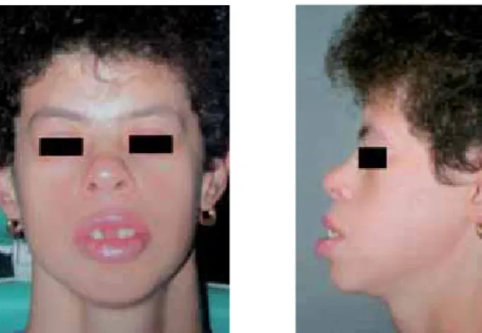
A 21 years old woman was admitted at Maxillofacial Surgery, Galeazzi Hospital, Milan, Italy in December 2003. She had no previous history of congenital mal-formations. She presented with difficulties in eating, some mild speech problems and impairment of psycho-motor development. In the craniofacial complex we found thick eyebrows, bitemporal narrowing, periorbi-tal sinking, short nose, wide nasal tip, malar hypoplasia, large philtrum, thick lips and prominent ear lobes (Fig. 1, 2). She had a skeletal III Class (Fig. 3), diastema of the incisors (Fig 4), severe periodontal disease, inclusion of the right central upper incisor and absence of the lower left second molar (Fig 5). She also had macro-glossia.
The patient had deficit of cardiac electric conduction but no morphological defects. No additional anomalies were detected.
The final diagnosis of WBS was established, based on
June 2011; 2(3) Grecchi F. et al.
JOURNALofOSSEOINTEGRATION
>
92
Fig. 1, 2 Patient’s frontal and lateral views. Fig. 3 Pre-treatment teleradiography.
the clinical features described above and supported by the fluorescent in situ hybridization test.
Pre-surgical orthodontic treatment
Pre-surgical orthodontic treatment was carried out with a straightwire edgewise technique using a 0.018-inch high-torque system.
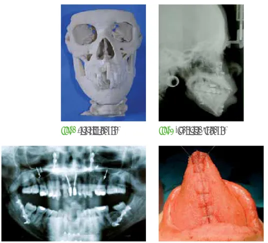
Dentoalveolar decompensation and harmonization of the upper and lower arches were the main treatment objectives. We maintained a space in the mandible midline as a sagittal osteotomy was planned to reduce the transverse dimension of the mandible. Standard 0.016 x 0.022-inch steel arches were used for stabiliza-tion during and after surgery. A stereolitographic model was performed in order to have a detailed model for surgical planning (Fig. 6).
Surgery
Orthognatic surgery was undertaken one year after. The maxilla was advanced at a Le Fort I of about 4 mm. The maxilla was fixed with two angled plates applied laterally to the pyriform aperture, one on each side. The lateral part of the maxilla was stabilized with wires. In addition, bilateral mandibular sagittal osteotomies were carried out together with a midline osteotomy (Fig. 7,d 8) and a partial glossectomy was performed (Fig. 9). Intermaxillary adaptation was supported by
applying soft elastics according to the concept of semi-rigid bone-fixation. Two-months post-surgery, the occlusion was Angle Class I with a well defined overbi-te and overjet.
Outcome
The healing was uneventful. Functional limitations or nerve disturbances did not occur. The miniplates remai-ned in situ.
DISCUSSION
The syndromal, skeletal, and dental malformations with the additional tongue dysfunction and macroglossia in WBS patients require individualized and complex treat-ment planning (6).
The following surgical interventions were taken into considerations: a partial glossectomy, an advancement of the maxilla at a Le Fort I level and the bilateral man-dibular sagittal osteotomies with a midline osteotomy. An enlarged tongue (macroglossia) can cause dento-musculoskeletal deformities, instability of orthodontic and orthognathic surgical treatment, and create masti-catory, speech, and airway management problems. Understanding the signs and symptoms of macroglos-sia will help identifying those patients who could
bene-JOURNALofOSSEOINTEGRATION
June 2011; 2(3)
Williams-Beuren Syndrome - Case report <
93 Fig. 6Stereolitography. Fig. 7Final teleradiograph.
Fig. 9The "keyhole" partial glosssectomy. Fig. 8 Final orthopantomograph.
rative period and the patient had a successful outcome.
REFERENCES
1. Axelsson S. Variability of the cranial and dental phenotype in Williams syndrome. Swed Dent J Suppl 2005;170:3-67.
2. Kaplan P, Wang PP, Francke U. Williams (Williams Beuren) syndrome: a distinct neurobehavioral disorder. J Child Neurol 2001;16:177-90.
3. American Academy of Pediatrics: Health care supervision for children with Williams syndrome. Pediatrics 2001;107:1192-204.
4. Lashkari A, Smith AK, Graham JM Jr. Williams-Beuren syndrome: an update and review for the primary physician. Clin Pediatr (Phila) 1999;38:189-208.
5. Mass E, Belostoky L. Craniofacial morphology of children with Williams syndrome. Cleft-Palate Craniofac J 1993;30:343-9.
6. Habersack K, Grimaldi B, Paulus GW. Orthodontic orthognathic surgical treatment of a subject with Williams Beuren syndrome a follow-up from 8 to 25 years of age. Eur J Orthod 2007;29(4):332-7.
7. Oncag A, Gunbay S, Parlar A. Williams Syndrome. J Clin Ped Dent 1995;19:301-4.
8. Herzberg J, Nakisbendi LA, Needleman HL, Pober B. Williams syndrome: oral presentation of 45 cases. Pediatr Dent 1994;16: 262-7.
9. Lowery MC, Morris CA, Ewart A, Brothman LJ, Zhu XL, Leonard CO, Carey JC, Keating M, Brothman AR. Strong correlation of elastin deletions, detected by FISH, with Williams syndrome: evaluation of 235 patients. Am J Hum Genet 1995;57:49-53.
10. Huang L, Sadler L, O’Riordan MA, Robin NH. Delay in diagnosis of Williams syndrome. Clin Pediatr 2002;41:257-61.
11. Kaplan P. Williams syndrome—does early diagnosis matter? Clin Pediatr 2002;41:277-8.
12. Wolford LM, Cottrell DA. Diagnosis of macroglossia and indications for reduction glossectomy. Am J Orthod Dentofacial Orthop 1996;110:170-7.
13. Egerton M. The management of macroglossia when associated with proguathism. Br J Plast Surg 1960;3:117-22.
14. Harvold EP. The role of function in the etiology and treatment of malocclusion. Am J Orthod 1968;54:883-98. fit from a glossectomy (reduction of tongue size) to
improve function, aesthetics, and treatment stability (12).
Several articles have appeared describing various methods for reducing the tongue size, including the midline wedge resection with the base in the anterior tongue, the midline elliptical excision, the marginal excision, and the "keyhole" or midline elliptical excision combined with an anterior wedge resection, have all been described (13).
The option of performing the reduction glossectomy first, as an isolated procedure, and secondly the ortho-gnathic surgery has the absolute indication when extensive orthodontics are necessary before orthogna-thic surgery, and the size of the tongue prevents the required orthodontic movements. Instead, in perfor-ming the orthognathic surgery and glossectomy in one surgical stage, it is usually helpful to complete the orthognathic surgery first. Once the orthognathic sur-gery is rigidly stabilized, a glossectomy can then be performed. Since a glossectomy generally causes a transient but significant increase in the size of the ton-gue, secondary to edema, performing the tongue pro-cedure last may allow the occlusion to be better esta-blished before the onset of edema. However, if the ton-gue is extremely large, the reduction glossectomy may need to be sequenced first, to allow the proper occlu-sion to be established when the orthognathic surgery is performed (12).
Harvold (14) demonstrated that reducing the tongue to a size much smaller than normal causes the dental arches to collapse lingually. There are potential risks and complications that can occur in reduction glossec-tomy including excessive bleeding, airway obstruction secondary to tongue edema, anesthesia of the tongue and loss of taste. These can develop secondary to lin-gual nerve injury, motor dysfunction secondary to hypoglossal nerve injury, decrease of tongue mobility secondary to scarring, salivary duct injury, and residual speech and masticatory problems (12).
In the case reported the "keyhole" partial glossectomy was performed in combination with the orthognatic surgery. No complication was recorded in the
post-ope-June 2011; 2(3) Grecchi F. et al.
JOURNALofOSSEOINTEGRATION
>