Sonoelastography in the diagnosis
of tendinopathies: an added value
Stefano Galletti Francesco Oliva Stefano Masiero Antonio Frizziero Riccardo Galletti Cosima Schiavone Vincenzo Salini Michele Abate
Department of Medicine and Science of Aging, Uni-versity “G. d’Annunzio” Chieti - Pescara, Italy
Corresponding author: Michele Abate
Department of Medicine and Science of Aging, Uni-versity “G. d’Annunzio” Chieti - Pescara
Via dei Vestini, 31 66013 Chieti, Italy E-mail: [email protected]
Summary
Background: sonoelastography helps in the
de-tection of abnormalities not yet evident on B-mode exam.
Methods: in this observational study, we report a
collection of cases of symptomatic patients with-out alterations at ultrasound imaging but with evi-dence of pathological findings at sonoelastogra-phy. Patients, with clinical history suggestive for tendinopathies or surgically treated, and negative at the ultrasound exam, were submitted to sonoe-lastography. Out of 846, 632 patients with positive ultrasound exam were excluded. Sonoelastogra-phy was therefore performed in the remaining 214.
Results: the examination was positive in 168 cases:
78 patients were affected with shoulder diseases, while elbow pathology was observed in 31 subjects; patellar, Achilles and plantar fascia disorders were reported in 19, 27, and 13 patients, respectively.
Conclusion: sonoelastography can reveal tendon
abnormalities of clinical relevance in a high per-centage of cases, where the ultrasound exam was negative, making the method a complementary tool to ultrasound evaluation.
KEY WORDS: imaging, sonoelastography, tendon, tendinopathy, ultrasound.
Introduction
Ultrasound elastography (EUS) is a new technique re-cently introduced in the clinical practice that allows qual-itative evaluations and quantqual-itative measurements of the mechanical properties of tissues1, 2. It is based on the
principle that tissue compression produces changes within it and that the displacement (strain), depending on the elastic properties, is less pronounced in hard than in soft tissues. Strain EUS, the most commonly method used, allows the direct visualization, on B-mode image, of strain distribution map or “elastogram”, where blue, red, and yellow/green colours indicate hard, soft, and intermediate tissue stiffness, respectively2.
EUS has been employed for musculo-skeletal dis-eases showing, in general, a good correlation with the clinical and ultrasound (US) examination, in agreement with the histopathological features of the lesion3-5.
It is well known that, in some cases, it is difficult or even impossible to distinguish pathological tissues using conventional US because these tissues show the same echogenicity of the surrounding healthy structures6. In these cases, EUS could detect or
dif-ferentiate abnormalities, which are thought to corre-spond to sub-clinical changes not yet evident on B-mode evaluation, providing supplementary informa-tion useful for diagnostic, therapeutic (ultrasound guided procedures) and follow-up purposes.
However, only few systematic studies have compared EUS with traditional US3-5, 7, 8; in these studies little
information has been reported about the characteris-tics of lesions, which more frequently remain unde-tected by US, but are made evident by EUS. For in-stance, EUS could detect areas of cleavage inside tendons after partial tears, or scar tissue not strong enough to withstand load, or could provide early in-formation on the functional recovery after surgery. Aim of the present paper was to report a collection of illustrative cases of symptomatic patients without evi-dent alterations at US imaging but with evidence of pathological findings at EUS.
Materials and methods
The study was perfomed according to the Declaration of Helsinki and to the ethical standard of the Muscles, Ligaments and Tendons Journal9, and informed
writ-ten consent was obtained from all the patients. Subjects referred to our Ultrasound Services (Rizzoli
To-niolo Clinic, Bologna) with clinical history suggestive
for tendinopathies (pain, tenderness and/or functional limitation) or surgically treated for tendon tears and in the late phases of rehabilitation, and negative for acute pathology, were enrolled.
We excluded patients with a positive history of sys-temic inflammatory arthritis (rheumatoid, psoriatic and reactive arthritis, arthritis associated with inflam-matory bowel diseases, and spondiloarthritis), suffer-ing from severe osteoarthritis of the scanned district, malignancy, endocrinopathies and severe chronic diseases (renal, hepatic, cardiac, etc.).
Clinical examination was aimed at evaluating the presence of pain during the previous week, local ten-derness, functional limitation of the involved district and of the surrounding tissues (i.e. joints, muscles, ligaments and subcutaneous tissue), and, where pos-sible (palpable tendons), tendon thickening. Pain, at rest and during common activities of daily living, was assessed using 10 cm visual analogue scale (VAS), with 0 representing no pain and 10 representing the worst pain.
Afterwards, participants underwent an US and Colour Doppler (CD) evaluation of the affected region, using a high-resolution, multi-frequency (6-15 MHz) linear array transducer (Hitachi Preyrus). The US criteria adopted for the diagnosis of tendinopathies were the following.
The presence of dishomogeneous hypo- or hypere-choic thickening, diffuse or focal, of the tendon, asso-ciated with loss of the normal fibrillar pattern and/or irregularity of the tendon margins, was interpreted as sign of degeneration10.
Fluid within and patchy thickening of the paratenon, associated or not with irregularities of tendon mar-gins, were classified as peritendinitis11, while an
en-thesopathy was reported when, at US scans, focal tendon thickening, abnormal tendon echotexture, cal-cifications, bone erosions, and bursal fluid distension at the site of tenderness were observed12.
Involvement of bursae was diagnosed when accumu-lation of anechoic fluid, with or without hypoechoic swelling of the synovia, appeared within it13.
Presence of neovascularization was estimated, by means of CD and graded as (0), (1+), (2++), (3+++), (4++++), according to the appearance of vessels in-side the tendon14. To avoid artifacts, sensitivity was
optimised for low flow, and colour gain was set just below the noise level.
When the US scan was not suggestive for tendon dis-eases, a real-time EUS examination (Hitachi Preyrus, Philips IU22) was carried out.
EUS was performed with the tendon not in extension by applying light repetitive compression, both in the longitudinal and transverse plane, with the hand-held transducer over the region of interest; the size of the EUS window was selected in relationship to the size of the tendon to be examined. Force applied was set according to the quality factor of the equipment and was displayed on the screen: the visual indicator fa-cilitated the acquisition by showing the average strain applied. During the examination, B-mode and EUS
images were displayed side by side on the monitor, with EUS superimposed on B-mode image as a colour-coded, real-time picture. Colour scale, which represented the relative stiffness of the tissues, ranged from red (soft tissue), yellow/green (interme-diate stiffness) to blue (hard tissue). The most repre-sentative EUS image (defined as the adequate depic-tion of tissue structure and constant reproducdepic-tion of the scanned images) of at least three concordant was chosen and recorded on communication system for a further analysis.
Care was taken to hold the probe perpendicular to the target tissue to avoid anisotropy and tissue shift-ing when performshift-ing US and EUS respectively. Both the US and EUS evaluations were performed by the same radiologist (GS) with ultra-decennial experi-ence in musculo-skeletal imaging.
The demographic and clinical characteristics of the EUS positive and negative subjects were compared in general and for each region (shoulder, elbow, patellar, Achilles, and plantar fascia). Data are reported as mean ± SD for continuous variables, whereas categori-cal and dichotomous variables are reported as fre-quencies and percentage. The two-sample Student’s t-test was used to compare continuous variables, when the distribution of data was normal; the Wilcoxon’s rank sum test was used otherwise. The χ2 test was used to evaluate associations between categorical da-ta. The significance level was determined at p < 0.05.
Results
846 subjects (50.1 ± 20.4, range: 13-83, M:F 501:345) with referred symptoms suggestive of tendinopathy were evaluated clinically and by means of sonogra-phy. In 74.7% (632/846) the US scan was positive for tendon abnormalities (data not reported), and they were excluded from the trial.
EUS was then performed in the remaining 214 (25.2%) subjects (Tendinopathy= 203; surgically treated= 11), whose B-mode and color doppler US scans were negative. The US negative patients (mean age: 49.6 ± 13.8, range: 13-81, M:F 116:98), were complaining of mild symptoms (VAS at rest: 1.8 ± 1.1; VAS during activities: 2.6 ± 0.8), with a mean symptoms duration of 2.4 ± 1.1 months (Tab. 1). Regarding the upper limbs, 97 (45.3%) shoulders and 38 (17.7%) elbows were evaluated, while at the lower limbs, 26 (12.1%) patellar tendons, 35 (16.3%) Achilles tendons, and 18 (8.4%) plantar fasciae were studied. Concerning the 11 surgically treated tendons, 7 Achilles (6 complete and 1 incomplete tears), 3 Patellar (jumpers’ knee), and 1 elbow extensors ten-dons (chronic tendinopathies) were noted (Tabs. 2, 3). The tendon structure showed a normal or non diag-nostic EUS pattern in 46 (21.4%) patients.
The results of EUS evaluation were the following. Shoulder: out of 97 tendons, 19 subject (19.5%) did not show any EUS abnormalities (symptoms related to gleno-humeral instability), while EUS was positive in the remaining 78 patients (80.4%).
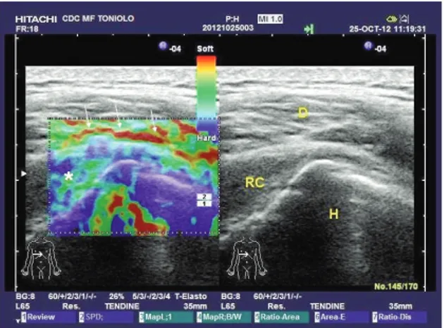
In the EUS positive group, 88.4% (69/78) patients were affected from post-trauma diseases: indeed, ro-tator cuff tendons appeared stiff (blue), and inflam-mation/oedema (red areas) of the sub-acromial bursa and surrounding tissues were associated (Fig. 1). Bi-ceps tendon disorders (red circle surrounding the ten-don) were reported only in 9 subjects (11.5%).
Elbow: of 38 tendons, EUS showed abnormalities in 31 subjects (81.5%). Signs of peritendinopathy (oedema and inflammation along the peritenon) were observed in 26 patients (83.8%); in the remaining 5 cases, enthesitis (blue areas at the attachment site of the tendon, oedema in the surrounding bursae and tissue) and post-surgery features (mixture of irregular Table 1. Demographic and clinical characteristics of included patients.
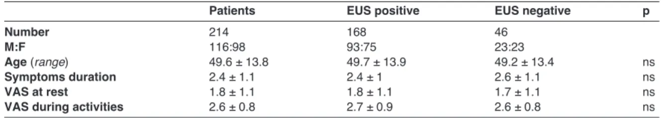
Patients EUS positive EUS negative p
Number 214 168 46
M:F 116:98 93:75 23:23
Age (range) 49.6 ± 13.8 49.7 ± 13.9 49.2 ± 13.4 ns
Symptoms duration 2.4 ± 1.1 2.4 ± 1 2.6 ± 1.1 ns
VAS at rest 1.8 ± 1.1 1.8 ± 1.1 1.7 ± 1.1 ns
VAS during activities 2.6 ± 0.8 2.7 ± 0.9 2.6 ± 0.8 ns
Table 2. Demopraghic and clinical characteristics of included patients, and EUS diagnosis of the upper limbs district. UPPER LIMBS
SHOULDER ELBOW
EUS pos EUS neg p EUS pos EUS neg p
Number 78/97 (80.4%) 19/97 (19.5%) 31/38 (81.5%) 7/38 (18.4%)
Age (range) 55.4 ± 12.8 56.6 ± 13.3 0.3 43.1 ± 11.6 43.7 ± 9.7 0.4 Symptoms duration 2.5 ± 1.2 2.5 ± 1.1 0.4 2.4 ± 1 3 ± 1.2 0.07
VAS at rest 1.7 ± 1 1.6 ± 1.1 0.3 1.9 ± 1.1 1.4 ± 1.1 0.1
VAS during activities 2.6 ± 0.7 2.5 ± 0.8 0.3 2.9 ± 1.1 2.7 ± 1.1 0.3 EUS diagnoses - Post-trauma 69/78 (88.4%) - Biceps tendon 9/78 (11.5%) - Peritendinopathy 26/31 (83.8%) - Enthesis 4/31 (12.9%) - Post-surgery 1/31 (3.2%)
Table 3. Demographic and clinical characteristics of included patients, and EUS diagnosis of the lower limbs district. LOWER LIMBS
PATELLAR ACHILLES PLANTAR FASCIA
EUS pos EUS neg p EUS pos EUS neg p EUS pos EUS neg p
Number 19/26 7/26 0.002 27/35 8/35 0.0000 13/18 5/18 0.01
(73%) (26.9%) (77.1%) (22.8%) (72.2%) (27.7%)
Age (range) 37.9 ± 16.7 38.1 ± 13.9 0.4 46.9 ± 12.5 48.5 ± 10.6 0.3 52.8 ± 6.3 50.2 ± 4.4 0.2 Symptoms duration 2.4 ± 1.1 2.7 ± 1.2 0.2 2.5 ± 1 3.1 ± 1.5 0.08 1.7 ± 0.5 1.8 ± 0.4 0.3 VAS at rest 1.9 ± 1.1 1.8 ± 1.1 0.3 2.1 ± 1.4 2.4 ± 1.1 0.2 1.8 ± 0.8 1.2 ± 0.8 0.1 VAS during activities 2.9 ± 0.9 2.7 ± 0.7 0.2 2.9 ± 1.1 3.1 ± 0.6 0.2 2.6 ± 0.8 2.2 ± 0.8 0.1 EUS diagnoses - Peritendinopathy 11/19 11/27 (57.8%) (40.7%) - Post-surgery 3/19 7/27 (15.7%) (25.9%) - Proximal tendinopathy 2/19 9/13 (10.5%) (69.2%) - Distal tendinopathy 2/19 3/27 (10.5%) (11.1%) - Pre-patellar thickening 1/19 (5.2%) - Midportion tendinopathy 6/27 4/13 (22.2%) (30.7%)
red and blue areas) were observed in 4 and 1 pa-tients, respectively.
Patellar: EUS alterations were observed in 19/26 pa-tients (73%). Eleven papa-tients (57.8%) complained peritendinopathy, while proximal and distal tendino -pathies were observed in 2 cases respectively; 3 post-surgery alterations and 1 pre-patellar thickening (red areas) were observed in the remaining patients (Fig. 2).
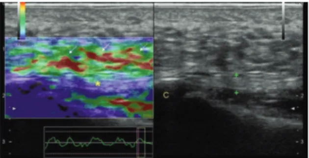
Achilles: 27/35 (77.1%) patients showed EUS posi-tive features: peritendinopathy (Fig. 3) was observed in 11 (40.7%) subjects, while EUS revealed signs of focal mid-portion and of distal tendinopathy (Fig. 4) in 6 (22.2%) and 3 cases (11.1%), respectively. Finally, post-surgery features (Fig. 5) were observed in 7/27 (25.9%) patients.
Plantar fascia: 13/18 (72.2%) patients were EUS positive for plantar fascitiis. Plantar fascia appeared
homogeneously blue (hard structures), probably re-lated to anelastic tissue alterations. Red areas (tis-sue edema) was present in the surrounding tis(tis-sue under the fascia (9 cases proximal and 4 cases mid-portion tendinopathy) (Fig. 6). In 5 cases (27.7%) the results of EUS evaluation were negative or in-conclusive.
Discussion
In several symptomatic subjects US examination fails to reveal tendon abnormalities of clinical rele-vance. In this observational study we illustrate a case series of patients with negative or inconclusive US exam where EUS allowed to show tendon pathologies.
Indeed, EUS abnormal features explaining the mild symptomatology of our patients were found in several tendons, such rotator cuff, elbow, patellar, Achilles and plantar fascia. In these patients we observed tis-sue softening (red-yellow) within the tendon, paratenon and surrounding tissues (fascia, bursae),
Figure 1. Post-trauma shoulder: in the left panel, EUS shows a stiff (blue, green colours) rotator cuff (*), expres-sion of initial fibro-adesive process; oedema and inflamma-tion (arrows) (red areas) are present at the site of sub-acromial bursa (synovial contusion without effusion). RC=
Rotator Cuff; D= Deltoid muscle; H= Humeral head. Figure 3. Achilles peritendinopathy: EUS reveals areas ofinflammation and oedema (red spots, arrows) in the ventral side of the tendon. Achilles tendon (calipers) is normal at US evaluation (right panel). C= calcaneal bone; *= anisotropy effect.
Figure 2. Patellar peritendinopathy: patellar tendon (calipers) appears normal on B-mode exam (right panel). At EUS evaluation, red areas (arrows) along the full length of the ventral portion of the tendon are expression of peritenon inflammation. P= patella; T= Tibia.
Figure 4. Distal Achilles tendinopathy: at the EUS exam, peritenon is severely inflamed (red areas, arrows); synovial
thickening (red spot) in the retrocalcaneal bursa (dot circle)
is present, while the distal portion of the tendon (*) is stiff (blue and green spots). Achilles tendon (calipers) is normal at US evaluation. C= calcaneal bone.
that might be explained by very early changes in tis-sue elasticity, probably due to histopatological alter-ations (oedema and inflammation)15, 16.
We also observed, in quite all cases of suspected plantar fasciitis, that plantar fascia, which showed a normal echogenic pattern at US evaluation, behaved as an hard tissue (blue), probably related to the in-creased content of type III collagen.
Our observations are in agreement with previous studies where EUS detected intra and peritendinous alterations17-19. Indeed, in a comparison study
be-tween healthy volunteers and patients suffering from lateral epicondylitis, De Zordo et al.4 found that
nor-mal tendons appeared blue (hard) in 96% of volun-teers, while tissue edema (red areas) was observed in 67% of patients (p< 0.001). In the same study, the authors observed that EUS was more sensitive than US in discovering early oedema and inflammation (red-yellow areas) in the collateral ligament (26 vs 21%) and paratenon (29 vs 13%).
Similar patterns were also described for Achilles ten-don20, 21where normal tendons3were found to be
ho-mogeneously hard in 93% of cases, while marked (red) and mild softening (yellow) was observed in 57 and 32% of patients, respectively3.
On the basis of these observations, EUS appears as a complementary method to US evaluation, because it can discover small changes in the elastic and mechani-cal properties of tissue1, 2, expression of pathology,
which are not evident on B-mode evaluation due to the same echogenicity of the surrounding healthy tissues6.
Only in few cases (46/214, 21.4%) the tendon’s struc-ture showed a normal or non diagnostic EUS pattern. We hypothesize that this can be due to a misleading clinical history (low pain threshold of the patient?) or to artifacts which masked subtle pathological findings. The potential advantages of EUS can be summarized as follows: first, it could be used to differentiate identi-cal grey-sidenti-cale images, better detecting at an early stage alterations which could progress to higher stages of tendinopathy. Second, it may be used as a tool al-lowing the subjects to modify exercise regimen to pre-vent further tendon damage. Third, it may be useful for therapeutic purposes and to monitor treatment effec-tiveness. EUS could be also applied in the athletic pop-ulation, but this requires further validation studies, be-cause the tendinous structures of athletes are partly dif-ferent from those of the non athletic subjects.
Despite these advantages, the method suffer the limi-tation of being in some way operator dependent in terms of application of pressure to the probe and the differentiation of artifacts from diagnostic image infor-mation in real time. At this regard, a visual indicator on the screen may give an optimal dynamic range of pressure, helping to decrease inter-observer variabili-ty and facilitate image acquisition. However, because in the present study all the evaluations were per-formed by the same radiologist, these features must be confirmed by investigations aiming the intra- and inter-observer variability of the method.
Some limitations of the present study must be acknowl-edged. First, this is not a randomized control study and a control group, made of asymptomatic subjects matched for age and sex, is lacking; second, we did not evaluate the controlateral side (probably normal) and not investigate differences between the sexes, and the dominant and non-dominant side (important in the ath-letic population); third, we did not follow up the evolu-tion of the EUS alteraevolu-tions; finally, no histopatological exam was available to confirm our results.
In addition, it must be added that the EUS systematic evaluation of all the patients (i.e. including those pos-itive to the traditional US) would have allowed to de-tect a higher number of tendon abnormalities. In conclusion, EUS may be considered a powerful di-agnostic adjunct to US and CD evaluation in the diag-nostic approach to tendinopathies.
References
1. Drakonaki EE, Allen GM, Wilson DJ. Ultrasound elastogra-phy for musculoskeletal applications. Br J Radiol. 2012; 85(1019):1435-1445.
Figure 5. Achilles tendon tenorraphy: at US evaluation, Achilles tendon (calipers) appears hypoechoic, patchy (dot circle) and thick; surgical suture is present (*).
Worthnoting, the EUS exam (left panel) shows an oedema-tous area (arrows) at the tear site, which indicate that the scar tissue is not strong enough to withstand load. Patchy Achilles tendon appears as a mixture of blue, green, red and yellow areas.
Figure 6. Plantar fascitiis: at B-mode evaluation, plantar fascia (calipers) appears normal, while at EUS exam it is stiff (*, blue areas). In the surrounding tissues (arrows), oedema and inflammation (red areas) can be observed. C= calcaneal bone.
2. Ophir J, Céspedes I, Ponnekanti H, Yazdi Y, Li X. Elastogra-phy: a quantitative method for imaging the elasticity of biologi-cal tissues. Ultrason Imaging. 1991;13(2):111-134.
3. De Zordo T, Chhem R, Smekal V, et al. Real-time sonoelas-tography: findings in patients with symptomatic achilles ten-dons and comparison to healthy volunteers. Ultraschall Med. 2010;31(4):394-400.
4. De Zordo T, Lill SR, Fink C, et al. Real-time sonoelastography of lateral epicondylitis: comparison of findings between pa-tients and healthy volunteers. AJR Am J Roentgenol. 2009; 193(1):180-185.
5. Sconfienza LM, Silvestri E, Cimmino MA. Sonoelastography in the evaluation of painful Achilles tendon in amateur athletes. Clin Exp Rheumatol. 2010;28(3):373-378.
6. Frey H. Realtime elastography. A new ultrasound procedure for the reconstruction of tissue elasticity. Radiologe. 2003; 43(10):850-855.
7. Wu CH, Chang KV, Mio S, Chen WS, Wang TG. Sonoelas-tography of the plantar fascia. Radiology. 2011;259(2):502-507.
8. Park GY, Kwon DR. Application of real-time sonoelastography in musculoskeletal diseases related to physical medicine and rehabilitation. Am J Phys Med Rehabil. 2011;90(11):875-886. 9. Padulo J, Oliva F, Frizziero A, Maffulli N. Muscles, Ligaments and Tendons Journal. Basic principles and recommendations in clinical and field science research. MLTJ. 2013;4:250-252. 10. Grassi W, Filippucci E, Farina A, Cervini C. Sonographic
imag-ing of tendons. Arthritis Rheum. 2000;43:969-976.
11. Gibbon WW, Cooper JR, Radcliffe GS. Distribution of sono-graphically detected tendon abnormalities in patients with a clinical diagnosis of chronic achilles tendinosis. J Clin Ultra-sound. 2000;28:61-66.
12. Kamel M, Eid H, Mansour R. Ultrasound detection of heel en-thesitis: a comparison with magnetic resonance imaging. J Rheumatol. 2003;30:774-778.
13. Gibbon WW, Cooper JR, Radcliffe GS. Sonographic incidence of tendon microtears in athletes with chronic Achilles tendi-nosis. Br J Sports Med. 1999;33:129-130.
14. Ohberg L, Lorentzon R, Alfredson H. Neovascularisation in Achilles tendons with painful tendinosis but not in normal ten-dons: an ultrasonographic investigation. Knee Surg Sports Traumatol Arthrosc. 2001;9:233-238.
15. Kannus P, Józsa L. Histopathological changes preceding spontaneous rupture of a tendon. A controlled study of 891 pa-tients. J Bone Joint Surg Am. 1991;73(10):1507-1525. 16. Järvinen TA, Kannus P, Paavola M, Järvinen TL, Józsa L,
Järvinen M. Achilles tendon injuries. Curr Opin Rheumatol. 2001;13(2):150-155.
17. Klauser AS, Faschingbauer R, Jaschke WR. Is sonoelastog-raphy of value in assessing tendons? Semin Musculoskelet Radiol. 2010;14(3):323-333.
18. Lalitha P, Reddy MCh, Reddy KJ. Musculoskeletal applica-tions of elastography: a pictorial essay of our initial experience. Korean J Radiol. 2011;12(3):365-375.
19. Botar Jid C, Vasilescu D, Damian L, Dumitriu D, Ciurea A, Dudea SM. Musculoskeletal sonoelastography. Pictorial es-say. Med Ultrason. 2012;14(3):239-245.
20. De Zordo T, Fink C, Feuchtner GM, Smekal V, Reindl M, Klauser AS. Real-time sonoelastography findings in healthy Achilles tendons. AJR Am J Roentgenol. 2009;193(2):W134-138.
21. Drakonaki EE, Allen GM, Wilson DJ. Real-time ultrasound elastography of the normal Achilles tendon: reproducibility and pattern description. Clin Radiol. 2009;64(12):1196-1202.