Contents lists available atScienceDirect
American Journal of Ophthalmology Case Reports
journal homepage:www.elsevier.com/locate/ajocCase report
Spontaneous retinal-choroidal anastomosis in a case of branch retinal vein
occlusion
Alessandro Arrigo
a, Adriano Carnevali
a,b, Riccardo Sacconi
a,c, Lea Querques
a,
Giuseppe Querques
a,∗, Francesco Bandello
aaDepartment of Ophthalmology, University Vita-Salute, IRCCS Ospedale San Raffaele, Milan, Italy bDepartment of Ophthalmology, University of“Magna Graecia”, Catanzaro, Italy
cEye Clinic, Department of Neurological, Biomedical, and Movement Sciences, University of Verona, Verona, Italy
A R T I C L E I N F O
Keywords:
Fluorescein angiography Choroidal neovascularization Retinal vein occlusion
Optical coherence tomography angiography
A B S T R A C T
Purpose: To report a case of a patient affected by branch retinal vein occlusion (BRVO) showing a possible spontaneous retinal-choroidal anastomosis (RCA) at the level of the fovea.
Observations: A 72 years old male patient with diagnosis of BRVO of left eye (LE) one year before came to our observation for a follow-up visit.
The patient underwent a complete ophthalmologic examination, includingfluorescein angiography, struc-tural optical coherence tomography (OCT), OCT-Angiography (OCT-A). Interestingly, OCT-A revealed the pre-sence of bloodflow suggestive of single collateral vessel descending from the deep capillary plexus up to and possibly below a hyperreflective subfoveal deposit.
Conclusions and importance: We hypothesize that it can represent a RCA, starting from retinal capillaries and reaching the choriocapillary and choroidal vessels in order to allow a vascular shunt between the 2 circulations and thus a new passage for the bloodflow. A similar condition is represented by laser-induced RCA for the treatment of non-ischemic central retinal vein occlusion in order to establish a new passage for the bloodflow.
1. Introduction
Branch retinal vein occlusion (BRVO) is a relatively common disease caused by thrombotic event.1It is associated with a number of systemic
risk factors and it can led to deep loss of visual function.2One of the
possible evolution of BRVO is the spontaneous development of col-lateral vessels (i.e. the creation of new ways to bypass the damaged site in order to restore a good bloodflow).1,2It was demonstrated that their
development and the accurate preservation when performing laser treatment have a good impact on visual function.3The positive effect of new circles on patients' visual acuity induced ophthalmologists to de-velop and propose a possible laser treatment of BRVO, namely laser induced retinal-choroidal anastomosis (RCA).4 This represents an at-tempt to induce the creation of an anastomosis between superficial retinal vessels and choroidal ones in order to establish a new passage for the bloodflow. In this study we describe the case of a patient af-fected by BRVO showing a possible spontaneous RCA at the level of the fovea. This finding was assessed by means of multimodal imaging protocol including optical coherence tomography (OCT) based meth-odologies.
2. Case report
A 72 years old male patient with diagnosis of branch retinal vein occlusion (BRVO) of left eye (LE) one year before (Fig. 1) presented for routinely follow-up control. The BRVO did not require any treatment. Medical clinical history included diabetes mellitus type 2, lymphoma under chemotherapy, obstructive sleep apnea syndrome, bilateral glaucoma and a previous BRVO in the right eye (RE) laser-treated. BCVA at baseline was 20/100 in RE and 20/40 in LE. After the BRVO, the patient reported a development of moderate macular edema, which did not require anti-VEGF injections, due to spontaneous resolution detected at the follow-up visit.
The patient underwent complete ophthalmologic examination, which included best-corrected visual acuity, structural spectral domain (SD-OCT) and multimodal fundus images (Spectralis, Heidelberg Engineering, Heidelberg, Germany), OCT angiography (OCT-A; PlexElite 9000 SS-OCT, Carl Zeiss Meditec, Inc., Dublin, USA) and ultra-wide field fluorescein angiography (California; Optos PLC, Dunfermline, UK).
BCVA was 20/100 in RE and 20/32 in LE, intraocular pressure was
https://doi.org/10.1016/j.ajoc.2018.06.009
Received 23 October 2017; Received in revised form 5 June 2018; Accepted 18 June 2018
∗Corresponding author. Department of Ophthalmology, University Vita-Salute, IRCCS Ospedale San Raffaele, Via Olgettina 60, Milan, 20132, Italy. E-mail address:[email protected](G. Querques).
American Journal of Ophthalmology Case Reports 11 (2018) 92–94
Available online 20 June 2018
2451-9936/ © 2018 The Authors. Published by Elsevier Inc. This is an open access article under the CC BY-NC-ND license (http://creativecommons.org/licenses/BY-NC-ND/4.0/).
12 mmHg in both eyes. Fluorescein angiography of both RE and LE showed extensive compensative circles due to long-lasting BRVO. Of note, fundus autofluorescence revealed the presence of a macular hyper-autofluorescent lesion, which corresponded to a hyper-reflective
subfoveal deposit on structural SD-OCT; thisfinding was not detected in any of the previous controls performed at our Institution. Moreover, an initial epiretinal membrane was detected on structural OCT, causing a partial lack of the foveal pit. OCT-A nicely showed the collateral tem-poral circles in the LE, and revealed the presence of bloodflow sug-gestive of single collateral vessel descending from the deep capillary plexus up to and possibly below the subfoveal deposit (Fig. 2).
3. Discussion
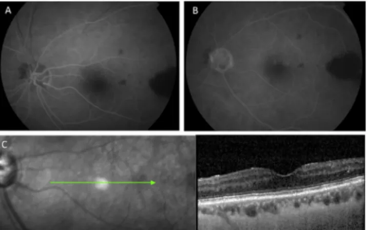
In this case OCT-A revealed the presence of blood-flow signal from a collateral vessel located in the deep capillary plexus up to and possibly below the subfoveal hyper-reflective deposit. We hypothesize that it can represent a RCA, starting from the superficial retinal capillaries and reaching the choriocapillary and choroidal vessels in order to allow a vascular shunt between the 2 circulations. A similar condition is re-presented by laser-induced RCA for the treatment of non-ischemic central retinal vein occlusion.4This represents an attempt to bypass the site of the occlusion by inducing the creation of a direct vascular communication between superficial retinal vessels and choroidal ones. In our case, the development of RCA might be secondary to the is-chemic stimulus caused by BRVO, thus justifying the absence of any foveal alterations (including macular edema or hemorrhages) one year before, during its acute phase, which could have been responsible for the hyper-reflective subfoveal deposit. The findings of the present study differed from other cases of RCA already described by our group.5 Fig. 1. Fluorescein angiography (FA), infrared image, and corresponding
structural spectral domain optical coherence tomography (SD-OCT) of the left eye 1 year before. A-B) FA demonstratesfilling delay in early phases. Late phase of FA shows areas of capillary nonperfusion with an hemorrhagic masking temporally. C) Infrared image and corresponding structural SD-OCT showing a normal foveal morphology without alterations of the internal retinal layers.
Fig. 2. Fluorescein angiography (FA), and structural spectral domain optical coherence tomography an-giography (OCT-A) of left eye. A-C) Early to late phase of FA reveals several hyperfluorescence and hypofluorescent spots corresponding to micro-aneurysms and hemorrhage masking, respectively, along with collateral circles. Note an abnormally deepen vessel (red circle) corresponding to possible retinal choroidal anastomosis on optical coherence tomography angiography. B1) Superficial plexus on 3 × 3 en-face OCT-A and corresponding OCT B-scan showed remodelling of foveal avascular zone, vas-cular tortuosity and ischemic areas. B2-B3) Deep plexus and avascular plexus on 3 × 3 en-face OCT-A showedflow in correspondence of red dot on OCT B-scan. (For interpretation of the references to colour in thisfigure legend, the reader is referred to the Web version of this article.)
A. Arrigo et al. American Journal of Ophthalmology Case Reports 11 (2018) 92–94
However, it cannot be excluded that thisfinding may represent an OCT-A artifact. Indeed, it is known that OCT-OCT-A analysis may suffer from a number of pitfalls related to its inability tofilter out from the particles' movement analysis the exclusive signal of the bloodflow. In this con-text, what OCT-A detected as bloodflow and thus a vessel might be the result of the movement of the material composing the foveal deposit. This may explain the absence of a clear foveal alteration onfluorescein angiography, which might be also related to resolution limits of the methodology. On the other hand, we excluded the possibility of artifact for a number of reasons. First of all, the adoption of swept source OCTA technology is known to strongly allow the reliable bloodflow detection, providing data less affected by artifacts if compared with older OCTA technologies. Moreover, althoughfluorescein angiography resulted less capable to anatomically defined the lesion, it showed a focal alteration in the region corresponding to the OCTA-based anastomosis, thus re-inforcing the reliability of ourfindings. Furthermore, we cannot defi-nitely exclude the possibility of a dilated vessel; however, the structural OCT morphology combined with OCTA features strongly support the hypothesis of a new anastomotic vessel. However, the slight BCVA improvement might be related with the development of the RCA, thus allowing a bloodflow support.
4. Conclusions
What we interpreted as RCA, could actually represent a projection artifact from the collateral down to the choriocapillary, Through an old hemorrhage (the hyper-reflective deposit) that developed close to the collateral.
In conclusion, this case reports on the presence of bloodflow from a single collateral in the deep capillary plexus descending up to and possibly below the subfoveal hyper-reflective deposit, suggestive of RCA. Further studies are needed to confirm these findings.
Patient consent
The patient provided written informed consent for publication of
this case report and any accompanying images. Funding
No funding or grant support. Conflicts of interest
All authors have nofinancial disclosures. Authorship
All authors attest that they meet the current ICMJE criteria for Authorship.
Acknowledgements None.
References
1. Karia N. Retinal vein occlusion: pathophysiology and treatment options. Clin Ophthalmol. 2010;4:809–816.
2. Jaulim A, Ahmed B, Khanam T, Chatziralli IP. Branch retinal vein occlusion: epide-miology, pathogenesis, risk factors, clinical features, diagnosis, and complications. An update of the literature. Retina. 2013;33(5):901–910.
3. Im CY, Lee SY, Kwon OW. Collateral vessels in branch retinal vein occlusion. Kor J Ophthalmol. 2002;16(2):82–87.
4. McAllister IL, Douglas JP, Constable IJ, Yu DJ. Laser-induced chorioretinal venous anastomosis for nonischemic central retinal vein occlusion: evaluation of the com-plications and their risk factors. Am J Ophthalmol. 1998;126(2):219–229.
5. Sacconi R, Bandello F, Querques G. Foveal chorioretinal anastomosis secondary to macular focal photocoagulation in diabetic retinopathy. Ophthalmol Retina. 2018https://doi.org/10.1016/j.oret.2017.10.004.
A. Arrigo et al. American Journal of Ophthalmology Case Reports 11 (2018) 92–94