1
Alma Mater Studiorum – Università di Bologna
DOTTORATO DI RICERCA IN Scienze biochimiche e biotecnologiche
Ciclo XXVIII
Settore Concorsuale di afferenza: 05/G1 FARMACOLOGIA, FARMACOLOGIA CLINICA E FARMACOGNOSIA Settore Scientifico disciplinare: BIO/14 FARMACOLOGIA
TITOLO TESI
EVALUATION OF TUMOR M2 PYRUVATE KINASE
AND ENDOCANNABINOID SYSTEM EXPRESSION IN
COLORECTAL PRENEOPLASTIC AND NEOPLASTIC
LESIONS:
POSSIBLE USE FOR NON INVASIVE DIAGNOSIS
Dottoranda: Cristina Zaccaro
Coordinatore Relatore
Prof. Santi Mario Spampinato Prof. Santi Mario Spampinato Supervisore
Prof. Dino Vaira
2
INTRODUCTION ... 4
Diet, lifestyle and cancer risk ... 5
CELLULAR METABOLISM AND CANCER
... 7Pyruvate kinase proteins . ... 8
Pyruvate kinase protein structure ... 8
Biochemical aspects of M2-pyruvate kinase ... 10
M2-PK role in cancer ... 11
Tetramer-dimer conversion of M2-PK ... 13
M2PK in blood samples ... 14
M2PK in stool samples ... 14
INFLAMMATION
... 15T
HE ENDOCANNABINOID SIGNALLING SYSTEM ... 16Endocannabinoid receptors ... 16
Endocannabinoids ligands ... 17
Endocannabinoid metabolism – hydrolysis ... 18
Endocannabinoid system in cancer cells ... 18
Endocannbinoids and Cycloxygenase-2 (COX-2) in Cancer ... 20
The endocannabinoid system in colorectal cancer ... 21
SCREENING RATES
... 22Fecal occult blood testing ( FOBT) ... 24
II. EXPERIMENTAL
METHODS
... 24Population ... 24
Eligible and not eligible criteria ... 25
MATERIALS
... 25Quesionnaires ... 25
Biopsie samples ... 25
Stool samples ... 26
Urine and blood samples ... 26
ANALYSIS
... 26Analysis of colonoscopic findings ... 26
3
Analysis of immunohistochemistry ... 27
Statistical analysis ... 28
RESULTS
... 29M2-PK and FOBT investigation on selected population ... 29
Diet, lifestyle and cancer risk ... 31
Measurement of faecal tumour M2-PK and faecal occult blood test ... 33
Immunohistological staining for localisation of Tumour M2-PK ... 34
Immunohistological staining for localisation of CB1 and CB2 receptors ... 36
Immunohistological staining for localisation of FAAH1 enzyme ... 39
DISCUSSION
... 41 M2-PK ... 42 Endocannabynoid system ... 45CONCLUSIONS ... 47
APPENDIX
... 49BIBLIOGRAPHY
... 544
I.
Introduction
Colorectal cancer (CRC) is a common malignancy especially in Western Europe, North America, Australia and New Zealand. Despite advances in surgery, chemotherapy and screening, it is still the second leading cause of cancer deaths in these affluent parts of the world (1). Prospective cohort data have linked dietary habits and lifestyle factors to CRC(2). Strong evidence show that immune cells, cytokines, and other immune mediators as well as disturbance of the host/microbiome mutualism play important roles in virtually all steps of colon tumourigenesis, including initiation, promotion, progression and metastasis(3,4). The normal colon function is fermentation of undigested food remnants such as starch and protein in order to extract energy from otherwise indigestible carbohydrates, production of vitamins, absorption of water and electrolytes and transport of waste products (faeces) to the rectum for excretion/defecation(5,6). Food remnants, intestinal secretions and digestive juices are metabolised by the bacteria (microbiome) in the colon(7).
In the bottom of each colonic crypt, 4-6 stem cells give rise to the enormous amount of colonocytes and have the potential of accumulating genetic and epigenetic changes(8)[10]. As a result of the ongoing and rapid proliferation, the colonocytes move from the lower parts of the crypts up towards the colonic lumen at a speed of approximately 1 cell position per hour. When colonocytes reach the luminal surface they are exfoliated. Thus, a crypt is fully renewed in 2-8 days(8,9). This makes the colonic mucosa the organ with the highest proliferation rate of all organs in mammals. The rapid cell replication requires a readily available supply of nutrients for tissue synthesis and the process is very responsive to dietary changes(10). The colon hosts a major part of the human microbiome consisting of approximately 0.5-1 kilo of bacteria of thousands of different and mostly anaerobic strains(4), both diet and environment can impact on function and composition of the gut microbiome(11). The age related proliferation of opportunistic bacteria could contribute to an
5
environment predisposing for diseases known to increase with age, such as colorectal cancer(12,13). Moreover, changes in the number, diversity and stability of commensal bacteria (dysbiosis), especially in the Clostridia group, also can alter normal physiological processes and lead to diseases(4,14).
Diet, lifestyle and cancer risk
It is proven that diet plays an important role in the development of CRC, and it is equally accepted that the malignant transformation of colonocytes is a reaction to a constant or prolonged exposure to carcinogens in the colon (fig.1). Luminal events in the colon, together with environmental exposure and genetics interact to create adenomas and carcinomas (15,16). Numerous cohort studies, with convincing evidence-based results, suppose that red meat and processed meat increase CRC risk, by approximately 10% for each 30 g/day increase of consumption. The cooking preparation of meat cause the formation of carcinogens such as N-nitroso compounds (NOC), heterocyclic amines and polycyclic aromatic hydrocarbons, and the contents of these compounds in the faecal matter are linked to inflammation and mucosal damage(17,18). Undigested remnants of dietary proteins and other nitrogenous compounds such as shed epithelial cells and nitrate/nitrite derived from processed meat, undergo bacterial degradation/fermentation producing ammonia, phenols and hydrogen sulfide. The presence of nitrogen facilitates the formation of NOC by nitrosation of nitrosamines and amides by bacterial decarboxylation of amino acids in the presence of a nitrosating agent.(19). NOC form DNA adducts (chemicals binding to the DNA), which can cause mutations in key oncogenes and tumour suppressor genes(20,21). Nonetheless, a high-fat diet impacts the microbiome in a way that favors the growth of pro-inflammatory microorganisms, this may link high-fat diet to intestinal inflammation and other diseases(4,22). Possible mechanisms could be related to high intake of animal fats, that results in increased volume of bile acids in the colon. Bile acids undergo bacterial degradation and
6
metabolism in the colon resulting in formation of secondary bile acids such as deoxycholic acid and lithocholic acid, which have been shown to be carcinogenic in experimental settings (23).
A high intake of dietary fibre, in particular cereal fibre and whole grains is associated with a reduced risk of colorectal cancer (24). Dietary fibre is the indigestible portion of food derived from plants and although a universally accepted definition for dietary fibre does not exist, it is generally agreed that the term means complex carbohydrates that are not digested in the upper part of the gastrointestinal tract. United States and United Kingdom health authorities recommend that adults consume 20-35 g of dietary fibres per day, but the average daily intake among the population in the Western World is only 12-18 g (15). When fibres reach the colon, the result is a partial or a total fermentation leading to the production of short chain fatty acids and gas, which affects gastrointestinal function. Short chain fatty acids reduce the intraluminal pH securing optimal conditions for colonocytes and decreasing the conversion of bile acids to secondary bile acids (14). Dietary fibres increase bulking by stimulating growth of normal gut flora (25), and reduce the time and concentration of carcinogens in contact with the bowel wall (26). The unwanted side effect of fibres is gas production, which may cause abdominal pain, bloating, and flatulence (27).
The process of human aging has an impact on the gut microbiome. The aged-type gut microbiome is typically characterized by a reduced biodiversity, an increased abundance of opportunistic facultative anaerobes, and a decreased number of species with anti-inflammatory properties (4). Aging itself involves chronic immune and anti-inflammatory disturbances causing a decline in immune system functionality, thus giving rise to a chronic inflammatory status, called “inflamm-aging”. The age-related proliferation of opportunistic bacteria could both contribute to and be nurtured by inflammation, in a sort of self-sustaining loop, possibly creating an environment for age-related diseases, such as CRC (28).
7
Inflammation may also represent a possible molecular link between host immune response, intestinal microbiome and genetic events in the development of CRC.
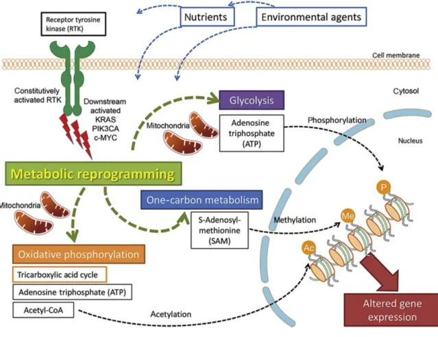
Fig. 1. Altered cellular energy metabolism in cancerogenesis. Reproduced with permission from Hagland et al. in Digestive Surgery, 2013.
Cellular metabolism and cancer
Nearly a century ago, the German physiologist and Nobel laureate Otto Warburg discovered an higher glucose flux through cancer cells(29). This glucose preference and metabolic switch observed in cancer cells is now recognized as one of the hallmarks of cancer (30). The increase in glycolysis is necessary to support rapid cell growth, and pathways emerging from the breakdown of glucose include the pentose phosphate pathway. Consequently, highly proliferative cells, must maintain a high carbon flux to sustain the high use of energy and require biomolecules to support the biosynthesis of new cells(31,32) which explains why
8
Pyruvate kinase proteins
One of the glycolytic enzymes which was found to be consistently altered during
tumourigenesis is pyruvate kinase. In 1980, Eigenbrodt supposed that pyruvate kinase M2 (M2-PK) was phosphorylated and inactivated by various newly-discovered proteins kinase, including the src protein tyrosine kinase (34). Eigenbrodt also suggested that inhibition of the pyruvate kinase step in glycolysis is necessary for channeling metabolites into the pentose-phosphate pathway to support nucleotide biosynthesis required by a rapidly dividing cells (35). Much attention has been paid on M2-PK because it is expressed in essentially all human cancers, and efforts have been made to use M2-PK as a cancer biomarker (36).
Pyruvate kinase protein structure
There are four mammalian pyruvate kinase isoforms (M1-PK,M2-PK, R-PK, L-PK) (37). Although a tissue may express more than one of them, individual cells generally express only one isoform at appreciable levels; most adult tissues express M2-PK, and expression of the other three isoforms is restricted to distinct tissues and cell types(38-40). The M1-PK isoform is found in tissues with high catabolic demand, such as muscle, heart, and brain. L-PK is the major isoform in the liver and a minor isoform in the kidney, which also expresses M2-PK. R-PK is found exclusively in red blood cells. M2-R-PK is the embryonic isoform and is expressed in many types of cancer. It is also expressed in normal proliferating cells, such as lymphocytes and colonocytes (39,41), but lack of proliferation does not necessarily mean lack of M2-PK expression.
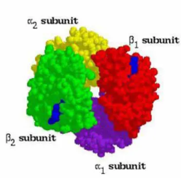
Mammalian pyruvate kinase is a tetrameric protein of identical subunits, which are arranged in a dimer-of-dimers configuration (Fig. 2,3). Each monomer contains one active site and is composed of three main domains – designated A, B, and C – plus a small N-terminal domain (42,43). The A domain is the largest domain, the active site is located in a cleft between the A
9
and B domains. The B domain is mobile and closes on the active site upon binding of the Mg2+-ADP substrate complex (44). The C domain is found on the opposite side of the A domain and contains the fructose-1,6-bisphosphate (FBP) binding pocket, present in the M2-PK, PKL, and PKR isoforms (45,46). FBP is a major allosteric activator of M2-PK. The C domains form the dimer–dimer interface of the fully-associated tetramer. Thus, differences in amino acids located at the dimer–dimer interface are responsible for differences in FBP binding and allosteric regulation of the M1-PK and M2-PK isoforms (46) (fig.3).
Pyruvate kinase is most active as a tetramer. Experiments show that M1-PK dimers retain catalytic activity, but that monomers are inactive (47). Unlike M1-PK, M2-PK is not a constitutive tetramer; the M2-PK isoform is subjected to reversible dissociation and inactivation when diluted in the absence of FBP ( 48). This is a key factor in tumour cells proliferation: when dissociated in the absence of FBP, the specific activity of M2-PK falls to only 4% of that found in an FBP-activated tetramer (48). Reversible activation of M2-PK triggered by FBP allows dynamic regulation of its enzymatic activity.
10
Fig3. Crystal structure of tetrameric M2-PK with bound ligands. The ribbon structure of tetrameric M2-PK (PDB 3BJF [8]) is shown, with a single subunit colored to represent individual domains. The A, B, C, and N-terminal domains are depicted in green, magenta, cyan, and yellow, respectively. Bound ligands are shown as gray spheres. In this structure, the catalytic site is occupied by K+, Mg2+, and oxalate (a PEP mimetic), and FBP is bound at its allosteric pocket. The binding site of amino acid binding between the A and C domains is indicated. The binding site for small molecule activators is also indicated.
Biochemical aspects of M2-pyruvate kinase
Pyruvate kinase catalyzes the direct transfer of phosphate from phosphoenolpyruvate (PEP) to ADP to produce ATP and pyruvate. This reaction is favourable due to the high energy released from PEP hydrolysis (49). During catalysis, the active site is occupied by both substrates (PEP and ADP, which is complexed with Mg2+) (50). A catalytic lysine residue stabilizes the penta-coordinate transition state that exists, as the phosphate is transferred directly from PEP to ADP. Tautomerization of enolpyruvate to the more stable ketoform of pyruvate contributes to the favourable energetics of phosphate transfer from PEP to ADP. In the absence of allosteric activators the M2-PK tetramers have low affinity for PEP and exhibit positive PEP cooperativity binding (dimeric form). Enzyme affinity for PEP at low PEP concentrations can be increased by binding of fructose-1,6-bisphosphate (FBP), an upstream glycolytic intermediate that is also thought to be the major allosteric activator of pyruvate kinase (51,52). Each M2-PK subunit contains one binding site that is specific for FBP and distinct from the active site (Fig. 3); FBP binding increase PEP binding affinity, promote tetramerization, and stabilize the enzyme tetramer in the active state. Many metabolites have been shown to have some effect on M2-PK activity (53) most of these effects are modest and
11
require high concentrations, thus calling into question their importance for the physiological regulation of M2-PK. The allosteric inhibitor phenylalanine (Phe) reduces both M1-PK and M2-PK activity by decreasing their affinity for PEP (54-56).
In vitro experiments have showed that M1-PK and M2-PK subunits are capable of forming
functional heterotetramers (57). This ability may explain why most individual cells restrict expression to a single isoform to retain the allosteric regulatory properties of that isoform. However, characterization of the tissue distribution of pyruvate kinase isoforms suggest that pyruvate kinase heterotetramers may exist in selected situations in vivo. Adult tissues express more than one pyruvate kinase isoform, but heterotetramers are not observed in most cases, providing biochemical evidence that expression of each isoform is usually restricted to a particular cell type (58).
M2-PK role in cancer
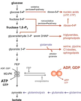
A proliferating cell must acquire and organize mass to replicate; this requires lipids for new membranes, carbohydrates for protein glycosylation, nucleotide precursors to support DNA replication and provide RNA for new ribosomes, amino acids and other cellular building blocks (59,60). ATP is critical to create and assemble these building blocks; but biosynthesis also need carbon and nitrogen precursors and the reducing equivalents necessary to process them (60,61). De novo lipid synthesis in particular requires significant expenditure of reducing power in the form of NADPH. Anabolic metabolism typically requires shunting of glucose carbon from glycolysis to biosynthetic pathways, most notably the pentose-phosphate pathway (PPP). The PPP produces ribose-5-phosphate and phosphoribosyl pyrophosphate (PRPP), which is required for nucleotide biosynthesis. The oxidative PPP also generates reducing equivalents in the form of NADPH. Glucose carbon is shunted from glycolysis for serine and glycine biosynthesis, and dihydroxyacetone phosphate is used to make glycerol-3-phosphate that can be further processed for generation of lipids. (fig 4). M2-PK controls the
12
final step of glycolysis, and its regulation serves to integrate intracellular signalling inputs with the metabolic state of the cell. Down-regulation of M2-PK activity and up-regulation of enzymes committing glucose to glycolysis have been thought to allow the accumulation of phosphorylated glycolytic intermediates, which are then available to spill into branching biosynthetic pathways (35).
Fig. 4. Glycolysis with debranching synthetic processes. A large level of the highly active tetrameric form correlates with high levels of ATP and GTP, high
ATP:ADP and GTP:GDP ratios as well as a high (ATP+GTP):(UTP+CTP) ratio. In contrast, high levels of the nearly inactive dimeric form of M2-PK correlate with low ATP and GTP levels, low ATP:ADP and GTP:GDP ratios as well as low (ATP+GTP):(UTP+CTP) ratio
Interestingly, cell culture experiments suggest that the ratio of pyruvate kinase activity to (2 x) phosphofructokinase activity is very high in neoplastic cells (62). These data suggest that the regulation of pyruvate kinase activity in proliferating cells may be particularly important to coordinate glucose metabolism with the synthesis of deoxy-nucleotides for DNA replication. Regulation of M2-PK activity by tyrosine kinase signalling is crucial for metabolic changes that support proliferative metabolism and tumour growth in several contexts (63,64). Following M2-PK deletion, pyruvate kinase expression is extremely low in proliferating tumour cells suggesting that low pyruvate kinase activity is important to support cancer cell proliferation in tumours. M2-PK, but not M1-PK, was also reported to play a role in cell cycle
13
progression via β-catenin trans-activation (65) and direct phosphorylation of histone H3 (66), mitotic progression and chromosome segregation.
Tetramer-dimer conversion of M2-PK
When M2-PK is mainly in the inactive dimeric form and not available for glycolytic ATP
production, energy can be provided by the degradation of the amino acid glutamine to
glutamate, aspartate, CO2, pyruvate, citrate and lactate, a pathway termed glutaminolysis (67).
Glutaminolysis and the truncated citric acid cycle have the metabolic advantage that a low
amount of acetyl CoA infiltrates into the citric acid cycle, thus it is saved for fatty acid and
cholesterol de novo synthesis. Fatty acids can be used for phospholipids synthesis or can be
released. Fatty acids and glutamate are immunosuppressive and may be capable of protecting
tumour cells from immune attacks (68). A key regulator of the M2-PK tetramer:dimer ratio is
not a stationary value in tumour cells and may oscillate depending on the concentration of key
metabolites as well as oncoproteins. A key regulator of the tetramer:dimer ratio of M2-PK and
the metabolic budget system is the glycolytic intermediate fructose 1,6-P2 (69).
High fructose 1,6-P2 levels induce the re-association of the inactive dimeric form of M2-PK
to the highly active tetrameric form. Consequently, glucose is converted to pyruvate and
lactate with the production of energy until fructose 1,6-P2 levels drop below a critical value
that allow the dissociation to the dimeric form.
Quantification of tumour M2PK
M2-PK is expressed in a variety of human cancers, so much attention has been paid to this enzyme and many efforts have been made to use it as a cancer biomarker (36). Tumour M2-PK (t-M2M2-PK) , the inactive dimeric form is in fact a characteristic metabolic tumour marker (70, 71,72). Initial studies in patients with lung, pancreas, liver, kidney and breast cancers
14
showed increased activity of M2PK in blood as well as cancer tissues and its role is emerging in the management of GI cancers (73-78). It can be measured in both blood and faeces.
M2PK in blood samples
Tumour M2-PK can be detected by a highly sensitive enzyme-linked immunosorbent assay (ELISA) which allows its the quantitative measurement in EDTA plasma samples. The test is based on two monoclonal antibodies which specifically react with t-M2-PK and do not cross-react with other isoforms of pyruvate kinase (types L, R, and M1) (79-81). t-M2-PK concentrations have been shown to be affected by haemolysis of blood sample, icterus and lipaemia. However, a correlation with the severity of these conditions has not been reported (82). Tumour M2-PK levels in EDTA plasma have been found to be elevated in bacterial infection as opposed to severe sepsis and polytrauma (83). Other benign conditions reported to have t-M2-PK elevations include: rheumatic diseases (84), diabetic nephropathy (85), chronic cardiac failure (86), inflammatory bowel disease (87) and acute and chronic pancreatitis (88). The suggested mechanism involves an increased glycolysis to meet the metabolic demand related to the stress of trauma and inflammatory reaction (88). Being t-M2-PK levels measured in EDTA plasma associated with these benign conditions, theyshould be interpreted with caution in GI tumours.
M2PK in stool samples
Tumour M2-PK can be measured in stools by an ELISA technique composed of the same monoclonal antibodies used in serum/plasma assay, which specifically react with tumour M2-PK in stool samples. In fact, CRC cells grow along the intestinal wall, when the tumor luminal surface is exfoliated; tumour cells can be found in stool samples, they can be used to measure tumour M2-PK levels. This new test does not depend on faecal blood, so it is also able to find non-hemorrhagic lesions. Effectively, in faecal material, the M2-PK epitope
15
remains stable more than 48 hours at room temperature and 1 year if stored at - 20 ° C. For this reasons, faecal determination of the M2-PK dimeric form was proposed in pilot studies forCRC diagnosis, obtaining very encouraging results (89-92).
INFLAMMATION
Inflammation may also represent a possible molecular link between host immune response, intestinal microbiome and genetic events in the development of CRC. Increased cell proliferation is mediated by prostaglandins and cytokines, which again are the result of an accelerated arachidonic acid (AA) metabolism. The AA metabolism is one of the major inflammatory pathways triggered by direct contact between the bowel wall and faecal water irritants, pro-inflammatory microorganisms and luminal carcinogens. The most important enzymes converting AA into pro-inflammatory cytokines are the cyclooxygenase enzymes (COX) and especially the inducible isoform COX2.
Related with the inflammation associated to the neoplastic development, is the generation of free radicals or reactive oxygen species (ROS). When present in excess, ROS are capable of damaging DNA and cellular macromolecules (93-98). In general, oxidative stress (OSS) can be defined as the imbalance between the generation of ROS and the availability of antioxidant defenses present in a system or biological compartment (99). A non-invasive method for direct measurement of overall OSS in humans has been developed, using a novel EPR
radical-probe. The probe, bis(1-hydroxy-2,2,6,6-tetramethyl-4-piperidinyl) decandioate
di-hydrochloride, quantitatively and instantaneously reacts with ROS (including superoxide) to yield the parent nitroxide, which is sufficiently persistent to be measured by EPR. This assay requires an average of 100 milligrams of tissue, blood or urine (more or less a drop), that could be obtained during other or clinical examination(100).
16
T
HE ENDOCANNABINOID SIGNALLING SYSTEMThe finding, in the early 1990s, of specific G-protein coupled receptors for the psychoactive component of Cannabis sativa (Δ)-D9-tetrahydrocannabinol (THC) (101), led to the discovery of a whole endogenous signalling system now known as the endocannabinoid system. During the past 15 years a remarkable amount of studies have been performed in order to understand the biological role of the endocannabinoid system and its regulatory functions in health and disease.
The endocannabinoid system consists of the cannabinoid receptors, their ligands (endocannabinoids) and regulatory enzymes (i.e. for their synthesis and inactivation).
Endocannabinoid receptors
Cannabinoid receptors are part of the “metabotropic membrane receptors” super-family, they are characterized by seven transmembrane domains, three intracellular loops, one extracellular amino-terminal domain with glycosylation sites and a intracellular carboxy-terminal domain that interacts, along with the third intracellular loop with a G-protein (fig5). Mammalian tissues contain at least two types of cannabinoid receptors: type 1 (CB1) and type 2 (CB2). CB1 receptors are mostly expressed in the central nervous system but also in most peripheral tissues including immune cells, the reproductive system, the gastrointestinal tract and the lung, while CB2 receptors are most abundant in the immune system, i.e. in tonsils, spleen, macrophages and lymphocytes (B-cells and natural killer cells) (102). Until recently, the only evidence for their expression in the digestive tract was the detection of CB2 messenger RNA in guinea-pig whole gut (103).
17
Fig5. CB1 e CB2 receptors are seven-transmembrane-domain proteins. The homology of the two receptors is about 44% (up to 82% in the transmembrane domains).
Endocannabinoids ligands
Endogenous ligands for the cannabinoid receptors are lipid molecules containing long-chain polyunsatured fatty acids, amides, esters and ethers, with different selectivity for the two receptor types (104,105). The most important among them are anandamide (arachidonylethanolamide) and 2-arachidonyl glycerol (2-AG) (fig.6). Anandamide is synthesized "on demand" from precursor N-Arachodonoil fosatidil-ethanolamine (NAPE) through the action of a specific phospholipase-D( PLD) (106). 2-AG is a product of the triacylglycerol metabolism and its synthesis is catalyzed by phospholipase C (PLC) (107). There is evidence that anandamide, and possibly also 2-AG, are removed from the extracellular space by a carrier mediated uptake process. Once inside the cell, anandamide is hydrolysed to arachidonic acid and ethanolamine by the enzyme fatty acid amide hydrolase (FAAH).
18 Endocannabinoid metabolism – hydrolysis
FAAH, an enzyme originally purified and cloned from rat liver microsomes (108) , catalyzes the hydrolysis of both AEA (as well as other long-chain fatty acid amides) and 2-AG to arachidonic acid and ethanolamine or glycerol, respectively. The structural and kinetic properties of FAAH have been widely reviewed in the literature (109) and several more or less selective FAAH inhibitors have also been developed (110) FAAH has an alkaline optimal pH and is found in microsomial membranes. Its hydrophobic domain is important for the formation of active oligomers, whereas its localization on intracellular membranes might be regulated by an SH-3 consensus, proline-rich sequence also necessary for enzymatic activity. The catalytic amino acid of FAAH has been identified as Ser241, and two other residues of the amidase consensus sequence, Ser217 and Cys249, participate in the enzymatic activity. (fig.7)
Fig.7Endocannabinoid system metabolism
Endocannabinoid system in cancer cells
Many tumour cells acquire the ability to synthesize autocrine growth factors to which they are responsive, thus reducing their dependence from exogenous growth stimulation. Furthermore, apoptosis is a potent mechanism that limits the expansion of tumour cells by triggering their suicide, while defects in apoptosis underpin both carcinogenesis and metastasis (111).
19
The endocannabinoids are ubiquitously synthesized molecules, with an emerging modulating activity on proteins and nuclear factors that regulate cell proliferation, differentiation and survival. This suggests that the endocannabinoid signaling system could be involved in the control of fundamental cellular processes.
CB1 receptors activation stimulates the ignition of the transduction pathway
RAF-1-MEK-ERK transduction pathway , which has the effect of
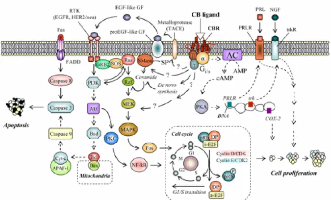
blocking the cell cycle in phase Go/G1-S limiting the cell proliferation. This happens through the activation of a Gi protein that blocks the adenylate cyclase, reducing cAMP cellular levels and, consequently, the action of PKA. This allows the activation of transduction pathways (ERK, MAP-K, etc.) that promote growth control and cell differentiation. CB1 stimulation also causes the suppression of Ras activity, this oncogene, if expressed, increases the amount of VEGF receptors. VEGF is a growth factor that induces angiogenesis. Cannabinoids also appear to be involved in the induction of apoptosis in tumour cells: it seems that the activation of the CB1 receptor causes an increase in intracellular ceramide, which, by increasing mitochondrial oxidative stress, induce the apoptotic mechanism. Apoptosis is also triggered by the activation of TRPV1 (transient receptor potential cation channel subfamily V member 1 or vanilloid receptor 1) by anandamide (AEA). This occurs through the increase of intracellular calcium, which activates the process of apoptosis. (fig 8)
20
Figure 8. Central growth factor signaling pathways in tumor cells involved in the molecular mechanisms of cannabinoid action.
Endocannbinoids and Cycloxygenase-2 (COX-2) in Cancer
COX-2 enzyme converts arachidonic acid to prostaglandins, prostacyclins and thromboxanes. COX-2 and prostaglandin E are commonly over-expressed in epithelial cancers including those found in the colon, lung, breast, and skin. In addition to metabolizing arachidonic acid, COX-2 catalyzes the conversion of AEA and 2-AG to ethanolamine-conjugated and glycerol-conjugated prostaglandins, respectively (112,113). Because these endocannabinoid-derived prostaglandins do not bind cannabinoid or prostaglandin receptors (114), increasing interest has developed in determining if these bioactive lipids mediate the cytotoxic effects of endocannabinoids. In colorectal carcinoma cells with elevated COX-2 expression, treatment with AEA resulted in increased E-series prostaglandin synthesis and cell death (115,116). Recently it has been shown that blockade of AEA degradation by inhibiting FAAH increased J-series prostaglandin synthesis and apoptosis. Thus, AEA-induced cell death in tumour cells which overexpress COX-2 appears to be caused by the conversion of AEA to cytotoxic
21
prostaglandins (115, 116,117,118). These responses were not reversed in the presence of CB1, CB2, or TRPV1 receptor antagonists.
Selective agonists and antagonists of CB1 and CB2, inhibitors of endocannabinoid hydrolysis, and cannabinoid analogs have been utilized to probe the pathways involved in the effects of the endocannabinoid system on cancer cell apoptosis, proliferation, migration, adhesion, and invasion.
The endocannabinoid system in colorectal cancer
Different molecules of the endocannabinoid system have been related to the CRC and adenomatous polyps. In 2003 Ligresti and colleagues demonstrated that differentiation of the colorectal cancer cell line CaCo-2, into non-invasive cells results in increased fatty acid amide hydrolase (FAAH) expression, decreased in endocannabinoid levels and no responsiveness to cannabinoids(119). They showed that either selective CB1 receptor stimulation (as previously found for breast and prostate cancer cells) or activation of both CB1 and CB2 receptors causes inhibition of proliferation. These findings are in agreement with the presence of both CBR subtypes in colon normal mucosa and CRC and suggest that endocannabinoids, present in high amounts in CRCs and particularly, in colorectal adenomas, might function as endogenous inhibitors of cancer growth. These data were also confirmed in vivo, where pharmacological enhancement of endocannabinoid levels (through inhibition of endocannabinoid hydrolysis) reduced the growth of tumour xenografts induced by the subcutaneous injection of rat thyroid transformed cells (120) as well as the development of precancerous lesions in the mouse colon (121).
Knowledge of the molecular factors involved in the CRC and in precancerous lesions is essential to identify possible markers for the identifications of these pathologies.
22
SCREENING RATES
Colorectal cancer is one of the commonest causes of cancer death in Europe (122). CRC pathogenesis is a multistep process that , in most cases, goes through adenoma-carcinoma sequence. Many forms of CRC may be prevented by routine control, which can detect precancerous neoplasms and early cancers before they undergo malignant transformation or metastasis (123). Prevention methods have the potential to detect both cancer and polyps, whereas detection methods generally show a low sensitivity for polyps and an even lower sensitivity for cancer. However, these are easier to execute and are more cost efficient. The United States Preventive Task Force recommends CRC screening for the average at-risk population, using a faecal occult blood test (FOBT), a periodic flexible sigmoidoscopy (FS), or a colonoscopy (124,125).
Fecal occult blood testing ( FOBT)
Fecal occult blood testing is based on the premise that polyps and cancer bleed more than normal mucosa. The amount of bleeding is related to the site of the cancer (highest blood loss from large lesions in the cecum and ascending colon) and increases with the size of the polyp and the stage of the cancer, therefore it can lead to diagnosis at an early stage.
(i-FOBT)
Fig 9. human haemoglobin structure.
The test is specific for human haemoglobin. It is based on antigen-antibody reaction using monoclonal antibodies. A latex reagent is prepared by sensitizing polystyrene latex particles
23
with rabbit IgGanti-human haemoglobin AOantibodies. If a stool sample contains haemoglobin, it reacts with the human anti Hb-AO antibodies that are bound to latex particles, thus producing a latex agglutination. This reaction is then analyzed in terms of optical density variation, the increase of which is directly proportional to HbAO concentration in the sample. Guidelines recommend the collection of two to three stool samples. Dietary restrictions and suspension of medication, such as aspirin, is controversial. FOBT must be done annually. Patients with positive results should receive a medical referral to undergo a colonoscopy (126). Unfortunately, colorectal screening is underused, and at least 40% of age-eligible adults do not adhere to up-to-date screening guidelines (127). Three strategies may improve colon cancer screening rates: convince the population about the importance of undergoing a screening test; achieve higher efficacy in standard screening tests and make them more available to the community (128). The current literature on Tumour M2-PK and endocannabinoid system molecules in CRC is limited but would justify further investigation of this novel cancer markers. They have a potential role in bowel cancer screening to be used as non-invasive screening test able to detect the pre-cancerous lesions, such as polyp adenoma. A proper screening trial is required in a consecutive, non selected population, to compare these markers with i-FOBT test and to evaluate their efficacy, feasibility and influence in cancer specific diagnosis. Further investigation, therefore, are needed to define their clinical role.
24
II. EXPERIMENTAL PART
METHODS
The aim of this study is to clarify whether FOBT, enzyme Tumor M2-PK and endocannabinoid system molecules (CB1, Cb2, FAAH), could represent diagnostic non-invasive markers, alone or in combination, for early diagnosis of CRC and its precancerous lesions (colorectal adenomatous polyps).
Population.
A prospective study was carried out in “S. Orsola-Malpighi” Hospital in Bologna in collaboration with “Giovanni XXIII” Hospital in Bari. It was conducted on 127 patient
undergoing colonoscopy in Digestive Endoscopy Units.
Eligible and not eligible criteria
All outpatients between 50 and 75 years, both gender, asymptomatic, undergoing screening for bowel diseases (i.e. those exposed to risk factors and/or with family history of crc) or patients attending our Units for abdominal symptoms related to organic bowel diseases were potentially eligible for the study. In particular, patients undergoing radiological or endoscopic procedures for diagnosis and/or clinical management of bowel lesions. Patients had to be able to understand the aim of the study and to answer the questionnaires. Before being enrolled each patient signed a consent form. Patients were excluded if they had undergone total colonoscopy in the previous 3 years, if they had been diagnosed with inflammatory bowel disease (IBD) or other gastrointestinal diseases, in case of coexisting serious illness or if they were on medication associated with intestinal inflammation or intestinal infection (specifically sought by stool culture). In addition, patients with inadequate colon cleansing or subjected to incomplete colonoscopy were excluded.
25
MATERIALS
Aim of this study was to use different techniques on different biological samples to evaluate markers for an early diagnosis of CRC.
Quesionnaires
Each patient gave us detailed individual information on dietary and lifestyle habits, family history and other risk factors by filling a questionnaire, helped by a trained interviewer.
In our Unit, all endoscopies were performed without anaesthesia by the same investigator (DV) using an Olympus GIF 100 video endoscope. Urine, blood, stool and biopsy samples were collected the day of the endoscopy. If they had to be shipped, dry ice or refrigerated boxes (-20°C) were used.
Biopsie samples
Biopsy specimens were obtained from both healthy and pathological tissue in patients affected with CRC or adenomatous polyps, and only in healthy tissue in “negative” patients .
The first biopsy was fixed in 4% paraformaldehyde, paraffin embedded and processed for histo-pathological and immunohistochemical evaluations. A second biopsy was quickly frozen for RNA or protein extraction and the third sample (about 0.2 g) was treated with 0.5 ml of standard saline solution containing hydroxylamine probe + 0.5 ml sterile distilled water to perform oxidative stress measurements.
Stool samples
The day before the endoscopy all patients collected a stool sample, that was rapidly frozen at-20°C. Faecal dimeric M2-PK (tumor M2-PK or t-M2-PK ) was detected using a quantitative
26
AG, Giessen, Germany); cut-off: 4 U/ml. Real Time PCR to evaluate CB1, CB2 and FAAH, was performed using RNAs isolated from exfoliated colonocytes using "immunomagnetic beads". FOBT was performed with latex agglutination method, using OC autosampling bottle 3”cuvettes (Eiken Chem, Tokio. Rivenditore Wellkang Tech, London), and analized by “DIANA oc sentor system”. cut-off 100ug/ml.
Urine and blood samples
Peripheral blood and urine samples were collected and immediately treated with the hydroxylamine probe. Samples (0.5 ml) were treated with 0.5 ml of standard physiological solution containing the hydroxylamine probe (1mM) and deferoxamine (1mM) as a metal chelating agent. After 5 min incubation at 37°C, samples were frozen in liquid nitrogen to stop any reaction until the EPR measurement for oxidative stress evaluation.
ANALYSIS
Analysis of colonoscopic findings
The result of each colonoscopy was classified according to the lesion with the worst prognosis. When no adenomatous polyps or cancer were found, colonoscopy was considered normal and histology was named “negative”. Adenomas were histologically classified as tubular, tubulovillous (25–75% villous component), villous or serrated subtypes. Advanced adenoma was defined as an adenoma of at least 10 mm in diameter, with high-grade dysplasia, villous or tubulovillous histological characteristics, or any combination of these. Cancer was defined as carcinoma if invading at least the submucosa across the muscularis mucosa (category 5 in the revised (Vienna classification of gastrointestinal epithelial neoplasia). In-situ and intramucosal carcinomas were classified as high-grade dysplasia. Cancers were graded in 4 stages (I-IV) according to the Tumour Node Metastasis classification . Adenomas treated by endoscopic polypectomy and found to harbour invasive
27
cancer (transformed adenomas) were classified as stage I cancers. These cases were named “positive” for histology.
Measurement of faecal tumour M2-PK and faecal occult blood test
All faecal samples were analysed at a single central laboratory (S. Orsola Hospital, Bologna, Italy) whit immuno latex agglutination faecal tests (HM-Jack, Kiowa; Olympus, Tokyo, Japan). They were processed using an automated reading technique “OC autosampling bottle 3” and analysed without rehydration by “ DIANA oc sentor system”, cut-off value was set at 100 ng/ml according to the results of a pilot trial (129). Faecal tumour M2-PK concentrations were determined using a commercially available sandwich ELISA based on two different monoclonal antibodies that specifically recognize the dimeric form of M2-PK (ScheBo Biotech AG, Giessen, Germany). A positive test result was defined as more than 4.0 U/ml, as indicated by the manufacturer.
Analysis of immunohistochemistry
The expression of M2-PK and endocannabinoid system markers (CB1, CB2, FAAH) was determined by a biotin free immunoenzymatic antigen detection system. This technique involves the sequential incubation of the colonic slides with an unconjugated rabbit or mouse primary antibody specific to the target antigen, an anti-rabbit or anti-mouse secondary antibody, a secondary antibody-HRP conjugate and substrate-chromogen (DAB ). DAB (3,3´-diaminodbenzidine) is a sensitive colorimetric substrate used with horseradish peroxidase (HRP) in immunohistochemistry applications. HRP catalyzes hydrogen peroxide oxidation of substrates such as DAB. Electrons are transferred by HRP from the DAB to the peroxide to yield an insoluble brown product.
Biopsies were formalin fixed, paraffin embedded, sectioned at 4 µm and mounted on glass
28
deparaffinised and rehydrated; a blocking step was performed whit protein block solution to
block nonspecific background staining and peroxidative block solution to suppresses
endogenous peroxidase activity (EXPOSE specific HRP/DAB detection IHC, ABcam). After that, slides were incubated whit primary antibody overnight at 4°C.
The primary Tumor M2-PK antibody (ab38237, Abcam, 1:30) is a rabbit polyclonal anti-human antibody directed against dimeric form of M2-PK (ScheBo Biotech UK Ltd, Basingstoke, UK); the primary anti- Cannabinoid receptor I (ab23703 Abcam, 1:250) and Cannabinoid receptor II (ab3561, Abcam, dilution 1:250) are rabbit polyclonal anti-human antibody directed respectively to Cannabinoid Receptor I and II. FAAH-1 antibody (ab128917, Abcam, dilution 1:250) is a mouse monoclonal anti-human antibody directed
against fatty-acid amide hydrolase1; Anti-Ki-67 primary antibody (ab833 Abcam, dilution
1:50) is a rabbit polyclonal anti-human antibody directed against ki-67 protein, a cellular marker for proliferation. The appropriate dilution for each primary antibody was determined by pilot experiments. Sections were visualized by adding a secondary rabbit or anti-mouse HRP conjugate antibody and chromogen substrate (DAB). Sections were finally counterstained in hematoxylin (Sigma, St. Louis, MO, USA) to visualize nuclei.
This trial was approved by “S.Orsola-Malpighi” Hospital Ethical Committee, according to the relevant institutional guidelines. Before being enrolled each patient read the information sheet and signed the consent format.
Statistical analysis
Statistical analysis for each test alone and their combination was performed by 2x2 tables, in comparison with the Gold Standard defined by colonoscopy-histology. Diagnostic accuracy was calculated in terms of sensitivity, specificity, positive predictive value (PPV) and negative predictive value (NPV). Analysis was performed in duplicate, considering as
29
obtained in the first case. This was carried out as a “blind” protocol, as the endoscopist was unaware of faecal tests results, and biologists were unaware of endoscopy reports. Data management, statistics and graphics (95% CIs, P<0.05) were performed using Ministat 2.1 software.
RESULTS
M2-PK and FOBT investigation on selected population
We previously analysed a population of 280 patients selected in this way: 46% (n. 126) “negative”, 16.8% (n. 47) CRC, 30.4% (n.85) high risk adenoma, 7.9% (n.22) low risk adenoma.
In these patients we evaluated diagnostic accuracy of M2-PK stool test alone or in combination with i-FOBT, compared with the Gold Standard. i-FOBT and tumour M2-PK concentrations in these different diagnostic subgroups were determined. Histology was normal (apart small hyperplasic polyps) in 126 patients; CRC was found in 47 and colorectal adenomous polyps in 107 patients (85 were “high risk” and 22 were “low-risk” adenomas) (tab. 1)
The concentrations of i-FOBT and tumour M2-PK in the different diagnostic subgroups of patients was determined (colon cancer/ advanced adenoma/ normal findings). For both markers, there were significant differences between patients with colon cancer and those with advanced adenoma (P<0.01) and between the groups with advanced adenoma and normal findings (P<0.01). Using the manufacturer’s cut-offs of 100 ng/ml faeces for i-FOBT, 4 U/ml for tumour M2-PK, the sensitivity, specificity PPV and NPV of the two tests for the detection of CRC are shown in Tables 2. i-FOBT showed the highest specificity and PPV (88.8%; 84.1–92.3 and 52.7% ; 39.8–65.3 respectively) ; M2-PK had the best sensitivity (87.2% ;74.8–94.0) and NPV (96.0%; 91.6–98.2). According to these results, i-FOBT+M2-PK was
30
the best combination of tests to predict cancer risk in our patients (Table 3). Patients who were found to be positive to both faecal tests had a 67% risk of developing colon cancer. In contrast, in those patients who were negative to both markers, the risk of cancer was as low as 4%. Patients with discordant test results (one positive and the other negative) had a risk of colon cancer of 16%, although it should be emphasized that the most common discordant results were the combination i-FOBT negative M2-PK positive (88 cases) rather than the opposite way round (only fifteen cases). So if we had tested our patients with i-FOBT test alone, as indicated by the screening program, 33 high risk adenoma and 14 CRC they would not have been detected.
For this reason we established to investigate this markers in association with other, in consecutive population to define their application in a context of CRC screening.
Tab.2 Accuracy of M2-PK and i-FOBT stool tests alone or in combination.
Sensitivity Specificity PPV NPV Diagnostic accuracy Likelihood ratio of a positive test Likelihood ratio of a negative test i-FOBT 61.7% (47.4–74.2) 88.8% (84.1–92.3) 52.7% (39.8– 65.3) 92.0% (87.7– 94.9) 84.3% (79.6– 88.1) 5.50 (4.9– 6.2) 0.43 (0.39– 0.48) M2-PK cut-off 4 U/ml 87.2% (74.8–94.0) 62.66% (56-67.7) 32.0% (24.6– 40.5) 96.0% (91.6– 98.2) 66.8% (61.1– 72.1) 2.33 (2.26– 2.40) 0.20 (0.14– 0.28) i-FOBT + M2-PK
(at least one positive)
91.5% (80.1–96.6) 57.08 (50,9-62,9) 30.1% (23.1– 38.0) 97.1% (92.7– 98.9) 62.9% (57.1– 68.3) 2.13 (2.08– 2.18) 0.14 (0.09– 0.24
31 Diet, lifestyle and cancer risk
One hundred thirty (130) consecutive patients were considered eligible for the study at the two participating centres during a 12-month period. Only 3 patients were dropped because of inadequate colon cleansing and/or for the lack of samples, so 127 patients were enrolled in this study (64 men/63 women; mean age 63, range 50–75 years).
For each patient detailed individual informationwere collected by filling a questionnaire in a
face to face interview. These included age, gender, dietary and lifestyle habits, comorbidity as chronic inflammatory diseases and history of medication use (aspirin, anticoagulants, hormones drugs) (Tab 4). The correlation of lifestyle factors and comorbidity with CRC risk
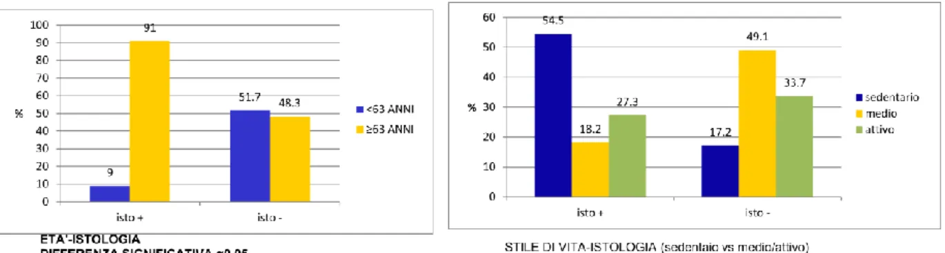
are shown in Table 5. The prevalence of colon lesions were significantly higher in patients of
an older age (>63 years) (91 vs. 48%; P<0.01, odd ratio 10,71) and sedentary lifestyle habits (64,5 vs. 17%; P<0.01, Odd Ratio 5,76) than in those without these factors. However, differences between gender (64 vs 49,1%, p 0,35, OD 1,81) , medication use and chronic inflammatory diseases, as well as red meat consumption were not statistically significant,
maybe this is due to the number of the analyzed population
.
(Tab2). In contrast consumptionof vegetable and fruit (at least three time/week) seems to prevent CRC (33% vs 5%; P<0,01, Odd Ratio 0,1).
32 fig. 10 Lifestyle factors correlation with CRC.
The result of each colonoscopy such as macroscopic features of polyps, size (measured before tissue fixation), location and number of lesions, were recorded by the endoscopist. A dedicated pathologist confirmed lesions on all the specimens by histological examination. When no adenomatous polyps or cancer were found, the diagnosis was considered “negative”. In accordance to TNM classification, CRC was found in 2 patients; 7 patients had high risk adenomas and 2 adenocarcinoma. Low-risk adenomas and hyperplasic polyps were found respectively in 10 and 4 patients, 7 with metaplasia, 4 had melanosis coli and only 1 had Crhon’s desease; histology was definite normal in 90 patients (tab 6). We used histological findings as the “gold standard”.
33
Measurement of faecal tumour M2-PK and faecal occult blood test
Using the manufacturer’s cut-offs of 100 ng/ml faeces for i-FOBT, 4 U/ml for tumour M2-PK, the sensitivity, specificity PPV and NPV of the two tests for the detection of CRC are shown in tables 7 and 8. i-FOBT showed the highest sensitivity 80.91 % ((95% CI: 74.1– 82.3) whereas M2-PK showed performances similar to i-FOBT in terms of specificity (74,14% ; (95% CI: 65.18-81.82), but markedly inferior in terms of sensitivity 63,64% (95% CI: 53.98 - 71.39); it showed best results to predict absence of lesions, VPN 95.56% (95% CI: 89-98.75) Performance of combined t-M2-PK and iFOBT tests is reported in table 9. In fact, patients with both positive tests have a 31.6% (95% CI: 23.10 to 39.81) risk of developing ccr; in contrast, in patients negative to both markers, cancer risk was as low as 2% (VPN 98.5%, 95% CI: 93.78 -99.66). M2-PK/ i-FOBT NUMERO CASI ISTOLOGICO VPP CI (95%) VPN (CI 95%) i-FOBT 19 (15%) 6+, 13- 31.6% 23.10% to 39.81% 64.4% 55.01% to 72.32% M2-PK cut-off 4 U/ml 39 (31%) 3+, 36- 7.7% 3.23% to 12.92% 92.3% 85.84% to 96.07% i-FOBT + M2-PK 69 (54%) 1+, 68- 1.5% 0.05% to 4.66% 98.5% 93.78 to 99.66%
(at least one positive)
34
Immunohistological staining for localisation of Tumour M2-PK
To investigate t-M2-PK expression in the large bowel, first of all biopsy sections were stained with haematoxylin and eosin to identify the morphology and verify the origin of the tissue. Immunohistological staining with monoclonal antibodies which specifically recognize the
dimeric form of M2-PK allows the visualization of the pyruvate kinase isoenzyme shift in
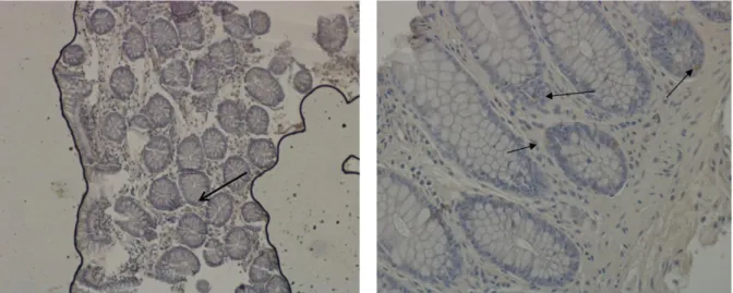
tumour cells. Negative controls were uniformly negative (fig 11a). The positive tissue controls all showed staining. Normal large bowel showed patchy staining of crypt cells (fig 11b). In low risk adenomas there was a signal in cytoplasm (fig 11c); high risk adenocarcinomas were strongly positive (++) in the cytoplasm (fig 11d). A strong signal was
observed in carcinomas. Thus, the amount of tumour M2-PK was found to increase in patients
with colorectal cancer cells as well as in stool samples and to correlate with pre neoplastic
35
Fig. 11. Distribution of M2PK immunoreactivity in preoneoplastic and neoplastic lesions in
colorectal cancer.
Fig.11a Negative controls Fig 11b Normal large bowel tissue
36
Immunohistological staining for localisation of CB1 and CB2 receptors
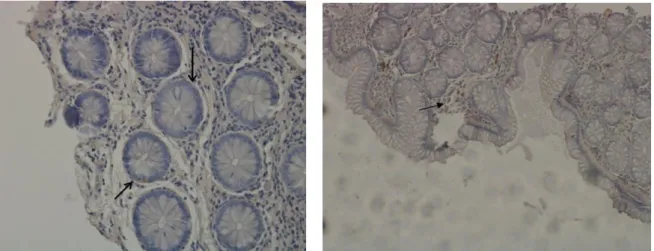
To investigate the expression of cannabinoid receptors in the large bowel, the expression pattern of CB1 and CB2 were studied immunocytochemically in different pathological stages as well as in normal tissue. DAB chromogen produce a brown staining on target epitopes (sections are counterstained with Hematoxylin). CB1 is expressed specifically in the epithelial crypts. Immunostaining with the AbCam CB1-receptor antibody showed intense positive expression in the absorptive epithelial cells with enhanced signal in microvilli, in particular on the apical surface facing the lumen. (Fig.12a). The goblet cells appear negative. Sub epithelial plasma cells in the lamina propria are weakly positive, this could be due to a blockage in antibody binding.
In low-risk adenoma (Fig. 12b), CB1 signal intensity appears very intense, though we observed a change of its location in epithelial cells. In hyperplasic polyps, it is possible to observe nuclei irregularities, hyperplasia and absence of CB1 receptor signal; in fact it is known that in advanced stages of carcinogenesis CB1 decreases until disappearing whereas it is still observed in the healthy tissue (Fig 12c,d ).
37
Fig. 12. Distribution of CB1 receptors immunoreactivity in preoneoplastic and neoplastic
lesions in colorectal cancer.
Fig. 12a Normal large bowel tissue Fig. 12b low risk adenomas
38
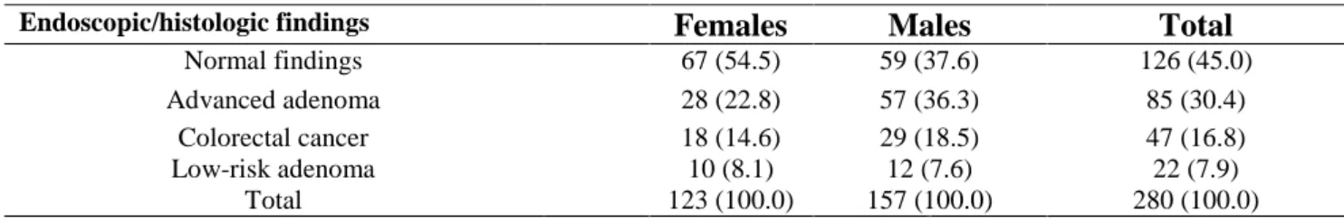
Similar tissue sections stained with the Abcam CB2-receptor antibody showed a different expression pattern. In normal colon tissue CB2 receptor is highly expressed in the subepithelial macrophages and plasma cells of the lamina propria, while the signal is not present in the epithelial crypts, nor on the apical surface of microvilli, in contrast to CB1 (fig 13a). The presence of CB2 receptor may depend on some degree of inflammation also present in healthy tissues. A low epithelial CB2 immunoreactivity was observed only in some samples (data not shown), this is probably due to normal variations in the inflammatory status of the gut, which is not evident macroscopically.
In patients with low-risk adenoma the CB2 signal is still intense, especially in areas surrounding the pathological tissue, where there is an ongoing inflammatory process(fig. 13b). In low risk adenoma it is possible to observe villi deconstruction, this is more evident in high risk adenoma (fig. 13c). However, CB2 signal decreases gradually and remains weak in the surrounding areas, where the healthy tissue presents macrophages. It is totally absent in carcinomatous tissue, probably due to the lack of an inflammatory response in the advanced stages of CRC (fig 13d).
39
Fig. 13. Distribution of CB2 receptors immunoreactivity in preoneoplastic and neoplastic lesions in
colorectal cancer.
Immunohistological staining for localisation of FAAH1 enzyme
In normal colonic tissue CB1 and CB2 receptors and FAAH enzymes are well expressed, as physiological regulators of the cell cycle. FAAH is the principal catabolic enzyme for a class of bioactive lipids called the fatty acid amides, such as anandamide.
Fig. 13a Normal large bowel tissue Fig. 13b low risk adenomas
40
In normal colon tissue the endocannabinoid-system activity, is regulated by the deactivating enzymes, FAAH, that hydrolyzes endocannabinoid ligands (AG and 2-AG), and indirectly decreases endocannabionid-system signalling (Fig14a). FAAH signal is well expressed already in low-risk adenoma (Fig. 14b), and increases in advanced stages as high risk adenoma and carcinoma.(Fig 14c and 14d)
An increased FAAH enzyme level in the advanced stages of CRC lower the availability of CB1 ligands, this reduces the inhibitory effects on cell proliferation induced by the endocannabinoid system.
Fig. 14. Distribution of FAAH receptors immunoreactivity in preoneoplastic and neoplastic lesions in
colorectal cancer.
Fig. 14a Normal large bowel tissue Fig. 14b low risk adenomas
41
The pivotal role of cell proliferation regulation has been demonstrated with Ki67 immunostaining. Antigen KI-67 is a nuclear protein associated with cellular proliferation through ribosomal RNA transcription. The fraction of Ki-67-positive tumor cells is often correlated with the clinical course of cancer.
Unfortunately so far it was not possible to perform the following tests: Real-Time PCR for CB1, CB2, FAAH enzymes in stool samples, nor expression of inflammatory molecules and oxidative stress in blood, biopsies and urine samples.
DISCUSSION
Colorectal cancer (CRC) is one of most frequently occurring forms of cancers worldwide and represents a high disease burden to society and second most common cause of death from malignant disease in Italy. Numerous epidemiological data from around the world, show major geographical variation with significantly higher risk in affluent societies and prospective cohort data have linked dietary habits and lifestyle factors to CRC(2). Risk factors for developing colon cancer include age, male sex, previous sporadic colonic polyps or FAP (Familial adenomatous polyposis), previous CRC and environmental factors, such as diet, weight and general lifestyle (130). In a consecutive population that we had previously analyzed, the prevalence of colon lesions were significantly higher in patients with an older age (>63 years) (91 vs. 48%; P<0.01, odd ratio 10,71) and sedentary lifestyle habits (64,5 vs. 17%; P<0.01, Odd Ratio 5,76) thesefinding arein accordance withthe literature (130).
It is proven beyond reasonable doubt that diet plays an important role in the CRC development, and it is equally accepted that the malignant transformation of colonocytes is a reaction to a constant or prolonged exposure to carcinogens in the colon. Luminal events in
the colon together with environmental exposure and genetics interact to create the conditions
42
Meat consumption increases the risk of CRC; this is the convincing evidence-based conclusion from numerous cohort studies. Numerous cohort studies and meta-analyses of more than 20000 cases of CRC show a 21%-28% increase in CRC risk with high intake of red and processed meat (15,16). However in our population we found a not statistically significant correlation between red meat consumption (3 time/week o more) and risk of developing adenomas/CRC; which was somewhat surprising and maybe due to the population size. In contrast, consumption of vegetable and fruit three time for week appears to lower CRC risk (33% vs 5%; P<0,01, Odd Ratio 0,1). When fibres reaches the colon, the result is a partial or a total fermentation leading to the production of short chain fatty acids and gas, which affects gastrointestinal function. Short chain fatty acids reduce the intraluminal pH securing optimal conditions for colonocytes and decreasing the conversion of bile acids to secondary bile acids (14). Dietary fibres increase bulking by stimulating growth of normal gut flora, and reduce the time and concentration of carcinogens in contact with the bowel wall (26).
There is an increasing interest among clinicians to understand risk factors, genetics and molecular biology of CRC . The precise molecular mechanism behind this phenomenon has yet to be defined, although some candidate pathways have been suggested.
M2-PK
In the bottom of each colonic crypt, 4-6 stemcells give rise to the enormous amount of colonocytes and host the potential of accumulating genetic and epigenetic changes (131). As a result of the ongoing and rapid proliferation, the colonocytes move from the lower parts of the crypts up towards the colonic lumen at a speed of approximately one cell position per hour.
When colonocytes reach the luminal surface they are exfoliated (132,133). Furthermore the
expression of the pyruvate kinase isoenzyme type M2, which can switch between a highly
active tetrameric form and a nearly inactive dimeric form, is an important metabolic sensor to
43
Therefore, different study in vitro, with human gastric carcinoma cell lines, had demonstrated
that tumour M2-PK reflects the metabolic activity and proliferation capacity of tumours.
By immunohistological staining with monoclonal antibodies which specifically recognize the
dimeric form of M2-PK, we observed its increase in patients with colonic precancerous and
cancerous lesions. This technique shows that normal large bowel has patchy staining of crypt cells (fig 11b). Weakly signal appear in the colonocytes cytoplasm, in patients with low risk adenomas (fig 11c). Immunostaining becomes very strong in high risk adenomas, still in the epithelial cell cytoplasm (fig 11d). It is important to note that this increase in
immunohistological signal correlates with tumor M2-PK concentrations (U/ml) detected with
quantitative ELISA test in stool samples where normal < low risk adenoma < high risk
adenoma < CRC.This suggest that the dimeric form of M2-PK (tumour M2-PK) is released
from gastrointestinal tumours into the stool of tumour patients, most likely by tumour necrosis
and cell turnover, providing the possibility of diagnostic application.
The quantification of the dimeric form of M2-PK in exfoliated colonocytes makes possible the use of t-M2-PK as a non-invasive early marker of CRC and as a tool for therapy control.
Many cancer types may be prevented by routine controls, that allow the detection of early carcinogenetic phases and especially a well-timed therapeutic approach before the malignant transformation and metastases spreading. CRC screening guidelines suggest annual i-FOBT testing; but unfortunately, colorectal screening is underused, and at least 40% of age-eligible adults do not adhere to up-to-date screening guidelines. It is essential to improve standard screening tests performances, to develop new and more sensitive diagnostic tools making them available as routine tests. For this reasons, we hypothesized that a combination of simple faecal tests, performed on a single stool sample, may help to identify patients exposed to higher risk of developing adenomas and/or CRC.
44
In a pivotal study we previously analyzed a selected population, using quantitative ELISA stool test (ScheBo® • Biotech AG, Giessen, Germany) for t-M2-PK detection and i-FOBT test. We defined as “positive group” patients with CRC and/or advanced adenoma and as “negative group” patients with normal histology and low risk adenoma. We observed statistically significant differences between these groups. The overall sensitivity of t-M2-PK test for CRC was 87% (95% CI: 75–94%); clearly higher than i-FOBT sensitivity, which was 62% (: 47–74%). t-M2-PK ELISA specificity was 63% (95% CI: 56–69%); lower than i-FOBT specificity, which was 89% (95% CI 84–92%). We found that i-FOBT and t-M2-PK test combination was the optimal ‘panel’ for faecal tests that yielded the highest diagnostic accuracy for CRC; indeed, sensitivity and NVP of this combination were 91% and 97%, respectively.
Considering the potential role of these faecal markers in detecting adenomas, especially the more advanced forms, which are at a higher risk of a rapid progression to cancer, we found that patients showing positivity to both i-FOBT and t-M2-PK tests had a 79.3% (PPV) risk of cancer; instead patients who resulted negative to both markers had a risk of cancer not greater than 4% (PPV). t-M2-PK marker crucial relevance is evident especially in discordant test results; it is important to remark that the most common discordant results were i-FOBT negative /t- M2-PK positive (88 cases), the opposite situation occurs only in 15 cases. If our patients had been subjected only to the i-FOBT test, as indicated by the screening program, 33 high risk adenoma and 14 CRC would not have been diagnosed.
Supported by these findings, we choose to continue with further investigations. In the context of CRC screening was important to evaluate whether this combination of tests would give excellent results also in a consecutive population. In this case stool t-M2-PK marker showed a lower sensitivity (74%) compared to previous results obtained in selected cases, although a valid VPN (95.56%) was obtained. Considering i-FOBT and M2-PK test combination, patients who were negative for both markers, had a cancer risk as low as 2% (NPV 98.5%).