Effect of cytochalasin B on the induction of chromosome
missegregation by colchicine at low concentrations in human
lymphocytes
Sandra Minissi
1,4, Bianca Gustavino
1,
Francesca Degrassi
2, Caterina Tanzarella
3and Marco Rizzoni
11Dipartimento di Biologia, Universita` di Roma ‘Tor Vergata’, Viale della
Ricerca Scientifica, 00133 Roma,2Centro di Genetica Evoluzionistica del CNR, Roma and31Dipartimento di Biologia, Universita` ‘Roma Tre’, Viale
Guglielmo Marconi 446, 00146 Roma, Italy
The aim of the present work was to investigate the possible
interference of cytochalasin B (cyt B) with low
concentra-tion treatment with colchicine in the inducconcentra-tion of
chromo-some/chromatid
loss
and
micronuclei
in
human
lymphocytes mitotically activated in vitro. Thus, cells from
a single female donor were treated with colchicine (10 or
25 nM, from 24 h after PHA addition to fixation at 66 h)
either in the presence or absence of cyt B. Single lagging
chromosomes/chromatids were scored in bipolar
ana-tel-ophases and greater damage (disrupted and c-anaphases)
was scored in cells at anaphase. Micronuclei were scored
in the first 4000 nuclei observed in both cyt B-treated (in
mononucleate and binucleate cells) and untreated cultures.
With the same criterion, FISH analysis was performed on
2000 nuclei where chromosome 7 and 11 centromeric DNA
probes were used in pairs. Our results showed that: (i) the
frequency of laggards and of micronuclei increased with
colchicine concentration but in the presence of cyt B there
was a lower frequency of both (with a mean reduction of
~49%); (ii) FISH analysis showed a colchicine
concentra-tion-dependent increase in nuclei with three spots for
chromosome 7; (iii) a colchicine concentration-dependent
increase in tetraploid cells was observed. This increase was
particularly remarkable (5-fold) in cells grown in the
presence of cyt B compared with cyt B-untreated cells. The
observed ‘cyt B effects’ can be explained if it is assumed
that in cytokinesis-blocked cells there is a shorter distance
between the poles. As a consequence: (i) laggards would be
engulfed in the nearest daughter nucleus with a consequent
lower induction of micronuclei; (ii) segregating sister
chromatids in heavily impaired anaphases would not travel
a sufficient distance to give rise to two daughter nuclei,
leading to an increased frequency of polyploid nuclei.
Introduction
Cytochalasin B (cyt B) is a chemical agent which inhibits
cytoplasmic cleavage (cytokinesis) without preventing nuclear
division (karyokinesis) (Carter, 1967). As a consequence, cells
that have divided once in the presence of cyt B can be easily
identified by the presence of two nuclei. It is noteworthy that,
under this condition, all the products of a mitosis are included
in the same cell.
In recent years, the cytokinesis-block (CB) method,
intro-duced by Fenech and Morley (1985), has been widely used in
the human lymphocyte micronucleus assay because it allows
4To whom correspondence should be addressed. Tel:139 6 72594812; Fax: 139 6 2023500; Email: [email protected]
restriction of the analysis to cells that have undergone one
mitotic division. Furthermore, the fluorescence in situ
hybrid-ization technique (FISH) with chromosome-specific
centro-meric DNA probes (Eastmond and Pinkel, 1990) on binucleate
cells has been shown to be a useful approach for studying
chromosome
missegregation
(loss
and
non-disjunction),
allowing recognition of micronuclei (MN) containing whole
chromosomes or identification of co-migration of sister
chromatids in the daughter nucleus (Zijno et al., 1994; Marshall
et al., 1996).
Based on the use of the CB method, recent work suggested
that in human lymphocytes treated with low concentrations of
spindle poisons, non-disjunction is induced more frequently
than loss events (MN) (Marshall et al., 1996; Minissi et al.,
1996; Zijno et al., 1996a; Sgura et al., 1997).
A prerequisite for such a conclusion is that cyt B treatment
does not interfere with the induction of chromosome
missegre-gation or, at least, the degree of interference should be known.
Earlier studies provided contradictory results on this issue.
According to these investigations, cyt B does not interfere
with MN induction (Fenech and Morley, 1985; Prosser et al.,
1988), but rather reduces MN induction in combined treatments
with colchicine (Antoccia et al., 1993) or changes the balance
between MN containing chromosome fragments and whole
chromosomes (Norppa et al., 1993; Surralle´s et al., 1996;
Falck et al., 1997).
The aim of the present work was to investigate the possible
interference of cyt B with the induction of chromosome/
chromatid loss and MN by colchicine (10 and 25 nM)
in cultured human lymphocytes in vitro. Such colchicine
concentrations were chosen because single chromosome
mis-distributions are induced and the c-mitotic effect does not
prevail (Gustavino et al., 1994). For our purposes,
ana-telophases of cells grown in the presence or absence of cyt B
were analysed for lagging chromosomes and chromatids, which
are the events leading to formation of MN. To obtain a more
general picture of the interference of cyt B with
colchicine-induced spindle damage, the ratio between disrupted anaphases
(in which two scattered groups of chromatids directed towards
the two poles can be distinguished) and c-anaphases (in which
only one group of chromatids can be distinguished) was
investigated. Moreover, the MN frequency per nucleus and the
distribution of hybridization centromeric signals (chromosomes
7 and 11) per nucleus were investigated in interphase cells
grown in the presence or absence of cyt B. For this purpose,
scoring was performed on randomly chosen activated
interphase nuclei (morphologically recognizable), regardless
of whether they were in mononucleate or binucleate cells. This
scoring criterion was chosen because in cyt B-untreated culture
it is impossible to distinguish interphase nuclei belonging to
cells that have undergone mitotic division, which are
compar-able with binucleate cells. Using this approach, it became
possible to compare randomized populations of activated nuclei
Fig. 1. Frequency (%) of single lagging chromosomes and chromatids in
bipolar ana-telophases of human lymphocytes treated in vitro with low concentrations of colchicine in the presence or absence of cyt B. The number of ana-telophases scored is shown in parenthesis below each column. The difference in the frequency of colchicine-induced laggards between cells treated and untreated with cyt B is significant at 0.05 (χ25 4.03). Data for statistical analysis were obtained by subtracting the respective control level from the two concentrations of colchicine and pooling the resulting data.
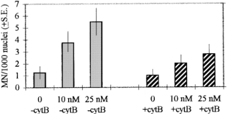
Fig. 2. MN frequency at interphase in human lymphocytes treated in vitro
with low concentrations of colchicine in the presence and absence of cyt B. The frequency is expressed per 1000 nuclei. At each concentration 4000 nuclei were scored. In cultures containing cyt B no distinction was made between mononucleate and binucleate cells. The difference in the frequency of colchicine-induced MN between cells treated and untreated with cyt B is significant at 0.01 (χ25 6.77). Data for statistical analysis were obtained by
subtracting the respective control level from the two concentrations of colchicine and pooling the resulting data.
Fig. 3. Human lymphocytes treated in vitro with low concentrations of colchicine (10 and 25 nM). (a) A normal anaphase; (b) a lagging chromatid in a
slightly disrupted anaphase (black arrow); (c) a disrupted anaphase; (d) a c-anaphase. Bars represent 10µm.
and was present until fixation. In order to obtain cytokinesis-blocked cells, 44 h after PHA stimulation cytochalasin B (Sigma) dissolved in DMSO was added to a final concentration of 6µg /ml. Three cultures were made in parallel for each concentration.
Both cyt B-treated and untreated cells were harvested 66 h after PHA stimulation. Lymphocytes were pelleted by centrifugation. After a mild hypotonic treatment (75 mM KCl for 2 min at room temperature) to preserve the cytoplasm, cells were gently fixed four times with methanol:acetic acid (3:1 for cyt B-untreated cultures and 5:1 in the case of cyt-B treated cultures). Fixed cells were stored at –20°C.
Giemsa stained slides
Slides for the analysis of anaphases and MN were stained with Giemsa (3% for 5–8 min). For each experimental point, the degree of spindle damage was studied scoring 100–200 anaphases for the frequency of normal bipolar, disrupted and c-anaphases; at least 100 bipolar ana-telophases were analysed for single lagging chromosomes and chromatids.
One hundred randomly chosen, activated cells in interphase were prelimin-arly scored for each point to estimate the frequencies of binucleate and multinucleate cells.
Micronucleus frequency was investigated, at each concentration, in the first encountered 4000, randomly chosen, activated interphase nuclei (morpho-logically recognizable) in both cyt B-treated (where nuclei were scored regardless of whether they belonged to mononucleate or binucleate cells) and cyt B-untreated cultures. MN were identified following the standard criteria (Heddle et al., 1983). As recommended by different authors, cyt B-induced multinucleate cells were not scored because they show a high micronucleus frequency and derive from multipolar divisions which frequently show aberrations in anaphase of cells that have divided more than once in the presence of cyt B (Lindholm et al., 1991; Norppa et al., 1993).
Slides were coded and scored blind by two scorers. Each scorer analysed half of the scored cells.
Fluorescence in situ hybridization (FISH)
FISH was performed using commercial centromeric DNA probes (Oncor) specific for the alphoid sequences of chromosome 7 (biotin-conjugated probe) and chromosome 11 (digoxigenin-conjugated probe). Chromosomes 7 and 11 were probed in pairs using FITC and rhodamine as fluorescent labels. Slides were pretreated with pepsin (Sigma) (50 µg/ml in 0.01 N
Fig. 4. Frequency of interphase nuclei with three hybridization signals for
either chromosome 7 or 11 in human lymphocytes treated in vitro with low concentrations of colchicine in the presence and absence of cyt B. Frequency is expressed per 1000 nuclei. For each concentration 2000 nuclei were scored. In cultures containing cyt B no distinction was made between mononucleate and binucleate cells. The difference in the frequencies of nuclei with three hybridization signals for chromosome 7 between cells treated and untreated with cyt B is not significant (χ25 1.00, P . 0.25).
The difference in the sum of frequencies of nuclei with three hybridization signals for either chromosome 7 or 11 between cells treated and untreated with cyt B is not significant (χ25 1.06, P . 0.25). Data for statistical analysis were obtained by subtracting the respective control level from the two concentrations of colchicine and pooling the resulting data.
HCl, 5 min at 37°C), dehydrated (3 min in cold 70, 90 and 100% ethanol) and denatured (70% formamide, 23 SSC, 2 min, at 70°C). The probes were denatured at 70°C for 5 min. Hybridization was performed overnight at 37°C in a moist chamber. The slides were washed in 50% formamide, 23 SSC at 42°C and then in 0.13 SSC at 60°C. The detection of chromosome 7 biotin-labelled probe was carried out with FITC–avidin (Oncor) and the fluorescence intensity was amplified using biotinylated anti-avidin antibody (Oncor), followed by an additional layer of FITC– avidin. The chromosome 11 digoxigenin-labelled probe was immunodetected using a mouse anti-digoxigenin antibody (Boehringer Mannheim) followed by an mouse–digoxigenin antibody (Boehringer Mannheim) and anti-digoxigenin–rhodamine antibody (Boehringer Mannheim). After immuno-detection slides were counterstained with DAPI (Sigma) and mounted in Vectashield (Vector laboratories).
The slides were examined with a Zeiss Axiophot microscope fitted with a FITC/rhodamine double bandpass filter set and a DAPI single bandpass filter set.
For the reasons explained above, analysis was restricted to monucleate and binucleate cells. For each experimental point, the first encountered 2000, randomly chosen nuclei were scored regardless of whether they belonged to mononucleate or binucleate cells (in cyt B-treated cultures). All nuclei with any number of hybridization signals were recorded. The frequencies of nuclei with three and four spots for one probed chromosome were estimated on all the four classes of nuclei which were found (1, 2, 3 and 4 spots). Nuclei with three hybridization signals for one chromosome always had two signals for the other. Nuclei with four hybridization signals for one chromosome always had four signals for the other, therefore they were classified as tetraploid nuclei. All the observed tetraploid nuclei were found in mononucleate cells, both in cyt B-treated and untreated cultures. Nuclei with one spot for one probed chromosome were considered mostly as artifacts due to signal overlap or poor probe penetration; their frequency ranged between 1 and 5%.
Slides were coded and scored blind by two scorers. Each scorer analysed half of the cells.
Differentially stained slides
For each experimental point, one slide was stained by the Hoechst1 Giemsa technique (Perry and Wolff, 1974) and a sample of 100 metaphases was analysed for the frequency of M1, M2 and M31 metaphases. Tetraploid metaphases were all M2and they were counted as two cells, being the result
of a lack of segregation of the two daughter cells following anaphase failure (colchicine) or cytokinesis failure (cyt B).
Slides were coded and scored blind by two scorers. Each scorer analysed half of the scored cells.
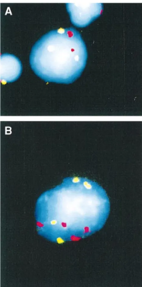
Fig. 5. Labelling of chromosome 7 (green) and 11 (red) centromeres by in situ hybridization in human lymphocytes treated in vitro with low
concentrations of colchicine. (a) A cell with three hybridization signals for chromosome 7; (b) a tetraploid cell, with four hybridization signals for both chromosomes 7 and 11. Bars represent 10µm.
Fig. 6. The frequency of interphase nuclei with four hybridization signals
for both chromosomes 7 and 11 in human lymphocytes treated in vitro with low concentrations of colchicine in the presence and absence of cyt B. Frequency is expressed per 1000 nuclei. At each concentration 2000 nuclei were scored; in cyt B-treated cultures nuclei were scored regardless of whether they belonged to mononucleate or binucleate cells. All scored tetraploid nuclei belonged to mononucleate cells. The difference in the frequency of colchicine-induced tetraploid cells between cultures treated and untreated with cyt B is significant at 0.001 (χ25 50.14). Data for statistical
analysis were obtained by subtracting the respective control level from the two concentrations of colchicine and pooling the resulting data.
colchicine in the presence (1) and absence (–) of cytochalasin B (cyt B) Colchicine concentration M1(2n) M2(2n) M2(4n) M3(2n) (6 cyt B) 0 nM –cyt B 58 42 0 nM1cytB 57 9 17 (χ25 0.0205, P . 0.75) 10 nM –cyt B 71 29 10 nM1cytB 67 9 12 (χ25 0.374, P . 0.5) 25 nM –cyt B 83 16 1 25 nM1cytB 77 9 7 (χ25 1.125, P . 0.25)
One hundred cells were scored per experimental point considering each 4n metaphase as two cells.
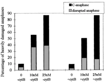
Fig. 7. Frequency (%) of disrupted and c-anaphases scored in human
lymphocytes treated in vitro with low concentrations of colchicine in the presence and absence of cyt B. 100 anaphases were scored with cyt B and 200 anaphases without cyt B. The difference in the frequency of colchicine-induced disrupted anaphases and c-anaphases between cultures treated and untreated with cyt B is significant at 0.005 (χ25 21.4 and 13.86,
respectively). Data for statistical analysis were obtained by subtracting the respective control level from the two concentrations of colchicine and pooling the resulting data.
Fig. 8. Interference of cyt B with chromosome content in daughter cells following colchicine-induced mitotic damage. In cytokinesis-blocked cells a shorter
distance between the poles is hypothesized. Following this hypothesis, laggards are engulfed in the nearest daughter nucleus with a consequent lower induction of micronuclei (A’ and A’’) and segregating sister chromatids in heavily impaired anaphases do not travel a sufficient distance to give place to two daughter nuclei, leading to polyploid restitution nuclei (B). The fate of a lagging chromatid (A’), of a lagging chromosome (A’’) and of a heavily impaired anaphase (B) are shown. Black bent arrows show engulfment of a chromatid/chromosome by the main nucleus. Column 1, misdistribution events; column 2, fate of misdistributed chromosomes/chromatids at telophase in the presence and absence of cyt B; column 3, karyotype composition of interphase daughter nuclei.
micronuclei was ~49% in the presence of cyt B, compared
with the corresponding frequencies observed in cyt B-untreated
cultures. Figure 3 shows examples of a normal anaphase
(a) and of a lagging chromatid at anaphase (b) in human
lymphocytes.
Both in the presence and in the absence of cyt B, FISH
analysis showed a concentration-dependent increase in the
frequency of nuclei with three hybridization signals for
chromo-some 7 (Figure 4). Such an increase was not observed for
chromosome 11. The reason for this apparent difference is not
understood at present and more studies are needed to verify
it. A photograph of a human lymphocyte with three spots for
chromosome 7 is shown in Figure 5. No significant difference
was found for the frequencies of three spot nuclei between cyt
B-treated and untreated cultures for chromosome 7.
By FISH analysis, a colchicine concentration-dependent
increase in tetraploid nuclei (four well-separated signals for
both chromosomes in the same nucleus; see Figure 5) was
observed (Figure 6). Such an increase was particularly
remark-able in cells grown in the presence of cyt B, where it was
5-fold higher than the corresponding frequency in cyt
B-untreated cultures.
An increase in the frequency of induced heavy damage at
anaphase (disrupted and c-anaphases) with increasing
concen-tration of spindle poison was observed (Figure 7). Both in the
presence and in the absence of cyt B a frequency of ~50%
damaged anaphases was recorded at 10 nM and of ~86% at
25 nM colchicine. However, in the presence of cyt B the
frequency of disrupted anaphases was about half of the
corresponding value observed in the absence of cyt B, while
the frequency of c-anaphases was remarkably higher. Thus, in
the presence of cyt B and colchicine treatment, the most
frequently induced damage is a lack of sister chromatid
segregation, with a frequency of 69% of c-anaphases at the
highest colchicine concentration. Figure 2 shows examples of
a disrupted (c) and a c-anaphase (d).
No significant difference was found in the frequency of M
1,
M
2and M
31cells between cell cultures grown in the presence
and absence of cyt B in colchicine-treated (10 and 25 nM)
and untreated lymphocytes (Table I). This result suggests that,
independently of the colchicine treatment, the addition of 6
MN frequency due to the omission of scoring of multinucleate
cells in cyt B-treated cultures is negligible; thus, considering
such underevaluation, an estimation of the reduction in MN
frequency due to the interference of cyt B is ~42%. The
present data should be confirmed in replicate experiments on
lymphocytes from different subjects, including male subjects.
Our results suggest that cyt B treatment interferes with
chromosome missegregation in human lymphocytes treated
in vitro with spindle poison (as first suggested by Eastmond
and Tucker, 1989; Migliore et al., 1989). The observed ‘cyt B
effects’ are likely to be explained if it is assumed that in
cytokinesis-blocked cells the absence of the actin ring interferes
with anaphase-B, leading to a shorter distance between the
poles, as suggested by Norppa et al. (1993), Surralle´s et al.
(1996) and Falck et al. (1997) and measured by Cimini et al.
(1997) in human fibroblasts. In Figure 8 the process and the
consequences of a shorter pole distance on daughter nuclei are
described.
According to this hypothesis laggards would be engulfed
in the nearest daughter nucleus, with a consequent lower
micronucleus yield and an underestimation of the frequency
of loss events in balance with non-disjunction (Figure 8A
9 and
A
99). A similar phenomenon could be hypothesized to explain
the lower frequency of MN containing whole autosomes
(Surralle´s et al., 1996) and acentric fragments (Falck et al.,
1997) in binucleate human lymphocytes.
On the basis of our estimations, starting from data on
laggard and MN frequency (see Appendix 1), a very small
increase in three spot cell frequency, due to the engulfment of
laggards into daughter nuclei, is to be expected. This explains
our results on three spot cells.
Another consequence of this hypothesis is that segregating
sister chromatids in heavily impaired anaphases would not
travel a sufficient distance to give rise to two daughter nuclei.
This may explain the transformation of disrupted anaphases
into c-anaphases and, consequently, the increased frequency
of polyploid nuclei we observed (Figure 8B). An increased
frequency of tetraploid cells was observed by Zijno et al.
(1996b) in human lymphocytes treated with another spindle
poison, vinblastin, in the presence of cyt B. The authors
proposed a similar model to explain such an effect: the lack
of a cleavage furrow could favour accidental nuclear fusion
after a badly damaged mitosis.
References
Antoccia,A., Tanzarella,C., Modesti,D. and Degrassi,F. (1993) Cytokinesis-blocked micronucleus assay with kinetochore detection in colchicine-treated human fibroblasts. Mutat. Res., 287, 93–99.
Carter,S.B. (1967) Effects of cytochalasins on mammalian cells. Nature, 21, 261–264.
human lymphocytes. Mutagenesis, 9, 17–21.
Heddle,J.A., Hite,M., Kirkhart,B., Mavournin,K., MacGregor,J.T., Newell,G.W. and Salamone,M.F. (1983) The induction of micronuclei as a measure of genotoxicity. Mutat. Res., 123, 61–118.
Lindholm,C., Norppa,H., Hayashi,M. and Sorsa,M. (1991) Induction of micronuclei and anaphase aberrations by cytochalasin B in human lymphocyte cultures. Mutat. Res., 260, 369–375.
Marshall,R.R., Murphy,M., Kirkland,D.J. and Bentley,K.S. (1996) Fluorescence in situ hybridisation with chromosome-specific centromeric probes: a sensitive method to detect aneuploidy. Mutat. Res., 372, 233–245. Migliore,L., Nieri,M., Amodio,S., Loprieno N. (1989) The human lymphocytes micronucleus assay: a comparison between whole-blood and separated-lymphocytes culture. Mutat. Res., 227, 167–172.
Minissi,S., Degrassi,F., Tanzarella C., Gustavino,B. (1996) Micronucleus induction and origin of trisomy in human lymphocytes as detected by centromeric FISH analysis on binucleate cells and chromosome counting in M2 metaphases. Atti Ass. Genet. Ital. Vol. XLII, Riccione (RN), 2–
5 October.
Norppa,H., Renzi,L. and Lindholm,C. (1993) Detection of whole chromosomes in micronuclei of cytokinesis-blocked human lymphocytes by antikinetochore staining and in situ hybridisation. Mutagenesis, 8, 519–525. Perry,P. and Wolff,S. (1974) New Giemsa method for the differential staining
of sister chromatids. Nature, 251, 156–158.
Prosser,J.S., Moquet,J.E., Lloyd,D.C. and Edwards,A.A. (1988) Radiation induction of micronuclei in human lymphocytes. Mutat. Res., 199, 37–45. Sgura,A., Antoccia,A., Ramirez.,M.J., Marcos,R., Tanzarella,C., Degrassi F. (1997) Micronuclei, centromere-positive micronuclei and chromosome nondjunction in cytokinesis blocked human lymphocytes following mitomycin C and vincristine treatment. Mutat. Res., 392, 97–107. Surralle´s,J., Falck,G. and Norppa,H. (1996) In vivo cytogenetic damage
revealed by FISH analysis of micronuclei in uncultured human T lymphocytes. Cytogenet. Cell Genet., 75, 151–154.
Zijno,A., Marcon,F., Leopardi,P. and Crebelli,R. (1994) Simultaneous detection of X-chromosome loss and non-disjunction in cytokinesis-blocked human lymphocytes by in situ hybridization with centromeric DNA probe; implications for the human lymphocyte in vitro micronucleus assay using cytochalasin B. Mutagenesis, 9, 225–232.
Zijno,A., Marcon,F., Leopardi,P. and Crebelli,R (1996a) Analysis of chromosome segregation in cytokinesis-blocked human lymphocytes: non-disjunction is the prevalent damage resulting from low concentration exposure to spindle poisons. Mutagenesis, 11, 335–340.
Zijno,A., Leopardi,P., Marcon,F. and Crebelli,R (1996b) Analysis of chromosome segregation by means of fluorescence in situ hybridization: application to cytokinesis-blocked human lymphocytes. Mutat. Res., 372, 211–219.
Received on January 5, 1998; accepted on September 17, 1998
Appendix 1. Calculation of the expected increase in the
frequency of nuclei with three hybridization signals due to
engulfment of lagging chromatids/chromosomes in cyt
B-treated cultures
The expected relative frequency of nuclei with three
hybridization signals due to engulfment of lagging chromatids/
chromosomes in binucleate cells after cyt B treatment can be
easily calculated on the basis of the hypothesis delineated in
Figure 8A’ and A’’. The equation is derived by multiplying
the probabilities of single events leading to the class of cell
of interest and by adding together the different probabilities of
pathways leading to it. The following assumptions were made:
(i)
all chromosomes act similarly, i.e. they have the same
probability, among them, to be lost or engulfed;
(ii)
the decrease in the frequency of laggards is due to
engulfment.
The following symbols are used in the equation:
cmsl, probability of chromosome loss, measured as the relative
frequency of ana-telophase with a single lagging
chromosome among all scored bipolar ana-telophases;
ctdl, probability of chromatid loss, measured as the relative
frequency of ana-telophase with a single lagging
chromatid among all scored bipolar ana-telophases;
eng, probability of a laggard being regained by a main nucleus
through engulfment;
i,
expected increase in the frequency of cells with three
hybridization signals for one probed chromosome.
Thus, the following equation can be written for binucleate cells
i
5 (0.5 cmsl 1 0.25 ctdl)eng/23.
The expected increases in the frequency of cells with
three hybridization signals for one probed chromosome were
calculated giving the following values to the variables of the
equation, on the basis of the empirical data (Figure 1).
dControl culture: cmsl
5 0%, ctdl 5 0.35%;
d
10 nM colchicine concentration: cmsl
5 1.48%, ctdl 5
1.48%;
d
![Assunzioni obbligatorie [dir. lav]](data:image/gif;base64,R0lGODlhAQABAIAAAP///wAAACH5BAEAAAAALAAAAAABAAEAAAICRAEAOw==)