UNIVERSITÀ DEGLI STUDI DI ROMA
"TOR VERGATA"
FACOLTA' DI SCIENZE MATEMATICHE, FISICHE E NATURALI
DOTTORATO DI RICERCA IN
BIOLOGIA CELLULARE E MOLECOLARE
XX CICLO
Role of ATM in Fas-induced apoptosis
in lymphoid cells
Venturina Stagni
Docente Guida/Tutor: Dott.ssa Daniela Barilà
Coordinatore: Prof. Gianni Cesareni
1
INDEX
INDEX ... 1
ABSTRACT ... 2
INTRODUCTION ... 3
ATM (ATAXIA-TELANGIECTASIA MUTATED) KINASE ... 3
Ataxia Telangiectasia... 3
ATM structure and function ... 4
ATM function in DNA damage signaling ... 5
ATM function in non-DNA damage signaling ... 7
ATM activation ... 8
Mechanism of ATM activation ... 8
ATM substrates ... 10
Proteomic approach for ATM substrates identification ... 12
ATM in the immune system and cancer... 13
DEATH RECEPTOR INDUCED APOPTOSIS: FAS PATHWAY ... 15
Apoptosis overview ... 15
Fas pathway ... 18
Fas pathway regulation: FLIP protein ... 20
FLIP function ... 21
Regulation of FLIP function and expression ... 23
Role of Fas pathway in the immune system and in cancer progression ... 24
AIM OF THE PROJECT ... 26
ROLE OF ATM IN FAS-INDUCED APOPTOSIS ... 26
MATERIAL AND METHODS ... 28
DNA CONSTRUCTS ... 28
ANTIBODIES AND OTHER REAGENTS ... 28
CELL CULTURE AND TRANSFECTIONS ... 28
ANALYSIS OF APOPTOSIS ... 29
ANALYSIS OF FAS-RECEPTOR LEVELS ... 29
FLOW CYTOMETRY OF PHOSHO-SER1981-ATM IN APOPTOTIC CELLS ... 29
IMMUNOFLUORESCENCE ANALYSIS ... 29
IMMUNOBLOTTING ... 30
CASPASE-8 ACTIVITY ASSAY ... 30
REVERSE TRANSCRIPTION-POLYMERASE CHAIN REACTION (RT-PCR) ... 30
STATISTICAL METHODS ... 30
RESULTS ... 31
ATM DEFICIENT CELLS ARE RESISTANT TO FAS-INDUCED APOPTOSIS ... 31
ATM KINASE ACTIVITY ENHANCES FAS-INDUCED APOPTOSIS ... 33
ATM KINASE ACTIVATION UPON FAS STIMULATION DOES NOT PLAY A MAJOR ROLE IN FAS-SENSITIVITY ... 34
ATM KINASE ACTIVITY PROMOTES CASPASE-8 ACTIVATION ... 36
ATM KINASE ACTIVITY DOWNREGULATES C-FLIP PROTEIN LEVELS ... 37
ATM KINASE ACTIVITY DOES NOT MODULATE FLIP MRNA LEVELS ... 41
ATM KINASE ACTIVITY MODULATES FLIP PROTEIN STABILITY ... 42
ATM KINASE ACTIVITY SENSITIZES HODGKIN LYMPHOMA CELLS TO FAS-INDUCED APOPTOSIS ... 43
DISCUSSION ... 44
FUTURE DIRECTIONS... 46
2
ABSTRACT
Ataxia Telangiectasia (A-T) is a rare cancer-predisposing genetic disease, caused by the lack of functional ATM kinase, a major actor of the Double Stand Break (DSB) DNA-damage response. A-T patients show a broad and diverse phenotype, which includes an increased rate of lymphoma and leukemia development. Fas-induced apoptosis plays a fundamental role in the homeostasis of the immune system and its defects have been associated with autoimmunity and lymphoma development.
We therefore investigated the role of ATM kinase in Fas-induced apoptosis. Using A-T lymphoid cells we could show that ATM deficiency causes resistance to Fas-induced apoptosis. A-T cells upregulate FLIP protein levels, a well-known inhibitor of Fas-induced apoptosis. Reconstitution of ATM kinase activity was sufficient to decrease FLIP levels and to restore Fas-sensitivity. Conversely, genetic and pharmacological ATM kinase inactivation resulted in FLIP protein upregulation and Fas resistance.
Both ATM and FLIP are aberrantly regulated in Hodgkin lymphoma. Importantly, we found that reconstitution of ATM kinase activity decreases FLIP protein levels and restores Fas-sensitivity in Hodgkin lymphoma derived cells.
Overall, these data identify a novel molecular mechanism through which ATM kinase may regulate the immune system homeostasis and impair lymphoma development.
ABSTRACT (in Italian)
L’Atassia Telangectasia (A-T) è una rara patologia genetica causata dall’assenza funzionale della chinasi ATM, la quale svolge un ruolo fondamentale nella risposta al danno a doppia elica del DNA. I pazienti A-T manifestano un’ampia gamma di fenotipi, tra i quali una maggior predisposizione allo sviluppo di linfomi e leucemie. L’apoptosi indotta da Fas gioca un ruolo fondamentale nel controllo dell’omeostasi del sistema immunitario e difetti nella via di trasduzione del segnale controllata dal recettore di morte Fas sono stati associati con lo sviluppo di linfomi e malattie autoimmuni.
Abbiamo quindi investigato il ruolo di ATM nell’apoptosi indotta da Fas. Nei nostri studi cellule linfoblastoidi che non hanno una proteina ATM funzionale sono sensibilmente resistenti alla morte indotta dal recettore Fas. Tale resistenza all’apoptosi correla con alti livelli di espressione nelle cellule di una proteina anti-apoptotica chiamata FLIP. La ricostituzione di queste cellule con una forma cataliticamente attiva di ATM è sufficiente per abbassare i livelli di FLIP e ripristinare la sensibilità all’apoptosi indotta da Fas. Invece, l’inibizione dell’attività chinasica di ATM, tramite inibitori farmacologici e/o genetici, porta all’innalzamento dei livelli di FLIP ed alla resistenza all’apoptosi indotta da Fas.
Sia ATM che FLIP sono regolati in modo aberrante nei linfomi di Hodgkins. Noi abbiamo scoperto che la ricostituzione dell’attività chinasica di ATM, in cellule derivate dai linfomi di Hodgkins, fa sì che i livelli di FLIP si abbassino e le cellule diventino sensibili all’apoptosi indotta da FAS.
Nel loro insieme questi dati hanno portato all’identificazione di un nuovo meccanismo molecolare attraverso il quale la chinasi ATM può regolare l’omeostasi del sistema immunitario e lo sviluppo di tumori a carico di questo apparato.
INTRODUCTION ATM (Ataxia-Telangiectasia Mutated) KINASE
3
INTRODUCTION
ATM (Ataxia-Telangiectasia Mutated) KINASE
Ataxia Telangiectasia
The ATM (Ataxia-Telangiectasia Mutated) protein was identified as the product of the gene that is mutated (lost or inactivated) in the human genetic disorder Ataxia-Telangiectasia (A-T) (Savitsky et al., 1995). A-T belongs to a group of diseases that are collectively known as ‘genomic instability syndromes’, each of which results from a defective response to a specific DNA lesion. A-T is characterized by cerebellar degeneration, which leads to severe and progressive neuromotor dysfuction, immunodeficiency, genomic instability, thymic and gonodal atrophy, a striking predisposition to lymphoreticular malignancies and extreme sensitivity to ionizing radiation and Double Stand Break (DSB) DNA damage -inducing agents (Fig.1) (McKinnon, 2004). This human disorder typically combines most of the hallmarks of a defective DNA-damage response, clearly pointing to the DSB as the lesion that elicits this defects. Indeed, cultured cells from A-T patients show a broad defect in responding to DSBs that span almost all of known branches of this response. The identification of ATM, which when mutated is the underlying cause of the disease, supported a rapid progression in the understanding of the molecular basis of this disease. Moreover, the striking clinical and cellular phenotype that is caused by ATM loss, clearly places this protein at the top position in the DSB-response cascade (Shiloh , 2003).
A-T patients suffer as a result of over 400 distinct ATM mutations, among them 85% are accounted for null mutations in the ATM gene (Becker-Catania et al., 2000). Thus approximately 85% of A-T suffers have no detectable ATM protein. There are a few reported genuine A-T cases with normal ATM protein levels; however, in these cases the protein is defective in its enzymatic activity (Stankovic et al., 1998).
Fig.1. Clinical symptoms, cellular phenotypes and cancer predisposition of individuals with mutations in the ATM (ataxia-telangiectasia mutated) gene.
INTRODUCTION ATM (Ataxia-Telangiectasia Mutated) KINASE
4
ATM structure and function
The ATM protein is a large molecule (approximately 350 kDa) which belongs to a conserved family of proteins, most of which possess a serine/threonine kinase activity (Shiloh Y., 2003). All of these proteins contain a domain typical of the lipid kinase phosphatidylinisitol 3-kinase (PI3K) (Fig.2), so they are dubbed ‘PI3K-like protein kinases’ (PIKKs). The PI3K domain harbours the catalytic site of the active protein kinases of the PIKK family. The ATM protein is characterized by the presence of other structural domains (Fig.2): the FAT domain, located N-terminally to the kinase domain, is conserved among PIKK family of proteins FRAP, ATR and TRRAP; the FATC domain, a 33aa conserved domain in the C-terminal of the protein. Recent works show that these two domains play a critical role in the regulation of ATM protein kinase activity (Bakkenist and Kastan,2003; Jiang X et al., 2006).The FAT domain of ATM contains serine 1981, the site that is autophosphorylated during ATM activation (Bakkenist and Kastan, 2003). The FATC domains binds to histone acetyltransferase TIP60
and this binding is important for ATM activation
(Jiang X et al., 2006). Instead the function of the large N-terminal portion of ATM is almost unknown, but this large portion is crucial to form productive complexes with some important ATM substrates proteins as p53 and c-Abl ( Khanna et al., 1998;Shafman et al., 1997), supporting the idea that the N-terminal of ATM is also essential for correct ATM function (Fernandes et al., 2005).The active protein kinases in the family, which are conserved from yeast to mammals, respond to various stress phosphorylating key proteins in the corresponding response pathways (Shiloh Y., 2003). They could therefore simultaneously affect several processes depending on the spectrum of their substrates. Four mammalian PIKKs are known to be involved in the DNA-damage response: the DNA-dependent protein kinase (DNA-PK), ATM, ATR and ATX .Whereas ATM and DNA-PK respond primarily to DSBs, ATR and ATX respond to both ultraviolet (UV) light damage (possibly UV-light-induced replication arrest) and DSBs, and ATR also responds to stalled replication forks. mTOR/FRAP is the only active kinase in this family that is not involved in the DNA damage response.
Fig.2. Size and common motifs in the human members of the PIKK family. The number of residues is indicated for
each protein. This family comprises six proteins, all of which (except for TRRAP) possess protein kinase activity. These proteins share three motifs: the FAT and FATC domains and the PI3K domain, which contains the phosphatidylinositol 3-kinase motif and harbours the catalytic site in the active 3-kinases of the family (see text).
INTRODUCTION ATM (Ataxia-Telangiectasia Mutated) KINASE
5
ATM function in DNA damage signaling
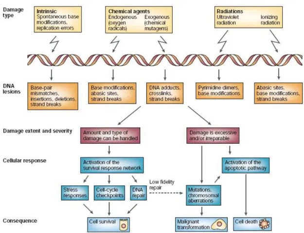
The greatest challenge to genome stability comes from DNA-damaging agents that can be either endogenous (occurring during normal cell metabolism) or exogenous (from the environment). Damaging agents such as radiation and reactive chemicals are capable of inducing a plethora of DNA lesions. Some of them are extremely cytotoxic if not repaired, whereas others are mutagenic and can affect the production, structure and function of cellular proteins, with consequences ranging from malfunction of the cell to malignant transformation. It is not surprising, therefore, that many mutagens are also carcinogens, and that there is a high correlation between their carcinogenic and mutagenic potencies. The basic cellular response is to arrest the cycle to prevent genome instability and to repair the damage, but the type and amount of damage might overwhelm the survival response machinery to the extent that programmed cell death (apoptosis) is initiated instead (Fig.3). The mechanism of this important choice between attempts at survival and programmed death is not entirely clear.
ATM is the prototype transducer of the Double Stand Break DNA damage. DSBs are naturally formed and sealed during processes such as meiotic recombination and the assembly of the T-cell receptor and immunoglobulin genes via V(D)J recombination, in T cells and B cells, respectively. It is safe to assume that cellular DSB repair mechanisms maintain continuous, low-level activity, ensuring that the occurrence and resealing of these breaks leave the cell unharmed. But when DSBs are inflicted on the genome by damaging agents, such as free radicals or ionizing radiation, their threat to cell life is sufficiently serious to set in motion, within minutes, a rapidly mounting, decisive DNA-damage response. Recent models depict the DSB response as developing through a series of steps (Fig. 3) (Shiloh Y., 2003).According to these models, DSBs might first be detected by sensor proteins that recognize the DNA lesion itself or possibly chromatin alterations that follow DNA breakage. The broken ends are then processed — their chemical nature is random, so they cannot serve directly as substrates for repair mechanisms. Then, the transducers are brought into action; these convey the damage signal to downstream effectors. It is this relay system from transducers to effectors that enables a single transducer to quickly affect the operation of many pathways .The transducers might also be involved in the assembly of DNA-repair complexes at the site of the damage , so DSB repair and signaling are probably concomitant and functionally linked . As mentioned above in the case of DSBs, the initial and primary transducer is ATM which transmits the message via a standard signalling mode: protein phosphorylation. In particular ATM kinase activity is induced upon DSB and modulates the cell cycle arrest and repair as well as the apoptotic response in case the damage is very extend preventing DNA replication in the presence of damaged DNA and genomic instability (Fig.3) (Shiloh Y., 2003).
INTRODUCTION ATM (Ataxia-Telangiectasia Mutated) KINASE
6
Fig. 3. Cellular responses to DNA damage. Different types of DNA damage cause different types of lesions, and
these, in turn, are handled by the cell in different ways. The outcome could be cell survival and resumption of the normal life cycle of the cell, cell death or malignant transformation. The mechanism of choice between attempt at survival and programmed cell death is not completely understood. The survival response is elaborate and encompasses many signalling pathways. (From Shiloh Y. 2003)
INTRODUCTION ATM (Ataxia-Telangiectasia Mutated) KINASE
7
ATM function in non-DNA damage signaling
Although the work of several labs supports the idea that ATM is a predominantly nuclear protein, involved in the signaling of DNA damage to the cell cycle checkpoint machinery, some
reports demonstrated that ATM is also a cytosolic protein (Watters, D et al. 1997) and that the kinase
activity of this protein is also activated by non-DNA damage signaling (Kastan MB & Lim DS., 2000; Lavin MF, 2000). For example Yang and Kastan (Yang DQ & Kastan MB., 2000) provide evidence that the kinase activity of this protein is also activated by insulin through a non-DNA damage signalling pathway to phosphorylate 4E-BP1 (PHAS-I), a regulator of protein synthesis. This report supports a more general signalling function of ATM in cell growth and proliferation .
Moreover, in the cytoplasm, ATM localizes to vesicles and interacts with β-adaptin (Lim et al., 1998)one of the components of the AP-2 adaptor complex, which is involved in clathrin mediated endocytosis of receptors (Robinson, 1994). This interaction between ATM and the vesicle associated proteins may play an important role in regulating vesicle and or protein transport in neurons. Dysfunction in these pathways may contribute to the progressive cerebellar degeneration of AT patients (Lim et al., 1998).
In post-mitotic neuronal tissue samples, it has even been shown that ATM is predominantly cytoplasmic (Oka and Takashima 1998; Barlow et al. 2000), which further indicates a role for cytoplasmic ATM in neuronal cell differentiation and survival. Finally a recent work on Science (Zhao-Hui Wu et al. 2006) demonstrated that ATM can traslocate in the cytosol upon the phosphorylation and the binding to NEMO protein, a regulatory subunit of IKK complex that plays a central role in regulating NFKB transcription factor.
Although the role of ATM kinase in the cytosol remains unclear, understanding this function of ATM might help to explain how mutations in the ATM gene cause the pleiotropic nature of the A-T phenotype. In fact, a role for ATM in intracellular signaling and in the cytosol has been suggested by several of the phenotypic changes observed in AT cells, such as the high level of growth factors required for cell growth (Shiloh, Y et al. 1983), the insulin resistance in AT patients (Bar, R. S et al. 1978), and the altered actin cytoskeleton in AT cells (McKinnon, P. J. & Burgoyne, L. A. 1985). In addition, uncharacterized accumulated cytoplasmic lipid vesicles and an increased number of lysosomes in AT patient samples have been observed in electron microscopic studies (Schoonderwaldt et al, 1977), suggesting altered lipid metabolism or altered lysosomal enzyme activity in AT cells.
So the large size of the ATM protein and its multiple subcellular localizations suggest that ATM may have more than one function.
INTRODUCTION ATM (Ataxia-Telangiectasia Mutated) KINASE
8
ATM activation
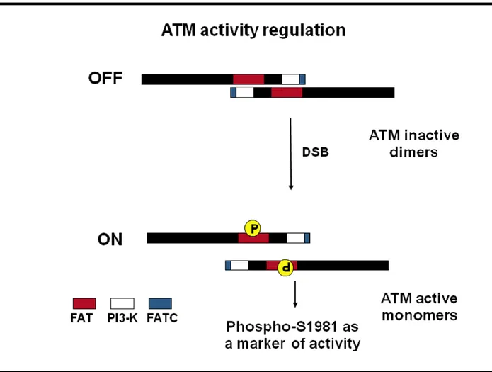
ATM is activated most efficiently by radiation and radiomimetic agents (Kurz et al. 2004). ATM resides predominantly in the nucleus in dividing cells, and responds swiftly and vigorously to DSBs by phosphorylating numerous substrate. The hallmark of ATM’s response to DSBs is a rapid increase in its kinase activity immediately following DSB formation. Researchers have long been impressed by the rapid phosphorylation of the many ATM substrates, which converts them within minutes to phosphorylated derivatives. A marked change in the activity of ATM would account for this massive process. Initial evidence indicated that ATM activation might involve autophosphorylation. A breakthrough in our understanding of this process came in a landmark publication by Bakkenist and Kastan (Bakkenist and Kastan,2003). They reported that ATM molecules are inactive in undamaged cells, being held as dimers or higher-order multimers. In this configuration, the kinase domain of each molecule is blocked by the FAT domain of the other (Fig. 4). Following DNA damage, each ATM molecule phosphorylates the other on a serine residue at position 1981 within the FAT domain , a phosphorylation that releases the two molecules from each other’s grip, into fully active monomers (Fig. 4). Within minutes after the infliction of as few as several Double Stand Breaks per turning genome, most ATM molecules become vigorously active.
Mechanism of ATM activation
There are different hypothesis about the molecular mechanism through which ATM is activated upon DSB DNA damage. Bakkenist and Kastan provide evidence that the signal for ATM activation might be chromatin alterations rather than direct contact of ATM with the broken DNA (Bakkenist and Kastan,2003). While it remains unclear whether the DNA double strand break per se or conformational change in chromatin resulting from the break initiates the process of ATM activation, other events associated with the process are better described. NBS1 (a component of the Mre11 complex) is conventionally thought to be a downstream substrate of ATM. However recent studies suggest that NBS1/MRN might function upstream of ATM, by recruiting ATM to the proximity of DNA damage sites and activating its enzimatic function (Uziel et al., 2003). In addition to the requirement for the Mre11 complex for ATM activation a number of post-translational modifications are also required. As mentioned above Bakkenist and Kastan showed that the phosporylation on S1981 on ATM plays an important role in its activation (Bakkenist and Kastan,2003). There is also evidence that dephosphorylation of ATM affects its enzimatic activity. Goodarzi et al. (Goodarzi et al. 2004) showed that the catalytic and scaffolding subunits of PP2A co-immunoprecipitated with ATM, in unirradiated cells, but dissociated from the complex after irradiation. These results suggested that PP2A associates with ATM in unperturbed cells to maintain it in an inactive state.
Moreover Sun Y. et al (Sun Y. et al., 2005) suggest a direct role for the histone acetyltransferase TIP60 in ATM activation in response to DNA damage. ATM in unstressed cells appeared to be associated through the FATC domain with TIP60. Upon DNA damage TIP60 is activated and directly acetylates ATM and this event is causal for kinase activation.
INTRODUCTION ATM (Ataxia-Telangiectasia Mutated) KINASE
9
Fig. 4. Model of ATM activation. In unstressed cells, the ATM kinase forms dormant dimers, distributed throughout the
nucleus. The ‘FAT’ domain of one ATM unit interacts with the enzymatic (kinase) domain of the other, and this probably locks ATM into a state in which it cannot interact with and phosphorylate its protein targets. Generation of DNA double-strand breaks (DSBs) by, for instance, ionizing radiation causes intermolecular modifications within ATM dimers that ultimately lead to their activation. The two bound ATM proteins phosphorylate each other on serine residue 1981 to form active monomers, and so this phosphorylation is consider a marker of ATM activity (Bakkenist and Kastan, 2003).
INTRODUCTION ATM (Ataxia-Telangiectasia Mutated) KINASE
10
ATM substrates
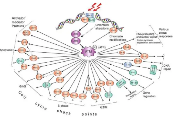
ATM-mediated phosphorylation either enhances or represses the activity of its targets, thereby affecting specific processes in which these proteins are involved. Similar to other active PIKKs (with the exception of mTOR/FRAP), ATM targets serine or threonine residues followed by glutamine (the ‘SQ/TQ’motif) (Traven A. and Heierhorst, 2005). Knowledge of the substrates of a protein kinase is essential to understanding its biological functions. So when ATM was identified as a member of the PIKK family, capable of phosphorylating a variety of substrates involved in Double Stand Break (DSB) DNA damage induced signalling, it was evident that it would play a key role in cell cycle checkpoint control, DNA repair and apoptosis (Fig.5) (Shiloh, 2003).
One of the first processes that is initiated by DSBs is the massive phosphorylation of the tail of a histone protein variant called H2AX(Redon et al., 2002). Foci of phosphorylated H2AX are rapidly formed at the DSB sites and are thought to be essential for further recruitment of repair factors, such as the MRN complex. H2AX phosphorylation , a very early event in the cascade induced by DSBs , was reported to be ATM dependent following DSB induction (Burma S. et al 2001).This process could therefore serve as a rapid and powerful mechanism for amplifying the damage signal via repeated cycles of H2AX phosphorylation, and recruitment of processing factors.
Another important substrate of ATM after DSB DNA damage is p53. A-T cells were defective in all the cell cycle checkpoints (Beamish and Lavin 1994). ATM plays a complex role in regulating the G1/S, S and G2/M checkpoints, directly phosphorylating substrates (Fig.5), activating other kinases to do this and ensuring rigid maintenance of the checkpoints by controlling signalling at multiple levels. This is illustrated for the G1/S checkpoint. When Kastan et al. (Kastan et al., 1992) demonstrated a defect in p53 stabilization in response to radiation exposure, p53 emerged as crucial mediator of G1/S checkpoint. They had previously shown that radiation exerted its influence on the G1/S checkpoint to delay the passage of cells into S-phase in order to facilitate DNA repair (Kastan et al., 1991). It is now history that ATM phosphorylates p53 on S15 and is responsible for other p53 phosphorylations that contribute to the efficiency of the transcriptional activation of p53 (Banin S. et al., 1998), responsible for G1/S checkpoint activation or induction of apoptosis (Lavin, 2006). ATM also phosphorylates and activates CHK2, a checkpoint kinase that phosphorylates p53 on Ser20 (Bartek, J. et al., 2001). This interferes with the p53–MDM2 interaction. The oncogenic protein MDM2 is both a direct and indirect inhibitor of p53, as it serves as a ubiquitin ligase in p53 ubiquitylation, which mediates its proteasome-mediated degradation. ATM also directly phosphorylates MDM2 on Ser395, which interferes with nuclear export of the p53–MDM2 complex, and hence the degradation of p53 (Khosravi et al. 1999). Finally, it has been reported that phosphorylations of p53 on Ser9 and Ser46 , and dephosphorylation of Ser376 , are ATM dependent as well, although the function of these changes is unknown (Saito S. 2002). This series of ATM-dependent modifications that activate and stabilize p53, although perhaps not complete , illustrates the elaborate way in which ATM handles a single effector, and indicates that ATM might regulate several effectors within the same pathway.
Most of these pathways have not been completely characterized, and the involvement of ATM substrates in them has been inferred simply from defective activation of specific checkpoints following abrogation of ATM-mediated phosphorylation of these proteins. It is possible that such proteins have a dual role in processes upstream and downstream of ATM. As mentioned above such is the case with NBS1 as well: on one hand, it is a component of the MRN complex that is thought to be
INTRODUCTION ATM (Ataxia-Telangiectasia Mutated) KINASE
11
involved in the initial processing of the DSB, and, on the other hand, it is a downstream effector of ATM in a checkpoint pathway.
So the emerging complex relationships between ATM and its substrates are drawing new flow charts for the DNA-damage signal that deviate from the traditional linear ones and assign to several proteins more than one role ‘upstream’ or ‘downstream’ in this chart (Fig.5).
INTRODUCTION ATM (Ataxia-Telangiectasia Mutated) KINASE
12
Proteomic approach for ATM substrates identification
The identification of downstream components of the ATM signaling network provide a necessary starting point for functional studies and will stimulate insights into diseases such as Ataxia Telangiectasia and the avoidance of cell cycle checkpoints in cancer, a critical early event in cancer progression.
The list of published ATM substrates is far from complete, and many ATM-dependent responses are likely to involve ATM targets that are unknown at present. Although a key to understanding any kinase network is the identification of the in vivo substrates, few techniques are available to identify protein kinasesubstrates so unbiased identification of kinase substrates is a difficult endeavor.Various techniques have emerged to identify phosphoproteins and kinase substrates (Ptacek et al., 2005; Dephoure et al., 2005), but few give in vivo confirmation or the sites of phosphorylation. However recently several groups made large-scale proteomic analysis of proteins phosphorylated by ATM kinase. Mu et al. (Mu et al., 2007) screened for potential ATM/ATR substrates using phospho-specific antibodies, against known ATM/ ATR substrates that recognize pSQ motifs, to immunoprecipitate potential new substrates. The proteins cross-reacting with phospho-specific antibodies, in response to DNA damage, were identified by mass spectrometry and the subset of candidate substrates for ATM/ATR-dependent phosphorylation was valitated in vivo. Interestingly, using this approach they identified proteins that belong to the ubiquitin-proteasome system (UPS) to be required in mammalian DNA damage checkpoint control thus revealing protein ubiquitylation as an important regulatory mechanism downstream of ATM/ATR activation for checkpoint control. Moreover recently Matsuoka et al. (Matsuoka et al., 2007) performed a large-scale proteomic analysis of proteins phosphorylated in response to DNA damage on consensus sites recognized by ATM and ATR and identified more than 900 regulated phosphorylation sites encompassing over 700 proteins. The identified proteins were annotated by the authors in gene ontology format (Fig.6). Functional analysis of a subset of this data set indicated that this list is highly enriched for proteins involved in the DNA damage response. More interestingly they identified also a large number of protein modules and networks not previously linked to the DNA damage response. This database paints a much broader landscape for the DNA damage response that was previously appreciated and opens new avenues of investigation into the responses to DNA damage in mammals.
Fig.6 Gene ontology analysis of the candidate ATM and ATR substrates (from Matsuoka et al., 2007). Gene ontology analysis of the candidate substrates was done with the PANTHER program. Of the 700 identified proteins, 421 were assigned with 769 biological processes. Proteins for which no biological process could be assigned were omitted from this display. Categories with more than 20 assigned proteins are shown.
INTRODUCTION ATM (Ataxia-Telangiectasia Mutated) KINASE
13
ATM in the immune system and cancer
Like the other genomic instability syndromes, A-T is a cancer-predisposing disorder. The higher cancer predisposition of A-T patients has been associated with the lack of DNA damage response, which results in genomic instability (Shiloh Y. 2003; Khanna KK. & Jackson SP, 2001) However, despite the nervous system being markedly affected in AT, the tumor types occurring in this disease are primarily lymphoma and leukemia (Gumy-Pause F. et al., 2004; Taylor AM, et al. 1996). This clinical feature is consistent with the central role of ATM in the management of the DNA DSBs generated during the immune system development and function in physiological conditions (Matei IR et al. 2006; Starczynski J et al 2003).
As mentioned above DNA damage checkpoints and repair of DNA double strand breaks (DSB) are necessary to preserve genomic integrity during V(D)J recombination, a process required to rearrange B-cell and T-cell receptor (TCR) gene segments. Although responsible for enormous diversity of the immune system, chromosome breakage and rejoining during V(D)J recombination contribute to oncogenic transformation in the context of defective DNA damage checkpoints and/or DNA DSB repair (Matei IR et al. 2006). Thus, impairments in ubiquitous DNA damage detection and/or repair pathway result in genomic instability. Defects in repair protein, as ATM kinase, involved in rejoining V(D)J recombination-induced DSBs preclude the generation of antigen receptor, profoundly compromising T- and B-cell development and causing severe immune deficiencies (Revy P et al. 2005). Moreover Atm-deficient mice show a striking predisposition to lymphoid malignancies, particularly thymic lymphomas, to which they succumb before the age of 1 year (Shiloh Y. 2001; Shiloh T. and Kastan MB 2001).
However, much of the literature on ATM mutations and cancer is not about A-T patients, but is, instead, on heterozygous carriers of A-T mutations. For more than two decades, ATM has been of interest to cancer epidemiologists and the public because of observations of cancer predisposition among A-T carriers. These studies pointed to a high incidence of malignancies, particularly breast cancer, among unaffected members of A-T families (Khanna, K.K., 2000). In view of the estimated 1– 2% frequency of A-T carriers in the general population, this observation has important implications for public health. A-T mutations lead, in most cases, to truncated, unstable protein products, and these ATM alleles therefore fail to produce any ATM at all. Carriers of such mutations have a reduced amount of otherwise functional ATM. Other A-T mutations lead to amino-acid substitutions (missense mutations) or in-frame deletions that produce a catalytically inactive protein. Cells of carriers of these mutations are expected to contain both functional and inactive ATM molecules in various ratios. A meticulous study of the type of ATM mutations in A-T families with high incidence of cancers disclosed a high frequency of missense mutations (Stankovic, T. et al 1998). The importance of missense mutations in predisposing carriers to cancer can be explained simply by the dominant- negative effect of the inactive version of the protein leading to a reduction in ATM function .
Further evidence of the importance of ATM missense mutations as cancer-causing genomic alterations came from the search for somatic ATM mutations in sporadic human tumours. Indeed, ATM was found to undergo somatic mutations in sporadic lymphoid tumours, behaving like a tumour-suppressor gene in these malignancies (Stankovic et al.2002). In particular ATM gene alterations, mainly missense mutations, have been reported frequently in adult lymphoid malignancies ( Gumy-Pause et al. 2004). More recently, childhood acute leukemias and Hodgkin disease were investigated for ATM gene alterations. ATM germline missense variants were frequently reported (Gumy Pause F et al. 2003; Liberzon E et al. 2004) and two studies showed that some of the detected variants were pathogenic, encoding functionally abnormal protein (Takagi M et al. 2004; Oguchi K et al. 2003) These results suggest that ATM is also involved in the pathogenesis of childhood lymphoid malignancies. These observations seem to reconcile the debate on the role of ATM mutations in
INTRODUCTION ATM (Ataxia-Telangiectasia Mutated) KINASE
14
genetic predisposition to cancer (Khanna, K.K., 2000; Stankovic, T. et al 1998; Gatti r. et al, 1999), and place the gene encoding the ATM protein well within the list of genes that are involved in cancer morbidity in the general population.
Thus understanding the contribution of ATM gene mutations to cancer predisposition in human populations will keep this gene in the spotlight of cancer epidemiology.
INTRODUCTION Death receptor induced apoptosis: Fas pathway
15
Death receptor induced apoptosis: Fas pathway
Apoptosis overview
Multicellular animals often need to get rid of cells that are in excess, or potentially dangerous. To this end, they use an active dedicated molecular programme, which is as important as cell division and cell migration and allows the organism to tightly control cell and tissue homeostasis. Various researchers during the past two centuries have observed this phenomenon and apoptosis is the term finally adopted, coined by Currie and colleagues in 1972 (Kerr et al., 1972).
Apoptosis is a genetically programmed, morphologically distinct form of cell death that can be triggered by a variety of physiological and pathological stimuli. This evolutionarily conserved form of cell suicide requires specialized machinery and the central components of this machinery is a proteolytic system involving a family of cysteinyl aspartate specific proteinases known as caspases (Earnshaw et al., 1999).
The dying cells shared morphologically features that can be seen under a microscope as this sequence:
1. The cell becomes circular because the cytoskeleton is digested by the caspases.
2. The chomatin undergoes degradation and condensation into compact patches against the nuclear envelope.
3. The nuclear double membrane started to be dismantle. The caspases begun to degrade the lamin that underlies the nuclear envelope.
4. The nuclear envelope becomes discontinous and the DNA inside it is fragmented. 5. The nucleus breaks into several discrete chromatin bodies or nucleosomal units. 6. Plasma membrane blebbings
7. The cell or the apoptotic bodies are phagocytesed.
This readily visible transformation (Fig.7) is accompanied by a number of biochemical changes like the externalization of phosphatidylserine at the cell surface and the activation of the caspase proteolityccascade.
INTRODUCTION Death receptor induced apoptosis: Fas pathway
16
Apoptosis can be triggered essentially in two distinct ways: intrinsic and extrinsic . In both cases effector caspases activated downstream are responsible for all the biochemical and morphological changes observed during apoptosis (Fig.8). Irreparable genome damage, caused by mutagens, pharmaceuticals or ionizing radiation, activates the intrinsic pathway, in which cytochrome c is released from the mitochondria and starts the assembly of the apoptosome. Apaf-1, an oligomeric cytoplasmic protein, binds the released cytochrome c and then undergoes an ATP-dependent conformational change that allows binding of pro-Caspase-9 through N-terminal CARD domains present in both molecules (Hofmann et al., 1997). This binding increases the local concentration of Caspase-9, leading to dimerization and promoting reorientations of the caspase’s activation loop (Renatus et al., 2001). In addition the juxtaposition of pro-Caspase-9 molecules in the complex permits transcatalytic cleavage (Srinivasula et al., 1998).
In the extrinsic pathway, the apoptotic signal is initiated by direct ligand-mediated activation of death receptors like CD95 at the cell surface and the apical caspases involved are pro-Caspase-8 and pro-Caspase-10 (Kischkel et al., 2001).
The death-receptor and mitochondrial pathways converge at the level of Caspase-3 activation. Cross-talk and integration between the death-receptor and mitochondrial pathways is provided by Bid, a apoptotic Bcl2 family member. Caspase-8 mediated cleavage of Bid greatly increases its pro-death activity, and results in its translocation to mitochondria, where it promotes cytocrome c exit. Nevertheless the contribution of this cross-talk is minimal and the two pathways operate largely independently each other (Hengartner, 2000) (Fig.8).
INTRODUCTION Death receptor induced apoptosis: Fas pathway
17
Fig.8. Two major apoptotic pathways in mammalian cells. (Hengartner, 2000). The death-receptor pathway (left
pathway in the figure) is triggered by members of the death receptor superfamily (such as CD95 and tumour necrosis factor receptor I). Binding of CD95 ligand to CD95 induces receptor clustering and formation of a death inducing signalling complex (DISC). This complex recruits, via the adaptor molecule FADD (Fas-associated death domain protein), multiple procaspase-8 molecules, resulting in caspase-8 activation. Caspase-8 activation can be blocked by recruitment of the degenerate caspase homologue c-FLIP. The mitochondrial pathway (right) is used extensively in response to extracellular cues and interna insults such as DNA damage These diverse response pathways converge on mitochondria, often hrough the activation of a pro-apoptotic member of the Bcl-2 family.
INTRODUCTION Death receptor induced apoptosis: Fas pathway
18
Fas pathway
CD95 (APO1/Fas) is the best characterized member of the tumor necrosis factor (TNF) superfamily of receptors like TNF and TRAIL receptors (Fig.9). Members of the TNF-R family have pleiotropic action. Depending on the signal and on the cell type these receptors can trigger proliferation, survival, differentiation or death. CD95 has three cysteine-rich extracellular domains and an intracellular death domain (DD) essential for signalling. The receptor mediates apoptosis when triggered by agonistic antibodies or its cognate oligomerizing ligand, CD95L, expressed on cell membranes or in a soluble form. CD95L belongs to a corresponding coevolved family of proteins, the family of TNF and its related cytokines (Peter et al., 1997).
Fig.9. Tumor necrosis factor superfamily (Curtin JF and Cotter TG., 2003).Schematic diagram depicting the death
receptors Fas, TNF-R1, DR3, TRAIL-R1, TRAIL-R2 and DR6 and the decoy receptors DcR1, DcR2 and DcR3. Ligands that are known to bind to these receptors are all shown and are predominantly membrane bound. Some death receptors can bind with more than one ligand and some ligands bind to more than one receptor as indicated. Important adaptor proteins that are recruited to each receptor and that are involved in signal transduction are also indicated.
INTRODUCTION Death receptor induced apoptosis: Fas pathway
19
CD95 receptors are expressed on the surface of cells as preassociated homotrimers (Papoff et al., 1999) and mutations on the domain that impairs this association cause the autoimmune disorder called ALPS, ‘Autoimmune Lympho Proliferative Syndrome’ (Siegel et al., 2000). Upon specific ligand binding (Wallach, 1999) there is assembly of a complex of adaptor proteins that leads to clustering of the two Fas homotrimers (Holler et al., 2003). This enables the adapter molecule FADD/MORT1 to interact with the Death receptor via homophilic interaction of the death domains (DD). FADD is a protein of 22 kDa containing two structurally similar protein motifs, the N-terminal Death-Effector Domain (DED) and the C-terminal Death-Domain (DD).
FADD molecules interact then with the death protease Caspase-8 (FLICE) via homophilic interaction of death effector domains (DED). The assembly of this proteins constitutes the death-inducing signalling complex (DISC) (Fig.10) (Medema et al., 1997).
When pro-Caspase-8 dimerizes, its activity is induced and it acquires the capability to cleave the adjacent dimer (Boatright, 2003) (Chang et al., 2003) (Donepudi, 2003). This starts the processing (Medema et al., 1997) (Muzio, 1996) of pro-Caspase-8 to form the active Caspase-8, and initiate the caspase cascade (figure 5). Upon prolonged triggering of CD95 with agonistic antibodies all cytosolic caspase-8 gets proteolitically activated. Physiological caspase-8 cleavage requires association with the DISC and occurs by a two-step mechanism. Initial cleavage generates a p43 and a p12 fragment further processed to a p10 fragment. Subsequent cleavage of the receptor-bound p43 results in formation of the prodomain p26 and the release of the active site-containing p18 (Medema et al., 1997). Pro-Caspase-8 activation is absolutely required to trigger this apoptotic response (Juo et al., 1998) and its catalytic activity has to be tightly regulated to avoid inappropriate activation and undesired cell death (Peter, 2004). Several mechanisms contribute to the control of the apoptotic response regulating caspase-8 activity. In particular the most important player in the control of caspase-8 activation and Fas signalling is FLIP protein that modulates the caspase-8 recruitment to the DISC (Peter, 2004). Moreover recently we demonstrated (Cursi et al., 2006) that tyrosine phosphorylation plays a central role in the control of caspase-8 processing and activity. We have shown that Src kinase directly phosphorylates Caspase-8 on Tyr 380. Src activity triggers endogenous Caspase-8 tyrosine phosphorylation and protects cells from Fas-induced apoptosis, suggesting that tyrosine phosphorylation directly modulates Caspase-8 activity and function (Cursi et al., 2006).
INTRODUCTION Death receptor induced apoptosis: Fas pathway
20
Fig.10. Fas pathway and DISC formation
.
Fas pathway regulation: FLIP protein
To avoid uncontrolled cell death or tissue damage, apoptosis is tightly controlled by a collection of inhibitors. Both viral and cellular inhibitors exist which block cell death at different levels. Proteins of the inhibitor of apoptosis (IAP) family directly bind and inhibit caspase-3, -6, -7 and -9 (Deveraux et al., 1999), whereas Bcl-2 family members regulate apoptosis that is induced by the mitochondrial pathway (Adams et al., 2001).
There have been several recent advances in defining the molecular and physiological function of a novel family of inhibitors of death-receptor induced apoptosis, called FLIP, also known as FLICE/caspase-8 inhibitory proteins. FLIP was originally identified as a viral gene product, viral FLIP (vFLIP), while investigators were searching genomes for proteins that contain a DED in an effort to identify molecules that interact with caspases (Thome, M. et al.1997; Hu, S. et al. 1997; Bertin, J. et al.1997). The vFLIPs each contain two DEDs and are members of a family of DED-containing proteins that includes FADD, caspase-8, caspase-10 and PEA15 (phosphoprotein enriched in astrocytes 15 kDa) (Fig.11).
Following the characterization of vFLIPs, the mammalian cellular homologue was identified and called cFLIP (also known as CASH, Casper, CLARP, FLAME, I-FLICE, MRIT and usurpin)(Irmler, M. et al.1997; Inohara, N. et al. 1997). As many as 11 distinct cFLIP splice variants have been reported, two of which are mainly expressed as proteins: the 26 kDa short form (FLIP-S) and the 55 kDa FLIP-L (Fig.11) (Tschopp, J et al. 1998; Golks, A et al. 2005; Krueger, A.et al2001). FLIP-S is similar in structure to the vFLIPs, except that the two DEDs of FLIP-S are followed by ~20 amino acids that seem to be crucial for its ubiquitylation and therefore its targeting for proteasomal degradation (Poukkula, M. et al., 2005). FLIP-L contains a longer C terminus than FLIP-S and this full-length form of cFLIP closely resembles the overall structure of caspase-8 and caspase-10 (Fig.11)
INTRODUCTION Death receptor induced apoptosis: Fas pathway
21
(Tschopp, J et al. 1998). However, the C-terminal portion of FLIP-L lacks caspase enzymatic activity, owing to the substitution of several amino acids, including the crucial cysteine residue in the Gln-Ala-Cys-X-Gly motif (where X denotes any amino acid) and the histidine residue in the His-Gly motif, both of which are necessary for caspase catalytic activity and are conserved in all caspases(Cohen, G. M.1997).
Fig.11. Molecular structure of viral and cellular FLIPs (Thome and Tschopp, 2001). Both herpes viral FLIP and the
short form of cellular FLIP (FLIP-S) consist essentially of two repeats of a death effector domain (DED).The long splice variant of c-FLIP (FLIP-L) contains a carboxy terminal inactive caspase-like domain which confers on the molecule an overall structural homology with caspase-8 and caspase-10. (FLIP, FLICE/caspase-8 inhibitory protein.)
FLIP function
The inhibition of Fas signaling is based on the ability of the DED domains of FLIP to compete with the DED domain caspase-8, thus preventing caspase-8 recruitment to the adaptor protein FADD and consequently preventing the binding of caspase-8 to the death-inducing signalling complex (DISC), which is required for caspase-8 activation (Fig.12) (Irmler, M. et al.1997). Therefore, vFLIPs and FLIP-S function as dominant-negative inhibitors of 8 by blocking recruitment of caspase-8 to the DISC, and as a result, block its subsequent processing and activation (Irmler, M. et al.1997) (Krueger, A.et al. 2001). FLIP-L function is more complex. Although FLIP-L can compete with caspase-8 for recruitment to the DISC, and can therefore inhibit caspase-8 activation downstream of CD95 ligation, it also forms a heterodimer with caspase-8 through interactions between both the DEDs and the caspase-like domains of the two proteins. The C-terminus of FLIP-L contains an activation loop that overlaps and exposes the enzymatic pocket of caspase-8. (Micheau, O. et al.2002). This allows partial auto-processing of caspase-8, which releases the p10 fragment to generate the p43 cleavage product but inhibits further cleavage to fully active caspase-8 (Krueger, A.et al. 2001). In this capacity, FLIP-L functions also as an activator of caspase-8 but then restricts the degree of
INTRODUCTION Death receptor induced apoptosis: Fas pathway
22
caspase-8 activation to a moderate, non-apoptotic range. This is probably crucial for the moderate caspase activation observed in activated T cells ( Ralph C et al.2006).
However ligation of CD95 induces the rapid recruitment of FADD, caspase-8 and the various forms of c-FLIP to the DISC. The ratio of total c-FLIP to caspase-8 in the DISC is generally higher than that observed in whole-cell lysates, even in cells that express very low levels of FLIP-L. This indicates that FLIP-L–caspase-8 heterodimers might have a higher affinity for CD95–FADD complexes than do caspase-8 homodimers. This phenomenon might also reflect, at least in part, a greater affinity of caspase-8 for FLIP-L than for other caspase-8 molecules. In agreement with this observation that homodimerization of a caspase-8 mutant that lacks DED domains is less efficient than heterodimerization of FLIP-L and caspase-8 molecules that both lack DEDs (Scaffidi et al., 1999).
Fig.12. Death-receptor signalling in the absence or presence of FLIP (Thome and Tschopp, 2001). Binding of a
trimeric ligand to a death receptor leads to recruitment of the adapter molecule Fas-associated death domain (FADD). In the case of Fas, tumour-necrosis-factor-related apoptosis-inducing ligand receptor 1 (TRAIL-R1) and TRAIL-R2, the binding of FADD is direct, whereas its recruitment to TNF-R1 is mediated by additional adapter molecules. These interactions are due to homophilic interactions between the death domains (DD) of receptors and adapter molecules. FADD has an additional domain, the death effector domain (DED), which serves to recruit caspase-8, again through homophilic interactions. (a)In the absence of FLIP, caspase-8 is recruited and initially activated by autocatalytic cleavage, followed by cleavage between the large and small subunit and between the caspase domain and the DED of the neighbouring caspase. As a result, the DED-containing amino-terminal fragment of caspase-8 stays transiently at the death-inducing signalling complex, whereas the active caspase-8 protease dimer is released into the cytoplasm to initiate the apoptotic cascade. (b) Viral FLIP (v-FLIP) and the short form of cellular FLIP (FLIP-S) have two DEDs and bind to FADD and caspase-8. v-FLIP and FLIP-S inhibit the processing of caspase-8 at the receptor level and protect the cells from apoptosis. (c) Long cellular FLIP (FLIP-L) has two DEDs and a caspase-like domain that lacks catalytic activity due to absence of a cysteine residue. In the presence of FLIP-L, both caspase-8 and FLIP-L are recruited and partially processed at the receptor level. Because no cleavage between the caspase domain and DED occurs, the partially processed proteins stay bound to the receptor and no active caspase-8 can be released into the cytosol .
INTRODUCTION Death receptor induced apoptosis: Fas pathway
23
Regulation of FLIP function and expression
The ability of cells to modulates the apoptotic response is crucial during development and differentiation. In this light, c-FLIP proteins are essential regulator of casapase-8 activity and play a central role in the control of death receptor signaling. Consequently, disturbances in c-FLIP expression have been implicated in several malignancies. For example, an elevated expression of c-FLIP is associated with resistance of many lymphomas tumours to Fas-induced apoptosis (Budd et al., 2006). Moreover an elevated expression of c-FLIP results in the escape of tumors from immune surveillance (Djerbi M. et al., 1999; Medema JP et al. 1999).
c-FLIP expression is carefully regulated at different levels. The transcriptional regulation is linked to a number of growth and survival promoting signaling pathways, including NF-kB (Kreuz s. et al, 2001), MAPK/ERK (Yeh JH et al, 1998) and Akt (Panka et al, 2001).
In addition to gene expression, it has been reported that the turnover of c-FLIP is actively regulated by ubiquitin-mediated degradation (Kim Y, et al, 2002)(Perez D. & White, E. , 2003). Although very little is known about the molecular mechanisms underlying this regulation, recently it has been identified the E3 ubiquitin ligase involved in the ubiquitination of Flip-L, ITCH (Chang et al., 2006). Chang et al. (2006) demonstrated that TNFa-mediated JNK activation accelerates turnover of the anti-apoptotic protein FLIP-L. This is not due to direct FLIP-L phosphorylation but depends on JNK-mediated phosphorylation and activation of the E3-ubiquitin ligase ITCH, whichs specifically ubiquitinates FLIP-L and induces its proteasomal degradation.
INTRODUCTION Death receptor induced apoptosis: Fas pathway
24
Role of Fas pathway in the immune system and in cancer progression
The death receptor system is essential for the regulation of the lymphoid system homeostasis ( Krammer PH., 2000). One of the principal roles of Fas receptor is regulating the immune response and this is the most clearly characterized function of Fas receptor. A number of studies have highlighted the multiple modes by which Fas receptor signalling can regulate T cell and B cell development, maturation and deletion (Newton et al., 2000; Rathmell JC ., 1996; Bras A et al.1997). It is assumed that the negative selection process of B as well as T cells in the germinal center (GC) and thymus, respectively, depends on Fas system (Siegel RM. Et al., 2000). Moreover several mouse mutations have been identified that cause complex disorders of the immune system, manifested as lymphadenopathy and autoimmunity. One is the recessive lpr (lymphoproliferation) mutation. The symptoms of the disease arising from lpr are similar to those in systemic lupus erythematosus. The mutations lprcg (allelic to lpr) and gld (generalized lymphoproliferative disease) cause a very similar disease. In all three cases, aberrant T cells accumulate. In lpr mice a splicing defect results in the greatly decreased expression of CD95. In lprcg mice a point mutation in the intracellular death domain of CD95 abolishes the transmission of the apoptotic signal. In gld mice a point mutation in the carboxy terminus of CD95L impairs its ability to interact successfully with its receptor. Thus, a failure of Fas-induced apoptosis accounts for the complex immune disorder in lpr and gld mutant mice (Nagata, S., 1998). In humans a similar disease with a dysfunction of the CD95–CD95L system (type Ia ‘autoimmune lymphopro liferative syndrome’ (ALPS) has been reported. Children with ALPS (or Canale Smith syndrome) show massive, non-malignant lymphadenopathy, an altered and enlarged T-cell population and a severe autoimmunity (Fisher, G. H. et al. , 1995). In some cases (type II ALPS), defective CD95-mediated apoptosis is observed without mutations in CD95 or CD95L (Wang, J. et al 1999). This suggests the existence of defects that affect CD95 signalling , for example mutations of altered expression of some components of this pathway.
Indeed, resistance to apoptosis is believed to be one of the hallmarks of cancer (Hanahan D & Weinberg RA, 2000). Most cancer cells are relatively resistant to apoptosis mediated through Fas, with molecular defects being identified at several levels of the apoptotic signaling pathway (Fig.13) (Houston A. & O’Connell J, 2004). In particular Fas mutations where have been identified in lymphomas, especially those deriving from GC B cells (Muschen M et al. 2002). A common mechanism employed by cells to decrease sensitivity to Fas-mediated apoptosis is to regulate cell surface expression of Fas (Ivanov et al., 2003). Alternatively, cells may secrete an antagonistic ‘decoy’ receptor (Pitti et al, 1998). The Fas signal can also be inhibited at the level of the DISC via increased expression of cFLIP (FLICE-inhibitory protein), which can inhibit interaction of caspase-8 and -10 with the DISC (Irmler et al. 1997), or reduced expression of FADD (Tourneur et al., 2003) or caspase-8 (Fulda et al., 2001). Thus, because of their insensitivity to Fas-mediated apoptosis, tumor cells can express FasL without undergoing apoptosis (Houston et al., 2003).
However, the control of Fas pathway imparted by the isoforms of the caspase-8-related FLICE-inhibitory protein (FLIP) is of particular interest for cancer progression. There is growing evidence that FLIP can act as a tumor progression factor (Thome et al., 2001; Igney & Krammer, 2002). For example, FLIP expression correlates with resistance against death receptor-induced apoptosis in a variety of B-cell lymphomas, and FLIP-transfected tumor cell lines develop more aggressive tumors in vivo (Thome et al., 2001; Igney & Krammer, 2002).In particular there are a lot of work that demonstrated that Hodgkin/Reed Sternberg (HRS) cells, the malignant cells of classical Hodgkin’s lymphoma (cHL, a common human lymphoma), are resistant to Fas-induced apoptosis (Re et al. 2000) because in these cells there is an aberrant upregulation of FLIP proteins (Dutton et al. 2004)
INTRODUCTION Death receptor induced apoptosis: Fas pathway
25
(Mathas et al., 2004). Moreover Fas resistance has been proposed to play an active role in the development of HRS cells (Re et al., 2000).
The particular relevance of FLIP for apoptosis-resistance has been pointed in recent reports showing that decreased expression of FLIP is sufficient to confer sensitivity against death receptor induced apoptosis (Dutton et al. 2004; Mathas et al., 2004). Future studies will show whether selective decreases in FLIP expression accounts for differential sensitization of tumor cells and normal cells for death receptor-induced apoptosis (Dutton A, Young LS, Murray PG , 2006). Interesting administering chemotherapeutic drugs to sensitize cells that are resistant to death receptor–induced apoptosis often correlates with decreased expression of FLIP (Wajant, et al.,2002).
The identification of defective steps in Fas receptor signalling pathways in tumours, might be useful to design therapies able to overcome the resistance of tumours to Fas ligand and thus improve the clinical outcome of patients when used in combination with standard chemotherapy. Understanding the complex regulation of Fas-mediated apoptosis is crucial to this process.
Fig.13. Mechanisms of resistance to Fas-mediated apoptosis (Houston A.& O’Connell J, 2004). Fas-mediated apoptosis
can be inhibited at different points in the apoptotic signalling pathway. Cells may secrete soluble ‘decoy’ receptors, such as sFasL or DcR3, which can bind to FasL and inhibit FasL-induced apoptosis. FADD-like interleukin-1b-converting enzyme inhibitory protein (FLIP) binds to the DISC and prevents the activation of caspase-8; reduced expression of FADD or caspase-8 can also inhibit Fas signalling. IAPs present in the cytosol can bind to and inhibit caspases, whereas upregulation of Bcl-2 or Bcl-xL can render type II cells resistant to Fas-mediated apoptosis. Cyto c, cytochrome c; IAP, inhibitor-of-apoptosis protein.
AIM OF THE PROJECT Role of ATM in Fas-induced apoptosis
26
AIM OF THE PROJECT
Role of ATM in Fas-induced apoptosis
Ataxia Telangiectasia (A-T) is an autosomal recessive disorder characterized by cerebellar progressive neurodegeneration leading to ataxia, dilatation of blood vessels in the eye and facial area (telangiectasia), sensitivity to -irradiation, high incidence of tumorigenesis in the lymphoid system and deficiency in immunoresponses. A-T pathology is characterized by the loss of functional ATM protein kinase (Shiloh, 2003). Following DNA damage, ATM is rapidly activated, (auto)phosphorylated (Bakkenist et al., 2003) and, in turn, it phosphorylates a number of substrates which all contribute to cell growth arrest or, alternatively, apoptosis (Shiloh, 2003). The higher cancer predisposition of A-T patients has been associated with the lack of DNA damage response, which results in genomic instability (Khanna KK, Jackson SP, 2001). The immune system is the major target of tumor development in these patients, and lymphoma and leukemia are very frequent (Gumy-Pause et al. 2004; Taylor et al., 1996). In particular ATM is an interesting candidate for a tumor suppressor gene. A-T patients show an increased predisposition to develop cancer, in particular, neoplasms of the lymphoid system including both B- and T-cell tumors (Taylor et al.1996). The risk of these patients developing leukemia is approximately 70 times higher than in the normal population (Morrell et al.1986). Moreover mutational inactivation of the ATM gene recently has been demonstrated in T-prolymphocytic leukemia (T-PLL) and a subset of B-cell chronic lymphocytic leukemias (B-CLL) in patients without A-T history, indicating a tumor suppressor function of ATM in both sporadic leukemias (Stilgenbauer et al., 1997; Stoppa-Lyonnet et al., 1998; Stankovic T, et al.1999; Gilad S. et al., 1996). ATM is a key regulator of the cellular response to DNA double-strand breaks induced by irradiation or physiological processes, such as V(D)J recombination (Shiloh, 2003). Indeed most of the lymphoma developed in A-T patients are characterized by aberrant VDJ recombination (Matei et al., 2006). However molecular mechanisms by which ATM inactivation may act as a tumorigenic promoter are not very clear.
The death receptor system, in particular Fas pathway, is essential for the regulation of the lymphoid system homeostasis (Krammer PH, 2000). Resistance to death receptor–mediated apoptosis is supposed to be important for the deregulated growth of B cell lymphoma. Several lines of evidence indicate the importance of this system for the balance between B cell proliferation and apoptosis (Defrance et al. 2002). Indeed, mice lacking functional Fas expression suffer from autoimmunity and increased incidence of B cell lymphomas (Adachi et al. 1995; Davidson et al., 1998). Patients with mutations that impair the function of proteins involved in Fas-dependent apoptosis develop the autoimmune lymphoproliferative syndrome (ALPS), which predisposes them to autoimmune disorders and to lymphoma development (Fleicher et al., 2001; Straus et al., 2001). Finally, Fas mutations where identified in lymphomas, in particular those deriving from GC B cells (Muschen et al., 2002).
However, it is quite astonishing that, despite the fact that ATM and Fas play a central role in the control of the immune system and in lymphoma development, any relationship between these pathways has been reported in literature. Interestingly, there are many type of lymphomas that are resistant to Fas induced apoptosis (Re et al., 2000) and that express aberrantly low level of ATM kinase (Starczynski et al., 2003). Moreover, there is a close linkage between Fas impairment and the development of those tumors that are more frequent in A-T patients.
Thus, taking into account these observations, the aim of this project is to investigate whether any relationship exists between Fas and ATM signalling pathways. In particular, in this work we will
AIM OF THE PROJECT Role of ATM in Fas-induced apoptosis
27
present data supporting the idea that ATM kinase is an essential component to modulate Fas sensitivity through the control of FLIP proteins stability. Furthermore, we test the hypothesis that the ability of ATM kinase to modulate FLIP protein levels plays a central role in the pathogenesis of cancer as Hodgkin’s lymphoma.
MATERIAL AND METHODS
28
MATERIAL AND METHODS
DNA constructs
pcDNA3-Flag-ATMWT, pcDNA3-Flag-ATMKD were kindly provided by M. Kastan. shFLIP construct and its control were kindly provided by H. Walczak (Ganten TM et al. 2001).pCR3.V64-Flag-FLIPL was kindly provided by J Tschopp (Irmler et al., 1997).
Antibodies and other reagents
The following antibodies and reagents were used: phosphoSer1981-ATM (Rockland), anti-ATM (MAT3, generously provided by Y.Shiloh), anti-phosphoSer15-p53 (Cell Signaling), anti-p53 (Santa Cruz, Pab240), anti-phosphoThr68-Chk2 (Cell Signaling), anti-Chk2 (kindly provided by D. Delia), anti-pS139 H2A.X (UBI), anti-Fas IgM monoclonal antibody (CH11; UBI), anti-Flag (Sigma), anti-Caspase-8 (clone 5F7, MBL), anti-FLIP(S and L) (H-202 Santa Cruz), anti-active Caspase-3 (Cell Signaling), caspase-inhibitor zVAD (Biomol), NCS (kindly provided by Y.Shiloh), KU-55933 (kindly provided by KUDOS), Cycloheximide (SIGMA) .
Cell culture and transfections
C3ABR, L6 cells (kindly provided by M. Lavin and Y. Shiloh) and HL-derived cell line, L428 (kindly provided by Martin Kronke) were cultured in RPMI 1640 medium with 10 mM HEPES, 1.0 mM sodium pyruvate, 10% fetal bovine serum. C3ABR (wild-type) (Lavin et al., 1981) and L6 (derived from A-T patients) (Gilad et al., 1996) cells were Epstein-Barr virus-transformed
lymphoblastoid cell lines (EBV-LCL). L428 was derived from plural effusion that was histologically
confirmed as HL (Shaadt et al, 1980).
C3ABR and L6 cells were stably transfected by electroporation. C3ABR and L6 cells were diluited with medium to a density of 5X10⁵ cells/ml. After 24 hours 10X106 cells were washed twice with serum-free RPMI1640 medium and electroporeted using 20 g of the indicated construct. Electroporetion was performed using a Gene-Pulser II (Bio-Rad laboratories) with 950 µF and 0.18 kV; cells were then incubated in culture media. Trasfection-associated cell death was of 30-50% so after 12 hours density gradient separation with Lympholyte-H (Cedarlane cat.no.: CL5010) was used to eliminated cellular debris and dead cells. Trasfection efficiency of C3ABR cells was 5-10% and of L6 cells it was 3-5%. Stably transfected cells were selected in the presence of 500 g/ml G418.
HL-derived cell line, L428 was transiently trasfected using the Nucleofactor unit supplied by AMAXA GmbH . In brief, 4X10⁶ L428 cells were pelleted at 1000 rpm for 10 minutes: following resuspension in 100 µl of freshly prepared Nucleofactor kit L (amaxa cat no:VCA-1005), 1 µg of plasmid DNA pEGFP-C3 and 4 µg of pcDNA3 or ATMWT or pcDNA3-Flag-ATMKD were added. Subsequently cells were pulsed using program X-001;cells were then incubated in culture media and after 24 hours were stimulated with anti-Fas antibody. Trasfection efficiency of L428 cells was 50-70% and trasfection-associated cell death was negligible.
MATERIAL AND METHODS
29
Analysis of apoptosis
C3ABR, L6, L6pCDNA, L6-Flag-ATM-wt, L6-Flag-ATM-Kin- and L6-shFLIP cells lines (5x10⁵ per ml) were treated to undergo apoptosis with 250 ng/ml anti-Fas antibody. Where indicated in western blot and immunofluorescence analysis cells were also treated with NCS (100 ng/ml for 1h) or stimulated in the presence of 40 M zVAD caspase-inhibitor, which was added 30 min before stimulation with Fas.
Apoptosis was quantificated by propidium iodide (Sigma) nuclear staining or by the analysis of Annexin V (Pharmigen) exposure using a FACScan (Becton Dickinson). Staning with propidium was performed as described by Kalejta et al. (Kalejta et al. 1999).Staining with Annexin V was performed with 1µg AnnV-PE in 100ul AnnexinV binding buffer (10mM Hepes pH 7.5, 150mM NaCl, 5mM KCl, 1mM MgCl2 and 2 mM CaCl2) following incubation for 30’ at RT.
Specific apoptosis was determined as follows: (% of apoptotic cells with anti-Fas - % of apoptotic cells without anti -Fas) / (100 - % of apoptotic cells without anti -Fas).
Analysis of Fas-receptor levels
To analyze the expression of Fas protein 1X10⁶ cells per ml were incubated for 30 min RT with mouse anti-human Fas antibody 1:100 (APO1,Transduction Laboratories). Next, cells were reacted with PE-conjugated goat anti-mouse IgG(H+L)1:200 (Pharmigen) for 30 min at RT. Cells were analyzed using a flow-cytometer. For each cellline incubation with PE-conjugated alone served as negative controls. Mean fluorescence intensity of cell stained with anti-Fas used to compare the level of Fas expression.
Flow cytometry of phosho-Ser1981-ATM in apoptotic cells
Our protocol is a variation of a recent method used to evaluate phosphoepitope status by flow cytometry (Perez et al. 2004). 5 x 10⁵ cells were fixed in 4% formaldehyde and incubated 15 min at 4 C. They were then permeabilized by resuspending with vigorous vortexing in 1 ml ice-cold MeOH and incubated at -20°C O/N. Cells were washed and resuspended in PBS-Tween 0.5% containing 5% NGS containing anti-phospho-Ser1981-ATM (monoclonal) and anti-active-Caspase-3 (polyclonal) primary antibodies and incubated for 1 h at room temperature. After washing and repeating the process with anti-mouse-AlexaFluor488 and anti-rabbit-AlexaFluor633 conjugated secondary antibodies, flow cytometry was evaluated with FACScan (Beckton Dickinson).
Immunofluorescence analysis
C3ABR, L6-pCDNA, L6-FlagATM-wt and L6-Flag-ATM-Kin- cells line were fixed, permebilized and Immunofluorescence were carried out as previously described (Tritarelli et al., 2004). Flag-ATM protein was visualized with monoclonal anti-Flag (Sigma, 1:500) followed by fluorescein-conjugated mouse antibody (Alexis, 1:200) in blocking buffer. Phospho-S1981ATM was labeled with anti-pS1981 ATM (Rockland, 1:1000) followed by rhodamine-conjugated anti-rabbit diluted (Alexis, 1:600) or by fluorescein-conjugated anti-rabbit antibody diluted 1:200. Nuclei were visualized with Hoechst 33342 (Molecular Probes) diluted 1:20,000 in PBS-0.1% Triton X-100.