UPPER TRIASSIC (NORIAN-RHAETIAN) HYPERCALCIFIED SPONGES
FROM THE LUT BLOCK, EAST CENTRAL IRAN
FATEMEH AMIRHASSANKHANI1, BABA SENOWBARI-DARYAN2& KOOROSH RASHIDI3
Received: March 05, 2014; accepted: October 08, 2014
Key words: Triassic, Nayband Formation, Reef, Sponges, Jur-assic, Ab-e Haji Formation, Lut Block, Central Iran.
Abstract. In order to study the hypercalcified sponges in reefal deposits of the Nayband Formation in Lut Block, the Garm Ab section near the village of Mehran Kushk, located about 20 km north-east of Ferdows city, was sampled. Eight horizons of reefal limestone beds are exposed in this section. The most important reef builders are hypercalcified sponges with some representatives of hexactinellids, scleractinian corals and other reef organisms. The field and lab-ob-servations on rock units, sedimentary facies and faunal assemblages indicate the middle Norian-Rhaetian as the age of the reef horizons. Twenty-three sponge taxa, including 15 of the chambered sphinctozo-ans, 2 of hexactinellids sponges and 8 non-chambered inozoan were identified. The majority of recognized sponges are reported from the Nayband Formation from the other localities in central Iran. One new species identified as Cryptocoelia maxima n. sp. was recovered and is described here.
Introduction
The Nayband Formation, with some bioconstruc-tions, is a widespread sedimentary unit in central Iran. The central Iran is divided in four tectonic Blocks (from west toeast including Yazd, Posht-e Badam, Tabas and Lut Blocks) (Aghanabati 2004). Douglas (1929) firstly described the type locality of Nayband Formation from Tabas Block. The type section of the Nayband Forma-tion reaches a thickness of about 2195 meters (BroÈnni-mann et al. 1971) and is located along the southern flank of the Nayband Mount, near the town of Naybandan (StoÈcklin & Setudehnia 1991; Aghanabati 2010). The Nayband Formation in central and eastern part of Iran overlies the Middle Triassic dolostone of Shotori
For-mation with disconformity and is overlain by siliciclas-tic sediments of Early Jurassic Ab-e Haji Formation.
Due tothe facies similarities, it is difficult toiden-tify the contact of the Nayband Formation from the overlying Ab-e Haji Formation in the type locality (Aghanabati 2004) and in the Garm Ab section, too.
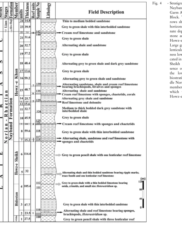
The type section of Nayband Formation consists of five members, including Gelkan, Bidestan, Howz-e Sheikh, Howz-e Khan and Qadir members in ascending order.
Both, the Gelkan (915 m thick) and Howz-e Sheikh (365 m thick) members are composed of grey to green siltstone and sandstone. The Bidestan (450 m thick) and Howz-e Khan (465 m thick) members are composed of sponge-coral or coral-sponge construc-tions of biohermal and biostromal types and siliciclas-tics. The Qadir member (up to1000 m thick) is com-posed of coal-bearing shale (Beagin et al. 1976; Kluyver et al. 1983a; FuÈrsich et al. 2005).
The sedimentological and palaeontological char-acteristics of the first four members of the type section of Nayband Formation point to the marine environ-ment. Thin coal beds in the Qadir member support mainly the continental to deltaic environments with only few marine incursions (Kluyver et al. 1983b).
The Bidestan member contains bioconstructions mainly dominated by sponge, while scleractinian corals, especially of dendroid types, are the most important reef builder in the Howz-e Khan member (FuÈrsich et al. 2005).
Numerous lithological, sedimentological, pa-laeoenvironmental and palaeontological studies have
1 Department of Geology, Science and Research Branch, Islamic Azad University, Tehran, Iran. E-mail: [email protected] 2 Geozentrum Nordbayern, FG Palaeoumwelt, UniversitaÈt Erlangen-NuÈrnbnerg, Loewenichstr. 28, 91045 Erlangen, Germany.
E-mail: [email protected]
been accomplished on the Nayband Formation. Some of the most important palaeontological works, dealing with sponges were published by Senowbari-Daryan et al. (1997), Senowbari-Daryan & Hamedani (1999), Se-nowbari-Daryan (2003, 2005a, b), Rashidi & Senowbari-Daryan (2011), and Senowbari-Senowbari-Daryan et al. (2011). Geographical and geological setting of the studied area
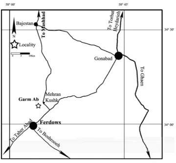
Geographically the studied area is situated about 3 km southwest of the town Burun, near Mehran Kushk,
almost 15 km northeast of Ferdows city (GPS-data: 34ë 7' 59.40" N; 58ë 12' 8.43" E; altitude of 1500 meters above sea level; Figs 1, 2). The locality can be reached by car, taking the road from Ferdows to the Garm Ab (Fig. 2).
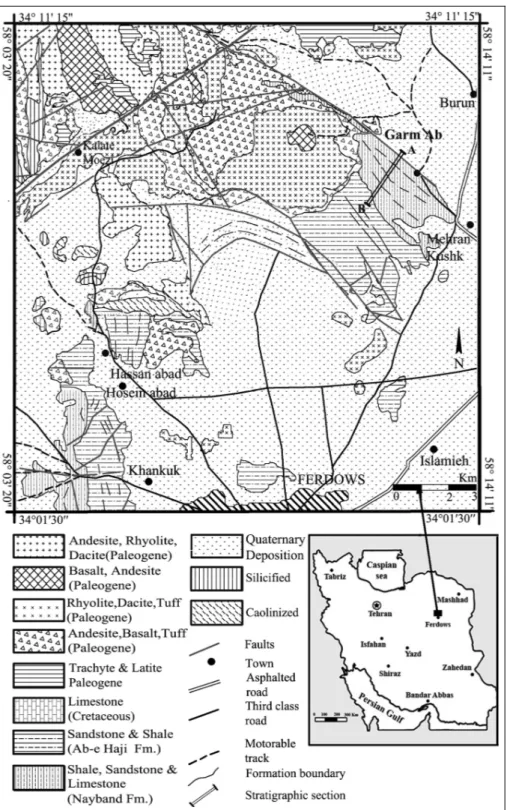
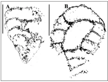
Geologically, the studied section is a part of the Lut Block. It consists of an alternating sequence of reef carbonates and grey to green pencil shale, referred to the Nayband Formation. The unit contains a rich macrofauna, such as sponges (including sphinctozoans, inozoans, chaetetids, spongiomorphids, and rare hexac-tinellids), scleractinian corals, crinoids, gastropods, bi-Fig. 1 - Geological map and location of the studied section of Garm Ab in the northeast of Ferdows. (Modified from Eftekhar-Nejad et al. 1977 and Purlatifi 2004).
valves and microfauna, mostly foraminifers. The lower contact of Nayband Formation in the Garm Ab sec-tion is composed of grey to green siltstone and dark grey sandstone with ripple marks, comparable with the Gelkan member in the type locality. The top of Nay-band Formation is marked by the disappearance of the reef carbonates (Eftekhar-Nejad et al. 1977; Purlatifi 2004).
The thickness of Nayband Formation decreases from west to east central Iran (Aghanabati 2010; Kluy-ver et al. 1983b; FuÈrsich et al. 2005). The Garm Ab section (eastern part of Iran) is about 870 meters thick. This might indicate the influence of additional factors, e.g. tectonic events and climatic fluctuations (FuÈrsich et al. 2005).
The Garm Ab section consists of eight horizons of sponge dominated reef carbonate beds (Figs 3, 4) and some reefal limestone. Generally, they reach thicknesses of about 2 meters and more, with some lenticular reefal limestone of smaller size in micritic matrix, embedded within the Nayband Formation. The grey colored car-bonates are classified as boundstone, floatstone, and packstone. Siliciclastic rocks,
in-cluding dark grey and thin beds of sandstone and grey to green shale, separate the horizons of reefal limestone.
The spherical hydrozoan Heterastridium, mostly with twospecies, namely H. conglo-batum Reuss and H. loconglo-batum Reuss, are common macrofossil in the first four units in the low-er part of the section (Fig. 4). The occurrence of
Heterastri-dium corresponds to its presence in the Bidestan mem-ber of the type locality (Kluyver et al. 1983b; FuÈrsich et al. 2005). The presence of Heterastridium supports bios-tratigraphically the Middle Norian age (late Alaunian) for this part of the Nayband Formation.
The middle part of the section is composed of thick layers of grey to green pencils shale and siltstone with disappearing of Heterastridium. The reefal lime-stone became reduced in this part and there are few lenticular reef carbonate with less than 2 meter in thick-ness, rather corresponding to the Howz-e Sheikh mem-ber.
Most of the carbonate deposits are developed in the last part of the section (seven horizons of reef car-bonate beds) as reefal limestone which is more similar to Howz-e Khan member in type locality of Nayband Formation (Fig. 4). Based on the lithological and bios-tratigraphical data (e.g. sponges and foraminifers), this part of the section, from rock unit number 4 to the top in the stratigraphic column, should refer to the Late Norian-Rhaetian.
The Nayband Formation ends with the disappear-ance of the carbonates. According to Purlatifi (2004) and Eftekhar-Nejad et al. (1977), the Nayband Forma-tion is overlain by the siliciclastics of the Ab-e Haji Formation, dated as Early Jurassic (Figs 1, 3). Reefal limestone with high diversity and biotic associations such as sponges, corals and other reef builders and reef dwellers like benthic foraminifers characterize the se-quence, indicating tropical to subtropical, warm-water settings at low latitudes in shallow-water, which is agreement with other studies (Senowbari-Daryan 1996; FuÈrsich et al. 2005).
Methods
Sponges, described in this paper were collected from the reefal carbonates of the Nayband Formation. All sponges were studied in large-sized thin-sections (10 x 15 cm and 7.5 x 10 cm). Isolated sponge were cut in longitudinal and cross sections and studied in polished slabs as well as in thin sections.
Fig. 2 - Geographical positions of the studied section in the north-east of Ferdows.
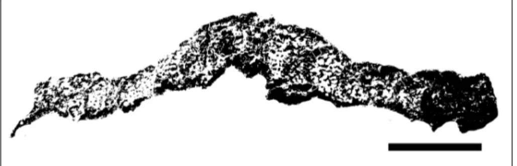
Fig. 3 - View of the Garm Ab section showing the Upper Triassic Nayband Formation with reefal carbonate beds (such as layer 4 and layer 6 shown in the photograph), overlain by the siliciclastic sediments of the Lower Jurassic Ab-e Haji Formation in beside of igneous rocks.
Depository
All illustrated specimens and thin sections will be deposited in the collection of the department of Geology Payame Noor University (in the Paleontological Museum), Yazd, Iran; material marked with Am-F-10-100.
Palaeontology
The systematic description of sponges is based on the classification of Finks & Rigby (2004). Because of the polyphyletic nature of Sphinctozoa and Inozoa (Se-nowbari-Daryan & Rigby 2011), the systematic cate-gories for classification of these sponges are used here for their morphological descriptions.
``Sphinctozoa''
ClassDemospongiaeSollas, 1875 Order Agelasida Verrill, 1907 Family Sebargasiidae de Laubenfels, 1955
Genus Amblysiphonella Steinmann, 1882 Remarks. Amblysiphonella is a long lasting sponge genus, occurring abundant in Permian and in Triassic time interval. It is, however, a relatively rare sponge genus in the Nayband Formation, particularly in the investigated reefs or reefal limestones of the Garm Ab section. Huckriede et al. (1962) reported the genus for the first time from the reef carbonates near the town of Kerman (South Iran) in the Bolbolu section of Fig. 4 - Stratigraphic column of the Nayband Formation of the Garm Ab section in the Lut Block. The small black ar-rows show reefal carbonate horizons; most of the carbo-nate deposits as reefal lime-stone are developed in the Howz-e Khan member. Large grey arrows show the lenticular reefs, with thick-ness lower than 2 meters lo-cated in Gelkan and Howz-e Sheikh members. The pre-sence of Heterastridium in the lower part, supports biostratigraphically the Mid-dle Norian same as Bidestan member in the type locality which was disappeared in Howz-e Sheikh member.
the Nayband Formation. Senowbari-Daryan & Hame-dani (1999) described twospecies as A. cf. steinmanni (Haas, 1909) and A. cf. tubifera from the Nayband For-mation near Wali Abad, central Iran. The new species A. najafiani and additionally three informal species were described by Senowbari-Daryan (2005a). Two new spe-cies ± A. torabii and A. bisiphonata ± and three informal species were described by Rashidi & Senowbari-Daryan (2011) from the Nayband Formation, in the northeast of Esfahan, central Iran. AlsoSenowbari-Daryan et al. (2011) added one more informal species from the reef limestones near Yazd, central Iran to the content of the genus Amblysiphonella. The following two species of the genus were found in the studied section.
Amblysiphonellasp. 1 Pl. 1, fig. 3 Material: One incomplete specimen only.
Description. The species is composed of ring-shaped chambers, arranged around the two axial tubes (recognizable on the chamber roof of the last chamber in Pl. 1, fig. 3) as spongocoel allowing the attribution of the specimen tothe genus Amblysiphonella. The avail-able specimen reaches a length 8.4 mm with a diameter of 5.6 mm. The first chamber is cut marginally with oval appearance. The last chamber is broken. The height of the chambers is 1.5 mm. Chamber walls are thin, with a thickness of 0.06 mm to 0.15 mm and are pierced by small and un-branched pores. The spongocoel (diameter 1.8 mm) is composed of at least two tubes, running internally through the sponge. Chamber interiors are without vesiculae and other type of filling skeleton.
Remarks. The diameter of the whole sponge and the axial tubes of this species correspond to the dimen-sions of Amblysiphonella bisiphonata described by Ra-shidi & Senowbari-Daryan (2011). The thin chamber walls, however, distinguish this species clearly from A. bisiphonata. As there is only one incomplete specimen available, we refrain from describing a new species.
Amblysiphonellasp. 2 Pl. 1, fig. 2A-B
Material: Three specimens in twothin sections.
Description. The marginally longitudinal section of one species illustrated in Pl. 1, fig. 2A exhibits at least five chambers, appearing rectangle in longitudinal sec-tion. The two downward pointed chamber roofs around the spongocoel of the middle chambers indicate the ax-ial spongocoel of the reticulate type and therefore the attribution of the species to the genus Amblysiphonella. The sponge reaches a height of 4 mm. Diameter of the
chamber is about 1.1 mm. Height of a middle chamber is about 0.7 mm, increasing moderately during the growth. Narrow pores of chamber walls are hardly re-cognizable. Diameter of the spongocoel is 0.5 mm and thickness of chambers walls 0.05 mm.
Comparison. Amblysiphonella sp. 2 differs from Amblysiphonella sp. 1 by the small sponge diameter and by the low chamber heights.
? Family Thaumastocoeliidae Ott, 1967 ? Subfamily Enoplocoeliinae Senowbari-Daryan, 1990
Genus Musandamia
Senowbari-Daryan & Bernecker, 2010
Type species: Musandamia omanica Senowbari-Daryan & Bernecker, 2010.
Musandamia gosaukammensis (Senowbari-Daryan, 1994)
Pl. 1, fig. 5A, B; Fig. 5
1994 Enoploecoelia? gosaukammensis n. sp. - Senowbari-Dar-yan, p. 670, pl. 1, figs 1-5.
2011 Musandamia gosaukammensis (Senowbari-Daryan) - Ra-shidi and Senowbari-Daryan, p. 325, pl. 1, fig B: arrow, D, E-G.
Material: Twospecimens in thin sections.
Description. This species is one of the smallest thalamid sponges in the investigated material. Like Am-blysiphonella, it is composed of several ring-like cham-bers, arranged around an axial spongocoel. The typical characteristic of the sponge and the genus is the ambi-siphonate type (sensu Seilacher 1962) of the spongocoel formation.
Specimen in Pl. 1, fig. 5A shows a longitudinal sec-tion, which has a length of 6.1 mm and is composed of four globular chambers. Diameter of the sponge and the chambers is almost constant, being 2.5 mm. Height of the chambers varies between 0.8 and 1.1 mm. Spongo-coel and chamber walls are about 0.1 mm thick. The communication of the chamber interiors with the spon-gocoel is constructed by a large opening only, situated in the middle of the chamber endowalls. The sponge surface exhibits spine-like elements (length of the ``spines'' 0.1-0.16 mm), reflecting the horizontally run-ning rips on the outer surface of the sponge skeleton.
Remarks. The sponge diameter of the specimens from Garm Ab section is moderately larger than those specimens described as Enoplocoelia? gosaukammensis from the Norian-Rhaetian reef of Gosaukamm, Austria by Senowbari-Daryan (1994) and from those specimens described by Rashidi & Senowbari-Daryan (2011) from the Nayband Formation, northeast of Esfahan. All other features of Garm Ab specimen correspond to the specimens from the two mentioned localities.
Occurrence. The Norian-Rhaetian reef of Gosau-kamm, Austria (Senowbari-Daryan 1994), the Nayband Formation in central Iran (Rashidi & Senowbari-Daryan 2011). The second species of Musandamia, Musandamia omanica, was reported from Norian reef of Jebel Agah, Oman by Senowbari-Daryan & Bernecker (2010).
Family Salzburgiidae Senowbari-Daryan & SchaÈfer, 1979 Salzburgia Senowbari-Daryan & SchaÈfer, 1979
Salzburgiasp. Pl. 1, fig. 1; Fig. 6 Material: One specimen only.
Description. This porate and cylindrical speci-men reaches a length of about 23 mm and is composed of four spherical chambers (semicircular in section) of almost constant diameter and height of 3 mm. The inner part of the chamber walls is laminated (Fig. 6), but the outer part exhibits labyrinthic pore system. Chamber exowalls are thinner (about 1.4 mm) than the doubled chamber roofs, reaching thicknesses of up to 2.5 mm. In addition to the labyrinthic pore system the exowalls of individual chambers are pierced by usually one (in sec-tion) rimmed ostium of about 0.55 mm in diameter, only the youngest chamber exhibits two ostia. From the chamber roofs some drop-like skeletal elements hang intothe chamber interior, indicating the presence of an axial canal.
Remarks. Two-layered chamber walls (laminated inner part, labyrinthic canal system of the outer layer) are characteristic for the genus Salzburgia (Senowbari-Daryan & SchaÈfer, 1979). This feature supports the
at-Fig. 5 - Longitudinal section through Musandamia gosaukam-mensis Rashidi and Senowbari-Daryan (was drawn from Pl. 1, fig. 5A) showing chambers with horizontal rips (spine-like appearance in the longitudinal section) of the outer surface. Scale = 10 mm.
Fig. 6 - The longitudinal section of Salzburgia sp. (Pl. 1, fig. 1) shows the lamination in the internal part of the chamber walls, the labyrinthic canal system of the external part of the chamber walls, and some rimmed ostia in the exowalls (one of them is shown with black arrow). Scale = 10 mm.
tribution of the species to the genus Salzburgia. The rimmed ostia of the Iranian species, however, were not observed in original material of the alpine species (S. alpina). Because there is only one specimen in the collection, the determination of the species is not car-ried out.
Family Solenolmiidae Engeser, 1986 Subfamily Solenolmiinae Senowbari-Daryan, 1990
Genus Solenolmia Pomel, 1872 Type species: Scyphia? manon MuÈnster, 1841.
Solenolmiasp. (n. sp.?) Pl. 2, fig. 5 Material: One specimen.
Description. The one sectioned specimen of this sponge is cut in oblique section and illustrated in Pl. 2, fig. 5. Because of the position of the section, the true length of the cylindrical or moderately inverse conical specimen cannot be ascertained. The height of the ob-lique section is about 48 mm with a diameter of at least 40 mm and is composed of six chambers. The low chambers with a maximum height of 6 mm are cres-cent-like and their interiors contain fiber skeleton of reticulate type. Chamber exo-, inter- and endowalls are about 1.6 mm thick and pierced by single and evenly distributed pores of about 0.2 mm. An axial spongocoel of about 11.5 mm in diameter passes (most probably) internally through the whole sponge.
Remarks. Solenolmia occurs in Permian and Triassic time interval and is an abundant genus in Ladi-nian-Carnian reefs (Senowbari-Daryan & Garcia Belli-do 2002). All known species of the genus are small-sized, never reaching a diameter of 40 mm. In addition, those species described from the Norian-Rhaetian reefs of Iran by Senowbari-Daryan (2005a) are distinctly smaller than the species from the Garm Ab locality. Based on the large diameter of the described specimen, it represents most probably a new species. However, because of scarce material it is described here as uncer-tain species.
Genus Deningeria Wilckens, 1937 Type species: Deningeria mirabilis Wilckens, 1937
Deningeria sp. Pl. 2, figs. 1, 3; Pl. 8, fig. 1; Fig. 7
Material. Numerous specimens (observed in the field) and in thin sections.
Description. This species seems to be one of the most abundant thalamid sponges in the studied section. Several specimens were observed in the field and in thin sections but only two specimens of them are studied in thin sections (Pl. 2, figs 1, 3) and one specimen from the field (Pl. 8, fig. 1) is documented. The description of the species is mainly based on investigations of the thin sections.
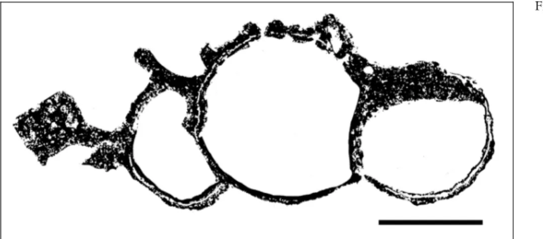
The sponge is composed of numerous spherical to hemispherical chambers, arranged one above the other (moniliform, Fig. 7). The chambered construction is hardly recognizable from the outside. The sponge reaches a length of more than 15 mm with a diameter of up to 2.5 mm in thin section. Chamber interiors are filled with reticulate fiber skeleton. Height of the cham-bers is variable. The thicknesses of the chamber walls are about 0.25 mm and pierced by numerous pores, un-evenly distributed. An axial canal is lacking in the illu-strated specimens.
Remarks. Wilckens (1937) described twospecies of the genus Deningeria as D. camerata and Deningeria mirabilis from the Upper Triassic of Seran, Moluccas. D. mirabilis is documented by a section of three cham-bers with reticulate fiber skeleton within the chamber interiors. Because detailed information about this ``spe-cies'' is lacking, we propose to limit the species-name mirabilis only to this specimen.
Wilckens (1937: 201) defined the genus diagnosis of Deningeria as vague. Soit is not clear if Deningeria possesses an axial canal or not. He wrote on page 201: ``An axial tube seems tobe present, but the available material does not allow a decision for sure''.
Family Colospongiidae Senowbari-Daryan, 1990 Synonymy: Colospongiidae Boiko (in Boiko et al. 1991), Para-uvanelliidae Wu 1991, Imbricatocoeliidae Wu 1991.
Fig. 7 - Deningeria sp. Longitudinal section through numerous chambers exhibiting the reti-culate filling structures with-in the chamber with-interiors and the perforated chamber walls. There are twotubes indicat-ing the possibly branchindicat-ing of the sponge in the margin of the large chamber (middle of the skeleton). Scale = 10 mm
Subfamily Colospongiinae Senowbari-Daryan, 1990 Genus Colospongia Laube, 1865
Type species: Manon dubium MuÈnster, 1841.
Colospongia sp. Pl. 2, fig. 4; Fig. 8D
Material: At least twospecimens in thin sections.
Description. Section through five spherical and moniliformly (the last two chambers are arranged be-side others) arranged chambers of this sponge (Fig. 8D) exhibits chamber walls with equally and evenly distrib-uted single pores of about 0.09 mm. The maximum height of the chambers is 1.8 mm with diameters of about 3.5 mm. Chamber walls are thin and correspond almost to the pore diameter. Chamber interiors are without filling skeleton.
Remarks. Colospongia is a long-lasting sponge genus occurring from Carboniferous (KuÈgel 1987) up to Upper Triassic. In Norian-Rhaetian reefs of the Nay-band Formation, it occurs rarely.
Subfamily Corymbospongiinae Senowbari-Daryan, 1990
Genus Parauvanella
Senowbari-Daryan & Di Stefano, 1988 Type species: Parauvanella paronai Senowbari-Daryan & Di Stefano, 1988
Parauvanella ferdowsensisSenowbari-Daryan, 2005a Pl. 3, fig. 1; Pl. 8, fig. 6; Fig. 8A
2005a Parauvanella ferdowsensis nov. sp. - Senowbari-Daryan, p. 177, pl. 1, figs 1-8, pl. 2, figs 1-2, pl. 15, fig. 1/P; text-figs 8-9.
2011 Parauvanella ferdowsensis Senowbari-Daryan - Senow-bari-Daryan and Link, p. 677, fig. 6f.
Material: Twospecimens (one naturally weathered specimen and one in thin section).
Description. Specimens of this asiphonate species are composed of several spherical-subspherical cham-bers with uviform arrangement (chamcham-bers are above and beside others, Fig. 8A). In marginal sections, the number of chambers appears lower as in the specimen illustrated in Pl. 3, fig. 1. Chamber heights are almost the same size as the chamber widths, measuring usually 1.5 mm in diameter. The thin chamber walls (thickness about 0.15 mm) are pierced by un-branched pores of about 0.2 mm in diameter. Chamber interiors are with-out any secondary filling skeleton.
Occurrence. Parauvanella ferdowsensis in known from the Norian-Rhaetian reefs of the Nayband For-mation (Senowbari-Daryan 2005a). Recently, it was de-scribed from the Norian of the Taurus Mountains, southern Turkey by Senowbari-Daryan & Link (2011). Parauvanella spinosa Rashidi & Senowbari-Daryan, 2011
Pl. 3, fig. 2; Fig. 8B
2011 Parauvanella spinosa nov. sp. - Rashidi and Senowbari-Daryan, p. 315, pl. 1, fig. J; pl. 9, fig. G/1; pl. 10, fig. F/1)
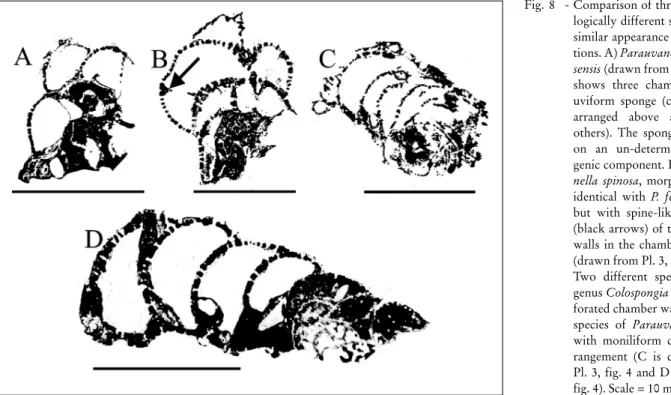
Fig. 8 - Comparison of three morpho-logically different species with similar appearance in thin sec-tions. A) Parauvanella ferdow-sensis (drawn from Pl. 3, fig. 1) shows three chambers of an uviform sponge (chamber are arranged above and beside others). The sponge is grown on an un-determinable bio-genic component. B) Parauva-nella spinosa, morphologically identical with P. ferdowsensis, but with spine-like extension (black arrows) of the chamber walls in the chamber interiors (drawn from Pl. 3, fig.2). C-D) Twodifferent species of the genus Colospongia exhibit per-forated chamber walls (like the species of Parauvanella), but with moniliform chamber ar-rangement (C is drawn from Pl. 3, fig. 4 and D from Pl. 2, fig. 4). Scale = 10 mm.
Material: One specimen only.
Description. The sponge morphology, chamber arrangement and the perforation pattern of the chamber walls corresponds to those of P. ferdowsensis. P. spinosa differs from P. ferdowsensis by the spine-like extensions of the chamber walls into the chamber interior as shown in (Fig. 8B).
Occurrence. In the Norian-Rhaetian reef of the Nayband Formation in central Iran (Rashidi & Senow-bari-Daryan 2011) and now in Garm Ab section of the Lut Block.
Subfamily Kashanelliinae Senowbari-Daryan, 2005b Genus Kashanella Senowbari-Daryan, 2005a Type species: Kashanella irregularis Senowbari-Daryan, 2005a
Kashanella irregularisSenowbari-Daryan, 2005a Pl. 3, figs 3, 5-7
2005a Kashanella irregularis nov. sp. - Senowbari-Daryan, p. 181-182, pl. 5, fig. 4, pl. 19, fig. 6, pl. 20, fig. 7, pl. 21, figs. 6-7.
2009 Kashanella irregularis - Senowbari-Daryan, p. 115, pl. 2, figs H-I, pl. 3, figs A/2, H-I, pl. 15, fig. E.
2011 Kashanella irregularis - Rashidi and Senowbari-Daryan, p. 316, pl. 2, figs K-L, pl. 4, figs E, H-I.
Material: Numerous specimens in several thin sections. Description. This irregularly growing sponge is composed of several irregular chambers, usually with moniliform arrangement. Highly characteristic of the sponge is the labyrinthic canal system of the chamber walls with ``spongy'' appearance. For detailed descrip-tion for the species see Senowbari-Daryan (2005a).
Remarks. Senowbari-Daryan & Link (2011) de-scribe a second species of the genus as Kashanella cylin-drica from the Norian of southern Taurus Mountains.
Occurrence. Kashanella irregularis is known from the Norian-Rhaetian reefs of the Nayband For-mation, Iran, (Rashidi & Senowbari-Daryan 2011), the Gosaukamm in Austria (Senowbari-Daryan 2009), and from the Norian reefs of Taurus Mountains in southern Turkey (Senowbari-Daryan & Link 2011).
Family Annaecoeliidae Senowbari-Daryan, 1978 Genus Annaecoelia Senowbari-Daryan, 1978 Type species: Annaecoelia maxima Senowbari-Daryan, 1978
Annaecoelia parvaSenowbari-Daryan, 2005a Pl. 4, fig. 1
2005a Annaecoelia? parva nov. sp. - Senowbari-Daryan, p. 184, pl. 29, figs 4, 6.
2011 Annaecoelia parva - Rashidi and Senowbari-Daryan, p. 318, pl. 2, fig. D, pl. 4, fig. L.
Material: Two specimens in one thin section.
Description. The small specimen of this sponge has a length of 6.7 mm and width of about 3.8 mm. It is composed of spherical chambers with diameters of about 1.6 mm. The chamber walls are with 0.09 mm very thin and pierced by extremely fine pores of 0.02 mm in diameter.
Remarks. This species was assigned tothe genus Annaecoelia with some doubts by Senowbari-Daryan (2005a). Following the opinion of Rashidi & Senow-bari-Daryan (2011) the question mark is taken away alsoin this paper.
Occurrence. Annaecoelia parva is known only from the Norian-Rhaetian reefs of the Nayband For-mation in Iran (Senowbari-Daryan 2005a; Rashidi & Senowbari-Daryan 2011).
Family Cryptocoeliidae Steinmann, 1882 Genus Cryptocoelia Steinmann, 1882 Type-species: Cryptocoelia zittlei Steinmann, 1882
Cryptocoelia maximan. sp. Pl. 1, fig. 6; Pl. 2, fig. 2; Pl. 4, fig. 6; Fig. 9
Derivatio nominis: Named after the large size of the species. Holotype: Specimen illustrated in Pl. 2, figs. 2 (thin section, Am-F-17).
Paratypes: Pl. 4, fig. 6, and Pl. 1, fig. 6 (thin sections, Am-F-28, 35).
Locus typicus: Nayband Formation, situated about 3 km southwest of the town Mehran Kushk, almost 15 km northeast of the Ferdows city, reef number 7 (34ë 7'59.40"N, 58ë12'8.43"E).
Stratum typicum: Upper Triassic (Rhaetian).
Diagnosis: Large species, stump conical-shaped with a trabecu-lar, without recognition of the internally crescent like low chambered construction, spongocoel pro-? pseudosiphonate?, filling skeleton tra-becular.
Material: Three specimens in thin sections, Am-F-17, 28, 35. Description. The skeleton of this distinctly large thalamid sponge (height of holotype at least 60 mm, diameter of the top is about 110 mm) is externally not recognizable. Internally the sponge is well chambered and individual low chambers are crescent-shaped. Height of the chambers varies between 1 and 2.5 mm. The most of the chamber walls are secondarily thick-ened but the well preserved parts show chamber walls of 1 mm (Pl. 2, fig. 2 lower part left). Chamber walls are pierced by numerous unevenly distributed single pores of 0.15 mm. Chamber interiors contain scattered pillar-like filling skeleton recognizable in well preserved parts (Pl. 2, fig. 2 lower part left). The holotype seems to be branched at the upper part. Our statement is based on a) asymmetrical position of the spongocoel pro-? pseudo-siphonate?, b) sediment filling in center of the top and c)
fracture of the chamber walls and the left part of the sponge, d) turning of the chamber wall in the axial re-gion. Recognition of the spongocoel (7 mm in diameter) as pro- or pseudosiphonate is not possible.
The description above is based on observations of the holotype. The two paratypes in Pl. 1, fig. 6 and Pl. 4, fig. 6 show almost identical characters, although, the spongocoel of the holotype could not be observed in the paratypes. The specimen illustrated in Pl. 1, fig. 6 clearly shows the pillar like filling skeleton in some chambers.
Comparison. Cryptocoelia maxima n. sp. differs from the known species of the genus by its large dimen-sion.
``Sphinctozoa''gen. et sp. indet Pl. 8, fig. 2; Fig. 10
Material: Three specimens in thin sections.
Description. Three specimens of this chambered sponge are cut in one sample exhibiting all features of the species. The longitudinal section through three spherical chambers of the specimen in Pl. 8, fig. 2,
ex-hibits thin chamber walls, which are partly perforated and partly imperforated. Also the two cross sections show this feature. One of the cross section exhibits in-ternally a perforated wall, not comparable with vesicu-lae or other types of filling skeleton compared within the chamber interiors of other sphinctozoans.
Such chambered sponges with partly perforated and partly imperforated chamber walls are not usual in this group of sponges. Most probably, this species can be attributed to the close neighborhood of the genus Co-lospongia. It is described as gen. et sp. ind. in this paper.
Inozoans
ClassDemospongiaeSollas, 1875 Order Agelasida Verrill, 1907
Family Sestrostomellidae de Laubenfels, 1955 Genus Sestrostomella Zittel, 1878 Sestrostomella robustaZittel, 1878
Pl. 5, figs 1, 4-5 1878 Sestrostomella robusta. Zittel, p. 41.
Fig. 9 - Comparison of pillar-like filling structures of Cryptocoelia maxima n. sp. (within several chamber interiors). (A is drawn from Paratype, Pl. 1, fig. 6; B is drawn from Holotype, Pl. 2, fig. 2 and C is drawn from Paratype, Pl. 4, fig, 6). In all three samples, the filling skeletons within the different chambers and even in different parts of the same chamber contain trabecular-filling skeleton. Not toscale.
Fig. 10 - ``Sphinctozoa'' gen. et sp. in-det. The moniliform sponge shows chamber walls (partly doubled) partly perforated and partly imperforated (middle chamber). It is drawn from Pl. 8, fig. 2. Scale = 10 mm.
1997 ?Sestrostomella robusta - Senowbari-Daryan, Seyed-Ema-mi & Aghanabati, p. 310, pl. 4, figs. 1-7, pl. 6, fig. 6, pl. 8, fig. 6. (cum syn.).
2009 Sestrostomella robusta - Senowbari-Daryan, p. 126, pl. 9, fig. B.
2011 Sestrostomella robusta - Senowbari-Daryan, Rashidi and Beitollah. p. 8, pl. 2, figs. B-C, F.
2011 Sestrostomella robusta - Rashidi and Senowbari-Daryan, p. 333, pl. 7, fig. D.
Material: Numerous naturally isolated specimens and several specimens within the carbonate rocks.
Description. This solitary and branched (Pl. 5, figs 1, 4-5) sponge is one of the most abundant inozoan species in the studied section. The largest specimen in collection is at least 20 mm long (Pl. 5, fig. 4), reaching a diameter of almost 14.5 mm (Pl. 5, fig. 1). The most characteristic of the genus and species is the axial canal bundle, which is composed of several individual tubes of about 0.5 (0.4-0.7) mm in diameter passing internally through the sponge. The tubes show a distinct wall, pierced by exhalant canals and additionally by small pores (Pl. 5, figs 1A, B). Inhalant canals connect the
sponge interior with the outside. The sponge wall is composed of reticulate fiber skeleton, filling the space between the inhalant and exhalant canals. In some speci-mens the fibers show, particularly at the periphery of the skeleton, an orientation to the up- and outward of the sponge.
Occurrence. Sestrostomella robusta is known from Ladinian to Norian and Jurassic-Cretaceous de-posits of several localities in the world (see Dieci et al. 1968; Hurcewicz 1975; Riedel & Senowbari-Daryan 1991; Senowbari-Daryan et al. 2011; Rashidi & Senow-bari-Daryan 2011). It is an abundant sponge in Norian-Rhaetian reefs, embedded within the Nayband Forma-tion in central Iran and has been described from several localities (see synonymy list).
Sestrostomellacf.robustaZittel, 1878 Pl. 5, figs 2, 6; Pl. 8, figs 3, 5; Fig. 11
Material: Twospecimens in thin sections and several naturally weathered rock surface in longitudinal, oblique and cross sections were cut on the reef number seven.
Fig. 11 - A) Longitudinal section through a multi-branched specimen of Sestrostomella cf. robusta, exhibiting the spongocoel composed of bundle of tubes (arrows) similar to Sestrostomella robusta Zittel. B-C) The cross and longitudinal sec-tions through a naturally isolated specimen (see Pl. 5, fig. 2) show the identical characteristics like fig. A. Scale = 10 mm.
Description. The dichotomously multi-branched (Pl. 5, figs 2, 6; Pl. 8, figs 3, 5) specimens show several tubes, axially arranged as spongocoel (Fig. 11). Dia-meters of the individual tube are about 1 mm (Pl. 5, fig. 2A). The diameter of the whole tube bundle de-pends from the distribution of the tubes in the sponge, with more than 5 mm (Pl. 8, fig. 5). Numerous speci-mens, one beside of others were observed in the field (Pl. 8, figs 3, 5).
Comparison. This species is differentiated from Sestrostomella robusta by its smaller dimension and the dichotomous branching pattern.
Family Auriculospongiidae Termier and Termier (in Termier et al., 1977)
Subfamily Auriculospongiinae Termier & Termier, 1977 Genus Anguispongia Senowbari-Daryan, 2005b Type species: Anguispongia parva Senowbari-Daryan, 2005b Anguispongia parvaSenowbari-Daryan, 2005b
Pl. 5, fig. 3
2005b Anguispongia parva nov. sp. - Senowbari-Daryan, p. 266-267, pl. 2, figs 1-6; pl. 3, figs 1-3; text-fig. 6.
2011 Anguispongia parva - Rashidi and Senowbari-Daryan, p. 330, pl. 6, figs H-I/1.
2011 Anguispongia parva - Senowbari-Daryan et al., p. 278, pl. 1, fig. K.
Material: Numerous specimens in the first reefal bed (see Fig. 4).
Description. The skeleton of this sheet-like or tabular sponge without any conspicuously inhalant or exhalant canals is composed of fiber skeleton. As shown by Senowbari-Daryan 2005b (pl. 3, figs 1-3 and text-fig. 6) the fiber skeleton exhibits different appearances in sections perpendicular or parallel to the sheets. Sheets reach dimensions of about 40 mm (in investigated sec-tions) and more (according to Senowbari-Daryan 2005b up to 100 mm) with thicknesses of about 6 mm. For a detailed description of the species see the original de-scription.
Occurrence. Until now, Anguispongia parva is known only from the Norian-Rhaetian reefs of the Nayband Formation in Iran.
Genus Molengraaffia Vinassa de Regny, 1915 Type species: Molengraaffia regularis Vinassa de Regny, 1915
Molengraaffia regularisVinassa de Regny, 1915 Pl. 6, figs 4?, 5
1915 Molengraaffia regularis n. g. n. f. - Vinassa de Regny, p. 80, p. 64 (2), figs 1-3.
2005b Molengraaffia regularis - Senowbari-Daryan, p. 265, pl. 1, figs 1-6; pl. 6, figs 4-8, text-fig. 5 (cum syn.).
2009 Molengraaffia regularis - Senowbari-Daryan, p. 122, pl. 10, figs B-D. (cum syn.).
2011 Molengraaffia regularis - Rashidi & Senowbari-Daryan, pl. 8, fig. A/1.
Material: Numerous specimens in thin sections.
Description. The club- to sheet-like skeleton of Molengraaffia regularis is composed, like the preceding species, by a fiber skeleton without recognizable inha-lant or exhainha-lant canals. In sections parallel to the sheets the fiber skeleton shows indistinctly linear arrangement of fiber skeleton (Pl. 6, fig. 5).
The specimen illustrated in Pl. 6, fig. 4 is branched and exhibits an identical (see detail with magnification in Pl. 6, fig. 4) fiber skeleton like other specimens of the species. Because branched specimens of this sponge are not known, species assignment is doubtful.
Occurrence. Senowbari-Daryan (2009) noted the following Upper Triassic localities, from which Molen-graaffia regularis was described: Karakoram, Bakony, Caucasia, Northern Calcareous Alps (Gosaukamm and Hohe GoÈll, Austria), Greece (unpublished material of him) and Iran.
Family Preperonidellidae Finks & Rigby, 2004 Sufamily Preperonidellinae Finks & Rigby, 2004
Genus Preperonidella Finks & Rigby, 2004 Type species: Preperonidella magna Rigby & Senowbari-Daryan, 1996
Preperonidella norica(Senowbari-Daryan, Seyed-Emami & Aghanabati, 1997)
Pl. 7, figs 3-4; Fig. 12E
1997 Radiofibra norica n. sp. - Senowbari-Daryan, Seyed-Ema-mi and Aghanabati, p. 299, pl. 1, figs 1-7; pl. 2, figs 1-6.
2009 Peronidella norica - Senowbari-Daryan, p. 124, pl. 11, figs A, C-D, G. (cum syn.)
2011 Peronidella norica - Rashidi and Senowbari-Daryan, p. 334, pl. 5, fig. D.
Material: Twospecimens.
Description. This large-sized (compared with the next described species) species of Preperonidella in the investigated material exhibits diameters between 15 and 21 mm (moderately smaller than the original material). The length of specimens is not known, but according to the original description it reaches length up to 120 mm (Senowbari-Daryan et al. 1997). An axial spongocoel of 2.8-4 mm in diameter passes internally through the whole sponge. The thick sponge wall is composed of
fine reticular skeleton. Inhalant and exhalant tubes are lacking, but the typical very narrow, irregular and wavy interfiber spaces run radially through the sponge wall (Fig. 12E).
Occurrence. Preperonidella norica is known from the Norian-Rhaetian reefs of Gosaukamm, Austria and from the time equivalent reefs of the Nayband Forma-tion in Iran. P. norica occurs also in Norian-Rhaetian reefs in Greece and in Turkey (unpublished material of BSD).
Preperonidella iranicaSenowbari-Daryan, 2003 Pl. 6, fig. 7; Pl. 7, figs 1-2, 5-6; Figs 12A-D
1980 Preperonidella fischeri FluÈgel, 1962 nom nud. - Senow-bari-Daryan, p. 43, pl. 7, fig. 2.
2003 Preperonidella iranica n. sp. Senowbari-Daryan, p. 63, pl. 1, figs 1-6; pl. 2, figs 1-6; pl. 3, figs 1-3.
Material: Numerous specimens in thin sections.
Description. The multi-branched species of this Preperonidella-species is usually smaller (about 10 mm) than the preceding one, but large specimens up to 21 mm were observed. An axial spongocoel attains dia-meters between 0.8 mm and 2.5 mm. Typical for this species is the distinct and perforated wall around the spongocoel. The sponge wall is composed of reticulate fiber skeleton. Exhalant canals pierce the spongocoel wall and are extended as short tubes into the sponge wall, which seems to be typical for this sponge (Pl. 7, fig. 5).
Remarks. The diameters of some specimens from Garm Ab are moderately larger than the dimensions of specimens of the original description, but other charac-ters are identical.
Occurrence. Until now, Preperonidella iranica is known from the Norian-Rhaetian reefs of Nayband Formation in Iran and from the Rhaetian of Austria (Senowbari-Daryan 2003).
Subfamily Permocorynellinae Rigby & Senowbari-Daryan, 1996
Genus Permocorynella Rigby & Senowbari-Daryan, 1996 Permocorynella maximaSenowbari-Daryan,
Seyed-Emami & Aghanabati, 1997 Pl. 6, fig, 6; Fig. 13
1997 Permocorynella maxima nov. sp. - Senowbari-Daryan, Seyed-Emami & Aghanabati, p. 302, pl. 3, figs 1-8; pl. 6, fig. 5; pl. 7, figs 1-3, 6, text-fig. 7 (cum syn.).
Fig. 12 - A-D) Some longitudinal and cross sections through Preperonidella iranica ex-hibiting the distinct spon-gocoel wall (black arrows), a characteristic feature of the species. E) Cross sec-tion of Preperonidella nor-ica showing some radially arranged and curved inter-fiber spaces (``tube-like'') (black arrow). Scale = 10 mm.
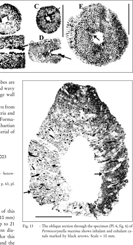
Fig. 13 - The oblique section through the specimen (Pl. 6, fig. 6) of Permocorynella maxima shows inhalant and exhalant ca-nals marked by black arrows. Scale = 10 mm.
2009 Permocorynella maxima - Senowbari-Daryan, p. 125, pl. 9, fig. E, pl. 12, figs D-E, pl. 17, fig. A/1.
2011 Permocorynella maxima - Rashidi and Senowbari-Daryan, p. 331, pl. 5, figs A-C, E.
2011 Permocorynella maxima -Senowbari-Daryan et al., p. 276, pl. 2, figs A, D, G.
Material: Three specimens (two specimens on the rock surface and one isolated specimen in thin section).
Description. The description of the species is based on the observation on the specimen in thin sec-tion. According to Senowbari-Daryan et al. (1997), the cylindrical to moderately conical specimens of this sponge reaches almost a diameter of 30-40 mm. The maximum diameter of the specimen from Garm Ab is 42 mm, close to the dimension of the original descrip-tion. The axial spongocoel measures 6 mm. The thick sponge wall exhibits two types of more or less radial running canals: inhalant canals, ending tothe sponge surface and exhalant canals to the spongocoel. Diameter of canals varies between 0.3 mm and 0.6 mm. Both canals show an indistinct tiny wall with pores of about 0.01 mm in diameter. The space between the canals is filled by the skeletal fibers of a reticulate type.
Occurrence. P. maxima is known from the Nor-ian-Rhaetian reefs of Gosaukamm, Austria (described as Corynella by Wurm 1982: see Senowbari-Daryan 2009) and from several reef localities of the Nayband Forma-tion in Iran (see synonymy).
Family Maeandrostiidae Finks, 1971 Genus Maeandrostia Girty, 1908
Maeandrostia? sp. Pl. 6, fig. 3 Material: One specimen only.
Description. The longitudinal section of the spe-cimen illustrated in Pl. 6, fig. 3 is composed of a thin outer wall with some coarse fiber skeleton in the sponge interior. The wall contains some rare openings. The ske-leton reaches a diameter of 3 mm and a length of 18 mm. The specimen seems tobe branched.
Remarks. Meandrostia is a ``typically'' Permian sponge genus. The only species from the Triassic depos-its has been described by Senowbari-Daryan et al. (1993) as Meandrostia triassica from the Anisian of Do-lomites, Italy. The genus is not known from Upper Triassic deposits.
ClassHexactinellidaSchmidt, 1870 Order Innaecoeliida Boiko, 1990 Family Craticulariidae Rauff, 1893
Remarks. The family Craticulariidae Rauff (1893) is referred to the order Hexactinosa Schrammen, 1903 by Finks and Rigby (2004). Based on the chambered construction of the genus Casearia and other cham-bered hexactinellids they are classified as Innaecoeliida by Senowbari-Daryan & FuÈrsich (2013). This classifica-tion is used in this paper.
Genus Casearia Quenstedt, 1858
Remarks. Casearia (= Caesiospongia) was the only one representative of the chambered hexactinellid sponge for a long time, occurring abundant in the Upper Jurassic of southern Germany. The genus was described from the Carnian of China by Wu (1989a), Rigby et al. (1998), and by Senowbari-Daryan & Amirhassankhani (2012) from the Norian-Rhaetian of Iran. A revision of the genus Casearia and description of further species from the Jurassic of central Iran were published by Se-nowbari-Daryan & FuÈrsich (2013).
Caseariacf. iranicaSenowbari-Daryan & Amirhassankhani, 2012
Pl. 4, figs 2, 4 B; Figs 14A-B
2002 Casearia sp. - Senowbari-Daryan & Garcia-Bellido, p. 1516, figs 5C, 5E.
2011 Caesaria sp. - Senowbari-Daryan et al., p. 276, pl. 1, fig. I. 2012 Casearia iranica n. sp. - Senowbari-Daryan & Amirhas-sankhani, pl. 1, figs 1-4; pl. 3, figs 2-3, 7.
Material: Twospecimens in thin sections.
Description. There is only one incomplete speci-men available (Pl. 4, fig. 2) which exhibits a spongocoel (6 mm in diameter) of retrosiphonate type passing in-ternally through the whole sponge. The poorly pre-served outer annulation corresponds to the internal seg-mentation. Both heights and widths of the chambers are less than 2 mm (Pl. 4, fig. 2; Fig. 14B and Pl. 4, fig. 4 B; Fig. 14A). The diameter of the sponge is 8.3 mm. Cham-ber walls are 0.3 mm thick and pierced by small pores. They are formed by coarse and loosely packed hexac-tine lattices (Pl. 4, fig. 2).
Remarks. The dimensions (chambers widths and heights) of specimens from Garm Ab are smaller than Casearia iranica described by (Senowbari-Daryan et al. 2011) and by Senowbari-Daryan & Amirhassankhani (2012) from the Norian-Rhaetian reefs of Iran. Based on the small dimensions of specimens from Garm Ab, they are identified as Casearia cf. iranica.
Occurrence. Casearia iranica is known exclu-sively from the Norian-Rhaetian reefs within the Nay-band Formation in Iran.
Casearia vezvanensisSenowbari-Daryan & Amirhassankhani, 2012
Pl. 8, fig. 4
1999 Casearia articulata (Schmidel, 1780) - Senowbari-Daryan & Hamedani, p. 94, pl. 7, figs 1-4.
2012 Casearia vezvanensis n. sp. - Senowbari-Daryan and Amirhassankhani, p. 252, pl. 2, fig. 4; pl. 3, fig. 4; text-fig. 4
Material: One naturally weathered specimen in carbonate rock. Description. The poorly preserved skeleton of this species has a length of more than 60 mm with nu-merous chambers. The width of each chamber is 8 mm, four-times larger than the height. The diameter of the sponge is 23 mm with outer annulations corresponding to the internal segmentation. Chamber walls are formed by coarse and loosely packed hexactine lattices (Pl. 8, fig. 4). Diameter of the spongocoel is 5 mm.
Occurrence. Casearia vezvanensis is only known from the Norian-Rhaetian reefs embedded within the Nayband Formation in Iran.
Discussion and conclusion
The occurrence and systematic positions of hy-percalcified sponge in the construction of reef-type de-posits in Garm Ab section has been beneficial for pa-leogeographic distributions of sponges, paleodepositio-nal conditions and comparing taxa in different strati-graphic levels during the upper Triassic.
Hypercalcified sponge shows a widespread global distribution during Norian-Rhaetian time in warm tro-pical conditions (Stanley 2001; FluÈgel & Kiessling 2002). They are known from numerous localities in southern
and northern America (Panthalassa), Europe (Northern Tethys) and Asia (Cimmeria such as Turkey and central part of Iran), (Senowbari-Daryan & Maurer 2008).
Norian-Rhaetian sponge associations of the Tethys are best developed in Northern Calcareous Alps (Shepherd et al. 2012). They formed reef belts in plat-form-edge positions facing the open marine northwes-tern Tethys basins, or as smaller patch reefs in intraplat-form basins or on the platintraplat-form ramps (Bernecker 2005). Reefs within the Nayband Formation also were formed on carbonate ramps in suitable depths below the wave base (Senowbari-Daryan 1996).
Taxa here studied, except hexactinellid sponges, are closer to Northern Calcareous Alps in Austria (Se-nowbari-Daryan 2003) than to other parts of Upper Triassic terrenes and it could suggest similar environ-mental conditions during Norian-Rhaetian. From Tur-key though, two taxa, Kashanella irregularis and Para-uvanella ferdowsensis, have been previously reported and only Parauvanella ferdowsensis has been identified from North America (Tab. 1).
Fig. 14 - A-B) Sections through two specimens of Casearia cf. iranica. A) An incomplete and marginally axial section (Pl. 4, fig. 4B). B) Oblique section exhibiting the ring-shaped chamber and the wide spongocoel (Pl. 4, fig. 2). Both specimens show thin chamber walls formed by hexactine lattice. Scale = 5 mm.
Middle East (Cimmeria) Current Geographic Distribution Europe (Northern Teth ys) North Ameri ca
(Panthalassa) Turkey Iran, Nayband
Formation
Taxa: this study Occurrences: Paleobiology Database (pbdb.org) Anguispongia parva B, D, M, T Annaecoelia parva B, T Casearia iranica* D Casearia vezvanensis* D Cryptocoelia maxima n. sp. Kashanella irregularis A + B, D, M Molengraaffia regularis A B, D, M, T Musandamia gosaukammensis A B Parauvanella ferdowsensis Al + D, M, T Parauvanella spinosa D Permocorynella maxima A B, D, M, T Preperonidella iranica A D, K, M, T Preperonidella norica A B Sestrostomella robusta A, I B, D, K, M, T
Tab. 1 - Current geographic distribution of hypercalcified and hexactinellid sponge taxa described in this paper and their occurrence in other Norian-Rhaetian localities. Turkey (+occurrence), Iran (different localities of the Nayband Formation): B= Bagherabad area, D= Delijan area, K= Kerman area, M= Marawand area, T= Tabas area, Europe (Northern Tethys): A= Austria, I= Italy, North America (Panthalassa) Al= Alaska, (based on Paleobiology Data-base, pbdb.org) and *Hexactinellids (Senowbari-Daryan & Amirhassankhani 2012).
Beyond Salzburgia and Cryptocoelia maxima n. sp., all described sponge taxa are known form the reef carbonates of other localities of Nayband Formation including Bagherabad, Delijan, Kerman, Marawand and Tabas area, (Tab. 1) being Norian-Rhaetian in age. Remarkable is the absence of Iranothalamia, Neva-dathalamia and Grossotubenella ± three abundant hy-percalcified sponges ± in other known Nayband local-ities. In this regard, the sponge fauna of Garm Ab sec-tion is very similar to the sponge associasec-tion known from the Rhaetian section exposed south of the town of Bagherabad, north of Esfahan (Rashidi & Senowbari-Daryan 2011), where these genera are alsoabsent.
In contrast to the abundance of hexactinellid sponges in Carnian reefs of China (Wu 1989a, 1989b; Wu & Xiao 1989; Rigby et al. 1998), this group of sponges is extremely rare in Norian-Rhaetian reefs in
the northwestern Tethys (Alps and adjacent areas, Se-nowbari-Daryan & Zankl 2010). Furthermore, the chambered hexactinellied sponges occur rarely in the time equivalent reefs in the central and northern Tethys (Caucasus: Boiko 1990; Iran: Senowbari-Daryan & Ha-medani 1999; Senowbari-Daryan & Amirhassankhani 2012). However, hexactinellid sponges in the Garm Ab section were found in the reefal limestone beds R1 to R6 (Tab. 2). The occurrence of the hexactinellid sponges was also reported from the reefal limestone near the town of Delijan (Tab. 1) in the western part of central Iran (Senowbari-Daryan & Amirhassankhani 2012) and it could testify for similar depositional con-dition in the east and west parts of central Iran during the Upper Triassic.
Isolated spicules of hexactinellid sponges occur in most thin sections in micritic matrix indicating the quick destructibility of this group after death. In gen-eral, hexactinellids are indicative of deep-water environ-ments in the Palaeozoic, the Mesozoic and Cenozoic (e.g. Nose et al. 2013; Wendt et al. 1989; Krautter 1997). On the other hand, the association of sponges, corals and other reef builders and reef dwellers like benthic foraminifers with hexactinellids in the Garm Ab section suggests a deposition more closed to shallow water environment.
Sphinctozoans, inozoans and some hexactinellid, from the eight horizons of reefal limestone in different stratigraphic levels, indicate Norian-Rhaetian age. Based on sponges and other organisms (e.g. Heterastri-diums) the stratigraphical age of the Garm Ab section continues from the Middle Norian to Rhaetian. The faunal assemblage cannot define the Norian/Rhaetian boundary.
The most abundant sponges occur in the first reefal carbonate horizon at the lower part of the section (Tab. 2). The abundance of sponges decrease to the younger horizons of the section. Two inozoan genera, Preperonidella, Molengraaffia and only one sphinctozo-an genus Kashsphinctozo-anella were found in the last reefal layer. Tectonic events and climatic fluctuations may cause ad-ditional feeding of siliciclastic sediments and an over-supply of nutrients, which could have limited growth of taxa during the end of Triassic.
Acknowledgements. The authors really appreciate Prof. Dr. M. Gaetani for comments and suggestions that greatly improved our manuscript. We alsowant tothank toM. Niazi and A. Vafadari for their help during the fieldwork. We would also like to thank the staff of Ardakan University, whogave us the permission tofinish the thin section in their lab. We appreciate and thank one anonymous reviewer and Dr. H. Lehnert for useful comments and suggestions.
Reef Numbers Sponges R1 R2 R3 R4 R5 R6 R7 R8 Salzburgia sp. * Amblysiphonella sp.1 * Amblysiphonella sp. 2 * * Cryptocoelia maxima n. sp. * * Musandamia gosaukammensis * * Colospongia sp. * Kashanella irregularis * * * * * * Casearia cf. iranica * Casearia vezvanensis * Deningeria sp. * * Sestrostomella robusta * * * * * Sestrostomella cf. robusta * Molengraaffia regularis * * * * Solenolmia sp. * * Anguispongia parva * Parauvanella ferdowsensis * * Parauvanella spinosa * Maeandrostia? sp. * * Annaecoelia parva * Permocorynella maxima * * * Preperonidella norica * Preperonidella iranica * * * * * Hexactinellida * * * * *
Tab. 2 - Occurrence and abundance of sphinctozoan and inozoan sponges in reefal horizons of the Garm Ab section. The first horizon (R1) at the bottom of the section offered the most abundant sponge species. Apparently the abundance of the sponge decrease during the formation time of Garm Ab section from bottom to the top (from horizons R1 to R8).
PLATE 1
Fig. 1 - Salzburgia sp. Marginally axial longitudinal section through a specimen exhibiting four chambers. Chamber walls are internally laminated (see Fig. 6), externally are pierced by labyrinthic canal system. The rimmed ostia in the exowalls are cut in two chambers. The drop-like skeletal elements hanging from the chamber roofs indi-cate to the possibly axial spongocoel. Chamber inter-walls are doubled and thick. Thin section Am-F-15. Fig. 2 - Amblysiphonella sp. 2. A) The oblique longitudinal
sec-tion through several chambers shows axial spongocoel cut in the middle chambers. B) Oblique cross section through a specimen in the same thin section. Both speci-mens are poorly preserved. Thin section Am-F- 14. Fig. 3 - Amblysiphonella sp. 1. Longitudinal section through an
incomplete specimen was colonized on a chaetetid sponge. Thin section Am-F-12.
Fig. 4 - Amblysiphonella cf. sp. 2. Oblique longitudinal section through a specimen exhibiting perforated walls of ring-shaped chambers. It differs from Amblysiphonella sp. 2 (Fig. 2) with relatively wide spongocoel and bigger size of specimen. Thin section Am-F-42.
Fig. 5 - Musandamia gosaukammensis (Senowbari-Daryan). A) The longitudinal section through the sponge shows the ring-shaped chambers, the ambisiphonate type of the spongocoel and the horizontally ripped exowalls appear-ing as spine-like extensions in section. B) Oblique cross section through a second specimen showing the spine-like elements of the exowalls in part. Thin section Am-F-38. Fig. 6 - Cryptocoelia maxima n. sp. (Paratype), ?Oblique
?Long-itudinal section, cut through an internally well cham-bered specimen. The chamcham-bered nature is not recogniz-able from the outside of the sponge. The walls of cres-cent-like low chambers are clearly perforated. The filling structure within the chamber interiors as trabecular (pil-lar-like), especially within the young chambers is well recognizable. Thin section Am-F-35.
Scale in figs 1-5 = 5 mm, in fig. 6 = 10 mm.
PLATE 2
Fig. 1 - Deningeria sp. Longitudinal section through numerous chambers. Because of the coarse and abundant filling skeleton within the chamber interiors the chamber roofs are not well recognizable. Thin section Am-F-58. Fig. 2 - Cryptocoelia maxima n. sp. (Holotype) ?Marginal
?longi-tudinal section exhibiting the crescent-like low cham-bers pierced by an apparently narrow ?spongocoel (for further information see the explanation of fig. 6 in Pl. 1). Thin section Am-F-17.
Fig. 3 - A) Deningeria sp. Similar section like fig. 1 (detail ex-planation see fig. 1) B) Colosponiga sp. Thin section Am-F-13.
Fig. 4 - Colospongia sp. Longitudinal section through several spherical chambers with evenly perforated chamber walls. The sponge encrusted by its basal part another undeterminable sponge with reticular fiber skeleton. Thin section Am-F-56.
Fig. 5 - Solenolmia sp. (n. sp. ?). Oblique section through several crescent-like chambers with perforated walls. Chamber interiors contain reticular fiber skeleton. The axial canal with own wall is cut. Thin section Am-F-20.
Scale in all figs = 10 mm, except in fig. 3, 4 = 5 mm. PLATE 3
Fig. 1 - Parauvanella ferdowsensis Senowbari-Daryan. Marginal section through three globular chambers (sub-globular in section) exhibiting thin and evenly perforated chamber walls. The sponge encrusted a biogenic component. Thin section Am-F-59.
Fig. 2 - Parauvanella spinosa Rashidi and Senowbari-Daryan. Section through several chambers exhibiting thin and evenly perforated walls. The spine-like extended skeletal elements from the chamber walls into the chamber inter-iors are cut in two chamber roofs. Thin section Am-F-10. Figs. 3, 5-7- Kashanella irregularis Senowbari-Daryan. 3) Almost identical section through four moniliform chambers ex-hibiting the specific characteristic labyrinthic canal sys-tem of the chamber walls. Thin section Am-F-18. 5) Section through three chambers with relatively thick outer walls. Thin section Am-F- 27. 6) Thin section Am-F-39. 7) Section through a specimen exhibiting clearly the thick exowalls and thin interwalls like fig. 5. Thin section Am-F-57.
Fig. 4 - Longitudinal section through several low chambers with perforated chamber walls (compare Fig. 8C). Thin sec-tion Am-F-77.
Scale in all figs = 5 mm.
PLATE 4
Fig. 1 - Annaecoelia parva Senowbari-Daryan. Longitudinal sec-tion through several glomerately arranged chambers of a poorly preserved specimen. Thin section Am-F-23. Fig. 2 - Casearia cf. iranica Senowbari-Daryan and
Amirhassan-khani. Longitudinal section through several ring-shaped chambers arranged around a relatively wide axial spon-gocoel (for observation turn the photo 90oin clockwise direction, compare with Fig. 14B). Thin section Am-F-11.
Fig. 3 - Hexactinellid sponge gen. et sp. indet. Section through a non-chambered specimen builds by hexactine lattice. Thin section Am-F-11.
Fig. 4 - A) Casearia sp. Longitudinal section exhibiting some crescent-like low chambers. B) Casearia cf. iranica Se-nowbari-Daryan and Amirhassankhani. Marginally longitudinal section through an incomplete specimen ex-hibiting the poorly recognizable chamber walls. Thin section Am-F-30.
Fig. 5 - Cross section through a hexactinellid sponge gen. et sp. indet. The extremely wide spongocoel is surrounded by the thin sponge wall. Numerous large and radially run-ning operun-nings connected the spongocoel with the outside of the sponge. Thin section Am-F-34.
Fig. 6 - Cryptocoelia maxima n. sp. (Paratype) Oblique longitu-dinal section through a relatively well preserved speci-men showing pillar-like infilling within some chambers. Thin section Am-F-28.
PLATE 5
Figs 1, 4-5 - Sestrostomella robusta Zittel. 1) Longitudinal (A) and cross (B) sections from the same specimen exhibiting the tubes of the axial canal bundle concentrated in the center and the oblique running exhalant tubes ending into the tubes of the canal bundle. The thin and perfo-rated walls around the tubes are clearly recognizable. Thin section Am-F-16. 4) Longitudinal (A) and cross section (B) through the same specimen exhibiting clearly the axial canal bundle composed of several individual tubes. Thin section Am-F-22. 5) The cross to oblique section through a specimen shows the axial canal bundle, some vertically running tubes of the spongocoel (circles in the center of the sponge). At least an inhalant canal (left in the photograph) is cut and the reticular fiber skeleton of the sponge wall is clearly visible. Thin section Am-F-43.
Figs. 2, 6 - Sestrostomella cf. robusta. 2) Longitudinal section (A) of a branched specimen, cross section (B) through the same specimen exhibiting similar characteristics like Fig. 6. The tubes of the spongocoel are scattered through the sponge wall. Thin section Am-F-52. 6) Longitudinal sec-tion through a dichotomously multi-branched specimen exhibiting similar characteristics like another illustrated specimen in Pl. 8, fig. 5. The tubes of the spongocoel are scattered from the center through the wall to periphery of the sponge. Thin section Am-F-71.
Fig. 3 - Anguispongia parva Senowbari-Daryan. Sections per-pendicular (A) and parallel to the tabular sponge (B) of the same specimen exhibit the reticular fiber skeleton without any inhalant and exhalant canals. Thin section Am-F-21.
Scale in all figs = 10 mm.
PLATE 6
Figs 1-2, 4 - ?Molengraaffia sp. 1) Sections through two, not exactly determinable specimens. Thin section Am-F-25. 2) Sec-tions through two specimens exhibiting reticular fiber skeleton without other sponge features. Thin section Am-F-47. 4) Section through a dichotomously multi-branched specimen exhibiting similar or almost identical fiber skeleton (see the magnification) like Molengraaffia (compare fig. 5), but branched specimens of this genus are not known. Thin section Am-F-53.
Fig. 3 - Maeandrostia? sp. The longitudinal section of an appar-ently branched specimen shows the thin exowall of the sponge pierced by large openings. Internally the sponge contains some coarse fiber skeleton. Thin section Am-F-88.
Fig. 5 - Molengraaffia regularis Vinassa de Regny. The longitu-dinal section, pependicular to the tabular sponge exhibits the linear arrangement of the fiber skeleton. Thin section Am-F-66.
Fig. 6 - Permocorynella maxima Senowbari-Daryan, Seyed-Emami and Aghanabati. Oblique section through a rela-tively well preserved specimen exhibiting the axial spon-gocoel and numerous inhalant and exhalant canals pas-sing through the thick sponge wall composed of reticular fiber skeleton. Thin section Am-F-24.
Fig. 7 - Preperonidella iranica Senowbari-Daryan. The cross and oblique sections through dichotomously and multi-branched specimens show the sponge wall, composed of reticular fiber skeleton. The thick spongocoel wall with white appearance is pierced by openings. Thin sec-tion Am-F-29.
Scale in figs 1-2 and 4-6 = 10 mm, in figs 3 and 7 = 5 mm. PLATE 7
Figs 1-2, 5-6 - Preperonidella iranica Senowbari-Daryan 1) A - Cross section of the same specimen, whose longitudinal sec-tion is illustrated in B. Both secsec-tions show the thick sponge wall and the axial spongocoel with distinct wall pierced by several openings. Thin section Am-F-33. 2) Similar sections as in Fig. 1 exhibiting similar characteristics of the sponge. Thin section Am-F-62. 5) Cross section of a specimen exhibiting some ra-dially arranged exhalant tubes, ending tothe narrow spongocoel. Thin section Am-F-73. 6) Several oblique sections exhibit similar features of the sponge like other specimens. The narrow axial spongocoel is re-markable. Thin section Am-F-82.
Figs 3-4 - Preperonidella norica (Senowbari-Daryan, Seyed-Emami and Aghanabati). 3) Cross (A) and (B) long-itudinal sections from the same specimen. Both sec-tions show the thick sponge wall and the axial spon-gocoel without a wall. The connection between the spongocoel and the sponge wall is produced by the interfiber spaces. Thin section Am-F-26. 4) Cross sec-tion exhibiting similar sponge characteristics like Fig. 3A. Thin section Am-F- 31.
Scale in all figs = 10 mm.
PLATE 8
Fig. 1 - Deningeria sp. Longitudinal section of a specimen on a naturally weathered rock surface. Internally the species exhibits clearly the chambered construction with reticu-lar fiber skeleton within the chamber interiors. The sponge seems to be asiphonate (sample FR2).
Fig. 2 - Sphinctozoan sponge gen. et sp. indet. Longitudinal and cross sections through three specimens show partly per-forated and partly imperper-forated chamber walls. Chamber interiors contain rare vesiculae, separating the cements from the sediment. Thin section Am-F-36.
Fig. 4 - Casearia vezvanensis Senowbari-Daryan & Amirhassan-khani. Longitudinal section on the naturally weathered rock surface shows well preserved, crescent-like low chambers on the left side of the photographs. The cham-bered construction of the species is hardly or even not recognizable from the outside of the sponge (sample FR1).
Figs 3, 5 - Sestrostomella cf. robusta Zittel. Several longitudinal, ob-lique and cross sections cut on naturally weathered rock surface (samples FR7). The dichotomously multi-branched specimen (fig. 5) and all other specimens in both photos show the several axially arranged tubes as spongocoel.
Fig. 6 - Parauvanella ferdowsensis Senowbari-Daryan. Section through several chambers of a specimen on naturally weathered rock surface. The thin chamber walls are pierced by evenly distributed single pores (sample FR4). Scale in all figs = 10 mm.
R E F E R E N C E S
Aghanabati A. (2004) - Geology of Iran. Geological Survey of Iran, Tehran, 586 pp.
Aghanabati A. (2010) - Stratigraphic Lexicon of Iran, vol. 3 (Triassic). Geological Survey of Iran, Tehran, 727 pp. Beagin Y., Jahanbakhsh F., Golubev S. & Sadovikov G. (1976) - Stratigraphy of the Triassic-Jurassic coal-bearing deposits of Alborz. Unpublished Report, Na-tional Iranian Steel Corporation vol. ``Technoexport'' 51 pp., Tehran.
Bernecker M. (2005) - Late Triassic reefs from the North-west and South Tethys: distribution, setting, and bio-tic composition. Facies, 51: 442-453.
Boiko E.V. (1990) - Miogoobrazie skeletiij structur u cam-jeriij gubok. Iscopaemie problematiki SSSR (On the diversity of skeletal structure of Porifera Camerata. Problematic fossils of SSSR). Academia Nauk SSSR. Siberian Otdel., Inst. Geol. Geophys. Trudy, 783(60): 119-129.
Boiko E. V., Belyaeva G. V. & Zhuravleva I. T. (1991) -Sfinktozoa fanerozoya territorii SSSR [Phanerozoic sphinctozoans from the Territory of the USSR]. Siber-ian Department, Institute of Geology and Geophy-sics. Academy of Sciences of Tajikistan SSR, Institute of Geology., 224 p., 36 fig., 64 pl., Moscow.
BroÈnnimann P., Zaninetti L., Bozorgnia F., Dashti G.R. & Moshtaghian A. (1971) - Lithostratigraphy and fora-minifera of the Upper Triassic Nayband Formation, Iran. Rev. MicropaleÂontol., 14(5): 7-16.
Dieci G., Antonacci A. & Zardini R. (1968) - Le spugne cassiane (Trias medio-superiore) della regione dolo-mitica attorno a Cortina d'Ampezzo [Sponges from the Cassian Formation (Middle-Upper Triassic) from the Dolomites near Cortina d'Ampezzo]. Boll. Soc. Paleont. It., 7(2): 94-155.
Douglas J.A. (1929) - A marine Triassic fauna from eastern Persia. Quart. J. Geol. Soc., 85: 624-650.
Eftekhar-Nejad D., Valeh R., Ruttner A., Nabavi M.H., Maieni N. & Haghipour N. (1977) - Geological of Ferdows Quadrangle Map, 1:250000. Geological Sur-vey of Iran, Tehran.
Engeser T. S. (1986) - Nomenklatorische Notiz zur Gattung Dictyocoelia Ott 1967 (``Sphinctozoa'', Porifera). Neues Jahbr. Geol. PalaÈontol., Monat. 10: 587-590. Finks R. M. (1971) - Sponge zonation in the west Texas
Permian. Smithsonian Miscellaneous Contributions, Paleontology, 3: 285-300.
Finks R. M. & Rigby J. K. (2004) - Hypercalcified Sponges. In: Kaesler R. L. (Ed.) - Treatise on Invertebrate Pa-leontology, Part E, Porifera (revised), 3: 585-764. The Geological Society of America and the University of Kansas, Lawrence.
FluÈgel E. and KiesslingW. (2002) - Triassic reef patterns. In: Kiessling W., FluÈgel E. & Golonka J. (Eds) - Phaner-ozoic Reef Patterns. SEPM Spec. Pub., 72: 391-463. FuÈrsich F.T., Hautmann M., Senowbari-Daryan B. &
Seyed-Emami K. (2005) -The Upper Triassic Nayband and
Darkuh Formations of east central Iran: Stratigraphy, facies patterns and biota of extensional basins on an accreted terrane. Beringeria, 35: 53-133.
Girty G. H. (1908) - On some new and old species of Car-boniferous fossils. Proc. U. S. Nat. Mus., 34: 281-303. Haas O. (1909) - Bericht uÈber neue Aufsammlungen in den Zlambach-Mergeln der Fischerwiese bei Alt-Aussee. Beitr. PalaÈontol. Geol. OÈsterreich-Ungarns und Ori-ents, 22: 143-167.
Huckriede R., Kursten M. & Venzlaff H. (1962) - Zur Geo-logie des Gebietes zwischen Kerman und Sagand (Iran). Beiheft Geol. Jahr., 51: 1-197.
Hurcewicz H. (1975) - Calcispongea from the Jurassic of Poland. Acta Palaeontol. Pol. 20(2): 223-291.
KuÈgel H. W. (1987) - Sphinctozoen aus den Auernigschich-ten des Nassfeldes (Oberkarbon, Karnische Alpen, OÈsterreich). Facies, 16:143-156.
Kluyver H.M., Tirrul R., Chance P.N., Johns G.W. & Meix-ner H. M. (1983a) - Explanatory Text of the Nayban-dan Quadrangle map, 1: 250000. Quadrangle J8. Geo-logical Survey of Iran. No. 18, 143 pp., Tehran. Kluyver H.M., Tirrul R., Chance P.N., Johns G.W. &
Meix-ner H.M. (1983b) - Explanatory Text of the Nayban-dan Quadrangle map. 1: 250000, Quadrangle J9: Geo-logical Survey of Iran, 175 pp., Tehran.
Krautter M. (1997) - Aspekte zur PalaÈoÈkologie postpalaÈo-zoischer KieselschwaÈmme. Profil 11: 199-324, Stutt-gart.
Laube G. C. (1865) - Die Fauna der Schichten von St. Cas-sian. Ein Beitrag zur PalaÈontologie der alpinen Trias, I. Abtheilung: Spongitarien, Corallen, Echiniden und Crinoiden. Denkschriften Kaiserl. Akad. Wissensch., Math-naturwiss., Kl. 24: 223-296.
Laubenfels M. W. De. (1955) - Porifera. In: Moore R. C. (Ed.) - Treatise on Invertebrate Paleontology. Part E, Archaeocyatha and Porifera. Geological Society of America and University of Kansas: 21-112, New York and Lawrence, Kansas.
MuÈnster G. Graf zu. (1841) - Beschreibung und Abbildung in den Kalkmergelschichten von St. Cassian gefun-dene Versteinerungen. BeitraÈge zur Geognosie und Petrefactenkunde des suÈdoÈstlischen Tirols, vorzuÈglich des Schichten von St. Cassian, Beyreuth, 151 pp. Nose M., VodraÂzÏka R., FernaÂndez L.P. & MeÂndez-Bedia I.
(2013) - First record of chambered hexactinellid sponges from the Palaeozoic. Acta Palaeontol. Pol., published online: http://dx.doi.org/10.4202/app.2012.0112. Ott E. (1967) - Segmentierte KalkschwaÈmme (Sphinctozoa)
aus der alpinen Mitteltrias und ihre Bedeutung als Riffbildner im Wettersteinkalk. Bayer. Akad. Wissen. mathem.-naturwissen. Klasse, Abh. (n. ser.) 131: 1-96. Pomel A. (1872) - PaleÂontologie ou description des animaux fossiles de la Province d'Oran, Zoophytes, fascicule 5, Spongiaires. Tipographie et Lithographie, Perrier, 256 pp., Oran.