Page 1 of 8
Sonic Hedgehog Therapy in a Mouse Model of
Age-Associated Impairment of Skeletal Muscle Regeneration
Andrea Piccioni,
1,2Eleonora Gaetani,
1Valentina Neri,
1,2Ilaria Gatto,
1Mariangela Palladino,
1,2Marcy Silver,
2Roy C. Smith,
2,4Igor Giarretta,
1Enrico Pola,
3Lynn Hlatky,
4and Roberto Pola
1,2,41Department of Medicine, A. Gemelli University Hospital, Catholic University School of Medicine, Rome, Italy.
2Division of Cardiovascular Research, St. Elizabeth’s Medical Center, Tufts University School of Medicine, Boston, Massachusetts. 3Department of Geriatric, Neurological and Orthopaedic Sciences, A. Gemelli University Hospital, Catholic University School of
Medicine, Rome, Italy.
4Center of Cancer Systems Biology, St. Elizabeth’s Medical Center, Tufts University School of Medicine, Boston, Massachusetts.
Address correspondence to Roberto Pola, MD, PhD, Center of Cancer Systems Biology, CBR4, St. Elizabeth’s Medical Center, Tufts University School of Medicine, 736 Cambridge Street, Boston, MA 02135. Email: [email protected]
Sonic hedgehog (Shh) is a morphogen regulating muscle development during embryogenesis. We have shown that the Shh pathway is postnatally recapitulated after injury and during regeneration of the adult skeletal muscle and regulates angiogenesis and myogenesis after muscle injury. Here, we demonstrate that in 18-month-old mice, there is a significant impairment of the upregulation of the Shh pathway that physiologically occurs in the young skeletal muscle after injury. Such impairment is even more pronounced in 24-month-old mice. In old animals, intramuscular therapy with a plasmid encoding the human Shh gene increases the regenerative capacities of the injured muscle, in terms of Myf5-positive cells, regenerating myofibers, and fibrosis. At the molecular level, Shh treatment increases the upregulation of the prototypi-cal growth factors, insulin-like growth factor-1 and vascular endothelial growth factor. These data demonstrate that Shh increases regeneration after injury in the muscle of 24-month-old mice and suggest that the manipulation of the Shh pathway may be useful for the treatment of muscular diseases associated with aging.
Key Words: Sonic hedgehog—Skeletal muscle regeneration—Aging.
Received September 10, 2012; Accepted April 19, 2013 Decision Editor: Rafael de Cabo, PhD
S
ONIC hedgehog (Shh) is a morphogen regulating crucial epithelial–mesenchymal interactions during embryo-genesis (1,2). Over the last 10 years, we have demonstrated that the Shh pathway, which was considered generally silent in postnatal life, is upregulated in the adult in experimental models of myocardial and skeletal muscle ischemia (3–5). We have also shown that, in postnatal life, Shh regulates angiogenesis and that Shh therapy may be used to improve revascularization in porcine and rodent models of coro-nary artery disease and peripheral limb ischemia (4–6). More recently, we have found that Shh not only regulates angiogenesis in the adult but also plays a functional and regulatory role on adult myogenesis. For instance, we have shown that Shh is de novo expressed after injury and dur-ing regeneration of the adult skeletal muscle in mice and that Shh inhibition impairs the activation of the myogenic regulatory factors Myf5 and MyoD, decreases the upregula-tion of insulin-like growth factor (IGF)-1, and reduces the number of muscle satellite cells (MSCs) at injured site (7). Also, Shh inhibition results in muscle fibrosis, increased inflammatory reaction, and compromised motor functional recovery after injury (7).Here, we demonstrate that the activation of the Shh pathway in response to mechanical and toxic injury of the
skeletal muscle is significantly impaired in old mice com-pared with young animals. We also demonstrate that direct intramuscular injection of a plasmid encoding the human Shh gene (phShh) induces functional activation of the Shh signaling pathway in the aged muscle and results in increased regeneration and repair after injury.
Methods
Animals and Experimental Models of Muscle Injury
We used young (8–12 weeks old), 18-month-old, and 24-month-old C57BL/6J male mice. Mice were purchased from Charles River Laboratories and housed in our ani-mal facility until the age required for the experiments. Two injury models were used: (i) mechanical crush and (ii) cardiotoxin (CTX) injection of the tibialis anterior (TA) muscle. Injuries were carried out as previously described (7). In both models, contralateral TA muscles were used as internal controls. Muscle injuries were performed under anesthesia. Mice were sacrificed at various time points with an overdose of ketamine. All the experiments were approved by our Institutional Review Boards and Ethics Committees.
by guest on June 19, 2013
http://biomedgerontology.oxfordjournals.org/
Analysis of the Activation of the Shh Pathway in Response to Injury
Mechanical crush and CTX injection of the TA muscle were carried out unilaterally as described (7). Twenty-five young, 18-month-old, and 24-month-old mice were used for each experimental model. Five mice in each model were sacrificed at each of the following time points: days 0 (before injury), 2, 4, 7, and 14. These time points were chosen because they encompass both early and late phases of muscle repair after injury and have been established previously (7). After sacrifice, muscles were harvested and the activation of the Shh pathway was evaluated by quantification of Shh and Gli1 messenger RNA (mRNA) levels by real-time reverse transcription-PCR (RT-transcription-PCR). The primer sequences were as follows: Shh forward, 5′-GAGCAGACCGGCTGATGACT-3; Shh reverse, 5′-AGAGATGGCCAAGGCATTTAAC-3′; Gli1 forward, 5′-TTGTCCAGCTTGGATGAAGG-3′; Gli1 reverse, 5′-CCCAGACGGCGAGACAC-3′. One-step real-time RT-PCR was performed using the ABI PRISM 7700 Sequence Detection System (Applied Biosystems). Total RNA (2–20 ng) isolated from each muscle was added to a reaction mixture containing forward primer, reverse primer, TaqMan probe, and TaqMan One-Step RT-PCR Master Mix Reagents (Applied Biosystems) in a final volume of 50 μL. We used 18S rRNA as a stable endogenous reference gene to normalize the target mRNA expression. TaqMan probes were labeled with a fluorescent reporter dye (FAM) at the 5′ end and a quencher dye (TAMRA) at the 3′ end. Thermal cycling conditions were as follows: 1 cycle of 30 minutes at 48°C for reverse transcription; 1 cycle of 10 minutes at 95°C for activation of DNA polymerase; 40 cycles of 15 seconds at 95°C for denaturation and 1 minutes at 60°C for annealing and extension. The expression levels of target gene and 18S rRNA in each sample were calculated based on the standard curve generated with the Mouse Universal Reference Total RNA (BD Biosciences Clontech). The expression level of target gene was then normalized by the expression level of 18S rRNA using TaqMan Ribosomal RNA Control Reagents (Applied Biosystems) to control the quantity of the isolated RNA. Real-time RT-PCR analyses were performed in duplicate.
Assessment of phShh Expression In Vivo
The phShh has been described previously (5). Also, we have shown already the beneficial effects of phShh therapy in experimental models of myocardial ischemia in rats and pigs and peripheral limb ischemia in mice (5,6). In these pre-vious experiments, 200 µg phShh were able to induce func-tional activation of the Shh pathway in the murine ischemic hind limb (6). Based on these data and on the relative muscle masses of murine hind-limb and murine TA muscle, we cal-culated a dose equivalent of phShh (40 µg phShh) to be used in our experimental models of TA CTX injury. To confirm
the ability of this treatment regimen to induce functional activation in the injured TA muscle of aged mice, a total of twenty 18-month-old mice underwent unilateral injury of the TA muscle by CTX injection. Immediately after injury, 10 mice received an intramuscular injection of 40 µg phShh. The remaining 10 mice received an intramuscular injection of an equal dose of empty plasmid and were used as controls. Mice were sacrificed 4 and 7 days after injury and muscles were harvested. Expression of Gli1 was detected by real-time RT-PCR, as described earlier. Results are expressed as the ratio between Gli1 expression in the treated leg versus the untreated leg. We also performed PCR to detect expression of human Shh in muscles treated with 40 µg phShh. Primers sequences for PCR amplification for human Shh were as follows: forward, 5′-GAGCAGACCGGCTGATGACT-3′; reverse, 5′-AGAGATGGCCAAGGCATTTAAC-3. PCR experimental conditions were as follows: 5 minutes at 94°C; 1 minute at 94°C, 1 minute at 60°C, 1 minute at 72°C for 40 cycles; 72°C for 7 minutes. Expression of mouse β-actin was used as housekeeping gene. PCR products were analyzed by 2% agarose gel electrophoresis.
Quantification of Activated Muscle Satellite Cells, Newly Formed Myofibers, and Fibrosis
A total of sixty 24-month-old mice were used for these experiments. All animals underwent unilateral CTX injury of the TA muscle, as described earlier. Of these mice, 30 were treated, immediately after injury, with 40 µg phShh, injected into the injured TA muscle. The remaining 30 mice received an equal dose of empty plasmid, immedi-ately after injury, by direct injection in the injured TA mus-cle. For the analysis of activated MSCs, 10 mice in each treatment group were sacrificed 4 days after injury. For the analysis of newly formed myofibers, 10 mice in each treatment group were sacrificed 7 days after injury. For the analysis of fibrosis, 10 mice in each treatment group were sacrificed 14 days after injury. Activated MSCs were identified by positive immunostaining for Myf5, as pre-viously described (7). To identify Shh-responding cells, we used a rabbit polyclonal anti-Gli1 antibody (Abcam). Newly formed myofibers were identified by the presence of centrally located nuclei, as previously described (37). Fibrosis was evaluated by Van Gieson staining, as previ-ously reported (7). Calculations were done on 5 sections per muscle. Analyses were performed in a blinded fashion by two independent investigators.
Analysis of the Expression Levels of Growth Factors in the Injured Muscle of Old Mice Treated With phShh and Controls
Specimens of the TA muscles from the mice sacrificed at Days 4 and 7 after injury were used for assessing the protein levels of IGF-1 and vascular endothelial growth factor
by guest on June 19, 2013
http://biomedgerontology.oxfordjournals.org/
(VEGF)165 by ELISA (R&D Systems), as previously described (3–7). Results are presented as ratio between the injured muscle and the contralateral side.
Statistical Analysis
All data are expressed as mean value ± SD. Statistical comparisons of means were performed by Student’s t test. A p value < .05 was considered statistically significant.
Results
Age-Dependent Impairment of the Upregulation of the Shh Pathway in the Skeletal Muscle in Response to Injury
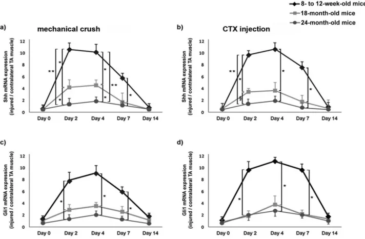
Upon mechanical crush and CTX injury of the skeletal muscle, young mice displayed significant increase of Shh mRNA level (Figure 1). In both experimental models, Shh mRNA levels were about 10 times higher in injured mus-cles compared with contralateral uninjured TA musmus-cles at Days 2 and 4 after injury. At Day 7 after mechanical crush, Shh levels were about six times higher in injured muscles
compared with the uninjured TA muscles (Figure 1a). Similarly, 7 days after CTX injection, Shh levels were about eight times higher in injured muscles compared with uninjured tissues (Figure 1b). At Day 14 after injury, Shh mRNA expression was not different in injured and control TA muscles (Figure 1a and b). In 18-month-old mice, the time course of Shh expression upon injury was similar to that observed in young animals, but Shh levels were sig-nificantly lower than those observed in young mice in both injury models (*p < .01; Figure 1a and b). An even more pronounced impairment of Shh upregulation after injury was observed in 24-month-old mice (Figure 1a and b). In these mice, Shh upregulation was significantly lower than in 18-month-old animals at Days 2 and 4 after crush (*p < .01), as well as at Day 2 after CTX injury (§p < .05).
Also, it was significantly lower than in young mice (8–12 weeks old) at 2, 4, and 7 days after injury in both experi-mental models (**p < .001, *p < .01).
Because Gli1 is the principal transcription factor of the Shh pathway, its expression constitutes evidence of func-tional activity of the Shh pathway (3). For this reason, the
Figure 1. Impaired activation of the Sonic hedgehog (Shh) signaling pathway in injured muscles of 18- and 24-month-old mice. Shh real-time PCR was performed at Days 0, 2, 4, 7, and 14 after mechanical crush (a) and cardiotoxin (CTX) injury (b) of the tibialis anterior (TA) muscle. Real-time PCR for Gli1 was performed at the same time points for both experimental models as well (c and d). The ratio between Shh and Gli1 mRNA levels in the injured TA muscle and the contralateral TA muscle increased significantly in young mice at Days 2, 4, and 7 after injury (*p < .01). In 18- and 24-month-old mice, Shh and Gli1 upregulation after both mechanical and toxic injury was significantly lower compared with young mice (*p < .01, **p < .001). In addition, Shh upregulation was reduced in 24-month-old mice compared with 18-month-old mice at Days 2 and 4 after crush (*p < .01) and at Day 2 after CTX injury (§p < .05).
by guest on June 19, 2013
http://biomedgerontology.oxfordjournals.org/
mRNA levels of Gli1 were also analyzed. We found that both mechanical and toxic injuries of the skeletal muscle were fol-lowed by significant upregulation of Gli1 mRNA in young mice at Days 2, 4, and 7 after injury, with the highest expres-sion level being observed at Day 4 (>8-fold increase in injured TA muscles compared with control TA muscles; Figure 1c and d). Gli1 upregulation was instead significantly impaired in 18-month-old mice and 24-month-old mice at 2, 4, and 7 days after injury in both experimental models (*p < .01; Figure 1c and d).
Shh Gene Therapy Is Able to Induce Functional Activation of the Shh Pathway in the Injured Skeletal Muscle of Old Mice
A basic tenet of this study is that we can overexpress functional Shh in a controllable manner in mouse mus-cle tissues. To prove this, we used a total of 20 old mice, which received an intramuscular injection of 40 μg phShh (n = 10) or empty plasmid (n = 10) immediately after CTX injury. Mice were sacrificed 4 days after injury. We found significantly higher expression levels of Gli1 in muscles of old mice injected with phShh compared with those injected with empty plasmid (p < .01; Figure 2a). Such levels of expression were similar to those observed in young mice at this time points after CTX injury (Figure 1d). We also observed strong expression of human Shh in muscles of old mice treated with 40 µg phShh (Figure 2b).
Shh Gene Therapy Increases the Number of Myf5-Positive Cells in the Site of Regeneration, Enhances the Number of Regenerating Myofibers, and Reduces Fibrosis in 24-Month-Old Mice
We tested the hypothesis that, in 24-month-old mice, Shh gene therapy increases the regenerative capacities of the
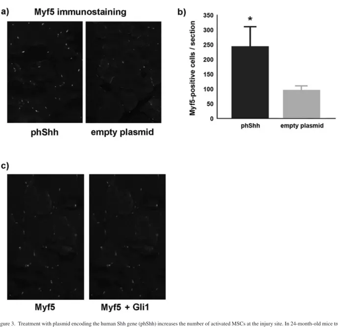
skeletal muscle after injury with CTX. First, we looked at the number of Myf5-positive cells in the site of injury. Myf5 is a myogenic regulatory factor expressed by activated MSCs and represents a biological marker of regeneration. We found a significantly higher number of Myf5-positive cells in the muscles of mice treated with phShh compared with those of mice treated with empty plasmid (Figure 3a and b). In the group treated with phShh, many Myf5-postive cells also displayed positive staining for Gli1 (Figure 3c), indicating direct response of MSCs to the Shh ligand.
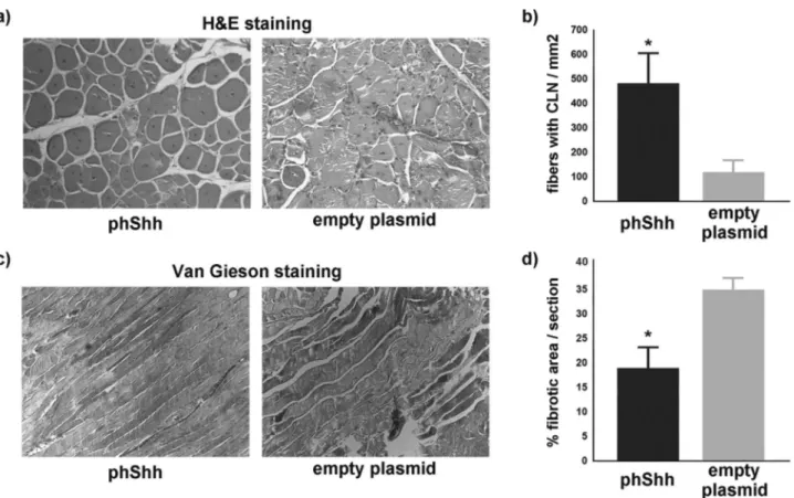
Upon activation, MSCs fuse to form young myotubes, which are characterized by centrally located nuclei. Staining with hematoxylin and eosin demonstrated a significantly higher number of fibers with centrally located nuclei in the phShh-treated group compared with controls (Figure 4a and b). Finally, we assessed the extent of injury-induced mus-cle fibrosis by performing a Van Gieson staining 14 days after injury. Significantly reduced extent of fibrotic tissue was detected in the TA muscles of mice treated with phShh compared with those of mice treated with the empty plas-mid (Figure 4c and d).
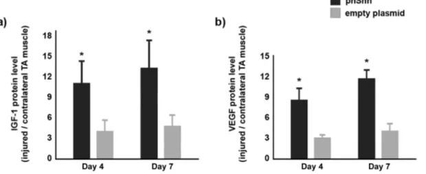
Shh Gene Therapy Increases Local Expression of IGF-1 and VEGF165 in the Injured Skeletal Muscle of 24-Month-Old Mice
We have previously shown that Shh regulates the expression of different families of angiogenic and myogenic growth factors (3–7). Here, we investigated whether Shh gene therapy has such ability also in the injured skeletal muscles of 24-month-old mice. For these analyses, we used specimens from mice sacrificed 4 and 7 days after injury. At both time points, the ratio between IGF-1 and VEGF165 levels, measured by ELISA, in the injured muscle compared with the contralateral muscle was significantly higher in
Figure 2. Treatment with plasmid encoding the human Shh gene (phShh) induces functional activation of the Shh pathway in the injured tibialis anterior (TA) muscle of 18-month-old mice. Immediately after cardiotoxin (CTX) injection, phShh (40 µg) was administered to the injured TA muscle of 18-month-old mice and induced significant increment of Gli1 expression compared with empty plasmid at Day 4 after injury (*p < .01) (a). Four days after injection of 40 µg phShh, expres-sion of human Shh is detectable in the TA muscle of one 18-month-old mouse, whereas no human Shh expresexpres-sion may be found in the TA muscle injected with the empty plasmid (b).
by guest on June 19, 2013
http://biomedgerontology.oxfordjournals.org/
the phShh-treated group than in the empty plasmid-treated group (Figure 5).
Discussion
The repair of an injured tissue is a complex biological process involving the coordinated activities of tissue-resident and infiltrating cells in response to local and systemic signals. Following acute tissue injury, inflammatory cell infiltration and activation/proliferation of resident stem cells are the first line of defense to restore tissue homeostasis. However, many cellular and molecular mechanisms underlying efficient muscle repair are dysregulated in the aging muscle and eventually lead to impaired regeneration in response to injury (8–16). Indeed, the progressive incapacity of regeneration machinery to replace damaged muscle is a
hallmark of age-related muscle loss or sarcopenia (16,17). A better understanding of impaired regenerative capacities represents an important first step for the development of therapeutic approaches. The results of our study hint at Shh as a novel player in the molecular mechanisms that underlie impaired regeneration in the aging skeletal muscle. The demonstration of impaired activation of the Shh pathway in response to injury in the skeletal muscle of 18- and 24-month-old mice is interesting because it is consistent with the concept that important mechanisms of muscle repair are affected by aging. The molecular and cellular mechanisms underlying this phenomenon remain to be elucidated, but this is beyond the scope of our study. Further investigation is needed to understand whether impaired activation of the Shh pathway depends on altered
Figure 3. Treatment with plasmid encoding the human Shh gene (phShh) increases the number of activated MSCs at the injury site. In 24-month-old mice treated with phShh (40 µg) after cardiotoxin (CTX) injury of the tibialis anterior (TA) muscle, there is a significant higher number of activated (Myf5-positive) MSCs at the site of injury (244.6 ± 65.6 vs 99.7 ± 11.2 Myf5-positive cells per section, *p < .01) (a and b). Many Myf5-positive cells (green staining) are also immunopositive for the Shh target gene Gli1 (red/yellow staining) (c).
by guest on June 19, 2013
http://biomedgerontology.oxfordjournals.org/
function or expression of the Shh receptors or transcription factors, increased expression of negative modulators of the Shh pathway, or other mechanisms. However, our findings suggest that dysregulation of the Shh pathway might be a novel contributor to deficient muscle regeneration associated with aging.
In the present study, we also show that phShh is able to induce functional activation of the Shh pathway in the injured skeletal muscle of old mice, resulting in Gli1—the principal transcription factor of the Shh pathway—expres-sion levels that are similar to those observed in young mice upon induction of injury. Our study demonstrates that Shh therapy activates an efficient regenerative process in response to injury and leads to increased number of acti-vated MSCs and new myofibers. Not surprisingly, it also results in decreased fibrosis. The beneficial effects of Shh therapy may be due, at least in part, to direct stimulation of MSCs, as indicated by the fact that, in mice receiving Shh therapy, Myf5-positive cells express the Shh target gene Gli1. This is consistent with previous findings from our group and others, demonstrating that Shh is able to act on adult MSCs in vitro (7,18,19). It is also consistent with the fact that, in the embryo, a Gli1-binding site in the Myf5 epaxial somite enhancer is necessary for the specification of epaxial muscle progenitor cells and that Gli1 interacts
with several important myogenic pathways, such as those regulated by Wnt, Frizzled, Numb, and β-catenin, during embryonic myogenesis (20–24). In addition, it has been recently demonstrated that Shh induces mitogen-activated protein kinase/extracellular signal-regulated kinase and phosphoinositide 3-kinase-dependent Akt phosphorylation in adult myoblasts in vitro and that Shh-induced Akt phos-phorylation is required for its promotive effects on muscle cell proliferation and differentiation (18).
An additional mechanism through which Shh therapy exerts beneficial effects in the injured muscle may be the upregulation of IGF-1 and VEGF165. In the last decade, IGF-1 has emerged as a growth factor with a remarkably wide range of actions and a tremendous potential as a therapeutic in attenuating the atrophy and frailty associ-ated with muscle aging and diseases (25). In addition, IGF-1 is a protein with potent anti-apoptotic functions in skeletal muscle cells (26,27), thus its upregulation by Shh therapy might also be responsible for the reduced extent of fibrotic tissue observed in Shh-treated muscles. Regarding VEGF, it is well accepted that angiogenic cytokines have direct effects on muscle cells and myogenesis (6,28–32). In this respect, it is intriguing to note that some VEGF-induced intracellular mechanisms, such as the phospho-inositide 3-kinase/Akt and mitogen-activated protein
Figure 4. Treatment with plasmid encoding the human Shh gene (phShh) increases the number of regenerating myofibers and reduces fibrosis after cardiotoxin (CTX) injury. Representative images of fibers with centrally located nuclei (CLN) in cross-sections of 24-month-old mice treated with phShh and empty plasmid (a). The number of regenerating myofibers was significantly higher in the phShh-treated group (485.5 ± 120.7 vs 123.4 ± 72.4 fibers with CLN/mm2, *p < .01) (b).
Treatment with phShh also results in decreased percentage of fibrotic area per section (18.8% ± 6.1% vs 34.9% ± 2.3%, *p < .01) (c and d).
by guest on June 19, 2013
http://biomedgerontology.oxfordjournals.org/
kinase signaling pathways, in addition to be important for endothelial cell survival, migration, and proliferation, are also involved in muscle survival, differentiation, and regeneration (25,33–37).
In this scenario, it is reasonable to hypothesize that, in the course of muscle regeneration, Shh gene therapy exerts its beneficial effects both directly—on MSCs—and indirectly through the upregulation of growth factors such as IGF-1 and VEGF, which are eventually responsible for activating and/or enhancing myogenesis. In summary, our findings demonstrate that Shh therapy increases the regenerative capacities of the aging muscle and merits further investigation for its potential therapeutic utility in muscular diseases of the elderly.
Funding
This work was supported by the National Institutes of Health (1R21HL089684 to R.P.); the “Fondazione Roma” (RCSNE3 to R.P.); the Italian Department of University and Research (MIUR) (FIRB-IDEAS RBID08MAFS to R.P.).
References
1. Borycki AG, Brunk B, Tajbakhsh S, Buckingham M, Chiang C, Emerson CP Jr. Sonic hedgehog controls epaxial muscle determina-tion through Myf5 activadetermina-tion. Development. 1999;126:4053–4063. 2. Krüger M, Mennerich D, Fees S, Schäfer R, Mundlos S, Braun T.
Sonic hedgehog is a survival factor for hypaxial muscles during mouse development. Development. 2001;128:743–752.
3. Pola R, Ling LE, Silver M, et al. The morphogen Sonic hedgehog is an indirect angiogenic agent upregulating two families of angiogenic growth factors. Nat Med. 2001;7:706–711.
4. Pola R, Ling LE, Aprahamian TR, et al. Postnatal recapitulation of embryonic hedgehog pathway in response to skeletal muscle ischemia. Circulation. 2003;108:479–485.
5. Kusano KF, Pola R, Murayama T, et al. Sonic hedgehog myocardial gene therapy: tissue repair through transient reconstitution of embry-onic signaling. Nat Med. 2005;11:1197–1204.
6. Palladino M, Gatto I, Neri V, et al. Pleiotropic beneficial effects of Sonic hedgehog gene therapy in an experimental model of peripheral limb ischemia. Mol Ther. 2011;19:658–666.
7. Straface G, Aprahamian T, Flex A, et al. Sonic hedgehog regulates angiogenesis and myogenesis during post-natal skeletal muscle regen-eration. J Cell Mol Med. 2009;13:2424–2435.
8. Lewis DM, Schmalbruch H. Effects of age on aneural regeneration of soleus muscle in rat. J Physiol. 1995;488:483–492.
9. Rivard A, Fabre JE, Silver M, et al. Age-dependent impairment of angiogenesis. Circulation. 1999;99:111–120.
10. Rivard A, Berthou-Soulie L, Principe N, et al. Age-dependent defect in vascular endothelial growth factor expression is associ-ated with reduced hypoxia-inducible factor 1 activity. J Biol Chem. 2000;275:29643–29647.
11. Pola R, Aprahamian TR, Bosch-Marcé M, et al. Age-dependent VEGF expression and intraneural neovascularization during regeneration of peripheral nerves. Neurobiol Aging. 2004;25:1361–1368.
12. Marsh DR, Criswell DS, Carson JA, Booth FW. Myogenic regulatory factors during regeneration of skeletal muscle in young, adult, and old rats. J Appl Physiol. 1997;83:1270–1275.
13. Dennis RA, Przybyla B, Gurley C, et al. Aging alters gene expression of growth and remodeling factors in human skeletal muscle both at rest and in response to acute resistance exercise. Physiol Genomics. 2008;32:393–400.
14. Goldspink G. Age-related loss of muscle mass and strength. J Aging Res. 2012;2012:158279.
15. Serrano AL, Muñoz-Cánoves P. Regulation and dysregulation of fibro-sis in skeletal muscle. Exp Cell Res. 2010;316:3050–3058.
16. Vinciguerra M, Musaro A, Rosenthal N. Regulation of muscle atrophy in aging and disease. Adv Exp Med Biol. 2010;694:211–233. 17. Cossu G, Mavilio F. Myogenic stem cells for the therapy of primary
myopathies: wishful thinking or therapeutic perspective? J Clin Invest. 2000;105:1669–1674.
18. Elia D, Madhala D, Ardon E, Reshef R, Halevy O. Sonic hedge-hog promotes proliferation and differentiation of adult muscle cells: involvement of MAPK/ERK and PI3K/Akt pathways. Biochim Biophys Acta. 2007;1773:1438–1446.
19. Koleva M, Kappler R, Vogler M, Herwig A, Fulda S, Hahn H. Pleiotropic effects of Sonic hedgehog on muscle satellite cells. Cell Mol Life Sci. 2005;62:1863–1870.
20. Borycki A, Brown AM, Emerson CP Jr. Shh and Wnt signaling pathways converge to control Gli gene activation in avian somites. Development. 2000;127:2075–2087.
21. Gustafsson MK, Pan H, Pinney DF, et al. Myf5 is a direct target of long-range Shh signaling and Gli regulation for muscle specification. Genes Dev. 2002;16:114–126.
Figure 5. Treatment with plasmid encoding the human Shh gene (phShh) increases expression of insulin-like growth factor (IGF)-1 and vascular endothelial growth factor (VEGF)165 after cardiotoxin (CTX) injury. Protein expression ratio (injured/contralateral muscle) of IGF-1 (a) and VEGF (b) is significantly increased both at Days 4 and 7 after injury in 24-month-old mice treated with phShh compared with controls (*p < .01).
by guest on June 19, 2013
http://biomedgerontology.oxfordjournals.org/
22. McDermott A, Gustafsson M, Elsam T, Hui CC, Emerson CP Jr, Borycki AG. Gli2 and Gli3 have redundant and context-dependent function in skeletal muscle formation. Development. 2005;132:345–357.
23. Holowacz T, Zeng L, Lassar AB. Asymmetric localization of numb in the chick somite and the influence of myogenic signals. Dev Dyn. 2006;235:633–645.
24. Borello U, Berarducci B, Murphy P, et al. The Wnt/beta-catenin path-way regulates Gli-mediated Myf5 expression during somitogenesis. Development. 2006;133:3723–3732.
25. Winn N, Paul A, Musaró A, Rosenthal N. Insulin-like growth factor isoforms in skeletal muscle aging, regeneration, and disease. Cold Spring Harb Symp Quant Biol. 2002;67:507–518.
26. Alessi DR, Andjelkovic M, Caudwell B, et al. Mechanism of activation of protein kinase B by insulin and IGF-1. EMBO J. 1996;15:6541–6551.
27. Mourkioti F, Rosenthal N. IGF-1, inflammation and stem cells: interactions during muscle regeneration. Trends Immunol. 2005;26: 535–542.
28. Smythe GM, Lai MC, Grounds MD, Rakoczy PE. Adeno-associated virus-mediated vascular endothelial growth factor gene therapy in skeletal muscle before transplantation promotes revascularization of regenerating muscle. Tissue Eng. 2002;8:879–891.
29. Germani A, Di Carlo A, Mangoni A, et al. Vascular endothelial growth factor modulates skeletal myoblast function. Am J Pathol. 2003;163:1417–1428.
30. Wagatsuma A, Tamaki H, Ogita F. Sequential expression of vascu-lar endothelial growth factor, Flt-1, and KDR/Flk-1 in regenerating mouse skeletal muscle. Physiol Res. 2006;55:633–640.
31. Messina S, Mazzeo A, Bitto A, et al. VEGF overexpression via adeno-associated virus gene transfer promotes skeletal muscle regeneration and enhances muscle function in mdx mice. FASEB J. 2007;21:3737–3746. 32. Ochoa O, Sun D, Reyes-Reyna SM, et al. Delayed angiogen-esis and VEGF production in CCR2-/- mice during impaired skel-etal muscle regeneration. Am J Physiol Regul Integr Comp Physiol. 2007;293:R651–R661.
33. Gerber HP, McMurtrey A, Kowalski J, et al. Vascular endothelial growth factor regulates endothelial cell survival through the hosphati-dylinositol 3’-kinase/Akt signal transduction pathway. Requirement for Flk-1/KDR activation. J Biol Chem. 1998;273:30336–30343. 34. Fujio Y, Guo K, Mano T, Mitsuuchi Y, Testa JR, Walsh K. Cell cycle
withdrawal promotes myogenic induction of Akt, a positive modulator of myocyte survival. Mol Cell Biol. 1999;19:5073–5082.
35. Bodine SC, Stitt TN, Gonzalez M, et al. Akt/mTOR pathway is a cru-cial regulator of skeletal muscle hypertrophy and can prevent muscle atrophy in vivo. Nat Cell Biol. 2001;3:1014–1019.
36. Rommel C, Bodine SC, Clarke BA, et al. Mediation of IGF-1-induced skeletal myotube hypertrophy by PI(3)K/Akt/mTOR and PI(3)K/Akt/ GSK3 pathways. Nat Cell Biol. 2001;3:1009–1013.
37. Takahashi A, Kureishi Y, Yang J, et al. Myogenic Akt signaling regu-lates blood vessel recruitment during myofiber growth. Mol Cell Biol. 2002;22:4803–4814.
by guest on June 19, 2013
http://biomedgerontology.oxfordjournals.org/