UNIVERSITY OF SASSARI
Department of Biomedical Science
PhD Course in Life Science and Biotechnologies
CICLE XXXI
PhD Course Coordinator: Prof. Leonardo A. Sechi
Oxidative stress in early stages of Chronic
Obstructive Pulmonary Disease
Tutor:
Prof. Ciriaco Carru Dr. Elisabetta Sotgiu PhD student:
LIST OF CONTENTS
1. ABSTRACT 3
2. INTRODUCTION 4
2.1 COPD 4
2.2 Epidemiology 6
2.3 Diagnosis and classification 6
2.4 Exacerbations 10 2.5 Quality of life 11 2.6 Comorbidities 11 2.7 COPD management 12 2.8 COPD treatment 13 2.9 COPD pathology 14 2.10 COPD immunology 14
2.11 Inflammation and oxidative stress 16
2.11.1 Inflammation 16
2.11.2 Oxidative stress 18
3. AIM OF THE PROJECT 23
4. MATERIAL AND METHODS 24
4.1 Subjects recruitment 24
4.2 Study biomarkers 25
4.3 Sample collection 27
4.4 Biochemical analysis 27
4.4.1 -SH protein 27
4.4.3 Paraoxonase 1 28
4.4.4 Ergothioneine 28
4.4.5 Taurine 29
4.4.6 Glutathione 29
4.4.7 Arginines 30
4.4.8 Global DNA methylation 30
4.4.9 Tryptophan and kynurenine 31
4.5 Statistical analysis 31
5. RESULTS 33
5.1 Oxidative stress biomarkers results 33
5.2 Arginines results 36
5.3 Global DNA methylation results 38
5.4 Tryptophan and kynurenine results 39
6. DISCUSSION 42
6.1 Oxidative stress biomarkers 42
6.2 Arginines 44
6.3 Global DNA methylation 45
6.4 Tryptophan pathway 47
7. CONCLUSION 49
1. ABSTRACT
Chronic obstructive pulmonary disease (COPD) is a common respiratory condition characterized by an irreversible or partial irreversible airway obstruction. Since oxidative stress and inflammation play an important role in the pathophysiology of the disease, our target was to evaluate biomarkers index of these conditions in order to find out one or more biomarkers that can predict the onset and the progression of the pathology. We analyzed oxidative stress and inflammation biomarkers in 29 mild COPD, 14 moderate COPD and in 43 healthy controls. Results obtained show the decrease of PSH levels in COPD patients from the early stage of COPD and the increase is higher with COPD progression. Furthermore, COPD patients presented high ADMA/arginine ratio, low levels of global DNA methylation, high levels of kynurenine and kyn/trp ratio and low levels of tryptophan compared to healthy controls. In addition, the alterations of these biomarkers are further greater with the progression of the disease.
Concluding, our results underline the importance of oxidative stress in COPD presence and severity. Indeed, our data show the alteration of the pathways analyzed due principally to oxidative stress. So, it might be interesting to increase the number of subjects of the study and to include patients with severe form of COPD to fully characterized the impact of oxidative stress in this pathology.
2. INTRODUCTION
2.1 COPD
Chronic obstructive pulmonary disease (COPD) is a common preventable and treatable respiratory condition characterized by a progressive and not completely reversible airway obstruction and by persistent inflammation of the lungs to noxious particles and gases, as cigarette smoke.1
The characteristic symptoms of COPD are generally the presence of chronic cough, dyspnea and sputum production. These symptoms are usually common in all patients affected by airflow obstruction, as COPD and asthma.
COPD is currently the fourth leading cause of death in the world and the World Health Organization (WHO) predicts that it is going to be the third cause of death and the fifth cause of disability in the world by 2020.2 Currently, the WHO estimates that chronic obstructive pulmonary disease (COPD) affects 65 million individuals worldwide.3 Moreover, it currently affects about 10% of people over 45 years of age, rising to 50% in heavy smokers.4
The main risk factor for the onset of the COPD is cigarette smoke,5 but not all smokers develop the disease and not all people affected by COPD are smokers. SO, this suggests that also other factors could influence the onset and the progression of COPD. Moreover, other types of inhalants, such as chemical fumes, pollution and also the exposure to passive smoke, may contribute to the risk of developing COPD.6
COPD is usually described as a pathology characterized by the presence of both emphysema and chronic bronchitis (Fig. 1) and the contribution of these conditions vary from person to person. Emphysema is characterized by an enlargement of the distal
airspaces7, created by alveoli destruction, which causes a decreased level of oxygen and an increased level of carbon dioxide in the blood. Chronic bronchitis is defined by the production of cough and sputum for at least 3 months for 2 consecutive years.8
Fig 1: Effects of emphysema and chronic bronchitis in lungs affected by COPD
COPD prevalence, morbidity and mortality vary across countries and, inside the country, across different groups of people. This disease is the resultant of constant exposure to risk factors, primarily the exposure to cigarette smoke, but also the exposure to other respiratory irritants as chemical fumes, dusts and indoor and outdoor pollution.1 COPD is characterized by chronic inflammation, remodeling of the small airways and destruction of the lung parenchyma.5 The disease worsening is characterized by the
onset of exacerbations, which are usually associated with an increase in symptoms, airway inflammation and systemic inflammatory effects.6
2.2 Epidemiology
The most common cause of chronic airflow obstruction globally is smoking and exposure to environmental tobacco smoke.6 As a matter of fact, cigarette smoking represents the major etiological factor in COPD development, as more than 90% of COPD subjects are smokers, but not all smokers are affected by COPD, so other factors could be involved in COPD development.9 There is some evidence that chronic airway obstruction might be even due to the exposure to smoke from biomass burning, a dusty work environment, high levels of outdoor and indoor air pollution6 and also an history of tuberculosis10. Moreover, several studies have suggested that hereditability plays an important role in the disease, accounting for at least 30% of the variation in COPD risk. Indeed, subjects that smoke and that are first-degree relatives of COPD patients have more or less a threefold increased risk of developing the pathology compared with smokers from the general population, whereas subjects that do not smoke and are first-degree relatives of COPD patients have similar or low risks for developing COPD compared with non-smokers in the general population. These results indicate that genetics plays an important role in COPD development.6
2.3 Diagnosis and classification
People presenting respiratory symptoms, as cough, sputum expectoration and dyspnea, should be suspected to be affected by COPD. The exposure to cigarette smoke,
occupational and environmental pollutants and the presence of a family history for COPD could increase the suspicion to be affected by COPD. Generally, the presence of cough and mucus production is present in COPD patients many years before the onset of the pathology. On the other hand, some patients are diagnosed with COPD without the presence of these characteristic symptoms.1
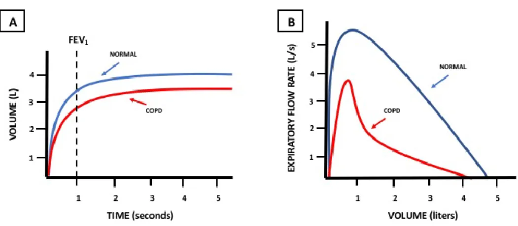
The diagnosis of COPD is confirmed by spirometry, which indicates the presence of airway obstruction on the basis of spirometric parameters, as FVC (forced vital capacity) and FEV1 (forced expiration volume in the first second). In healthy individuals, the value of FEV1/FVC is greater than 70%, while in presence of airway obstruction this value decreases under 70%. Moreover, FEV1 is characteristically low in COPD patients and in individuals with restrictive pulmonary diseases (Fig. 2). The FEV1/FVC ratio may lead to more frequent diagnosis of COPD in elderly people, as the reduction of respiratory function normally decreases with the progression of age.1
Fig 2: In part A, the characteristic decrease of FEV1 in people presenting airflow obstruction, as in COPD; in part B, the typical curve
representing airflow obstruction.
Moreover, the diagnosis of COPD requires FEV1/FVC ratio minor of 70% and the spirometry test should be repeated after inhaled bronchodilators administration, in order to distinguish if airflow limitation is poorly reversible, as in COPD patients, or
largely reversible, as in patients with asthma.11 Previously, spirometry was used to support the diagnosis of COPD, while now it is required to make a confident diagnosis.1 The Global Initiative for Chronic Obstructive Lung Disease (GOLD) draw up every year useful guidelines and reports, validating in many languages, regarding all information about chronic obstructive pulmonary disease. The document regarded is a global document and for this reason is not suitable for all countries of the world, so it has to be enhanced with country’s needs. According to the previous GOLD guidelines, COPD patients were classified into four stages, only on the basis of airflow obstruction severity:
1. STAGE GOLD 1 – mild (FEV1 ³80% of predicted); 2. STAGE GOLD 2 – moderate (FEV1 50-80% of predicted); 3. STAGE GOLD 3 – severe (FEV1 30-50% of predicted); 4. STAGE GOLD 4 – very severe (FEV1 <30% of predicted).12
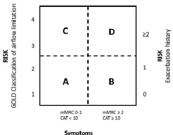
The last method indicated for COPD classification is a combined method and includes the assessment of symptoms and the airflow limitation. mMRC or CAT scale are used to indicate the presence of symptoms (maximum score: mMRC >2; CAT = 10). So, primarily patients are classified on the basis of symptoms or exacerbations, then on the basis of airflow limitation, according to the common classification evaluating the spirometric parameters.
So, patients can be classified as follow (see figure 3):
1. GROUP A (low risk, less symptoms): patients present mild or moderate airflow limitation and 0-1 exacerbation per year (mMRC 0-1 or CAT <10).
2. GROUP B (high risk, more symptoms): patients present mild or moderate airflow limitation and >2 exacerbation per year or >1 hospitalized exacerbation (mMRC >2 or CAT >10).
3. GROUP C (high risk, less symptoms): patients present severe or very severe airflow limitation and 0-1 exacerbation per year (mMRC 0-1 or CAT <10). 4. GROUP D (high risk, more symptoms): patients present severe or very severe
airflow limitation and >2 exacerbations per year or >1 hospitalized exacerbation (mMRC >2 or CAT >10).1
Fig 3: Combined COPD assessment.
This combined method reflects the complexity of the pathology better than the unidimensional analysis, as this one considers only airflow limitation and not the severity of symptoms.
2.4 Exacerbations
The progression of the pathology is usually characterized by exacerbations, which are defined as short periods of at least 48 hours of increased cough, dyspnea and sputum production. Exacerbations are often present in patients affected by COPD and this worsening of the pathology is mainly due to bacteria and/or virus infections, to smoke exposure, environmental pollutants and unknown factors.13 Exacerbations are characterized by different severity of symptoms and, on the basis of mild or moderate ones, the treatment used is different. For mild exacerbations is required treatment with bronchodilators, for moderate exacerbations systemic corticosteroids and antibiotics are required, while for severe ones could be necessary admission to hospital. Not all patients present the same severity and frequency of exacerbations. Frequency may rise with increasing severity of COPD.1
Exacerbations in COPD represent an important event in the worsening of the disease and they cause a reduction in life quality, an increase of disease progression and of the risk of death.14 Moreover, the presence of exacerbations and the COPD severity implicate the increase of the cost of care, as with the progression of the disease patients need more pharmacological treatments and hospitalizations.1
Because of their negative impact on the natural history of the disease, the prevention and the reduction of exacerbations represent a primary goal of treatment in COPD, as exacerbations and comorbidities contribute to the overall severity in COPD patients.12
2.5 Quality of life
COPD is characterized by the presence of common occurrence of exacerbations, which contribute to morbidity, death and health-related quality of life.15 Quality of life is a significant factor for COPD patients. Health status is defined as “the impact of health on
a person’s ability to perform and derive fulfilment from the activities of daily life. A patient’s self-reported health status thus includes health-related quality of life and functional status”16. COPD patients are characterized by an impaired health status, due to the severity of the pathology. The health status might be improved with starting polymedication, pulmonology visits, balanced diet, completing a rehabilitation program, smoking cessation and reducing exacerbations.6
2.6 Comorbidities
Even though COPD is a lung disease, it is usually associated with systemic manifestations and comorbidities. The most common comorbidities are ischemic heart disease, osteoporosis, depression, diabetes, skeletal muscle wasting, cachexia and lung cancer.17 So, COPD is not just a lung disease and many patients have compromised other body systems with important prognostic and therapeutic implications.6
The management of COPD comorbidities and infectious exacerbations, both viral and bacterial, is an important aspect for patients.
Comorbidities play an important role on COPD patient’s death. In particular, cardiovascular diseases markedly impact on disease morbidity, progression and mortality. Indeed, it is estimated that between 30% and 50% of COPD-related deaths
are caused by cardiovascular morbidity, such as coronary artery disease, hypertension and diabetes.18
Moreover, patients affected by COPD have exercise limitation due to skeletal muscle dysfunction, which is maybe due to the combination of atrophy and sarcopenia. The decrease of functional activity is a useful index to classify the severity of COPD, as it predicts mortality better than FEV1. Even people affected by mild COPD could present a decrease in exercise capacity.19
Furthermore, a significant part of COPD patients is characterized by low body mass index, with values below 21 kg per m2 associated with increased risk of death.20
The chronic inflammation in COPD patients may contribute to the onset of extrapulmonary complications, which enhance the presence of morbidity and mortality in patients affected by this pathology.21
2.7 COPD management
The reduction of symptoms and exacerbations is an important aspect in the management of COPD patients. Every individual patient needs to be monitored and evaluated singularly. To evaluate the monitoring of the disease, it is important to consider several aspects, as the presence of symptoms, the exposure to smoke, lung functionality, the presence and frequency of exacerbations, the treatments necessary to control symptoms and exacerbations, the history of hospitalization and the presence of comorbidities. All these aspects help the physicians to classify the health status of the patient, the severity of the pathology and the effectiveness of the pharmacological therapy.
2.8 COPD treatment
The first step for COPD treatment is absolutely smoking cessation, but also limitation of other risk factor, as exposure to occupational and environmental pollution, and to be underwent to influenza vaccinations yearly are recommended.22 Also, physical activity and rehabilitation are important aspects for COPD patients to improve their health and their pathological status. In this way patients improve, not only their physique, but also their tolerance to dyspnea and fatigue.23
The mainstay of pharmacological management of symptoms in COPD patients is bronchodilators treatment. Bronchodilators include b2-agonists and muscarinic receptor antagonists. It is preferable the treatment with inhaled long-acting bronchodilators of 12/24 hours of duration and, in particular, long-acting b2-agonists (LABAs) and long-acting muscarinic antagonists (LAMAs) are equally effective. The treatment with long-acting bronchodilators reduces breathlessness, improves long function, exercise capacity and health status.6
During exacerbations, systemic corticosteroids are indicated to improve lung functionality, in particular FEV1, and arterial hypoxemia, to reduce the risk of acute symptoms and to enhance the discharge from hospital. During exacerbations, in presence of bacterial infections it is also useful the treatment with antibiotics.1
If COPD patients present hypoxemia with a target saturation of 88-92%, an oxygen therapy should be needed.24
Although there is no an effective therapy for reversing airway obstruction in COPD patients, targeting biomarkers of oxidative stress and lung/systemic inflammation could be helpful in improving survival and quality of life in these patients.18
2.9 COPD pathology
The relevant pathological features of COPD are obstructive bronchiolitis, emphysema and mucus hypersecretion, which are mainly due to the persistent lung inflammation.25 Moreover, COPD is characterized by a reduction in FEV1 and the FEV1/FVC ratio, which progresses over time.6
COPD is characterized by progressive airflow limitation and this obstruction is due to remodeling and narrowing of small airway and destruction of lung parenchyma and, as a consequence, the destruction of the alveolar attachments of these airways as a result of emphysema. Inflammation plays a central role in these pathological changes and, although the molecular basis of inflammation is not yet fully understood, it is prospected that it may be probably determined by genetic and epigenetic factors. Respiratory irritants, as cigarette smoke, chemical fumes and dusts, may activate surface macrophages and airway epithelial cells to release chemokines that attract circulating monocytes, neutrophils and lymphocytes into the lungs. Inflammation caused by cigarette smoke persists even when smoking is stopped, suggesting that other mechanisms are involved.26
2.10 COPD immunology
Airways and lungs present innate mechanisms to prevent invasion of pathogenic microbes into the lower respiratory tract, represented by the epithelial barrier, mucociliary clearance, humoral factors and innate immunity cells, as macrophages, dendritic cells, monocytes, neutrophils, natural killer cells and mast cells.27
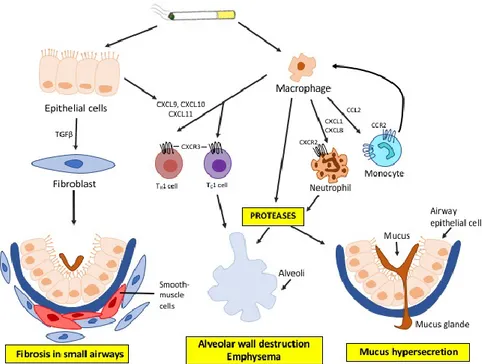
The persistent inhalation of cigarette smoke, biomass fuel and air pollutants activates pattern recognition receptors, as Toll-like receptors (TLRs), that enhance the innate immune response. The activation of immunity system causes not only the increased production of macrophages and neutrophils, but also the activation of epithelial cells in the airways and the secretion of mucus.27 (Figure 4)
Fig 4: The immunity activation in COPD
With the progression of the pathology there is the activation of adaptive immunity and, as a consequence, an increase of lymphocyte T and B production in the lungs. These cells are set into lymphoid follicles and these ones are involved in the activation of dendritic cells. With the severity of COPD there is also the activation of CD8+ cytotoxic T (Tc1) and CD4+ T helper (Th)1 cells in lung tissue.28 In COPD airways and lung parenchyma, CD8+ T cells predominate over CD4+ T cells, but their role in the pathogenesis of the disease is not yet certain.29
In airways there is also an increase of CD4+ Th17 cells, which are maybe involved in amplifying neutrophilic inflammation.30
2.11 Inflammation and oxidative stress
COPD is characterized by the presence of chronic systemic inflammation and systemic oxidative stress. Studies have shown an increase in serum levels of C-reactive protein (CRP), serum amyloid A, fibrinogen and several pro-inflammatory cytokines, as IL-8, IL-6 and TNFa. The increase of these markers is higher during exacerbations. About the presence of oxidative stress, there is an increase in H2O2 in the exhaled breath condensate (EBC) of patients affected by COPD and in smokers compared to non-smokers, and the increase is greater during exacerbations.18
2.11.1 Inflammation
Respiratory irritants, as smoke, dust and pollution, are responsible for lung inflammation, which is a normal answer of the lungs to noxious particles, but that in patients affected by COPD seems to be modified.1 In COPD patients inflammation is characterized by the presence of both innate immunity, with the activation of neutrophils, macrophages, mast cells, dendritic cells, eosinophils and natural killer cells, and adaptive immunity, characterized by T and B lymphocytes. Moreover, the activation of structural cells, including airway and alveolar epithelial cells, endothelial cells and fibroblasts, are involved in lung inflammation.26
Chronic inflammatory response in COPD may be orchestrated by macrophages and neutrophils. Macrophages are located in sites of alveolar wall destruction in patients
with emphysema. The number of macrophages in the parenchyma is correlated with emphysema severity.31 Macrophages play an important role in the pathophysiology of COPD, releasing inflammatory mediators, as TNF-a, CCL2, CXCL1, CXCL8 and reactive oxygen species (ROS). Since macrophages may be activated by cigarette smoke, this provide a cellular mechanism that links smoking with inflammation in COPD. Alveolar macrophages also secrete elastolytic enzymes, as MMP-2, MMP-9, MMP-12, neutrophil elastase taken up from neutrophils and cathepsins K, L and S.26
Neutrophil elastase plays an important role in breaking down the extracellular matrix and in stimulating the production and the secretion of mucin. The production of the mucin takes place via proteolytic cleavage of transforming growth factor a (TGFa). The high production of mucus and the impaired mucociliary clearance represent key factors for the onset of obstruction of airways in subjects affected by COPD.32
Most of the inflammatory proteins that are upregulated in COPD macrophages are regulated by the transcription factor nuclear factor kappa B (NF-kB), which is activated in alveolar macrophages of patients with COPD, particularly during exacerbations.33 The high number of macrophages that are present in lungs of smokers and in COPD patients is due to the recruitment of monocytes from the circulation in response to monocyte-selective chemokines CCL2 and CXCL1, which are increased in sputum and BAL of patients with COPD.34
As we said before, the inflammatory processes in COPD are also characterized by the presence of neutrophils, which are implicated in the development and progression of the pathology through the production of destructive mediators, as neutrophil elastase and matrix metalloproteinases.8 The abundant neutrophils, characteristic of COPD
condition, are due to an increase of CXC-chemokines production, as CXC-chemokine ligand 1 and CXCL8, whose receptor (CXCR2) is mainly expressed by neutrophils.28 The progressive airflow limitation in subjects affected by COPD is due to remodeling and narrowing of small airways and to lung parenchyma destruction.35 These COPD features are likely to be the results of chronic inflammation in the periphery of the lungs.36 Inflammation plays an important role in stable COPD, causing activation and alteration in the structural cells of the airways and lungs, and activation and recruitment of infiltrating inflammatory cells.37 Inflammatory cells in lower airways and lungs release several cytokines including IFN-γ, TNF-α, IL-1β, IL-6, IL-17, IL-18, IL-32 and TSLP and growth factors, such as TGF-β.38
Many of the cytokines and chemokines secreted by inflammatory cells are regulated by the nuclear factor-kB (NF-kB), which is activated in airway epithelial cells and macrophages, and it plays a key role in amplifying airway inflammation.39
The spread of the peripheral lung inflammation into the systemic circulation may contribute to the presence of various comorbidities, such as metabolic and cardiovascular diseases, in COPD patients.40
2.11.2 Oxidative stress
Oxidative stress is a normal process used by the body for the elimination of pathogens and toxic metabolites in physiological condition. When reactive oxygen species (ROS) are produced in excess compared to antioxidant defense mechanisms, oxidative stress causes harmful damage to cell structures, as lipids, proteins and DNA.41 ROS represent a large variety of free oxygen radical, like superoxide anion (O2-) and hydroxyl radical
(OH-) and also component of oxygen without unpaired electrons, such as hydrogen peroxide (H2O2). Normally, ROS are produced during normal metabolic processes in cells and when they are produced in excess cause harmful damages.42 ROS have three main roles: they are important in defense from pathogens, in the respiratory mitochondrial chain as part of the electron transport chain and, finally, they are involved in cell signaling.43
Free radicals are reactive molecules characterized by one or more unpaired electron(s) in their external shell. Oxygen plays an important role in the formation of these molecules.44 The terms reactive oxygen species (ROS) and reactive nitrogen species (RNS) refer to reactive radical and non-radical derivatives of oxygen and nitrogen, respectively.45
Because it is complicated to measure oxidative stress directly, it is more suitable to measure it by measuring its oxidation products, such as lipid peroxidation end products and oxidized proteins, and antioxidant molecules.46
In lipids, oxidative stress causes lipid peroxidation and one of the most abundant products is malondialdehyde, which inactivates many proteins through protein cross-linkages.47 In COPD, the increase of lipid peroxidation is maybe associated with pulmonary inflammation, emphysema development and alveolar destruction.
In proteins, reactive oxygen species are responsible for protein modifications, including changing of charge, the formation of disulphide bonds and the alteration of the tertiary structure, which can be reversible or irreversible. They can lead to reversible modifications as the formation of disulfides, persulfides, nitrosylation and s-glutathionilation, and to irreversible modifications as the formation of sulfinic and sulfonic acid, sulfonamides, protein carbonyls and nitrotyrosines.48
Moreover, oxidative stress is also responsible for DNA and RNA damages. The most common modification is the formation of the oxidized base 8-hydroxiguanosine, which inhibits DNA methylation, promotes microsatellite instability and accelerates telomere shortening.8
The presence of oxidative stress in COPD patients is generally due to chronic exposition to cigarette smoke, which represents the main risk factor for COPD and it is characterized by containing a high concentration of oxidants and reactive oxygen species.49 In chronic obstructive pulmonary disease patients, the presence of ROS is due directly to the presence of oxidants in cigarette smoke and to the release of ROS from macrophages and neutrophils. Moreover, these immune system cells, beyond ROS production, release several mediators that are responsible for the increase of inflammation.50 The increase of inflammation in COPD patients plays an important role in amplifying the oxidative stress in the lungs. The recruitment and activation of neutrophils, eosinophils, monocytes, and lymphocytes in the airways contributes to the ROS generation in response to inflammation. The activation of inflammatory cells produces O2·–, which is rapidly converted into H2O2 due to the action of superoxide dismutase (SOD), and ·OH, whose formation takes place in presence of Fe2+ as a secondary reaction.51 The ·OH radical is one of the most reactive chemical agents. It may act as a physiological intracellular agent that, in excess, is considered to be a risk factor for several respiratory diseases.52
The presence of H2O2 in the exhaled breath of COPD patients can be measured and it represents a direct measurement of oxidant burden in the lungs. COPD patients and smokers present higher levels of H2O2 than in non-smokers and levels of H2O2 further increase in presence of exacerbations.53
Normal lungs develop various endogenous antioxidant strategies to oppose and neutralize the damages caused by the deleterious effects of ROS. These strategies consist of both enzymatic and non-enzymatic mechanisms. Enzymatic antioxidant mechanisms include superoxide dismutase (SOD), catalase and glutathione peroxidase (Gpx), while non-enzymatic mechanisms are characterized by the activity of glutathione (GSH), vitamin C, vitamin E, albumin and uric acid.18 SOD exists in three forms and they have the role to remove superoxide anions, resulting in H2O2 production. The excess of H2O2 is not suitable and it represents the substrate of catalase and Gpx and it is converted into H2O and O2.43
Reactive oxygen species play a key role in COPD pathophysiology. First of all, ROS contribute to the activation of NF-κB and p38 MAPK, causing an increase in the expression of inflammatory genes and proteases. Moreover, ROS inhibit α1-antitrypsin, an endogenous antiprotease that is responsible for increased elastolysis. Furthermore, oxidative stress causes negative effects to DNA, but the DNA repair machinery works efficiently repairing them all. However, patients affected by COPD are not completely able to repair double-stranded DNA breaks and this could lead to an increased risk of developing lung cancer.54 Moreover, ROS cause protein damage through protein carbonylation, which is responsible for the generation of circulating autoantibodies that enhance lung inflammation and injury.55 Furthermore, ROS play an important role in the activation of transforming growth factor-β (TGFβ), which causes fibrosis56, and in reducing the expression and the activity of SIRT1, whose activity is reduced in COPD patient lungs. Moreover, ROS play a key role in maintaining genomic stability, regulating autophagy and protecting against cellular senescence and ageing.57 In addition, oxidative stress causes a reduction of HD2 expression and activity and this causes a
reduction of corticosteroid responsiveness in COPD patients.56 Furthermore, COPD patients frequently present defective endogenous antioxidant defense systems. One of the most relevant system is the transcription nuclear factor erythroid 2-related factor 2 (NRF2), which plays a key part in the regulation of multiple antioxidant and cytoprotective genes in response to oxidative stress. In COPD patients the function of NRF2 is impaired and not appropriately activated by oxidative stress, due to its increased acetylation as a result of reduced HD2 activity.58
Several studies are indicating the mitochondria as an important source of ROS in patients affected by chronic obstructive pulmonary disease. Moreover, mitochondrial function disruption in these patients leads to a reduction of intracellular ATP and an impaired oxidative phosphorylation.59 In airways epithelial cells, cigarette smoke induces mitophagy (the autophagic uptake of mitochondria), which causes mitochondrial deficiency and cell death.60
2. AIM OF THE PROJECT
Considering the large number of deaths caused by chronic obstructive pulmonary disease, it is necessary to implement the knowledge of the mechanisms involved in onset, progression and worsening of COPD. So, the aim of this project was to evaluate if there were some differences between COPD patients and healthy controls, in order to find out some biomarkers that could predict the onset and/or the progression of this disease.
Since COPD is an airway disorder characterized by a significant oxidative stress and inflammation, I evaluated biomarkers index of these conditions.
4. MATERIAL AND METHODS
4.1 Subjects recruitment
All subjects were enrolled in collaboration with the Respiratory Unit of the University of Sassari and each subject underwent to physical examination by the medical doctors, routine blood tests and spirometry. None of the patients had a previous diagnosis of COPD and none had a treatment with inhaled corticosteroids within 4 weeks prior to the study. Moreover, no patient was treated with long acting muscarinic antagonist, long or short beta-agonists (LABA or SABA) at the time of enrollment.
Data collected included forced vital capacity (FVC), FEV1 and FEV1/FVC ratio. Moreover, clinical and demographical information, as age, gender, body mass index, occupation and smoking status, were collected using a structure questionnaire.
We enrolled 43 COPD patients, divided in 29 mild COPD and 14 moderate COPD, and 43 sex- and age matched healthy controls. COPD was diagnosed and staged on the basis of physical examination, smoking history, respiratory symptoms and spirometric information according to the Global Initiative for Chronic Obstructive Lung Disease criteria. Healthy controls were selected from general population and chosen on the basis of age and gender. Exclusion criteria for the enrollment was the presence of concomitant inflammatory diseases such as infections, autoimmune disorders, cancer, liver, kidney and heart disease.
The study was in accordance with the principles of Declaration of Helsinki and it was approved by the Institutional Local Ethics Committee Azienda Sanitaria Locale n°1 of Sassari (Italy) (prot. 2175/CE del 21/04/2015). All subjects participating to the study provided written informed consent.
4.2 Study biomarkers
Oxidative stress biomarkers we analyzed were: glutathione, thiobarbituric acid-reactive substances, paraoxonase 1, -SH protein, taurine and ergothioneine.
• Glutathione (GSH) is the most important antioxidant molecule produced by the organism. It is located principally inside cells and it reduces organic hydroperoxides and protects lipids from peroxidation.
• Thiobarbituric acid-reactive substances (TBARS) derive from oxidative modification of polyunsaturated fatty acids and this causes the formation of aldehydes and in particular of malondialdehyde (MDA).
• Paraoxonase 1 (PON1) is an enzyme widely distributed among tissue and it is associated with HDL. It seems to play an important antioxidant role on protecting LDL by oxidation.
• -SH protein (PSH) refers to sulphydrilic groups located in plasma proteins and the most abundant reduced -SH group in plasma is that of human serum albumin, as it is the most plentiful plasma protein. PSHs represent an important scavenger of reactive oxygen and nitrogen species in the vascular compartment.
• Taurine (TAU) is a sulphur-containing amino acid and it is widely distributed in mammal tissues. It is important for its role as a neurotransmitter, an osmolyte and an antioxidant. Several studies suggest its role as an effective inhibitor of ROS generation.
• Ergothioneine (ERT) is an unusual sulphur-containing amino acid and it is widely distributed in higher plants and in organs of several animal species. In mammals, ERT is exclusively acquired by diet and it accumulates in tissues and cells exposed
to inflammation and oxidative stress. It is important for its antioxidant and scavenging activities.
In addition to oxidative stress biomarkers, we analyzed other important molecules, whose pathways are related to the presence of oxidative stress. On this way, we evaluated arginine and methylated arginines, global DNA methylation, tryptophan and kynurenine.
• Arginine and methylated arginines pathway are strictly linked to oxidative stress, in particular ADMA metabolism, since enzymes involved in ADMA formation (PRMTs) and degradation (DDAH) are redox sensitive. So, in presence of oxidative stress, PRMTs activity is enhanced, while DDAH activity is inhibited, causing an increase of ADMA concentration.
• Oxidative stress affects global DNA methylation, causing hypomethylation, through several ways: 1) the production of DNA base adducts, as 8-hydroxil-2’-deoxiguanosine and 0,6-methylguanine, that strongly inhibit the methylation of adjacent cytosine residues; 2) redox regulation of S-adenosylmethionine-dependent methyltransferases, whose activity is redox sensitive; 3) downregulation of methionine adenosyltransferase which catalyzes the synthesis of S-adenosylmethionine from the enzymatic addition of methionine to adenosine in an oxidant state; 4) glutathione depletion causes a S-adenosylmethionine depletion in folate/homocysteine pathway, leading to a decrease in DNA methylation.
• Tryptophan is an essential amino acid, which is introduced in the organism only by diet. Tryptophan is the precursor of important molecules, as melatonin, serotonin, quinolinic acid. In particular the last one is produced by the
kynurenine pathway. Tryptophan is converted into formylkynurenine through the activity of 2 enzymes: the tryptophan 2,3-dioxigenase (TDO) and the indoleamine 2,3-dioxygenase (IDO). TDO is mainly set in the liver and its activity is promoted in physiological condition. On the other hand, IDO is set ubiquitously in the organism, in particular in macrophages, astrocytes and neurons. It is active only in inflammation state, due to the presence of immune activation, infections and oxidative stress. The formylkynurenine originated by tryptophan is converted into kynurenine by the enzyme formamidase.
4.3 Sample collection
Blood samples were collected by venipuncture into evacuated EDTA-containing tubes and immediately processed. Whole blood aliquots were recovered and stored at -80°C and the remaining samples were centrifuged at 2500 rpm at 4°C for 10 minutes. The clear plasma supernatants were stored in aliquots and frozen at -80°C.
4.4 Biochemical analysis
4.4.1 -SH protein
Determination of -SH in plasma protein was performed by spectrophotometer using 5,5’-dithiobis-2-nitrobenzoic acid (DTNB) as titrating agent by measuring absorbance of conjugate at 405 nm. Moreover, we used a GSH standard curve to determine samples concentration. Levels of PSH are normalized with quantity of plasma proteins measured by Lowry’s method.61
4.4.2 Thiobarbituric acid reactive substances
Levels of TBARS were evaluated according to the method described by Esterbaurer and Cheeseman. This methodology measures aldehydes produced by lipid peroxidation induced by ROS, in particular the most abundant of them is malondialdehyde. Plasma had to be mixed with 10% trichloroacetic acid and 0.67% with thiobarbituric acid and heated at 95°C for 25 minutes in thermoblock heater. Then TBARS concentration was determined measuring the absorbance at 535 nm. The calibration standard curve was prepared using malondialdehyde as standard.62
4.4.3 Paraoxonase 1
The activity of paraoxonase 1 (PON1) was determined by measuring the increase of the absorbance at 412 nm, this is due to the formation of 4-nitrophenol using as substrate the paraoxon (O,O-diethyl-O-p-nitrophenyl phosphate). The activity of the paraoxonase 1 was evaluated using the molar extinction coefficient of 17 100/M cm and one unit (U) of enzyme activity was determined as 1 nmol of 4-nitropherol formed per minute.63
4.4.4 Ergothioneine
Ergothioneine was determined using capillary electrophoresis equipped with a laser-induced fluorescence detector (LIF). Ergothioneine in plasma is evaluated adding acetonitrile to precipitate proteins and, after mixing and centrifugation, derivatized with a solution of 5’-iodoacetoamide fluorescein (5’-IAF) and incubate for 30 minutes protected from light. Samples were diluted fifty times before the injection in capillary electrophoresis. The separation was performed with a current of 30 kV and a current limit of 300µA, in an uncoated fused silica capillary (ID 50 µm and length 60 cm) and
3.87 nl of sample injected. The separation was performed using a 20 mmol/l sodium phosphate tribasic dodecahydrate buffer, with a cartridge temperature of 15°C and voltage of 30 kV at normal polarity.64
4.4.5 Taurine
Taurine in plasma was derivatized with fluorescein isothiocyanate (FITC) at 100°C for 20 minutes. Analysis of FITC-taurine adduct was performed using capillary electrophoresis system (P/ACE 5510) equipped with a laser-induced fluorescence (LIF) detector (Beckman, Palo Alto, CA, USA). The separation was performed using an uncoated fused-silica capillary 75 µm I.D. and 40 cm effective length, and it was carried out in a 20 mmol/L tribasic sodium phosphate buffer, pH 11.8, 23 °C at normal polarity 22 kV.65
4.4.6 Glutathione
Glutathione (GSH) was determined in plasma samples using capillary electrophoresis equipped with a laser-induced fluorescence (LIF). For the determination of GSH, TBP (10%) was added and incubated for 10 minutes. Plasma proteins were precipitated adding TCA 10% and then centrifugated. Samples were derivatized adding a solution of Na3PO4 and a solution of 5’-iodoacetoamide fluorescein (5’-IAF) and incubated, protected from light, for 10 minutes. Samples were diluted 100-fold before the injection in capillary electrophoresis.
The separation was performed using an uncoated fused-silica capillary (75 µm ID and 57 cm of length). Analysis was performed with the injection of 14 nl of samples under nitrogen pressure (0.5 psi). The separation was performed using 5 mmol/L sodium
phosphate, 4 mmol/L boric acid as electrolyte solution with 75 mmol/L N-methil-D-glucamine.66
4.4.7 Arginines
Arginine and methylated arginines were measured using capillary electrophoresis UV detection. Plasma samples were added with a solution of homoarginine as internal standard and treated with acetonitrile/ammonia for protein elimination. The supernatants were dried and re-swollen in water before the injection in the instrument. The separation in capillary electrophoresis was performed using a capillary equipped with a diode array detector and using a field-amplified injection (FASI). The current was fitted at 30 kV with a current limit of 300 µA. The analysis was performed using an uncoated fused-silica capillary (I.D. 75 µm and length 60 cm) and a 50 mmol/L tris buffer at pH 2.30. The separation was performed using normal polarity (12 kV) and temperature of the cartridge at 15°C. Arginine and ADMA were evaluated at 200 nm, while SDMA at 190 nm.67
4.4.8 Global DNA methylation
Genomic DNA extraction was performed from whole blood by using QIAmp DNA Blood Mini Kit (Quiagen, Valencia, CA), in according to the instructions supplied by the manufacturer. After DNA extraction, DNA was checked for 260 nm and 260/280 nm UV adsorption to verify, respectively, DNA concentration and purity. The purified DNA obtained was exposed to hydrolysis using 90% acid formic, then samples were evaporated under vacuum and finally the dry residue containing the free bases was
dissolved in ultrapure water and stored at -20°C or immediately analyzed by capillary electrophoresis.
Capillary electrophoresis analysis was performed by PACE/MDQ system equipped with a diode array detector, as described in Sotgia 2008.68
4.4.9 Tryptophan and kynurenine
Tryptophan and kynurenine quantification were determined by capillary electrophoresis equipped with a UV detector, as described in Zinellu 2012.
A quantity of 100 µl of plasma sample was mixed with 50 µl of methyltryptophan, used as internal standard, and 1000 µl of cold acetonitrile. Then, after centrifugation, 1 ml of supernatant was evaporated under vacuum and the residue was dissolve in 80 µl of pure water. The sample was then injected in capillary electrophoresis.
The separation was performed using an uncoated fused-silica capillary (ID 75 µm and length 30 cm), a power supply of 30 kV with a limit current of 300 µA. The separation was performed using a 100 mmol/L of BTP buffer at pH 2.15, 12 kV at normal polarity and with a cartridge temperature of 20 °C.69
4.5 Statistical analysis
Statistical analyses were performed using MedCalc for Windows, version 15.4 64 bit (MedCalc Software, Ostend, Belgium) and SPSS for Windows, version 14.0 32 bit (IBM Corporation, NY, USA).
All results are expressed as mean values (mean ± SD) or median values (median and range). The distribution of the variables in the group of study was assessed by the Kolmorogov-Simirnov test. The statistical differences between controls and COPD
patients were compared using unpaired Student’s t-test or Mann-Whitney rank sum test, as appropriate. Correlation analysis between variables was performed using Pearson’s correlation or Spearman’s correlation as appropriate. Multiple comparisons were performed by One Way ANOVA. The Levene’s test was used to assess the equality of error variances and the Student-Newman-Keuls was used to access pairwise comparisons. Non-normally distributed variables were log10 transformed prior to being used with parametric tests. The normal distribution of residual was checked to access the goodness of fit of the transformations.
Logistic regression analysis with COPD absence vs. presence as dependent variable was conducted to determine associations between variables potentially involved in disease development. A further logistic regression analysis with mild or moderate condition as dependent variable was conducted to determine associations between COPD severity and variables potentially involved in disease progression.
We test the ability of Kyn/Trp ratio to discriminate between COPD patients and healthy controls using the receiver operating characteristic (ROC) curve analysis. Optimal cut-off values for sensitivity and specificity were identify according to the Youden Index. About Kyn/Trp ratio, we used the ROC curve analysis using Kyn/Trp ratio alone and in association with PSH and TBARS; ROC curves, areas under the curve (AUC) and cut-off values were determined with different combinations of these biomarkers.
5. RESULTS
5.1 Oxidative stress biomarkers results
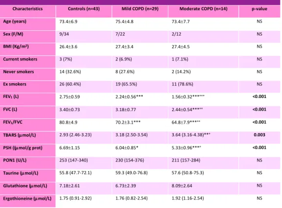
We evaluated levels of oxidative stress biomarkers in 43 COPD patients (mean age 74.8 ± 5.9 years, range 52-85 years), divided in 29 mild COPD and 14 moderate COPD, and 43 healthy controls, matched for gender, age and smoking status. In table 1 clinical, functional and biochemical parameters are shown.
Table 1: Clinical, functional and biochemical parameters of healthy subjects and COPD patients.
Characteristics Controls (n=43) Mild COPD (n=29) Moderate COPD (n=14) p-value
Age (years) 73.4±6.9 75.4±4.8 73.4±7.7 NS Sex (F/M) 9/34 7/22 2/12 NS BMI (Kg/m2) 26.4±3.6 27.4±3.4 27.4±4.5 NS Current smokers 3 (7%) 2 (6.9%) 1 (7.1%) NS Never smokers 14 (32.6%) 8 (27.6%) 2 (14.2%) NS Ex smokers 26 (60.4%) 19 (65.5%) 11 (78.6%) NS FEV1 (L) 2.75±0.59 2.24±0.56*** 1.56±0.32***°°° <0.001 FVC (L) 3.40±0.73 3.18±0.77 2.44±0.54***°° <0.001 FEV1/FVC 80.8±4.9 70.2±3.1*** 64.8±7.9***°° <0.001 TBARS (µmol/L) 2.93 (2.46-3.23) 3.18 (2.50-3.54) 3.64 (3.16-4.38)**° 0.003 PSH ((µmol/g prot) 6.69±1.15 6.04±0.85* 5.33±0.96***° <0.001 PON1 (U/L) 253 (147-340) 230 (154-376) 211 (157-284) NS Taurine (µmol/L) 55.8 (47.7-72.1) 59.3 (49.0-76.8) 57.6 (50.8-75.3) NS Glutathione (µmol/L) 7.18±2.61 6.73±2.39 8.09±2.64 NS Ergothioneine (µmol/L) 1.75 (0.91-2.92) 1.76 (0.82-2.54) 1.92 (1.16-2.54) NS *p<0.05 **p<0.01 ***p<0.001 vs controls
°p<0.05 °°p<0.01 °°°p<0.001 vs mild COPD obtained by ANOVA (Student-Newman-Keuls test for all pairwise comparisons or Krustall-Wallis test as appropriate)
Results obtained show that, as expected, COPD patients present a decrease of pulmonary functionality (lower FEV1 and FEV1/FVC) respect of healthy controls and the decrease is further greater with the progression of the disease.
Focusing on the first stage of the pathology, the only oxidative stress biomarker that is different between the two populations analyzed is PSH, showing a significative reduction of this biomarker in mild COPD patients.
Moreover, results show a significative difference in FEV1 and in FVC in males and females in mild COPD patients. About FEV1 females present mean value of 1.62 L (IQR 1.48-2.56), while males 2.36 L (IQR 2.23-2.65), with a p-value of 0.003; about FVC, females present a mean value of 2.32 L (IQR 2.15-3.13) and males of 3.41 (IQR 3.16-3.66) in males, with a p-value of 0.002. Univariate analysis shows a correlation between FEV1 and age (r = 0.49, p = 0.0067) and, furthermore, a positive correlation was found between FEV1 and PSH (r = 0.49, P = 0.007).
Analyzing the control population, a difference between males and females in FEV1 and FVC was found. About FEV1 in males there was a median of 2.64 L (IQR 2.46-2.86) and in females of 1.95 L (IQR 1.87-2.41), with a p value of 0.009; about FVC there was a median of 3.26 L (IQR 2.99-3.49) in males and of 2.56 L (IQR 2.34-2.91) in females, with a p value of 0.008.
In multiple linear regression analysis of gender, age, smoking status, BMI, TBARS, PON1, PSH, GSH, ergothioneine and taurine with FEV1 only gender (t = -3.36, p = 0.003) and age (t = - 3.11, p = 0.006) were independently associated with FEV1 in subjects affected by COPD. As in COPD patients, also in controls only age (t = -3.41, p = 0.003) and gender (t = -3.09, p < 0.006) were independently associated with FEV1.
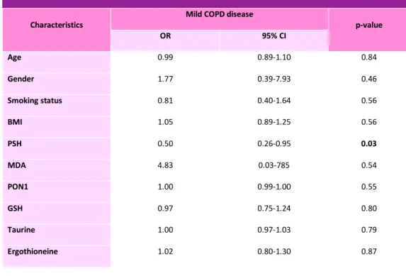
In table 2, it is displayed the multiple logistic regression analysis in healthy controls and in patients affected by mild COPD. This analysis, including as covariates smoking status, age, gender, BMI, PON1, PSH, GSH, taurine, ergothionine and TBARS, shows that only PSH was independently associated with the presence of mild COPD.
Table 2: logistic regression analysis showing ORs for mild COPD patients.
Characteristics Mild COPD disease p-value
OR 95% CI Age 0.99 0.89-1.10 0.84 Gender 1.77 0.39-7.93 0.46 Smoking status 0.81 0.40-1.64 0.56 BMI 1.05 0.89-1.25 0.56 PSH 0.50 0.26-0.95 0.03 MDA 4.83 0.03-785 0.54 PON1 1.00 0.99-1.00 0.55 GSH 0.97 0.75-1.24 0.80 Taurine 1.00 0.97-1.03 0.79 Ergothioneine 1.02 0.80-1.30 0.87
Furthermore, considering also the moderate population70, results obtained show that there is a further decrease of PSH levels in moderate COPD (p < 0.001 vs controls; p < 0.05 vs mild COPD) and levels of TBARS significantly increases with COPD presence and severity (p < 0.001 by ANOVA). In particular we found a significant difference between mild and moderate COPD (median 3.18 vs 3.64 umol/L, p < 0.05) and between controls and moderate COPD (median 2.93 vs 3.64 umol/L, p < 0.01).
About respiratory parameters, FEV1, FVC and FEV1/FVC further decrease with the progression of the disease. Values of FEV1 are in controls 2.75±0.59 L, in mild COPD 2.24±0.56 L, in moderate COPD 1.56±0.32 L, with a p- value < 0.001. Values of FVC are in controls 3.40±0.73 L, in mild COPD 3.18±0.77 L, in moderate COPD 2.44±0.54 L, with a p-value < 0.001. Moreover, FEV1/FVC ratio presents values of 80.4±4.9% in controls, 70.2±3.12% in mild COPD, 64.8±7.9% in moderate COPD, with a p-value < 0.001.
Analysis of multiple comparisons using ANOVA showed significant differences in PSH mean values between mild and moderate COPD (6.04±0.85 vs 5.33±0.96 μmol/g prot P<0.001) and between controls and moderate COPD (6.69±1.15 μmol/g prot vs 5.33±0.96 μmol/g prot, p<0.001).
5.2 Arginines results
An aspect we elaborated for the research project was to evaluate levels of arginine and methylated arginines in COPD patients (mild + moderate) and age- and gender- matched controls.70 As it is displayed in table 3, results obtained show that plasma levels of arginine were progressively lower in controls (median 79.8 umol/L), mild (median 70.4 umol/L) and moderate COPD (median 53.4 umol/L, p < 0.001). On the other hand, plasma concentrations of ADMA and SDMA were not significant different between COPD patients and healthy controls. As a consequence, ADMA/arginine ratio showed a significant increase according to COPD presence and severity.
Table 3: Arginines in healthy controls and COPD patients.
Characteristics Controls (n=43) Mild COPD (n=29) Moderate COPD (n=14) p-value Arginine (µmol/L) 79.8 (68.3-90.4) 70.4 (60.3-78.2)* 53.4 (41.4-59.8)***°° <0.001 ADMA (µmol/L) 0.488 (0.454-0.544) 0.505 (0.432-0.588) 0.513 (0.412-0.625) NS SDMA (µmol/L) 0.460 (0.395-0.590) 0.513 (0.429-0.594) 0.485 (0.456-0.577) NS ADMA/Arginine 0.0067 (0.0056-0.0077) 0.0075 (0.0053-0.0098) 0.0100 (0.0079-0.0177)***°° <0.001 ADMA/SDMA 1.07 (0.80-1.28) 0.98 (0.81-1.31) 1.12 (0.86-1.25) NS *p<0.05 **p<0.01 ***p<0.001 vs controls
°p<0.05 °°p<0.01 °°°p<0.001 vs mild COPD obtained by ANOVA (Student-Newman-Keuls test for all pairwise comparisons or Krustall-Wallis test as appropriate)
Univariate analysis in COPD subjects shows that FEV1 was correlated with age (r = -0.31, p = 0.43), PSH (r = 0.36, p = 0.016) and ADMA/arginine ratio (r = -0.43, p = 0.0001). On the other hand, in controls FEV1 was correlated only with age (r = -0.34, p = 0.036) and
adjusting for gender, age, smoking status, BMI, PSH, TBARS and ADMA/arginine ratio, displays that FEV1 was independently associated with gender (b = -0.44, p = 0.007), PSH (b = 0.33, p = 0.047) and ADMA/arginine ratio (b = -0.45, p = 0.005) in subjects affected by COPD. Multiple regression analysis in healthy controls shows that FEV1 was independently associated only with age (b = -0.38 p = 0.009) and gender (b = -0.68 p < 0.0001).
Multiple logistic regression analysis, after adjusting for gender, age, smoking status, BMI, PSH, TBARS and ADMA/arginine ratio, in all population, including mild and moderate COPD and healthy controls, only PSH and ADMA/arginine were independently associated with the presence of the pathology (Table 4).
Factor Total population p-value
OR 95% CI
PSH 0.44 0.25-0.77 0.004
ADMA/Arginine 172 2.27-13055 0.02
Table 4: Multiple logistic regression analysis of the total population (COPD + controls), after adjusting for age, gender, BMI, smoking status, ADMA/Arginine ratio, TBARS, PSH, PON and taurine.
The same statistical analysis in COPD population (mild + moderate) shows that beyond PSH and ADMA/arginine ratio also TBARS were independently associated with disease severity, as it is displayed in table 5.
Factor Moderate disease p-value
OR 95% CI
TBARS 481x1012 26-9x1027 0.030
PSH 0.0125 0.0003-0.4731 0.018
ADMA/Arginine 49x106 25-96x1012 0.016
Table 5: Multiple logistic regression analysis of the COPD population (mild vs moderate), after adjusting for age, gender, BMI, smoking status, ADMA/Arginine ratio, TBARS, PSH, PON and taurine.
5.3 Global DNA methylation results
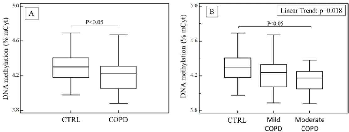
An important target we evaluated was the global DNA methylation in subjects affected by COPD and healthy controls.71 In part A of figure 5, the distribution plots of global DNA methylation show significantly lower levels of DNA methylation in COPD patients (range 3.88-4.67% mCyt; IQR 4.04-4.48% mCyt) than in controls (range 3.98-4.69% mCyt; IQR 4.18-4.40% mCyt).
Fig. 5: In part A are shown % of mCyt in DNA extracted from blood in healthy subjects and in COPD patients; in part B are shown % of mCyt in DNA extracted from blood in healthy subjects and COPD patients, after sorting in mild and moderate disease.
In part B of figure 5, levels of global DNA methylation in COPD patients, considering the severity of the disease, and in healthy controls are displayed. Global DNA methylation
mainly decreases with the severity of the pathology (4.14 ± 0.15% mCyt vs 4.23 ± 0.19% mCyt, p < 0.05).
Our data indicate that there are no significant correlations between global DNA methylation and oxidative stress indices.
In univariate logistic regression analysis, after adjusting for BMI, age, gender, smoking status, taurine, ergothioneine, PON1, PSH, GSH and TBARS, low levels of DNA methylation were independently associated with presence of COPD (OR 0.03, 95% CI 0.00 – 0.67; p = 0.028).
5.4 Tryptophan and kynurenine results
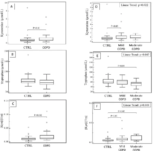
The last target of my research project was to evaluate if oxidative stress in COPD could affect tryptophan pathway and its degradation in kynurenine.72 As it is displayed in figure 6 (part A, B and C), results show that COPD patients present higher plasma levels of kynurenine (median 0.99 µmol/L; IQR 0.70 - 1.39 µmol/L vs 0.69 µmol/L; IQR 0.54 – 0.96 µmol/L, p = 0.006) and, consequently, higher kyn/trp ratio (median 0.0155 µmol/L; IQR 0.0115 – 0.0240 µmol/L vs 0.0115 µmol/L; IQR 0.0080 – 0.0140 µmol/L, p = 0.002) compared to healthy controls.
Figure 6: In part A, B and C are shown plasma levels of Kynurenine, Tryptophan and Kyn/Trp ratio in healthy subjects and COPD patients; in part D, E and F are shown plasma levels of Kynurenine, Tryptophan and Kyn/Trp ratio in healthy subjects and COPD patients after sorting for disease severity.
Moreover, dividing COPD population in mild and moderate COPD, plasma kynurenine concentrations further increase with the progression of the disease and lower levels of tryptophan become statistically significative as compare to mild COPD and healthy controls. Consequently, the increase of kyn/trp ratio was progressively higher with COPD severity (Fig. 6, part D, E and F).
In univariate correlation in all population (COPD +controls), FEV1 was correlated not only with age, gender and PSH, as we previously found, but also with kynurenine (r = -0.33; p = 0.004) and kyn/trp ratio (r = -0.39; p = 0.0007).
In multiple regression analysis, after adjusting for gender, age, smoking status, BMI, PSH and TBARS, the kyn/trp ratio was independently associated with FEV1 (b = -0.24; p = 0.049).
In univariate and multivariate analysis, there were no significant associations between kynurenine, tryptophan and kyn/trp ratio and indices of oxidative stress PSH and TBARS. We test the specificity, sensitivity and diagnostic accuracy of inflammation biomarker Kyn/Trp ratio to discriminate between COPD patients and healthy controls using the ROC curve analysis. Firstly, we tested the Kyn/Trp ratio alone and we obtained a cut-off of 0.013 with a specificity of 71% and a sensitivity of 65% (AUC: 0.703; 95% CI: 0.59– 0.80; p = 0.0006). Secondly, we tested the Kyn/Trp ratio in combination with the oxidative stress indices TBARS and PSH and results obtained considerably improved, with an AUC of 0.793 (95% CI: 0.687–0.877; p < 0.0001), sensitivity of 70% and specificity of 76%. ROC curve analysis obtained are shown in table 6.
Table 6: Prognostic accuracy of the kynurenine/tryptophan ratio alone or in combination with TBARS and PSH. Marker AUC 95% CI p-value Cutoff Sensitivity Specificity Kyn/Trp 0.703 0.589-0.801 0.0006 >0.013 65% 71% Kyn/Trp-TBARS 0.722 0.609-0.818 0.0002 >0.417 85% 58% Kyn/Trp-PSH 0.755 0.645-0.846 <0.0001 >0.700 42% 97% Kyn/Trp-PSH-TBARS 0.793 0.687-0.877 <0.0001 >0.531 70% 76%
6. DISCUSSION
Chronic obstructive pulmonary disease is a global health problem, as, according to World Health Organization, it is going to be the third leading cause of death and the fifth leading cause of disability in the world by 2020.2 Although COPD represents one of most global health problem, researchers have spent little attention on it.73 This is may be due to the fact that COPD is in general considered as self-inflicted disease for cigarette smoking and also because the pulmonary obstruction is usually considered irreversible. For this reason, the knowledge of the cellular, molecular and genetic mechanisms of COPD is not completely known.
COPD is characterized by inflammation in small airways and lung parenchyma, with activation of neutrophils and macrophages and increased number of inflammatory mediators in the airways. Moreover, in COPD there is an increase of oxidative stress, due to the not sufficient lung antioxidant systems. The increase of oxidative stress may be induced directly by cigarette smoke and indirectly by the release of reactive oxygen species from inflammatory cells. Indeed, studies of COPD subjects about cell profile in alveoli and small airways show an increase of several inflammatory cell types, as lymphocytes B and T, macrophages and neutrophils.74 The activation of these cells can generate anion superoxide (O2.-), probably through reduced nicotinamide adenine dinucleotide phosphate oxidase pathway.
6.1 Oxidative stress biomarkers
Several data suggest that both oxidative stress and inflammation greatly increase during the disease progression.75 So, we firstly focused on early stage of COPD, to find out a
sensitive and early biomarker of oxidative stress of the pathology. We assessed as oxidative stress biomarkers TBARS and GSH that were found to be altered in several case control studies in COPD and other pulmonary obstructive diseases as asthma76, and also the less studied biomarkers PSH and PON1.77-78 We studied biomarkers that were not considered in the context of COPD, as the antioxidants taurine and ergothioneine. In our study, we find a difference between the lung functional parameters FEV1 and FVC in males and in female both in patients and in controls. Moreover, simple correlation analysis showed FEV1 is correlated with age both in healthy controls and in COPD patients. Only in COPD patients we found a significant correlation between FEV1 and PSH, showing an inverse connection between FEV1 and oxidative stress. Furthermore, multiple logistic regression analysis showed, after adjusting for gender, age, smoking status, BMI, TBARS, GSH, PSH, ergothioneine and PON1, that PSH concentrations were independently associated with mild COPD. The presence of oxidative stress in the early stages of the pathology is indicated by lower levels of PSH in COPD patients, with a decrease of 10% compared to controls.
PSH represents an important indication of the antioxidant power of proteins and its decrease is the earliest indication of oxidative stress. Indeed, our results show that PSH was the only one biomarker of oxidative stress independently associated with mild COPD.
Afterwards, we studied oxidative stress biomarkers also in subjects affected by moderate COPD to analyze these biomarkers with the progression of the pathology. Results obtained showed a further decrease of PSH levels, as we had previous seen in mild COPD patients, and a significant increase of TBARS in moderate COPD patients.
So, results obtained analyzing biomarkers of oxidative stress in early stage of the disease showed a significantly decrease of PSH levels in mild COPD compared to controls and with the progression of the disease this decrease is further confirmed. Moreover, with the progression of the disease patients presented higher levels of TBARS compared to healthy controls. These results indicate the presence of significant oxidative stress since in the early stage of the pathology and it raises with the progression of the disease.
6.2 Arginines
Via NOS pathway, arginine metabolism contributes at the maintenance of airways function and tone through the production of nitric oxide.79 Dysregulation of the competing enzymes contributes to airway obstruction in asthma and in cystic fibrosis patients.80-81-82
We found that COPD patients had lower levels of arginine compared to healthy controls, and this decrease was greater in patients affected by moderate COPD. Moreover, categorizing patients on the basis of disease progression, only moderate COPD patients presented difference in ADMA/arginine ratio, supporting the hypothesis that arginine and ADMA could be involved in disease worsening. From several studies, it is plausible to suppose that COPD disease exacerbation states are associated with further increases of oxidative stress and ADMA/Arginine ratio. The imbalance between ADMA and arginine may be related to the presence of oxidative stress. Notably, the reduction in arginine levels in COPD patients is probably due to the increase of arginase activity stimulated by oxidative stress.83 Moreover, the depletion of arginine is maybe due also to the increase of neutrophils, a typical state in COPD patients, since these cells are characterized by the expression of high levels of arginase I in azurophilic granules. In
addition to arginase I, these granules maybe contain other constituents such as elastase.84 It is also known that the number of neutrophils increases with the progression and the worsening of the disease.85 This may greatly explain the further reduction of arginine concentrations observed in the moderate forms of COPD. In this study we did not assess the expression and the activity of these enzymes in lung tissue, but it is plausible that changes in arginine metabolites plasma concentration are caused by structural and functional lung alteration in COPD.
Moreover, the enzymes involved in the formation and in the degradation of ADMA, as PRMTs and DDAH, are redox sensitive.86 Oxidative stress acts through DDAH inhibition, PRMTs stimulation and increase of arginase activity and they are maybe responsible for an imbalance of ADMA/arginine ratio. This hypothesis is even support by the significant negative correlation observed ADMA/arginine ratio and PSH in the subjects analyzed in our study. Results we obtained show the reduction in arginine concentration and this is involved in the alteration of the ADMA/arginine ratio, even if a no significant increase in ADMA levels of about 3.5% has been found in all COPD subjects, with a rise of about 5.2% in moderate patients. It could be interesting to evaluate ADMA concentrations in more severe patients.
6.3 Global DNA methylation
Chronic inflammation of the small and distal airways plays a central role in the pathophysiology of COPD. Inflammation is characterized by the activation of neutrophils, macrophages, inflammatory mediators and anion superoxide, probably through an impairment of the nicotinamide adenine dinucleotide phosphate oxidase pathway.