Short threaded implants with an oxidized surface to restore posterior teeth: 1- to 3-year results of a prospective study.
11
0
0
Testo completo
(2) De Santis et al. Undoubtedly, implant surfaces, like implant designs, have undergone a series of evolutions over the years. As a consequence, new kinds of implant surfaces have been developed to improve both the quality of osseointegration and the amount of bone contact at the interface level; one of these is the oxidized surface. This surface is characterized by a highly crystalline and phosphate-enriched titanium oxide that results in a microstructured surface with open pores in the low micrometer range.6 This kind of surface has repeatedly been found to yield an enhanced bone response compared to machined implant surfaces, and the enhanced bone response to oxidized titanium results in faster and stronger osseointegration and thus better maintenance of implant stability compared to machined titanium implants.7–9 New implant designs have also been developed. Some implants retain the conventional tapered shape but incorporate a groove in the implant thread along the entire length of the implant. This feature enables an affinity for bone formation to be obtained within and along the groove, resulting in higher removal torque values.10,11 The greater osseointegration resulting from advances in surface and design has enabled implant length to be shortened, as proposed by Albrektsson et al12 and Brånemark et al.4 Short implants can be used, which were initially characterized as implants < 10 mm in the mandible and < 13 mm in the maxilla. Recently, Renouard and Nisand13 redefined the “short implant” as one with a “designed intrabony length” (ie, length of implant meant to support long-term osseointegration) of ≤ 8 mm. However, there are very few reports on the use of threaded dental implants of this length. The restoration of lost teeth in posterior areas is complicated by loss of permanent teeth at a young age, low bone quality (type III or IV), enhancement of bone resorption caused by mucous stimuli, and the presence of important anatomical structures, such as the inferior alveolar nerves or sinus cavities. Successful management of posterior areas has incorporated surgical techniques for augmentation of bone volume to permit the insertion of long implants to achieve predictable and reproducible results.14–16 However, as a result of developments in implant dentistry in recent years, short implants can now permit high success rates (95.5% to 100%)17–22 comparable to success rates seen with longer implants after bone grafting (94.5% to 100%).14,15 Different surfaces have been studied by different authors: Friberg et al17 investigated the survival rates of machined surfaces, Malò et al18 examined the survival rates of oxidized surfaces, Anitua et al19 studied the survival rates of acid-etched surfaces, Misch et al20. considered the survival rates of sandblasted surfaces, Deporter et al21 researched the survival rates of poroussintered surfaces, and Griffin and Cheung22 analyzed the success rates of hydroxyapatite-coated surfaces. In this preliminary study, the authors examined the efficacy of using short threaded implants with oxidized surfaces in treating partial edentulism in posterior areas. The performance of short implants with a length ≤ 8 mm, as a “designed intrabony length” ≤ 8 mm, is reported here after a follow-up of 1 to 3 years.. MATERIALS AND METHODS Patients. The patients chosen for treatment with short threaded implants were referred between October 2006 and July 2007 for single or partial implant-supported rehabilitation in posterior areas at three different centers. All patients had advanced alveolar bone resorption in the posterior areas of the maxilla or mandible that was planned to be treated with implant therapy. Patients had been advised that they were not candidates for long implants without extensive preparatory implant site development because of insufficient alveolar ridge height. However, all sites had a sufficient alveolar ridge width to receive implants at least 3.75 mm in diameter. The decision to use short implants was made after discussion with the patients and after obtaining informed written consent. The following criteria were used to select the patients in whom this kind of implant could achieve successful results: • Inclusion criteria: highly controlled oral hygiene, absence of acute infection in the oral cavity, residual bone volume at least 3.5 mm in width and 5 mm in height (for the maxilla) or 3.5 mm in width and 8 mm in height (for the mandible), and willingness to participate in an oral hygiene maintenance program • Exclusion criteria: insufficient bone volume, bruxism, smoking more than 20 cigarettes per day, abuse of alcohol, radiotherapy in the maxillofacial region, chemotherapy, liver diseases, blood diseases, kidney diseases, inflammatory and autoimmune diseases, immunosuppressed status, corticosteroid therapy, pregnancy, or insufficient oral hygiene Treatment was performed at three centers: the Department of Morphological and Biomedical Sciences, Section of Dentistry and Maxillofacial Surgery, University of Verona, and two private offices. All subjects provided written informed consent.. 394 Volume 26, Number 2, 2011 © 2011 BY QUINTESSENCE PUBLISHING CO, INC. PRINTING OF THIS DOCUMENT IS RESTRICTED TO PERSONAL USE ONLY.. NO PART OF MAY BE REPRODUCED OR TRANSMITTED IN ANY FORM WITHOUT WRITTEN PERMISSION FROM THE PUBLISHER..
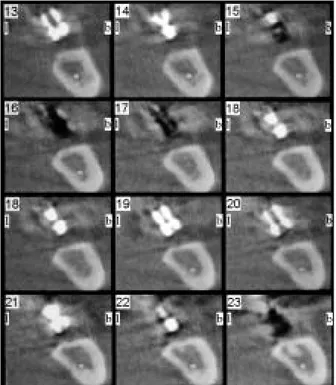
(3) De Santis et al. Implant System All implants used were short threaded implants (Brånemark System Mk III Shorty , Nobel Biocare; NobelSpeedy Shorty, Nobel Biocare) with a length of 7 or 8.5 mm and with an oxidized surface (TiUnite, Nobel Biocare). They were provided by the manufacturer and purchased by the clinicians. These implants are short versions of long threaded implants with oxidized surfaces, which are available in diameters of 3.75 or 4.0 mm for regular-platform implants (platform diameter: 4.1 mm) and 5.0 mm for wideplatform implants (platform diameter: 5.1 mm). They were machined from titanium alloy (Ti-6Al-4V) as a parallel design, with a designed intrabony length of 6.3 or 7.8 mm, and a standard 0.7-mm-high externalhex connection. All implants featured the oxidized surface described by Hall and Lausmaa.6. Treatment Planning. An initial examination of patients was performed to assess their oral condition and to evaluate the possibility of placing short implants. Particular attention was paid to the psychologic aspect of the patients in an attempt to understand what prompted them to approach oral rehabilitation without complex bone grafting, ie, vertical and vestibular onlay grafting or direct sinus elevation with consideration of the risk/ benefit ratio of short implants. After informed consent was obtained for the prosthetic plan, patients were examined to evaluate the inclusion and exclusion criteria, as well as other parameters required for the treatment planning. All patients were examined by means of panoramic and periapical radiographs and, if necessary, computed tomography (CT), which enabled assessment of the level of bone atrophy and the locations of anatomic reference points (Figs 1 to 4).. Surgical Procedures. The patients were submitted to a preventive protocol. This included amoxicillin and clavulanic acid (2 g) 1 hour before surgery, ketorolac tromethamine (20 mg) and chlordimethyldiazepam (0.8 mg) 30 minutes before surgery, and betamethasone sodium phosphate (1.5 mg/2 mL intramuscularly) an hour before the operation and twice daily thereafter for the following 3 days. Implants were placed in a two-stage approach after a full-thickness mucoperiosteal flap was raised, as described by the manufacturer. A flap technique is necessary to observe the underlying alveolar bone and adjacent anatomical structures and to place implants in the correct positions. In cases of alveolar ridges that were too small to receive 3.75- × 7.0-mm implants, special surgical procedures were performed to increase the available bone volume: split crest and. indirect sinus elevation techniques were the first choices of techniques to augment the ridge horizontally and vertically, respectively. These two techniques were accomplished as described by Simion et al23 and Summers,24 respectively. Implant sites were prepared using rotating burs and/or hand osteotomes. The rotary burs were made of surgical stainless steel and coated with an amorphous diamond layer and were used with external irrigation. The osteotomes were used in posterior maxillary sites with bone type III or IV or with bone height ≤ 8 mm. It is important to use an in-and-out motion and drill in bone for 1 to 2 seconds; to move the drill up without stopping the handpiece motor, which also allows the irrigation to flush away debris; and to proceed with this technique to the correct depth in accordance with bone quality and implant diameter. If rotating burs were used, these included: • A twist drill with a 2-mm-diameter tip to drill to the appropriate depth • A 2.4- to 2.8-mm-diameter twist step drill to continue site preparation • A 3.0- to 3.2-mm-diameter twist drill for mediumdensity bone (or 3.2- to 3.4-mm-diameter twist drill for dense bone) prior to placement of 3.75-mm implants and 4.0-mm implants, respectively, to drill to the appropriate depth (ie, the length of the implant being placed) • A 3.8-mm-diameter twist drill for medium-density bone (or a 4.2-mm-diameter twist drill for dense bone), in case of 5-mm-diameter implants • A screw tap, matching the diameter of the final twist drill, to pre-tap the implant site in medium/ dense bone During site preparation, a 2.0/2.4- to 2.8-mmdiameter directional indicator was used, as recommended, to check for correct orientation. A depth probe was also used to confirm the appropriate depth of the osteotomy. Great care was taken to ensure that the implant burs did not experience chatter (particularly in sites with a dense upper cortex) to guarantee that the osteotomy maintained the perfectly concentric shape needed to achieve the required initial tight press-fit of the implant. After the implant packaging was opened and the sterile inner vial was removed, the appropriate implant driver was connected to the handpiece, and the implant was picked up by applying light pressure on the implant driver. The implant was placed in the osteotomy site using low speed (25 rpm) and 30 to 45 Ncm of torque, and the implant was turned until it was fully inserted. At this point, the driver was removed with an easy upward motion. A cover screw was positioned The International Journal of Oral & Maxillofacial Implants 395. © 2011 BY QUINTESSENCE PUBLISHING CO, INC. PRINTING OF THIS DOCUMENT IS RESTRICTED TO PERSONAL USE ONLY.. NO PART OF MAY BE REPRODUCED OR TRANSMITTED IN ANY FORM WITHOUT WRITTEN PERMISSION FROM THE PUBLISHER..
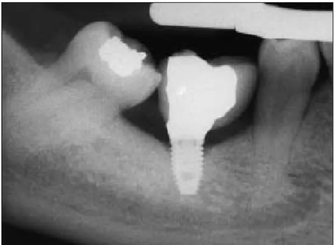
(4) De Santis et al. Fig 1 Preoperative panoramic radiograph obtained at 1 year before surgery.. Fig 2 Preoperative CT obtained 1 month before surgery.. Fig 3 Preoperative CT showing coronal sections of the mandible.. Fig 4 Preoperative CT showing parasagittal sections of the edentulous area.. Fig 5 Intraoral radiograph obtained after surgery.. on the implant using a Unigrip screwdriver (Nobel Biocare), and a tissue flap suture was performed to ensure submerged first-intention healing (Fig 5).. Prosthetic Procedures. The initial healing period ranged from 3 to 6 months, depending on the clinical circumstances and the treating clinician’s preference. After the healing period, a provisional healing abutment was then positioned on the implant to achieve adequate peri-. implant soft tissue healing. After soft and hard tissue healing was completed, implant-supported prostheses were created and fixed with screws or cement, depending on the treating clinician’s preference. Implants were restored with single crowns, crowns with a cantilever, or fixed prostheses, according to the patients’ needs and desires. However, cement-retained prostheses were usually used for single crowns, while screw retention was usually used in cases of fixed prostheses.. 396 Volume 26, Number 2, 2011 © 2011 BY QUINTESSENCE PUBLISHING CO, INC. PRINTING OF THIS DOCUMENT IS RESTRICTED TO PERSONAL USE ONLY.. NO PART OF MAY BE REPRODUCED OR TRANSMITTED IN ANY FORM WITHOUT WRITTEN PERMISSION FROM THE PUBLISHER..
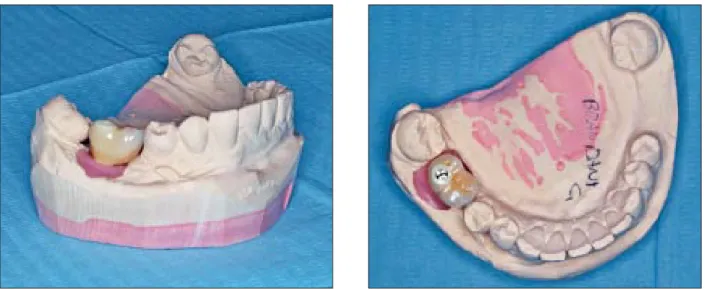
(5) De Santis et al. Figs 6a and 6b Lateral and occlusal views of the master model and zirconia-ceramic crown.. Figs 7a and 7b Clinical appearance before and after definitive rehabilitation with zirconia-ceramic crown.. All definitive restorations were placed into occlusion, where the occlusal surface was thoroughly modeled (Fig 6), so that it was in contact with reduced areas during lateral and protrusive excursions to reduce the dislocating vectorial components. More contacts were maintained in maximum intercuspation (Fig 7).. Postoperative Follow-up. Patients were included in a maintenance program to achieve optimal hard and soft tissue healing, which comprised professional oral hygiene every 6 months and twice-daily rinsing with chlorhexidine digluconate 0.2% during the first 2 weeks after implant placement. Clinical evaluations were performed weekly during the first month, after provisional restoration, and then monthly during the first 6 months after definitive restoration. Further examinations were performed every 6 to 9 months and consisted of analysis of soft tissue. health (Plaque Index, Gingival Index), evaluation of probing pocket depths, and radiographic examinations (panoramic or intraoral radiographs) (Figs 8 and 9).. Determinants of Success. An implant was considered successful if it met all the success criteria proposed by Albrektsson and Zarb25,26: (1) absence of any complaints such as pain, dysesthesia, or paresthesia in the implanted area, (2) absence of recurring peri-implant infection and/or suppuration, (3) absence of perceptible mobility of the implant, (4) absence of radiolucency at the implant-bone junction, and (5) absence of persistent peri-implant bone resorption greater than 1.5 mm during the first year of loading and 0.2 mm per year during the following years. The implants were considered successful in the absence of all of the aforementioned complaints at the most recent recall appointments. The International Journal of Oral & Maxillofacial Implants 397. © 2011 BY QUINTESSENCE PUBLISHING CO, INC. PRINTING OF THIS DOCUMENT IS RESTRICTED TO PERSONAL USE ONLY.. NO PART OF MAY BE REPRODUCED OR TRANSMITTED IN ANY FORM WITHOUT WRITTEN PERMISSION FROM THE PUBLISHER..
(6) De Santis et al. Fig 8 Intraoral radiograph after functional loading (baseline).. Fig 9 Intraoral radiograph 1 year after functional loading (follow-up).. All radiographs of implants from each subject’s last recall visit were reviewed independently by an oral radiologist at a magnification of ×6 for the measurements of marginal bone levels. Bone levels were assessed mesially and distally by identifying the lowest observed point of crestal bone in intimate contact with the implant; to quantify marginal bone loss, this was compared to the bone level observed at baseline. Clinical complications such as pain, dysesthesia, or paresthesia were assessed by interviewing the patients. Peri-implant infection with or without suppuration and implant mobility were assessed by clinical observation and pressure. Implant immobility was assessed by evaluating the mobility of the prosthesis, in cases of single-crown restoration; in cases of fixed prostheses, the immobility of each implant was assessed following removal of the prosthesis.. RESULTS. Statistical Analysis. All data were analyzed according to the Fisher exact test. A P value < .05 was considered statistically significant. Thus, it was possible to compare the success rates and the mean bone loss around short implants in grafted sites with those in nongrafted sites and determine the presence or absence of statistically significant differences between the two groups. The effects of implant length (7 mm versus 8.5 mm) and implant diameter (regular-platform versus wide-platform) on implant success were also evaluated. Finally, the implant success rate was investigated according to the kind of prosthesis placed: ie, single crown versus fixed cantilevered prosthesis versus fixed prosthesis.. Summaries of implant locations, types of prostheses placed, and time in function at the time of data collection for this report are presented in Tables 1 to 3. In total, records were analyzed for 107 implants in 46 subjects (aged from 36 to 78 years; 25 women and 21 men) who needed to restore single or multiple teeth. The university center contributed 56 implants in 23 subjects and had the longest follow-up times (12 to 38 months in function; mean follow-up 24.3 ± 8.7 months). Private office 1 (Verona, Italy) contributed 27 implants in 13 subjects (12 to 48 months in function; mean follow-up 28.4 ± 11.9 months), who were treated by an oral maxillofacial surgeon; and private office 2 (Mantua, Italy) contributed 24 implants in 10 subjects (12 to 32 months in function; mean follow-up 21.5 ± 6.8 months), who were treated by a general dentist. With regard to implant length, 7-mm-long implants and 8.5-mm-long implants were used in 69.2% (n = 74) and in 30.8% (n = 33) of cases, respectively. With respect to implant diameter, 3.75-mm, 4-mm, and 5-mm implants were used in 26.2% (n = 28), 51.4% (n = 55), and 22.4% (n = 24) of sites, respectively. Seventy-nine implants were used to replace molar teeth, and 28 replaced premolar teeth. Eighty-six implants (80.4%) were placed in the posterior mandible and 21 implants (19.6%) were placed in the posterior maxilla (Table 1). In 40.2% of cases, the implant site was grafted with heterologous bone at the time of implant placement using the split-crest or indirect sinus lift techniques. In the other 59.8% of sites, bone grafting was not required for implant placement.. 398 Volume 26, Number 2, 2011 © 2011 BY QUINTESSENCE PUBLISHING CO, INC. PRINTING OF THIS DOCUMENT IS RESTRICTED TO PERSONAL USE ONLY.. NO PART OF MAY BE REPRODUCED OR TRANSMITTED IN ANY FORM WITHOUT WRITTEN PERMISSION FROM THE PUBLISHER..
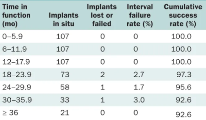
(7) De Santis et al. Table 1 Distribution of Implants According to Locations. Table 2 Distribution of Implants According to the Type of Prosthesis Placed. Site. Prosthesis type. First premolar. Mandible. Maxilla. Total. 11. 3. 14 (13.1%). No. of patients. No. of implants. 12. 29 (27.1%). Single crown. Second premolar. 24. 5. 29 (27.1%). Cantilevered. 9. 18 (16.8%). First molar. 35. 8. 43 (40.2%). Fixed prosthesis. 25. 60 (56.1%). Total. 46. 107 (100%). Second molar. 16. 5. 21 (19.6%). Total. 86. 21. 107 (100.0%). Table 3 Distribution of Implants According to Time in Function. Table 4 Cumulative Success Rate of the Short Implants Placed. Follow-up (mo). Time in function (mo). No. of patients. No. of implants. 12–17.9. 16. 34 (31.8%). 18–23.9. 7. 15 (14.0%). 24–29.9. 10. 25 (23.4%). 30–35.9. 5. 12 (11.2%). 36+. 8. 21 (19.6%). Total. 46. 107 (100.0%). Single crowns were used to restore 29 implants (27.1%), whereas another 60 implants (56.1%) were restored with a fixed prosthesis. The remaining 18 implants (16.8%) were restored with fixed cantilevered prostheses (Table 2). No surgical or prosthetic complications associated with implant placement were observed. All 107 implants became osseointegrated and functioned as planned. At the time of this report, these implants had been functioning for a mean follow-up of 24.6 ± 9.4 months (range, 12 to 48 months). All 107 implants were followed for at least 1 year, 58 were followed for at least 2 years, and 21 implants had a follow-up period of more than 3 years (Table 3). Only two implants were lost in two patients treated by the university center. The first patient had received a 4.1- × 7-mm implant in a maxillary first molar site; this patient had poor oral hygiene and a smoking habit (> 10 cigarettes/day), which likely resulted in the peri-implantitis and consequent implant loss at the 18-month follow-up. The second patient had received two 4.1- × 7-mm implants in the mandibular first and second molar sites after the split crest technique, which were restored with two single crowns; the mesial implant was lost because of overloading and related excessive crestal bone loss in the 32-month follow-up. After 24.6 ± 9.4 months of mean follow-up, the overall survival rate was therefore 98.1%.. Implants in situ. Implants lost or failed. Interval failure rate (%). Cumulative success rate (%). 0–5.9. 107. 0. 0. 100.0. 6–11.9. 107. 0. 0. 100.0. 12–17.9. 107. 0. 0. 100.0. 18–23.9. 73. 2. 2.7. 97.3. 24–29.9. 58. 1. 1.7. 95.6. 30–35.9. 33. 1. 3.0. 92.6. ≥ 36. 21. 0. 0. 92.6. Two additional implants were considered to have failed since they did not meet all the success criteria adopted in this study. One of these implants failed after 18 months because of excessive bone resorption caused by peri-implantitis, and the other failed at 24 months because of excessive bone resorption caused by occlusal overloading. While the first implant was successfully treated by means of a surgical approach, which consisted of peri-implant curettage and bone tissue regeneration with heterologous bone and a collagen membrane, the second was successfully treated with a strict maintenance program, including professional oral hygiene every 6 months. In all, the four failed implants, out of 107 placed, resulted in an overall success rate of 96.3% and a cumulative success rate of 92.6% (Table 4). The characteristics of the failed implants are provided in Table 5. With regard to implant length, 7-mm-long implants showed a success rate comparable to that of 8.5-mmlong implants (95.9% and 97.0%, respectively); with regard to implant diameter, implant success rates were 96.4% and 95.8% for regular-platform (4.1-mm) and wide-platform (5-mm) implants, respectively. Differences between 7-mm and 8.5-mm implants were not statistically significant (P = .0598); no significant differences were observed between implants with different platforms (P = .1344).. The International Journal of Oral & Maxillofacial Implants 399 © 2011 BY QUINTESSENCE PUBLISHING CO, INC. PRINTING OF THIS DOCUMENT IS RESTRICTED TO PERSONAL USE ONLY.. NO PART OF MAY BE REPRODUCED OR TRANSMITTED IN ANY FORM WITHOUT WRITTEN PERMISSION FROM THE PUBLISHER..
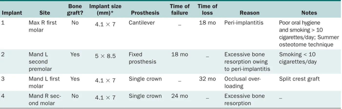
(8) De Santis et al. Table 5 Characteristics of the Failed Implants Implant. Site. Bone graft?. Implant size (mm)*. Prosthesis. Time of failure. Time of loss. 1. Max R first molar. No. 4.1 × 7. Cantilever. _. 18 mo. 2. Mand L second premolar. Yes. 5 × 8.5. Fixed prosthesis. 18 mo. _. 3. Mand L first molar. Yes. 4.1 × 7. Single crown. _. 32 mo. 4. Mand R second molar. No. 4.1 × 7. Single crown. 24 mo. _. Table 6 Distribution of Implants According to Crestal Bone Loss During Follow-up Period Mean bone loss (mm) < 0.1. Implants 5 (4.7%). 0.1–0.5. 71 (66.4%). 0.6–1.0. 21 (19.6%). 1.1–1.5. 5 (4.7%). 1.6–2.0. 2 (1.9%). 2.1–2.5. 1 (0.9%). > 2.5. 2 (1.9%). Total. 107 (100.0%). In contrast, the implant success rates were quite different with regard to the type of prosthesis placed: 93.1% for single crowns, 94.4% for cantilevered prostheses, and 98.3% for fixed prostheses. The success rate of implants restored with fixed prostheses was significantly higher than those of implants restored with single crowns or cantilevered restorations (P = .0322 and P = .0437, respectively). The radiographs obtained at baseline and every year of follow-up (Figs 8 and 9) revealed a mean marginal bone loss during functional loading of 0.6 ± 0.2 mm (range, 0.0 to 1.9 mm). Five implants (4.7%) showed no bone resorption. Most implants (n = 71; 66.4%) showed bone resorption ranging from 0.1 to 0.5 mm, and only three implants (2.8%) showed bone loss between 1.6 and 2.5 mm at the final follow-up examination. None of the osseointegrated implants showed a marginal bone loss greater than 2.5 mm, except for the two lost implants (Table 6).. Reason. Notes. Peri-implantitis. Poor oral hygiene and smoking > 10 cigarettes/day; Summer osteotome technique. Excessive bone resorption owing to peri-implantitis. Smoking < 10 cigarettes/day. Occlusal over loading. Split crest graft. Excessive bone resorption. _. With regard to crestal bone loss in grafted and ungrafted sites, no statistically significant differences were observed (P = .0678). In grafted sites, bone resorption was 0.8 ± 0.3 mm (range: 0.2 to 1.9 mm), while in ungrafted sites, it was 0.6 ± 0.3 mm (range: 0.0 to 1.6 mm). The implant success rate also did not vary significantly with respect to grafting at the implant site; only two of the four failed implants were placed in grafted sites (P = .1167).. DISCUSSION A recent redefinition of a short implant is one that has a designed intrabony length (ie, length of implant required to achieve and maintain osseointegration) of ≤ 8 mm.13 This definition was used in the present report. The threaded implants used here had a designed intrabony length of 6 or 7 mm, because they have a standard 1-mm-high external-hex connection, and therefore were considered short implants by the authors. They are characterized by an oxidized surface, which has repeatedly been found to induce an enhanced bone response compared to machined surfaces. The enhanced bone response appears to result in faster bone formation and a greater amount of bone in contact with the implant surface during healing, which results in greater osseointegration and in better maintenance of implant stability, despite the reduced implant length.7–9 Renouard and Nisand,27 in a retrospective study, reported that short threaded implants can be considered for prosthetic rehabilitation of severely resorbed arches. The oxidized surface yielded a higher success rate (97.6%, with one failed implant) than the machined surface (92.6%, with four failed implants), but this difference was not statistically significant.. 400 Volume 26, Number 2, 2011 © 2011 BY QUINTESSENCE PUBLISHING CO, INC. PRINTING OF THIS DOCUMENT IS RESTRICTED TO PERSONAL USE ONLY.. NO PART OF MAY BE REPRODUCED OR TRANSMITTED IN ANY FORM WITHOUT WRITTEN PERMISSION FROM THE PUBLISHER..
(9) De Santis et al. Frieberg et al,17 in a retrospective study with a long-term follow-up, reported that short implants with an oxidized surface yielded survival rates of 95.5% and 92.3% at 5 and 10 years, respectively. The authors showed that short implants without bone grafting can be a predictable means of treating the severely resorbed mandible. However, the study provided data only on patients whose implants were restored with fixed implant-supported prostheses. Malò et al18 demonstrated in a retrospective study that short implants with an oxidized surface represent a viable concept for both arches. They achieved survival rates of 96.2% and 97.1% at 5 years for implants that were 7 mm and 8.5 mm long, respectively. In their study, implants were placed using a one-stage delayed-function approach; definitive abutments were delivered at the time of surgery, and definitive prostheses were delivered 4 to 6 months later. Data were reported for 237 patients treated with fixed prostheses supported by 408 implants. The number of cases seems sufficient to confirm the effectiveness of short implants with an oxidized surface. Despite the limited number of cases treated and the short-term follow-up, the present report included data from different clinical situations: mandible and maxilla; implant restorations with single crowns, crowns supporting a cantilever, and fixed prostheses; patients enrolled at three different treatment centers; and the use of 7- and 8-mm-long implants. The acquired data support the possibility of using this kind of implant in the treatment of partially edentulous patients. Many authors have confirmed that not only 8.5-mm-long but also 7-mm-long implants constitute a reliable solution in severely resorbed arches17,18,28,29; others have even proposed the use of implants with lengths < 7 mm.30–33 Obviously, different authors have investigated different implant designs and surfaces, but many of them have validated the use of short implants as a predictable procedure. Ten Bruggenkate et al31 reported on the performance of 6-mm-long titanium plasma spray-coated implants using the nonsubmerged technique. Titanium plasma spray coating of bone-interfacing implants is known to increase the strength of osseointegration, at least as compared to machined-surface implants.34 After an initial healing interval of 4 months, implants were used to support a variety of prostheses. Seven implants failed during the study and 28 of 253 implants were lost during the follow-up period. The overall failure rate was 6% after 1 to 7 years in function, but the failure rate in the maxilla was 13%.31 Deporter et al30 reported on the efficacy of 5-mmlong sintered porous-surfaced implants placed in a submerged technique. Implants were restored with single crowns or as part of a fixed prosthesis with other. implants. Only two implants failed after functional periods of up to 8 years. Both of the failed implants were maxillary ones, resulting in a maxillary failure rate of 14.3%. It is worth noting that these implants have a “designed intrabony length” (ie, the sintered porous surface responsible for implant integration) of only 4 mm, which is much shorter than that of other implants. The same authors had already established in previous prospective or retrospective studies that short sintered porous-surfaced implants with a length of 7 mm would permit success rates of 98.1% to 100% with predictable results.21,35,36 In addition, it is important to remember that the purpose of short implants is to rehabilitate the severely resorbed posterior area, both in the maxilla and in the mandible, where proximity to the inferior alveolar nerve and to the sinus cavity may preclude the possibility of using osseointegrated implants without augmentation of bone volume, as with autogenous block grafting, guided bone regeneration, or nerve repositioning.37–39 Shorter implants offer several surgical advantages compared to longer implants: less need for vertical bone grafting, less time for treatment, lower cost of treatment, less discomfort, easier surgery, and fewer surgical risks (eg, sinus perforation, mandibular paresthesia, adjacent tooth injury). All of these factors make short implants a highly attractive restorative option.38 When short implants are used, ridge height is no longer a technical limitation for implant-supported prostheses. The most important limitation is ridge width, because a wide alveolar ridge is essential to retain implants with diameters of 4 or 5 mm; > 1 mm of cortical bone is necessary buccally and lingually, ie, a ridge width of more than 8 mm is required.30,39 In these cases, surgical techniques of ridge augmentation can be useful to improve success rates for short implants.30,33,36 The main consequence of reduced ridge height is an unfavorable crown/implant (C/I) ratio. The C/I ratio has been considered one of the prosthetic factors that may increase the risk of biomechanical complications, because unfavorable occlusal forces, such as overloading or nonaxial loading, have been reported to be a possible cause of these complications.40–42 A type of nonaxial loading is displayed by implant-supported reconstructions with high C/I ratios: the crown acts as a lever arm, creating a bending moment and transferring stress to the peri-implant crestal bone.43,44 This bone stress may, eventually, result in either crestal bone loss and/or technical complications.45,46 However, Tawil et al47 demonstrated that an increased C/I ratio did not prove to be a major complicating factor, although it was found to be increased by two to three times in nearly 87% of cases: The International Journal of Oral & Maxillofacial Implants 401. © 2011 BY QUINTESSENCE PUBLISHING CO, INC. PRINTING OF THIS DOCUMENT IS RESTRICTED TO PERSONAL USE ONLY.. NO PART OF MAY BE REPRODUCED OR TRANSMITTED IN ANY FORM WITHOUT WRITTEN PERMISSION FROM THE PUBLISHER..
(10) De Santis et al. peri-implant bone resorption was similar in all C/I ratio groups. Rokni et al48 reported a mean C/I ratio of 1.5, with 78.9% of implants having a C/I ratio between 1.1 and 2.0 and 10.0% of implants having a C/I ratio increased by two to three times; these authors demonstrated that neither C/I ratio nor estimated implant surface area affected steady-state crestal bone levels. Recently, Blanes44 systematically reviewed the occurrence of biologic and technical complications with respect to the C/I ratio of implant-supported restorations. A qualitative data analysis revealed that the survival rate of implant-supported reconstructions with a C/I ratio of more than 2 was 94.1%. In addition, peri-implant crestal bone loss seemed not to be influenced by the C/I ratio of the implant rehabilitation, except in one study. Technical complications related to implant-prosthetic components according to different C/I ratios were not found in any of the studies. The data presented here suggest that short implants with an oxidized surface may provide an alternative solution to these surgical procedures, reducing most of the risk related to aggressive and invasive surgery, and achieving an overall success rate of 96.3%. However, the number of sites treated was limited to 86 posterior mandibular implants in 37 patients and 21 posterior maxillary implants in 9 patients. This study has described the authors’ 1- to 3-year clinical experience with 7- and 8-mm-long implants. The overall survival rate of 98.1% was obtained in partially edentulous patients with severely resorbed alveolar ridges. Obviously, a larger prospective study with long-term follow-up is needed to provide more convincing evidence of the predictability and reproducibility of these implants.. CONCLUSIONS The short implants used in this retrospective study would appear to be successful because of their threaded design and oxidized surface and the accurate surgical procedures employed. Despite the limited number of cases, these preliminary results demonstrate the predictability and safety of such implants when used with careful treatment planning and an appropriate clinical protocol. The possibility of using short implants in clinical practice should be considered as an alternative to avoid surgical procedures such as bone grafts or the like and their related disadvantages. The information obtained from this study might help clinicians improve their decisionmaking with the aim of enhancing implant success, or it might provide them with suggestions regarding operative possibilities.. ACKNOWLEDGMENTS The authors wish to thank Professor Anthony Steele, Senior Lecturer in Medical English, University of Verona, for his invaluable assistance with the linguistic editing of this paper. The authors have no financial interest in any company or in any of the products mentioned in this article.. REFERENCES 1. Felton DA. Edentulism and comorbid factors. J Prosthodont 2009;18:88–96. 2. Prendergast PJ, Huiskes R. The biomechanics of Wolff’s law: Recent advances. Ir J Med Sci 1995;164:152–154. 3. Nocini PF, Chiarini L, De Santis D. La strategia operativa per la riabilitazione dell’apparato stomatognatico. In: Nocini PF, Chiarini L, De Santis D (eds). Trattato di chirurgia preprotesica e ingegneria tissutale. Bologna: Ed Martina, 2005:1–5. 4. Brånemark PI, Hansson BO, Adell R, et al. Osseointegrated implants in the treatment of the edentulous jaw. Experience from a 10-years period. Scan J Plast Reconstr Surg 1977;16 (suppl):S1–132. 5. Van Steenberghe D, Lekholm U, Bolender C, et al. Applicability of osseointegrated oral implants in the rehabilitation of partial edentulism: A prospective multicenter study on 558 fixtures. Int J Oral Maxillofac Implants 1990;5:272–281. 6. Hall J, Lausmaa J. Properties of a new porous oxide surface on titanium implants. Appl Osseointegration Res 2000;1:5–8. 7. Albrektsson T, Johansson C, Lundgren AK, Sul Y, Gottlow J. Experimental studies on oxidized implants. A histomorphometrical and biomechanical analysis. Appl Osseointegration Res 2000;1:21–24. 8. Ivanoff C-J, Widmark G, Johansson C, Wennerberg A. Histologic evaluation of bone response to oxidized and turned titanium micro-implants in human jawbone. Int J Oral Maxillofac Implants 2003;3:341–348. 9. Schüpbach P, Glauser R, Rocci A, et al. The human boneoxidized titanium implant interface: A microscopic, scanning electron microscopic, back-scatter electron microscopic, and energy-dispersive X-ray study of clinically retrieved implants. Clin Implant Dent Relat Res 2005;7(suppl):S36–S43. 10. Hall J, Miranda-Burgos P, Sennerby L. Stimulation of directed bone growth at oxidized titanium implants by macroscopic grooves: An in vivo study. Clin Implant Dent Relat Res 2005; 7(suppl):S76–S82. 11. Hanao G. The Tapered Groovy implant optimizes implant success in suboptimal clinical conditions. Dent Implantol Update 2006;17:1–4. 12. Albrektsson T, Brånemark PI, Hansson HA, Lindström J. Osteointegrated titanium implants. Requirements for ensuring a long-lasting, direct bone anchorage in man. Acta Orthop Scand 1981;52:155–170. 13. Renouard F, Nisand D. Impact of implant length and diameter on survival rates. Clin Oral Implants Res 2006;17:35–51. 14. Aghaloo TL, Moy PK. Which hard tissue augmentation techniques are the most successful in furnishing bony support for implant placement? Int J Oral Maxillofac Implants 2007; 22(suppl):S49–S70. 15. Rocchietta I, Fontana F, Simion M. Clinical outcomes of vertical bone augmentation to enable dental implant placement: A systematic review. J Clin Periodontol 2008;35(suppl): S203–S215.. 402 Volume 26, Number 2, 2011 © 2011 BY QUINTESSENCE PUBLISHING CO, INC. PRINTING OF THIS DOCUMENT IS RESTRICTED TO PERSONAL USE ONLY.. NO PART OF MAY BE REPRODUCED OR TRANSMITTED IN ANY FORM WITHOUT WRITTEN PERMISSION FROM THE PUBLISHER..
(11) De Santis et al. 16. Gutta R, Waite PD. Outcomes of calvarial bone grafting for alveolar ridge reconstruction. Int J Oral Maxillofac Implants 2009;24:131–136. 17. Friberg B, Grondahl K, Lekholm U, Brånemark PI. Long-term follow-up of severely atrophic edentulous mandibles reconstructed with short Brånemark implants. Clin Implant Dent Relat Res 2000;2:184–189. 18. Malò P, Da Araùjo Nobre M, Rangert B. Short implants placed one-stage in maxilla and mandibles: A retrospective clinical study with 1 to 9 years of follow-up. Clin Implant Dent Relat Res 2007;9:15–21. 19. Anitua E, Orive G, Aguirre JJ, Andía I. Five-year clinical evaluation of short dental implants placed in posterior areas: A retrospective study. J Periodontol 2008;79:42–48. 20. Misch CE, Steignga J, Barboza E, Misch-Dietsch F, Cianciola LJ, Kazor C. Short dental implants in posterior partial edentulism: A multicenter retrospective 6-year case series study. J Periodontol 2006;77:1340–1347. 21. Deporter DA, Todescan R, Watson PA, Pharoah M, Pilliar RM, Tomlinson G. A prospective human clinical trial of Endopore dental implants in restoring the partially edentulous maxilla using fixed prostheses. Int J Oral Maxillofac Implants 2001;16: 527–536. 22. Griffin TJ, Cheung WS. The use of short, wide implants in posterior areas with reduced bone height: A retrospective investigation. J Proshtet Dent 2004;92:139–144. 23. Simion M, Baldoni M, Zaffe D. Jawbone enlargement using immediate implant placement associated with a split-crest technique and guided tissue regeneration. Int J Periodontics Restorative Dent 1992;12:462–473. 24. Summers RB. Sinus floor elevation with osteotomes. J Esthet Dent 1998;10:164–171. 25. Albrektsson T, Zarb GA. Determinants of correct clinical reporting. Int J Prostodont 1998;11:517–521. 26. Albrektsson T. On long-term maintenance of the osseointegrated response. Aust Prosthodont J 1993;7(suppl):S15–S24. 27. Renouard F, Nisand D. Short implants in the severely resorbed maxilla: A 2-year retrospective clinical study. Clin Implant Dent Relat Res 2005;7(suppl):S104–S110. 28. Grant BT, Pancko FX, Kraut RA. Outcomes of placing short dental implants in the posterior mandible: A retrospective study of 124 cases. J Oral Maxillofac Surg 2009;67:713–717. 29. Fugazzotto PA. Shorter implants in clinical practice: Rationale and treatment results. Int J Oral Maxillofac Implants 2008;23:487–496. 30. Deporter D, Ogiso B, Sohn DS, Ruljancich K, Pharoah M. Ultrashort sintered porous-surfaced dental implants used to replace posterior teeth. J Periodontol 2008;7:1280–1286. 31. Ten Bruggenkate C, Asikainen P, Foitzik C, Krekeler G, Sutter F. Short (6-mm) nonsubmerged dental implants. Results of a multicenter clinical trial of 1 to 7 years. Int J Oral Maxillofac Implants 1998;13:791–798. 32. Gentile MA, Chuang SK, Dodson TB. Survival estimates and risk factors for failure with 6 × 5.7-mm implants. Int J Oral Maxillofac Implants 2005;20:930–937. 33. Donohoe DF, Morgan VJ. One-stage placement of a 6.0 × 5.7 mm short implant and its restoration with an Integrated Abutment Crown. S Afr Dent J 2008;63:314–317.. 34. Takemoto M, Fujibayashi S, Neo M, Suzuki J, Kokubo T, Nakamura T. Mechanical properties and osteoconductivity of porous bioactive titanium. Biomaterials 2005;26:6014–6023. 35. Deporter D, Pilliar RM, Todescan R, Watson P, Pharoah M. Managing the posterior mandible of partially edentulous patients with short, porous-surfaced dental implants: Early data from a clinical trial. Int J Oral Maxillofac Implants 2001; 16:653–658. 36. Deporter DA, Caudry S, Kermalli J, Adegbembo A. Further data on the predictability of the indirect sinus elevation procedure used with short, sintered, porous-surfaced dental implants. Int J Periodontics Restorative Dent 2005;25:585–593. 37. Ferrigno N, Laureti M, Fanali S. Inferior alveolar nerve transposition in conjunction with implant placement. Int J Oral Maxillofac Implants 2005;20:610–620. 38. Misch CE. Short versus long implant concepts: Functional surface areas. Oral Health 1999;89:13–21. 39. Spray JR, Black CG, Morris HF, Ochi S. The influence of bone thickness on facial marginal bone response. Stage I placement through Stage II uncovering. Ann Periodontol 2000; 5:119–128. 40. Rangert BR, Sullivan RM, Jemt TM. Load factor control for implants in the posterior partially edentulous segment. Int J Oral Maxillofac Implants 1997;12:360–370. 41. Berglundh T, Persson L, Klinge B. A systematic review of the incidence of biological and technical complications in implant dentistry reported in prospective longitudinal studies of at least 5 years. J Clin Periodontol 2002;29(suppl):197–212. 42. Bragger U, Karoussis I, Persson R, Pjetursson B, Salvi G, Lang NP. Technical and biological complications/failures with single crowns and fixed partial dentures on implants: A 10-year prospective cohort study. Clin Oral Implants Res 2005;16:326–334. 43. Kitamura E, Stegaroiu R, Nomura S, Miyakawa O. Bio mechanical aspects of marginal bone resorption around osseointegrated implants: Considerations based on a threedimensional finite element analysis. Clin Oral Implants Res 2004;15:401–412. 44. Blanes RJ. To what extent does the crown-implant ratio affect the survival and complications of implant-supported reconstructions? A systematic review. Clin Oral Implants Res 2009;20(suppl):67–72. 45. Misch CE, Suzuki JB, Misch-Dietsh FM, Bidez MW. A positive correlation between occlusal trauma and peri-implant bone loss: Literature support. Implant Dent 2005;14:108–116. 46. Wennerberg A, Carlsson GE, Jemt T. Influence of occlusal factors on treatment outcome: A study of 109 consecutive patients with mandibular implant-supported fixed prostheses opposing maxillary complete dentures. Int J Prosthodon 2001;14:550–555. 47. Tawil G, Aboujaoude N, Younan R. Influence of prosthetic parameters on the survival and complication rates of short implants. Int J Oral Maxillofac Implants 2006;21:275–282. 48. Rokni S, Todescan R, Watson P, Pharoah M, Adegbembo AO, Deporter D. An assessment of crown-to-root ratios with short sintered porous-surfaced implants supporting prostheses in partially edentulous patients. Int J Oral Maxillofac Implants 2005;20:69–76.. The International Journal of Oral & Maxillofacial Implants 403 © 2011 BY QUINTESSENCE PUBLISHING CO, INC. PRINTING OF THIS DOCUMENT IS RESTRICTED TO PERSONAL USE ONLY.. NO PART OF MAY BE REPRODUCED OR TRANSMITTED IN ANY FORM WITHOUT WRITTEN PERMISSION FROM THE PUBLISHER..
(12)
Figura
+2
Documenti correlati
Novel endogenous missense mutations in TDP- 43 RRM2 and LCD To study the effects of mutations in different domains of TDP-43 in vivo, at physiological levels, we selected two
quello di Rembrandt e la prima versione della Vergine delle Rocce di Leonardo sono al Louvre e di certo erano ben noti a Iłłakowiczówna dai suoi giovanili soggiorni a Parigi.. Non
Interestingly, we found that, within the group of dependent users, being female predicted volume reduc- tions of those regions (i.e., lateral OFC, cerebellar white matter), over
For language teaching, different profiles and documents have been proposed such as The European Profile for Language Teacher Education (Kelly et al. 2004),
The rest of the paper is organized as follows. In Section 2 we present a Bayesian nonparametric approach which clusters SNPs based on the observed numbers of counts of minor alleles
La forte connessione dell’architettura alla città, la ricerca della forma ottenuta dalla fisicizzazione delle infinite relazioni che concorrono a formarla, così come l’idea
In the present study, the microbial diversity associated to IMCs infecting economically important marine molluskan species was interrogated by a 16S rRNA gene amplicon sequencing
Biblioteca Filosofica © 2007 - Humana.Mente, Periodico trimestrale di Filosofia, edito dalla Biblioteca Filosofica - Sezione Fiorentina della Società Filosofica Italiana, con
