Freely available online open Access
BJR
Article focus
to provide the incidence of cut-out in a consecutive series of 571 patients treated via cephalomedullary nailing of proximal femoral fractures.
to provide the reliability of predictors for screw cut-out in intertrochanteric hip fractures.
Key messages
the incidence of cut-out across the sam-ple was 5.6%, women having a higher risk of incurring the complication.
A significantly higher risk of cut-out was correlated with lag-screw tip positioning in the upper part of the femoral head in the anteroposterior (AP) radiological view, posterior in the latero-lateral (ll) radiological view, and in the Cleveland peripheral zones.
the tip-apex distance (tAD) and the calcar- referenced tip-apex distance (CaltAD) were found to be highly significant predic-tors of the risk of cut-out at cut-offs of 30.7 mm and 37.3 mm, respectively, but the
A six-year retrospective analysis of
cut-out risk predictors in cephalomedullary
nailing for pertrochanteric fractures
CAN the tiP-APex DistANCe (tAD) still be CoNsiDereD the best
PArAmeter?
Objectives
Intramedullary fixation is considered the most stable treatment for pertrochanteric fractures of the proximal femur and cut-out is one of the most frequent mechanical complications. In order to determine the role of clinical variables and radiological parameters in predict-ing the risk of this complication, we analysed the data pertainpredict-ing to a group of patients recruited over the course of six years.
Methods
A total of 571 patients were included in this study, which analysed the incidence of cut-out in relation to several clinical variables: age; gender; the Ao Foundation and orthopaedic Trauma Association classification system (Ao/oTA); type of nail; cervical-diaphyseal angle; surgical wait times; anti-osteoporotic medication; complete post-operative weight bearing; and radiologi-cal parameters (namely the lag-screw position with respect to the femoral head, the cleveland system, the tip-apex distance (TAD), and the calcar-referenced tip-apex distance (calTAD)). Results
The incidence of cut-out across the sample was 5.6%, with a higher incidence in female patients. A significantly higher risk of this complication was correlated with lag-screw tip positioning in the upper part of the femoral head in the anteroposterior radiological view, posterior in the latero-lateral radiological view, and in the cleveland peripheral zones. The tip-apex distance and the calcar-referenced tip-apex distance were found to be highly significant predictors of the risk of cut-out at cut-offs of 30.7 mm and 37.3 mm, respectively, but the former appeared more reliable than the latter in predicting the occurrence of this complication.
Conclusion
The tip-apex distance remains the most accurate predictor of cut-out, which is significantly greater above a cut-off of 30.7 mm.
cite this article: Bone Joint Res 2017;6:481–488.
Keywords: Pertrochanteric fractures, Cut-out, tip-apex distance, Calcar-referenced tip-apex distance
ReseARCh
doi: 10.1302/2046-3758.68.bJr-2016-0299.r1
Bone Joint Res 2017;6:481–488. Received: 4 November 2016; Accepted: 10 May 2017 G. Caruso, M. Bonomo, G. Valpiani, G. salvatori, A. Gildone, V. Lorusso, L. Massari University of Ferrara, Ferrara, Italy g. Caruso, mD, orthopaedic surgeon, m. bonomo, mD, orthopaedic surgeon, g. salvatori, mD, resident, l. massari, mD, Professor of trauma and orthopaedic surgery, Department of morphology, surgery and experimental medicine, university of Ferrara, via borsari 47, 44121 Ferrara Fe, italy.
g. valpiani, bsc, PhD, statistician, research and innovation office,
A. gildone, mD, orthopaedic surgeon, orthopaedic and traumatology Department,
v. lorusso, mD, orthopaedic surgeon, orthopaedic and traumatology Department, Azienda ospedaliero-universitaria di Ferrara Arcispedale sant’Anna, via Aldo moro 8, 44124, Cona, Ferrara Fe, italy.
Correspondence should be sent to g. Caruso;
former appeared more reliable than the latter in pre-dicting the occurrence of this complication.
strengths and limitations
strengths: to our knowledge, this is the largest con-secutive series available in the literature concerning this topic.
limitations: the study is limited by the systematic bias associated with retrospective studies.
Introduction
Fractures of the proximal femur place a heavy burden on orthopaedic departments worldwide, and the annual incidence of this type of injury is growing every year.1
Nevertheless, there is considerable controversy regarding the optimal implant for fixing these fractures. extra-medullary and intraextra-medullary fixation are among the treatment options, with the preferred option for stable trochanteric fractures being a sliding or dynamic hip screw, rather than intramedullary nailing. A sliding or dynamic hip screw has an advantageous cost-benefit ratio in addition to comparable functional outcomes.2,3
the management of unstable proximal femoral fractures is still the subject of some debate, but intramedullary devices have become the benchmark in recent times as the minimally invasive surgery, the dynamic femoral neck screw, and the early post-operative weight-bearing involved lead to more rapid functional recovery.4
that being said, mechanical complications of this intervention are not uncommon, and cut-out failure after intramedullary and extramedullary treatment has an inci-dence of between 1.4% and 19%, depending on fracture type and implants used.5,6 Although the biomechanical
principles of intramedullary devices suggest that the reduction in lever arm moment in unstable fractures should be associated with lower cut-out rates, this has been experimentally proven not to be the case.7
early research showed that the risk of cut-out after internal fixation of intertrochanteric hip fractures can be raised by several variables, including fracture type, frac-ture reduction and lag-screw position in the femoral head.8,9 As far back as 1959, Cleveland et al10 introduced
a system that divided the femoral head into nine zones, and others have since reported screw cut-out in relation to the screw position in the femoral head and the reliabil-ity of the Cleveland dividing system in this regard.
baumgaertner et al11 were the first to indicate that the
tip-apex distance (tAD), the distance between the femo-ral head apex and the lag-screw tip, was a predictor of cut-out. they concluded that the tAD should be less than 25 mm, and that the lag-screw should be placed centrally in both latero-lateral (ll) and anteroposterior (AP) radio-graphs. this was followed by Kuzyk et al’s12 introduction
of the calcar-referenced tip-apex distance (CaltAD) and their recommendation of inferior placement of the lag-screw on the anteroposterior radiograph and central
placement on the lateral view. indeed, they showed that anterior and posterior lag-screw positions produce the lowest stiffness and load-to-failure rates, while the infe-rior position yielded the highest axial and torsional stiff-ness. however, the reliability of these measures has recently been questioned,13-16 and at this point there is
no conclusive evidence as to the reliability of predictors for screw cut-out in intertrochanteric hip fractures.
hence, the aim of this study was to evaluate the asso-ciation between cut-out, and tAD, CaltAD, and several other technical and clinical factors in patients treated via intramedullary nailing for proximal femoral fractures. Patients and Methods
this retrospective observational cohort study was con-ducted on consecutive patients treated via cephalomed-ullary nailing of proximal femoral fractures at our hospital’s Department of orthopaedics and traumatology in the period between January 2009 and August 2015. Data pertaining to 1065 trochanteric and subtrochanteric fracture patients were collected and the following patients were excluded: those without complete follow-up three months after surgery; those under the age of 70 years; those who sustained pathological fractures; and those who died within three months of surgery without evidence of cut-out. After the exclusion of patients with poor quality post-operative radiographs precluding accurate radiograph assessment, a total of 571 patients were reviewed in the study (Fig. 1).
these 571 patients were subdivided by age at surgery, gender, laterality, fracture classification according to the Ao/otA system without the application of subgroups (the fractures were grouped into classes 31-A1, 31-A2 and 31-A3), surgical waiting times, type of implant, neck
Patients treated from 01 January 2009 to 31 August 2015 for:
- Fractures 31-A according AO/OTA classification - Osteosynthesis with cephalomedullary nail
ELIGIBLE CRITERIA (n =1065)
NOT ELIGIBLE CRITERIA (n=494) - Aged < 70 yrs
- Pathological and atypical fractures - Poor quality of post-operative radiographs
- Patients who did not undergo a complete follow-up at 90 days post-operatively without the complication cut-out
- Patients who died within three mths post-operatively INCLUDED PATIENTS INTO ANALYSIS (n = 571) CUT-OUT YES n = 32 (5.6%) CUT-OUT NO n = 539 (94.4%) Fig. 1
Flowchart describing the inclusion and exclusion criteria of the study accord-ing to the Ao Foundation and orthopaedic trauma Association classification system.
angle for lag-screw entry, medication prior to trauma (vitamin D, bisphosphonates, corticosteroids), and weight-bearing after osteosynthesis. the tAD, CaltAD and lag-screw position according to the Cleveland femo-ral head dividing system10 were evaluated on post-
operative standard anteroposterior radiographs of the hip (with the leg positioned at an internal rotation of 15°) and lateral radiographs (taken with the contralateral hip flexed and abducted). For the purposes of this study, the nine Cleveland zones were reduced to three, specifically a central area, taken as a reference category,17 and two
peripherals, denoted “+” (in green) and “x” (in yellow) (Fig. 2). Post-operative radiographs were obtained within 24 hours of surgery, and full weight-bearing was allowed in cases in which good reduction and fixation had been achieved. the post-operative quality of fracture reduction was classified as good, acceptable or poor, according to the three-grade system proposed by baumgaertner et al.11 A good reduction was taken as normal or slight
valgus alignment on the anteroposterior radiograph, < 20º of angulation on the lateral radiograph, and ⩽ 4 mm of displacement. All patients were routinely eval-uated for clinical and radiological parameters a minimum of one month and three months after surgery, and data pertaining to these and the last follow-up available were analysed. Cut-out complications were identified through radiological and clinical evaluation, and radiographs and the relevant measures were evaluated with the aid of software Carestream vue Picture Archiving and Communication system (PACs, version 11.4.1.1102) as highlighted in Johnson et al.18 Fracture reduction quality,
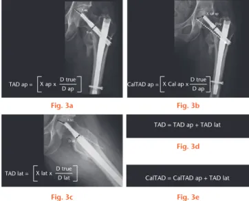
tAD, CaltAD, and lag-screw position were assessed on immediate post-operative radiographs in anteroposterior (AP) and latero-lateral (ll) views. A single observer – a consultant trauma surgeon (m.b.) – measured the tAD and the CaltAD (Fig. 3), the screw position according to
Cleveland et al,10 and the fracture reduction, in order to
eliminate inter-observer variability.
the study was approved by the local university hospital human subject research ethics Committee, and data collection and analysis were performed in compli-ance with the Declaration of helsinki.
statistical analysis
the shapiro-Wilk test was used to assess the assumption of normality, and data were expressed as mean and sD or as median (interquartile range, iQr), according to their distribution. Categorical data are presented as numbers (%). Percentages were compared using the chi-square test, and continuous data via the student t-test or mann-Whitney test, as appropriate. A receiver operating charac-teristic (roC) curve was used to investigate the diagnostic accuracy of tAD and CaltAD in predicting screw cut-out, and to identify the optimal cut-off point of these param-eters for distinguishing between patients with and with-out screw cut-with-out.19 odds ratios (or) and 95% confidence
intervals (95% Ci) were estimated using an unadjusted logistic regression model which considered the lag-screw cut-out to be a dependent variable and the demographic, surgical, pharmacological and radiological parameters to be independent variables. odds ratios and 95% confi-dence interval (Ci) were also calculated for an adjusted logistic regression analysis to assess the strength of asso-ciations between the dependent variable screw cut-out and several potential predictors (the independent varia-bles: gender; age; anti-osteoporotic therapy; weight-bearing; Cleveland classification and tAD). multivariate logistic regression analysis was conducted on the female SUPERIOR (1) (2) (3) (4) (5) (6) (7) (8) (9) INFERIOR A N T E R I O R P O S T E R I O R Reference category Fig. 2
Diagram showing the modified Cleveland system10 used in our study. Nine
areas were reduced to three, specifically the central (reference category) and two peripherals denoted “+” (in green) and “x” (in yellow).
TAD ap = X ap xD true
D ap CalTAD ap = X Cal ap x
D true D ap
TAD lat = X lat x D true
D lat
TAD = TAD ap + TAD lat
CalTAD = CalTAD ap + TAD lat
D ap X ap X cal ap D ap X lat D lat Fig. 3a Fig. 3c Fig. 3b Fig. 3d Fig. 3e
a) tip-apex distance calculated on anteroposterior radiograph (tAD ap); b) tip-apex distance as referenced to the calcar calculated on the anteroposte-rior radiograph (CaltAD ap); c) tip-apex distance calculated on the lateral radiograph (tAD lat); d) apex distance (tAD); e) calcar-referenced tip-apex distance (CaltAD). D true is the known diameter of the lag-screw (10.5 mm). D ap is the calculated diameter of the lag-screw on the anteroposterior radiograph. D lat is the calculated diameter of the lag-screw on the lateral radiograph.
population data as we found a statistically gender-related difference. this was stratified by age range (< 85 and
⩾ 85 years). All analyses were performed using stata 12.1 se software.(stata statistical software: release 12. College station, texas),and all tests were two-sided. statistical significance was set at a p-value of < 0.05. Results
Among the 571 patients with pertrochanteric fracture of the proximal femur who were reviewed in this study, lag-screw cut-out was observed in 32 cases (an incidence of 5.6%), in line with the range reported in the literature.20-22
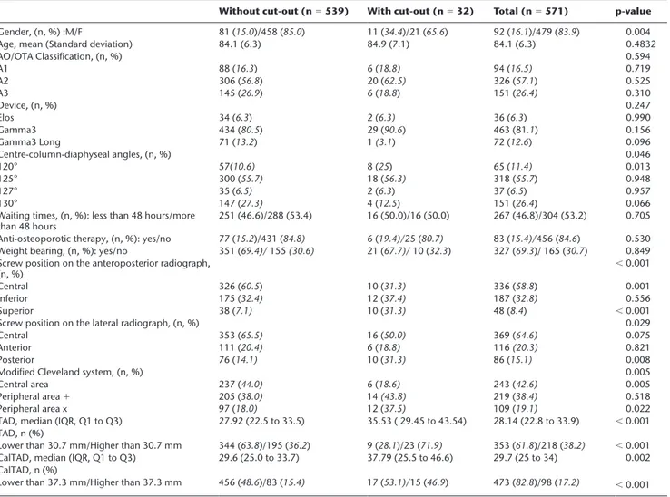
As shown in the demographic data and patient character-istics presented in table i, there was a prevalence of female subjects among those affected (83.9%), and a greater percentage of Ao/otA type 31-A2 fractures (57.1% of the total) than types A3 (26%) and A1 (17%). As for the surgical device, the gamma 3 trochanteric nail (stryker trauma gmbh schönkirchen, germany) was used in 81% of cases, the gamma 3 long nail (stryker trauma gmbh) in 12.6%, and the elos nail (intrauma s.r.l, turin, italy) in 6.3%; the cervical-diaphyseal angle most commonly employed was 125°, in 55.7% of cases.
Table I. Demographic data and baseline characteristics of all patients with trochanteric fractures
Without cut-out (n = 539) With cut-out (n = 32) Total (n = 571) p-value gender, (n, %) :m/F 81 (15.0)/458 (85.0) 11 (34.4)/21 (65.6) 92 (16.1)/479 (83.9) 0.004
Age, mean (standard deviation) 84.1 (6.3) 84.9 (7.1) 84.1 (6.3) 0.4832
Ao/otA Classification, (n, %) 0.594 A1 88 (16.3) 6 (18.8) 94 (16.5) 0.719 A2 306 (56.8) 20 (62.5) 326 (57.1) 0.525 A3 145 (26.9) 6 (18.8) 151 (26.4) 0.310 Device, (n, %) 0.247 elos 34 (6.3) 2 (6.3) 36 (6.3) 0.990 gamma3 434 (80.5) 29 (90.6) 463 (81.1) 0.156 gamma3 long 71 (13.2) 1 (3.1) 72 (12.6) 0.096 Centre-column-diaphyseal angles, (n, %) 0.046 120° 57(10.6) 8 (25) 65 (11.4) 0.013 125° 300 (55.7) 18 (56.3) 318 (55.7) 0.948 127° 35 (6.5) 2 (6.3) 37 (6.5) 0.957 130° 147 (27.3) 4 (12.5) 151 (26.4) 0.066
Waiting times, (n, %): less than 48 hours/more
than 48 hours 251 (46.6)/288 (53.4) 16 (50.0)/16 (50.0) 267 (46.8)/304 (53.2) 0.705 Anti-osteoporotic therapy, (n, %): yes/no 77 (15.2)/431 (84.8) 6 (19.4)/25 (80.7) 83 (15.4)/456 (84.6) 0.530 Weight bearing, (n, %): yes/no 351 (69.4)/ 155 (30.6) 21 (67.7)/ 10 (32.3) 327 (69.3)/ 165 (30.7) 0.849 screw position on the anteroposterior radiograph,
(n, %) < 0.001
Central 326 (60.5) 10 (31.3) 336 (58.8) 0.001
inferior 175 (32.4) 12 (37.4) 187 (32.8) 0.556
superior 38 (7.1) 10 (31.3) 48 (8.4) < 0.001
screw position on the lateral radiograph, (n, %) 0.029
Central 353 (65.5) 16 (50.0) 369 (64.6) 0.075
Anterior 111 (20.4) 6 (18.8) 116 (20.3) 0.821
Posterior 76 (14.1) 10 (31.3) 86 (15.1) 0.008
modified Cleveland system, (n, %) 0.005
Central area 237 (44.0) 6 (18.6) 243 (42.6) 0.005
Peripheral area + 205 (38.0) 14 (43.8) 219 (38.4) 0.518
Peripheral area x 97 (18.0) 12 (37.5) 109 (19.1) 0.022
tAD, median (iQr, Q1 to Q3) 27.92 (22.5 to 33.5) 35.53 ( 29.45 to 43.54) 28.14 (22.8 to 33.9) < 0.001 tAD, n (%)
lower than 30.7 mm/higher than 30.7 mm 344 (63.8)/195 (36.2) 9 (28.1)/23 (71.9) 353 (61.8)/218 (38.2) < 0.001 CaltAD, median (iQr, Q1 to Q3) 29.6 (25.0 to 33.7) 37.79 (25.5 to 46.6) 29.7 (25 to 34) 0.002 CaltAD, n (%)
lower than 37.3 mm/higher than 37.3 mm 456 (48.6)/83 (15.4) 17 (53.1)/15 (46.9) 473 (82.8)/98 (17.2) < 0.001 Ao/otA Ao Foundation and orthopaedic trauma Association classification system; A1,A2,A3, type of fracture according to Ao/otA classification; tAD, tip-apex distance; CaltAD, calcar-referenced tip-tip-apex distance;iQr, interquartile range
chi-squared test was used for all p-values
SUPERIOR (1) (2) (3) (4) (5) (6) (7) (8) (9) INFERIOR A N T E R I O R P O S T E R I O R 1/8 2/26 7/14 2/47 6/243 2/46 3/61 8/100 1/26 Fig. 4
Diagram showing the number of cut-outs observed out of the total number of lag-screw positions in Cleveland’s10 nine areas.
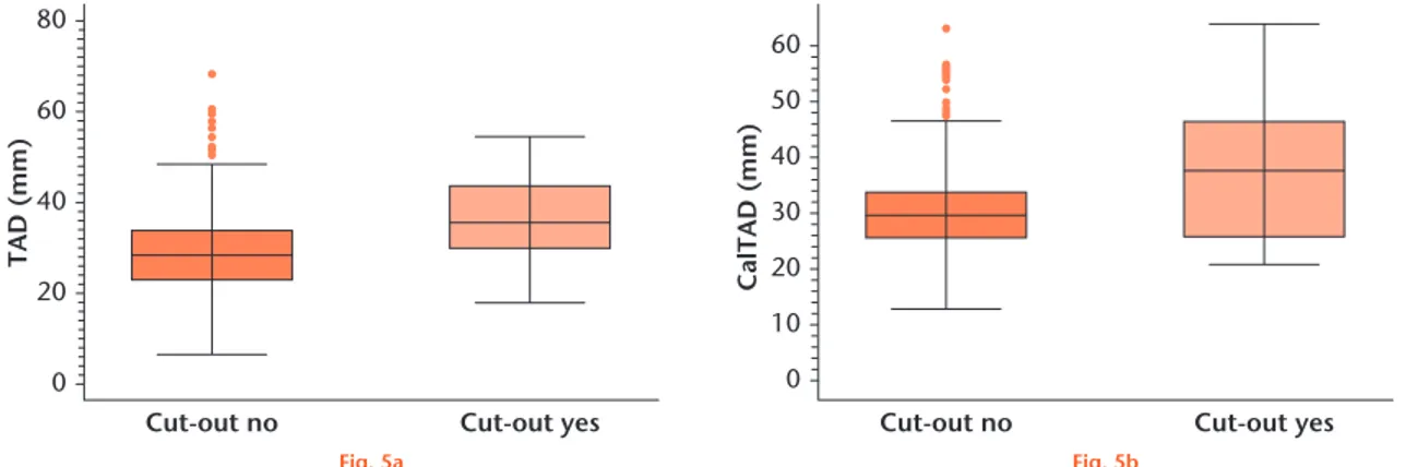
As shown in Figure 4, according to the classical Cleveland system, lag-screws were positioned most fre-quently in the centro-central area (42.5% of cases), fol-lowed by the infero-central area (17.5%). however, the Cleveland areas in which the highest incidence of cut-out was observed were the postero-superior (50%) and the antero-superior (12.5%). Figures 5a and 5b show the respective box plots created for the tAD and CaltAD measurements in relation to the presence or absence of cut-out. Calculation of the roC curves, specifically the area under the curve (AuC), showed the accuracy of tAD and CaltAD in predicting the risk of cut-out as AuC = 0.72 and AuC = 0.67, respectively (Figs 6a and 6b). thus, according to the AuC measurements calculated for our sample, tAD is a more reliable predictor of cut-out risk than CaltAD, albeit with only moderate and poor levels of accuracy, respectively. Application of the Youden test, which balances the highest values of sensitivity and speci-ficity, showed that the best cut-offs were 30.7 mm for
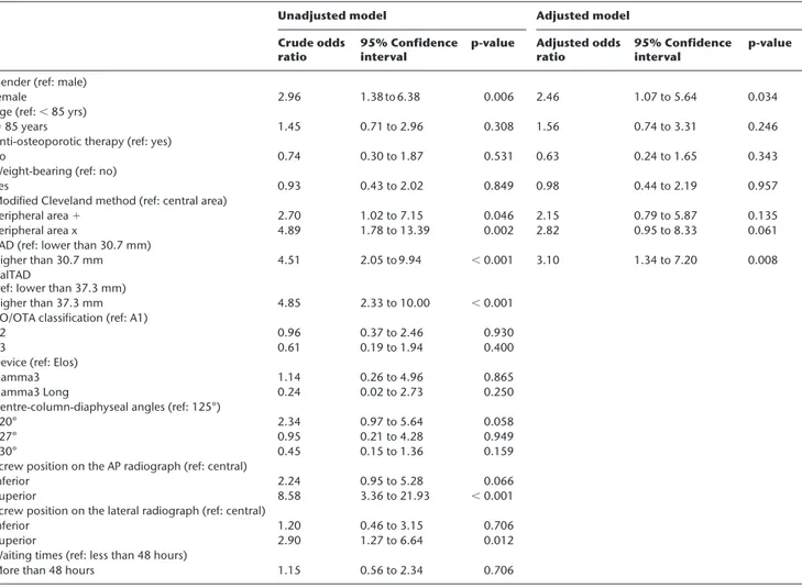
tAD and 37.3 mm for CaltAD. the results of the unad-justed and adunad-justed logistic regression analyses shown in table ii indicate a statistically significant gender-related difference, with the likelihood of cut-out being almost three times higher in females than in males. Furthermore, the probability of cut-out was statistically linked to the position of the lag-screw on the two radiological views, with significant differences between both the superior position in AP view (or 8.58; 95% Ci 3.36 to 21.93) and the posterior position in ll view (or 2.90; 95% Ci 1.27 to 6.64) with the centro-central position chosen as refer-ence category.17
using our modified version of the Cleveland system, the probability of cut-out increased by roughly five-fold when the lag-screw tip was located in peripheral zone “x”. tAD and CaltAD values were also highly significant when considered as dichotomous variables. in fact, the risk of lag-screw cut-out was 4.51 times greater when the tAD was above 30.7 mm, and 4.85 times greater when 0 Cut-out no 20 40 TAD (mm )60 80 Cut-out yes 0 Cut-out no 10 30 50 60 40 20 CalTAD (mm ) Cut-out yes Fig. 5a Fig. 5b
box plots showing the distribution of values and cut-out frequencies of a) tip-apex distance (tAD), and b) calcar-referenced tip-apex distance (CaltAD).
Fig. 6a Fig. 6b 0.00 0.00 0.25 0.50 Specificity Sensitivity 0.75 1.00 0.25 0.50 0.75 1.00 0.00 0.00 0.25 0.50 Specificity Sensitivity 0.75 1.00 0.25 0.50 0.75 1.00
graph showing the receiver operating characteristic curve obtained by plotting the sensitivity of a) tip-apex distance (tAD), and b) calcar-referenced tip-apex distance (CaltAD) against their respective specificity values.
CaltAD was above 37.3 mm. As shown in Figure 7, the probability of cut-out increased with increasing tAD, and by considering tAD as a continuous variable it was
therefore possible to estimate the increase in cut-out per unit increase in tAD. this analysis showed that for every millimetre of increase in tAD, the cut-out risk increased roughly 1.1-fold.
Finally, multivariate regression analysis on the female population stratified by age range showed that in women of less than 85 years of age, the risk of cut-out was increased 6.4-fold when the tAD was above 30.7 mm as shown in supplementary table i.
Discussion
before discussing the results obtained in this study, it is necessary to point out its limitations, which could have influenced clinical outcomes. the first is that the analysis was carried out retrospectively, on patients recruited over a long period of time. the second involves the com-parability of populations undergoing the two treatments, which may only be suggested by a retrospective study – i.e., weak evidence that would need to be confirmed by a specifically designed randomised clinical trial.
Table II. Association between baseline patient characteristics and cut-out according to logistic regression analysis, unadjusted and adjusted for potential
confounders
Unadjusted model Adjusted model
Crude odds
ratio 95% Confidence interval p-value Adjusted odds ratio 95% Confidence interval p-value gender (ref: male)
female 2.96 1.38 to 6.38 0.006 2.46 1.07 to 5.64 0.034
Age (ref: < 85 yrs)
⩾ 85 years 1.45 0.71 to 2.96 0.308 1.56 0.74 to 3.31 0.246
Anti-osteoporotic therapy (ref: yes)
no 0.74 0.30 to 1.87 0.531 0.63 0.24 to 1.65 0.343
Weight-bearing (ref: no)
yes 0.93 0.43 to 2.02 0.849 0.98 0.44 to 2.19 0.957
modified Cleveland method (ref: central area)
Peripheral area + 2.70 1.02 to 7.15 0.046 2.15 0.79 to 5.87 0.135
Peripheral area x 4.89 1.78 to 13.39 0.002 2.82 0.95 to 8.33 0.061
tAD (ref: lower than 30.7 mm)
higher than 30.7 mm 4.51 2.05 to 9.94 < 0.001 3.10 1.34 to 7.20 0.008
CaltAD
(ref: lower than 37.3 mm)
higher than 37.3 mm 4.85 2.33 to 10.00 < 0.001
Ao/otA classification (ref: A1)
A2 0.96 0.37 to 2.46 0.930
A3 0.61 0.19 to 1.94 0.400
Device (ref: elos)
gamma3 1.14 0.26 to 4.96 0.865
gamma3 long 0.24 0.02 to 2.73 0.250
Centre-column-diaphyseal angles (ref: 125°)
120° 2.34 0.97 to 5.64 0.058
127° 0.95 0.21 to 4.28 0.949
130° 0.45 0.15 to 1.36 0.159
screw position on the AP radiograph (ref: central)
inferior 2.24 0.95 to 5.28 0.066
superior 8.58 3.36 to 21.93 < 0.001
screw position on the lateral radiograph (ref: central)
inferior 1.20 0.46 to 3.15 0.706
superior 2.90 1.27 to 6.64 0.012
Waiting times (ref: less than 48 hours)
more than 48 hours 1.15 0.56 to 2.34 0.706
Ao/otA Ao Foundation and orthopaedic trauma Association classification system; A1,A2,A3, type of fracture according to Ao/otA classification; tAD, tip-apex distance; CaltAD, calcar-referenced tip-tip-apex distance; AP, anteroposterior
unadjusted and adjusted logistic regression tests were used for p-values
0 0 0.1 0.2 0.3 0.4 0.5 20 40 60 80 TAD (mm)
Predicted probability of cut-ou
t
Fig. 7
unlike previous studies,23 in which no apparent
influ-ence of gender on the incidinflu-ence of cut-out was found, in our sample there was a clear prevalence of complications in female patients, a disparity that, in our opinion, may be linked to the greater incidence and severity of osteo-porosis in this patient category. however, our data are in line with some of the literature8,9,11,22 on the subject of
cut-out incidence with respect to the Cleveland method of lag-screw positioning. specifically, we observed that positioning the lag-screw tip in the supero-posterior sec-tor was linked to a greater risk of cut-out. moreover, when using our modified version of the Cleveland classi-fication, we observed that the risk of cut-out increased almost five-fold if the lag-screw tip was positioned in the peripheral zone denoted “x”. indeed, baumgaertner et al11 found the highest rates of cut-out in the
postero-inferior and antero-superior zones, and consequently recommended a deep central insertion of the lag-screw. De bruijn et al23 also recommended centro-central or
low-central lag-screw placement with minimal tAD. these conclusions, however, differ from those of Kaufer, matthews and sonstegard24 who argued in favour of
lag-screw placement in the postero-inferior quadrant of the femoral head, as the crossing of tension and compression trabeculae in that area of the femoral head probably pro-vides the best bone for screw placement, thus improving proximal fragment control. this theory was recently tested in a biomechanical study by Kane et al17 who
found that the low central position with a tAD of greater than 25 mm provided equal if not superior stability to centro-central placement with the then optimal tAD of less than 25 mm.
our results also showed that tAD is a better predictor of cut-out risk than CaltAD, with their respective AuCs generated by roC analysis suggesting that tAD is more reliable than CaltAD in this regard. this, however, con-trasts with findings by Kashigar et al20 that CaltAD is the
only significant predictor of cut-out, which was not observed in patients with a CaltAD value lower than 20.98 mm. our case series, on the other hand, showed much higher cut-offs, of 37.3 mm for CaltAD and 30.7 mm for tAD, with respective increases in cut-out risk of roughly 4.8- and 4.5-fold above these thresholds. there is therefore a considerable discrepancy between our values and those traditionally found in the litera-ture,11,16 which identify 25 mm as the value above which
the risk of cut-out is increased. however, this discrepancy may in part be explained, as recently suggested in a mathematical simulation by li et al,16 by the fact that in
order to be accurate the cut-off proposed by baumgaertner et al11 should be corrected in relation to the diameter of
the femoral head, which varies according to gender and anthropometric characteristics, and among individuals. indeed, it is highly likely that variations in the quality of bone housing the lag-screw will affect its stability and therefore the risk of cut-out.
Another finding in our series was that the risk of cut-out is 6.4 × greater in women between 70 and 85 years of age if the tAD was greater than 30.7 mm - to our knowledge, the first such data reported in the literature. We hypothesise that in female patients below 85 years of age, the position of the lag-screw with respect to the fem-oral head is the factor most influential to the risk of cut-out, while in women above the age of 85 years other factors, more directly correlated with the bone quality and patient characteristics, come into play.
in the interests of comprehensiveness, we also ana-lysed more general variables of patients with pertrochan-teric hip fractures to see whether they would be of any interest. however, we observed no statistically significant differences in the incidence of cut-out related to Ao/otA fracture classification, laterality, type of device, pre-trauma anti-resorption medication, or surgical waiting times. As part of this analysis, however, we did find that there was no statistically significant correlation between the occurrence of cut-out and weight-bearing immedi-ately after surgery. As far as we know, this is the first time such a finding has been reported in the literature, and therefore further studies are warranted to confirm our results.
in conclusion, we find that tAD should still be consid-ered the most accurate predictive factor for cut-out among those suggested in the literature, as the increase in distance between lag-screw tip and femoral head apex is the main risk factor of this complication. in our case series, the cut-off for increased cut-out risk was 30.7 mm, unlike the 25 mm commonly cited in the literature,7,11,25,26 but our findings
do confirm reports that peripheral positioning of the lag-screw, specifically in the postero-superior portion of the femoral head, is associated with a greater risk of cut-out. in contrast, whether or not the patient was allowed to bear weight in the immediate post-operative period seemed to have no influence on the occurrence of this complication, although this latter finding may be the result of beta error and therefore requires confirmation.
supplementary material
A table showing multivariate regression analysis on the female population stratified by age range is available alongside this article online at www.bjr.bone-andjoint.org.uk
References
1. White SM, Griffiths R. Projected incidence of proximal femoral fracture in
England: a report from the NHS Hip Fracture Anaesthesia Network (HIPFAN). Injury 2011;42:1230-1233.
2. Swart E, Makhni EC, Macaulay W, Rosenwasser MP, Bozic KJ.
Cost-effectiveness analysis of fixation options for intertrochanteric hip fractures. J Bone Joint Surg [Am] 2014;96-A:1612-1620.
3. Parker MJ, Handoll HH. Gamma and other cephalocondylic intramedullary nails
versus extramedullary implants for extracapsular hip fractures in adults. Cochrane Database Syst Rev 2010;9:CD000093.
4. Yeganeh A, Taghavi R, Moghtadaei M. Comparing the Intramedullary Nailing
Method Versus Dynamic Hip Screw in Treatment of Unstable Intertrochanteric Fractures. Med Arch 2016;70:53-56.
5. Bojan AJ, Beimel C, Speitling A, et al. 3066 consecutive Gamma Nails. 12 years
experience at a single centre. BMC Musculoskelet Disord 2010;11:133.
6. Verhofstad MH, van der Werken C. DHS osteosynthesis for stable pertrochanteric
femur fractures with a two-hole side plate. Injury 2004;35:999-1002.
7. Walton MJ, Barnett AJ, Jackson M. Tip-Apex Distance as a Predictor of Failure
Following Cephalo-Medullary Fixation for Unstable Fractures of the Proximal Femur. Eur J Trauma Emerg Surg 2008;34:273-276.
8. Parker MJ. Cutting-out of the dynamic hip screw related to its position. J Bone Joint
Surg [Br] 1992;74-B:625.
9. Gundle R, Gargan MF, Simpson AH. How to minimize failures of fixation of
unstable intertrochanteric fractures. Injury 1995;26:611-614.
10. Cleveland M, Bosworth DM, Thompson FR, Wilson HJ Jr, Ishizuka T. A
ten-year analysis of intertrochanteric fractures of the femur. J Bone Joint Surg [Am] 1959;41-A:1399-1408.
11. Baumgaertner MR, Curtin SL, Lindskog DM, Keggi JM. The value of the tip-apex
distance in predicting failure of fixation of peritrochanteric fractures of the hip. J Bone Joint Surg [Am] 1995;77-A:1058-1064.
12. Kuzyk PR, Zdero R, Shah S, et al. Femoral head lag-screw position for
cephalomedullary nails: a biomechanical analysis. J Orthop Trauma 2012;26:414-421.
13. Mingo-Robinet J, Torres-Torres M, Martínez-Cervell C, et al. Comparative
study of the second and third generation of gamma nail for trochanteric fractures: review of 218 cases. J Orthop Trauma 2015;29:e85-e90.
14. Goffin JM, Pankaj P, Simpson AH. The importance of lag-screw position for the
stabilization of trochanteric fractures with a sliding hip screw: a subject-specific finite element study. J Orthop Res 2013;31:596-600.
15. Goffin JM, Jenkins PJ, Ramaesh R, Pankaj P, Simpson AH. What is the
relevance of the tip-apex distance as a predictor of lag-screw cut-out? PLoS One 2013;8:e71195.
16. Li S, Chang SM, Jin YM, et al. A mathematical simulation of the tip-apex distance
and the calcar-referenced tip-apex distance for intertrochanteric fractures reduced with lag-screws. Injury 2016;47:1302-1308.
17. Kane P, Vopat B, Heard W, et al. Is tip apex distance as important as we think? A
biomechanical study examining optimal lag-screw placement. Clin Orthop Relat Res 2014;472:2492-2498.
18. Johnson LJ, Cope MR, Shahrokhi S, Tamblyn P. Measuring tip-apex distance
using a picture archiving and communication system (PACS). Injury 2008;39:786-790.
19. Youden WJ. Index for rating diagnostic tests. Cancer 1950;3:32-35.
20. Kashigar A, Vincent A, Gunton MJ, et al. Predictors of failure for cephalomedullary
nailing of proximal femoral fractures. Bone Joint J 2014;96-B:1029-1034.
21. Andruszkow H, Frink M, Frömke C, et al. Tip apex distance, hip screw placement,
and neck shaft angle as potential risk factors for cut-out failure of hip screws after surgical treatment of intertrochanteric fractures. Int Orthop. 2012;36:2347-2354.
22. Hsueh KK, Fang CK, Chen CM, et al. Risk factors in cutout of sliding hip screw
in intertrochanteric fractures: an evaluation of 937 patients. Int Orthop 2010;34: 1273-1276.
23. De Bruijn K, den Hartog D, Tuinebreijer W, Roukema G. Reliability of predictors
for screw cutout in intertrochanteric hip fractures. J Bone Joint Surg [Am] 2012;94-A:1266-1272.
24. Kaufer H, Matthews LS, Sonstegard D. Stable fixation of intertrochanteric
fractures. J Bone Joint Surg [Am] 1974;56-A:899-907.
25. Baumgaertner MR, Solberg BD. Awareness of tip-apex distance reduces failure of
fixation of trochanteric fractures of the hip. J Bone Joint Surg [Br] 1997;79-B:969-971.
26. Pervez H, Parker MJ, Vowler S. Prediction of fixation failure after sliding hip
screw fixation. Injury 2004;35:994-998.
Acknowledgement
We would like to acknowledge m. Pizzato and A. valentini (collection and statistical processing of data).
Funding statement None declared
Author contribution
g. Caruso: Conception and experimental design, Data interpretation, manuscript preparation.
m. bonomo: Conception and experimental design, Data interpretation, manuscript preparation.
g. valpiani: Data Analysis, Data interpretation, manuscrit proofing.
g. salvatori: experimental design, manuscript proofing.
A. gildone: experimental design, manuscript proofing.
v. lorusso: experimental design, manuscript proofing.
l. massari: Conception design, manuscript proofing.
conflicts of Interest statement None declared
© 2017 Caruso et al. this is an open-access article distributed under the terms of the
Creative Commons Attributions licence (CC-bY-NC), which permits unrestricted use, distribution, and reproduction in any medium, but not for commercial gain, provided the original author and source are credited.