© 2018 Gynecology and Minimally Invasive Therapy | Published by Wolters Kluwer - Medknow
6
Original Article
I
ntroductIonIn case of abnormal uterine bleeding (AUB), guidelines indicate transvaginal ultrasound (TVS) evaluation of endometrial thickness as the first investigation. Ultrasound is a less invasive test and proves quite useful when having to discriminate cancer or hyperplasia depending on endometrial thickness in a postmenopausal patient.
Endometrial cancer is the most common cancer of the female genital tract in the developed countries.[1] It generally occurs in the postmenopausal women with AUB, but in 10% of cases, it could be asymptomatic[2] and should be discovered by incidental during annual ultrasound control for evaluating the presence of postmenopausal‑thickened endometrium. This thickness indicates an increased risk of malignancy or other benign pathology (hyperplasia, myoma, and polyp).[3]
As patients with AUB, asymptomatic postmenopausal women with an endometrial thickness >5 mm, found by ultrasound, should undergo a hysteroscopy and an endometrial biopsy, even if there is no consensus among authors about the definition of thickened endometrium for asymptomatic postmenopausal women.[4] In 2014, Giannella et al. showed that using an endometrial thickness cutoff value ≥4 mm, only 3% of performed hysteroscopies were useful for the detection of premalignant or malignant lesions.[5] According to Smith‑Bindman, in a postmenopausal woman without vaginal
Abstract
Backgrounds and Aims: This study aims to compare hysteroscopic and histological findings in asymptomatic postmenopausal patients with
thickened endometrium.
Materials and Methods: A retrospective study involving case records of 295 asymptomatic postmenopausal women with a thickened
endometrium >5 mm diagnosed at transvaginal ultrasound (TVS). Patients (women) underwent hysteroscopy with biopsy between 2009 and 2015, and they were followed up at National Cancer Institute of Bari and at University Hospital of Pisa. Sensitivity, specificity, positive predictive value (PPV), and negative predictive value (NPV) of hysteroscopy were evaluated.
Results: Inclusion criteria were TVS, hysteroscopy, and endometrial biopsy. When the hysteroscopic findings were normal, a sensitivity
of 100%, specificity of 98.6%, PPV of 95.2%, and NPV of 100% were achieved. For polyps and myomas, we found 100%, 98.7%, 99.5%, and 100%, respectively. In case of endometrial hyperplasia, a sensitivity of 66.7%, a specificity of 100%, a PPV of 100%, and a NPV of 98.1% were achieved. For endometrial cancer hysteroscopy, sensitivity, specificity, PPV, and NPV were 100%, 99.6%, 75%, and 100%, respectively.
Conclusions: Hysteroscopy allows an accurate diagnosis in benign endometrial pathology and suspect of malignant endometrial pathology
in postmenopausal women with thickened endometrium.
Keywords: Endometrial thickness, hysteroscopy, postmenopausal patients
Access this article online
Quick Response Code:
Website:
www.e‑gmit.com
DOI:
10.4103/GMIT.GMIT_10_17
Address for correspondence: Dr. Gianluca Raffaello Damiani, Department of Obstetrics and Gynecology, ASST Lecco, Leopoldo Mandic Hospital, Lecco, Italy. E‑mail: [email protected]
This is an open access article distributed under the terms of the Creative Commons Attribution‑NonCommercial‑ShareAlike 3.0 License, which allows others to remix, tweak, and build upon the work non‑commercially, as long as the author is credited and the new creations are licensed under the identical terms.
For reprints contact: [email protected]
How to cite this article: Trojano G, Damiani GR, Casavola VC, Loiacono R, Malvasi A, Pellegrino A, et al. The role of hysteroscopy in evaluating postmenopausal asymptomatic women with thickened endometrium. Gynecol Minim Invasive Ther 2018;7:6‑9.
The Role of Hysteroscopy in Evaluating Postmenopausal
Asymptomatic Women with Thickened Endometrium
Giuseppe Trojano, Gianluca Raffaello Damiani1, Vita Caroli Casavola, Rossella Loiacono2, Antonio Malvasi3, Antonio Pellegrino4, Valeria Siciliano5,
Ettore Cicinelli, Maria Giovanna Salerno4, Lorella Battini4
Department of Obstetrics and Gynaecology, University of Bari, 1Department of Obstetrics and Gynaecology, ASST Lecco, Leopoldo Mandic Hospital, Lecco, 2National
Cancer Research Centre, Istituto Tumori “Giovanni Paolo II”, 3Department of Obstetrics and Gynaecology, Santa Maria Hospital, Bari, 4Department of Obstetrics and
Gynaecology, AOUP Santa Chiara Hospital, Pisa, 5Institute of Clinical Phisiology, National Research Council of Italy, Rome, Italy
Gynecology and Minimally Invasive Therapy 7 (2018) 6‑9
Trojano, et al.: Hysteroscopy as a valid tool for detecting endometrial pathology in postmenopause
7
Gynecology and Minimally Invasive Therapy ¦ January-March 2018 ¦ Volume 7 ¦ Issue 1
bleeding, if the endometrium measures >11 mm, a biopsy should be considered as the risk of cancer is 6.7%, whereas if the endometrium measures ≤11 mm, a biopsy is not needed as the risk of cancer is extremely low.[6]
On the contrary, hysteroscopy is commonly considered as the gold standard technique for the evaluation of endometrial pathologies, allowing a direct view of the uterine cavity directly and to perform the biopsy.[7,8] It is less invasive than dilation and curettage (D and C) and does not need anesthesia.[9] The aim of this study is to compare the macroscopic hysteroscopic findings to histological findings in asymptomatic postmenopausal patients with incidental ultrasound discovery of thickened endometrium >5 mm.
M
aterIalsandM
ethodsThis is a retrospective study involving case records of 295 women undergone outpatient hysteroscopy and endometrial biopsy between 2009 and 2015 at the National Cancer Research Centre, Istituto Tumori “Giovanni Paolo II,” Bari, Italy, and at the Department of Obstetrics and Gynaecology 2, AOUP, Santa Chiara Hospital, Pisa, Italy. The general methodology of this study has been described in a companion article published by Loiacono et al.[10]
The patients were all postmenopausal and asymptomatic. None of them had positive personal history of cancer of the genital tract. None took hormone replacement therapy.
Menopause was defined as spontaneous cessation of menses for 1 year or more.
The median age in our case reports is 59.5 years old with an age range of 45–81 years. Most of the patients were suffering from comorbidity (diabetes mellitus, obesity, and hypertension), but this information was not available in all cases.
Each patient underwent TVS to define endometrial thickness. In a sagittal scan, the operator calculated the maximum distance between the two lines of the endometrium/myometrium interface. The cutoff used to suspect the presence of endometrial pathology was a maximum thickness >5.
The most experienced operator was always present during all procedures. Hysteroscopies were performed by vaginoscopic approach: without speculum, without local or general anesthesia, and with a 3- or 5-mm hysteroscope (30° view). Isotonic sodium chloride was used as distension medium with a pressure of 50–70 mmHg and flow (100–120 mmHg).[11,12] Hysteroscopic examination included inspection of the uterine cavity with a panoramic shot, visualization of both tubal ostia, and observation of the cervical canal by removing the hysteroscope.
Histological findings were classified as normal if they were atrophic or hypotrophic and as abnormal in cases of endometrial polyps, submucous myomas, endometritis, adenomyosis, endometrial hyperplasia, and endometrial cancer.[13]
Endometrial cancer was suspected if there were these hysteroscopic findings: atypical vessels, irregular necrotic tissue,[14] micropapillary or polypoid hypertrophy,[15] mammillations, cerebroid irregularities associated with irregular polylobular, friable excrescences with necrosis or bleeding.[16] Endometrial hyperplasia was suspected if there were these hysteroscopic findings: increased endometrial thickness, both localized or diffuse; cystic formations with a reduction of the interglandular space; and dilated superficial vessels.[13] The material was fixed in 10% formalin and sent to an associate pathologist for the histopathological examination; in each case pathologist was informed about ultrasonographic and hysteroscopic findings.
Statistical analysis
Hysteroscopic and histopathological findings were expressed as percentage. Cohen’s kappa coefficient (Cohen, 1960) was calculated to determine the power of concordance between histological diagnosis and hysteroscopy. Landis and Koch (1977) described intervals for the values of κ and associated different empirical concordance levels with these values. Empirical concordance levels according to Landis and Koch are κ <0 low; 0.00≤κ≤0.20 weak; 0.21≤κ≤0.40 sufficient; 0.41≤κ≤0.60 good; 0.61≤κ≤0.80 excellent; and 0.81≤κ≤1.00 almost perfect. To evaluate the ability of the test to correctly classify the patients, sensitivity, specificity, negative predictive values (NPV), and positive predictive value (PPV) were calculated.
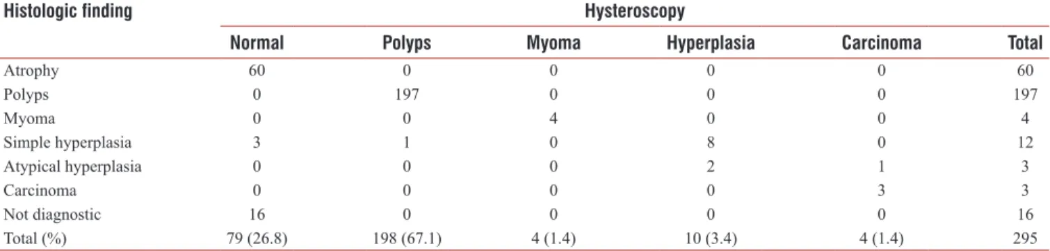
Table 1: Comparative breakdown of hysteroscopic and histological findings in asymptomatic postmenopausal women with a thickened endometrium
Histologic finding Hysteroscopy
Normal Polyps Myoma Hyperplasia Carcinoma Total
Atrophy 60 0 0 0 0 60 Polyps 0 197 0 0 0 197 Myoma 0 0 4 0 0 4 Simple hyperplasia 3 1 0 8 0 12 Atypical hyperplasia 0 0 0 2 1 3 Carcinoma 0 0 0 0 3 3 Not diagnostic 16 0 0 0 0 16 Total (%) 79 (26.8) 198 (67.1) 4 (1.4) 10 (3.4) 4 (1.4) 295
Trojano, et al.: Hysteroscopy as a valid tool for detecting endometrial pathology in postmenopause
8 Gynecology and Minimally Invasive Therapy ¦ January-March 2018 ¦ Volume 7 ¦ Issue 1
Ethical approval
The study was conducted in accordance with the Declaration of Helsinki and was approved by the local ethics committee of the institute. Informed written consent was obtained from all patients prior to their enrollment in this study.
r
esultsComparing hysteroscopic findings with histological results of 295 asymptomatic patients with thickened endometrium [Table 1], hysteroscopy showed normal findings in 79 (26. 8%) cases. Among these 79 cases, histology showed 60 cases of atrophy, 3 cases of simple hyperplasia, no case of atypical hyperplasia and endometrial carcinoma. In 16 cases of 79, endometrial sample was insufficient.
Histology confirmed the hysteroscopic diagnoses of 198 (67.1%) cases of polyps and 4 (1.4%) cases of submucous myoma. All samples were sufficient for histological diagnosis. Hysteroscopy showed 10 (3.4%) cases of hyperplastic endometrium; 8 of these cases resulted histologically as simple hyperplasia and 2 cases as atypical hyperplasia. None of the carcinoma cases was found. Each sample was sufficient for histological diagnosis. We suspected 4 (1.4%) cases of endometrial cancer by hysteroscopy, three of them were histologically confirmed. The last one was diagnosed histologically as atypical hyperplasia. Each sample was sufficient for histological diagnosis. The concordance between histological diagnosis and hysteroscopy was almost perfect; Cohen’s kappa equal to 0.86 (P < 0.001).
According to these data, hysteroscopy view of normal findings showed a sensitivity of 100%, a specificity of 98.6%, PPV 95.2, and NPV 100%.
The sensitivity, specificity, PPV, and NPV for hysteroscopic polyps and myomas were 100%, 98.7%, 99.5%, and 100%, respectively.
For endometrial hyperplasia, hysteroscopy showed a sensitivity, specificity, PPV, and NPV of 66.7%, 100%, 100%, and 98.1%, respectively.
The sensitivity, specificity, PPV, and NPV for endometrial cancer were 100%, 99%, 75%, and 100%, respectively. All of these data are shown in Table 2.
d
IscussIonHistological diagnosis and hysteroscopy showed almost perfect concordance. The sensitivity of hysteroscopy in case of atrophic or lipotropic endometrium was 100%, its specificity 98.6%, PPV 95.2%, and NPV 100%. The results were almost the same in case of polyps or myoma; sensitivity was 100%, specificity 98.7%, PPV 99.5%, and NPV were 100%. However, sensitivity and NPV decreased to 66.7% and 98.1%, respectively, for endometrial hyperplasia, whereas specificity and PPV were both at 100%. Sensitivity of hysteroscopy for carcinoma results was 100%, its specificity 99.6%, PPV 75.0%, and NPV 100%. According to literature, each postmenopausal patients showing an increased endometrial thickness (>5 mm), with or without AUB, should undergo to further investigation.[14] Conventionally, D and C was performed to arrow to an histological diagnosis.[9] Recently, outpatient hysteroscopy with biopsy starts to be considered as the gold standard investigation of ultrasound-thickened endometrium.[17] It allows both to visualize all uterine walls and to perform biopsy where endometrial lesions are localized.[18] Ceci et al. compared hysteroscopic and hysterectomy findings showing that hysteroscopy with targeted biopsy is more reliable than D and C.[19] In our experience, there was endometrial cancer close to left tubaric ostium <3 mm. It is necessary to underline that in almost 50% of ultrasound endometrial thickness >5 mm was not related to an endometrial pathology but hysteroscopic finding was a subseptate/arcuate uterus.
Outpatient hysteroscopy does not need hospitalization and anesthesia leading to important costs decrease.
In case of atrophic endometrium, the concordance between hysteroscopy and histology allows to avoid the execution of the biopsy by further reducing the costs.
On the other hand, some authors highlight that hysteroscopic normal findings are not enough to prove the absence of endometrial pathology, and they suggest to perform a biopsy in women with increased endometrial thickness, with or without AUB.[14,15,20,21] Endometrial sampling allows histological examination and immunohistochemistry for the final diagnosis.[22]
The thickened endometrium with hysteroscopic and histopathologic findings in asymptomatic postmenopausal women has been already evaluated in literature;[23] our results showed that hysteroscopy with targeted biopsy is useful not only in postmenopause woman with AUB but also in asymptomatic women with thickened endometrium in which both benign and malignant (3 cases on 295) were silent.
c
onclusIonsHysteroscopy allows an accurate diagnosis of benign endometrial pathology and suspect of malignant endometrial Table 2: Sensitivity, specificity, positive predictive value,
and negative predictive value (%) for hysteroscopy
Hysteroscopic
findings Sensitivity Specificity PPV NPV
Normal 100.0 98.6 95.2 100.0
Polyps-myoma 100.0 98.7 99.5 100.0
Endometrial hyperplasia 66.7 100.0 100.0 98.1
Carcinoma 100.0 99.6 75.0 100.0
PPV: Positive predictive value, NPV: Negative predictive value
Trojano, et al.: Hysteroscopy as a valid tool for detecting endometrial pathology in postmenopause
9
Gynecology and Minimally Invasive Therapy ¦ January-March 2018 ¦ Volume 7 ¦ Issue 1
pathology in postmenopausal women with thickened endometrium. In case of suspect of malignant pathology, hysteroscopy allows direct biopsy, histological result confirms in most of cases the hysteroscopic impression. Hysteroscopy avoids biopsy in case of atrophic endometrium.
Financial support and sponsorship
Nil.
Conflicts of interest
There are no conflicts of interest.
r
eferences1. Siegel R, Ma J, Zou Z, Jemal A. Cancer statistics, 2014. CA Cancer J Clin 2014;64:9-29.
2. Elfayomy AK, Habib FA, Elkablawy MA. Role of hysteroscopy in the detection of endometrial pathologies in women presenting with postmenopausal bleeding and thickened endometrium. Arch Gynecol Obstet 2012;285:839-43.
3. Lasmar RB, Dias R, Barrozo PR, Oliveira MA, Coutinho Eda S, da Rosa DB, et al. Prevalence of hysteroscopic findings and histologic diagnoses in patients with abnormal uterine bleeding. Fertil Steril 2008;89:1803-7. 4. Dreisler E, Sorensen SS, Ibsen PH, Lose G. Value of endometrial
thickness measurement for diagnosing focal intrauterine pathology in women without abnormal uterine bleeding. Ultrasound Obstet Gynecol 2009;33:344-8.
5. Giannella L, Mfuta K, Setti T, Boselli F, Bergamini E, Cerami LB, et al. Diagnostic accuracy of endometrial thickness for the detection of intra-uterine pathologies and appropriateness of performed hysteroscopies among asymptomatic postmenopausal women. Eur J Obstet Gynecol Reprod Biol 2014;177:29-33.
6. Smith-Bindman R, Weiss E, Feldstein V. How thick is too thick? When endometrial thickness should prompt biopsy in postmenopausal women without vaginal bleeding. Ultrasound Obstet Gynecol 2004;24:558-65.
7. Lalchandani S, Phillips K. Evaluation of endometrial cavity investigation option. Rev Gynecol Pract 2003;3:165-70.
8. Bourdel N, Modaffari P, Tognazza E, Pertile R, Chauvet P, Botchorishivili R, et al. Does experience in hysteroscopy improve accuracy and inter-observer agreement in the management of abnormal uterine bleeding? Surg Endosc 2016;30:5558-64.
9. Korkmazer E, Solak N, Üstünyurt E. Hysteroscopic assessment of postmenopausal endometrial thickening. Prz Menopauzalny 2014;13:330-3.
10. Loiacono RM, Trojano G, Del Gaudio N, Kardhashi A, Deliso MA, Falco G, et al. Hysteroscopy as a valid tool for endometrial pathology in patients with postmenopausal bleeding or asymptomatic patients
with a thickened endometrium: Hysteroscopic and histological results. Gynecol Obstet Invest 2015;79:210-6.
11. Damiani GR, Pellegrino A, Seghezzi U, Tagliabue R, Maggi F, Loverro G, et al. A dangerous trigger. Eur J Obstet Gynecol Reprod Biol 2012;165:133.
12. Damiani GR, Tartagni M, Crescini C, Persiani P, Loverro G, Von Wunster S, et al. Intussusception and incarceration of a fallopian tube: Report of 2 atypical cases, with differential considerations, clinical evaluation, and current management strategies. J Minim Invasive Gynecol 2011;18:246-9.
13. Arslan S, Aytan H, Gunyeli I, Koc O, Tuncay G, Tapisiz OL, et al. Office hysteroscopic evaluation of endometrium: Can we hit the target? Arch Gynecol Obstet 2005;271:200-2.
14. Kurosawa H, Ito K, Nikura H, Takano T, Nagase S, Utsunomiya H, et al. Hysteroscopic inspection and total curettage are insufficient for discriminating endometrial cancer from atypical endometrial hyperplasia. Tohoku J Exp Med 2012;228:365-70.
15. Lasmar RB, Barrozo PR, de Oliveira MA, Coutinho ES, Dias R. Validation of hysteroscopic view in cases of endometrial hyperplasia and cancer in patients with abnormal uterine bleeding. J Minim Invasive Gynecol 2006;13:409-12.
16. Daniele A, Ferrero A, Maggiorotto F, Perrini G, Volpi E, Sismondi P, et al. Suspecting malignancy in endometrial polyps: Value of hysteroscopy. Tumori 2013;99:204-9.
17. Lee DO, Jung MH, Kim HY. Prospective comparison of biopsy results from curettage and hysteroscopy in postmenopausal uterine bleeding. J Obstet Gynaecol Res 2011;37:1423-6.
18. Bettocchi S, Ceci O, Di Venere R, Pansini MV, Pellegrino A, Marello F, et al. Advanced operative office hysteroscopy without anaesthesia: Analysis of 501 cases treated with a 5 Fr. Bipolar electrode. Hum Reprod 2002;17:2435-8.
19. Ceci O, Bettocchi S, Pellegrino A, Impedovo L, Di Venere R, Pansini N, et al. Comparison of hysteroscopic and hysterectomy findings for assessing the diagnostic accuracy of office hysteroscopy. Fertil Steril 2002;78:628-31.
20. Tinelli R, Tinelli FG, Cicinelli E, Malvasi A, Tinelli A. The role of hysteroscopy with eye-directed biopsy in postmenopausal women with uterine bleeding and endometrial atrophy. Menopause 2008;15:737-42. 21. Loffer FD. Hysteroscopy with selective endometrial sampling compared
with D and C for abnormal uterine bleeding: The value of a negative hysteroscopic view. Obstet Gynecol 1989;73:16-20.
22. Caponio MA, Addati T, Popescu O, Petroni S, Rubini V, Centrone M, et al. P16(INK4a) protein expression in endocervical, endometrial and metastatic adenocarcinomas of extra-uterine origin: Diagnostic and clinical considerations. Cancer Biomark 2014;14:169-75.
23. Kalampokas T, Gregoriou O, Grigoriadis C, Iavazzo C, Zervakis A, Sofoudis C, et al. Comparing transvaginally defined endometrial thickness with hysteroscopic and histopathologic findings in asyptomatic postmenopausal women. Eur J Gynaecol Oncol 2012;33:508-11.