UNIVERSITÀ DEGLI STUDI DI SALERNO
Dipartimento di Farmacia
Dottorato di ricerca
in Biologia dei Sistemi
Ciclo XIV
Role of Annexin A1 in tumor progression
Dottoranda Dott.ssa Raffaella Belvedere
Tutor Chiar.mo Prof. Antonello Petrella
SUMMARY Summary 1 ABSTRACT Abstract 4 ABBREVIATIONS Abbreviations 6 Chapter I. PANCREAS 1.1 Pancreatic anatomy 10
1.1.2 The exocrine portion 12
1.1.3 The endocrine portion 14
1.2 Pancreatic development 16
1.3 Pancreatic functions 18
1.3.1 The exocrine portion 18
1.3.2 The endocrine portion 21
Chapter II. PANCREATIC CARCINOMA
2.1 Introduction: epidemiology, etiology and symptoms 24
2.2 The genomic landscape of PC 25
2.2.1 KRAS (Kirsten Rat Sarcoma Oncogene) 25
2.2.2 CDKN2A (Cyclin-Dependent Kinase inhibitor 2A) 27
2.2.3 TP53 28
2.2.4 SMAD4 29
2.3 Staging of PC 31
2.4 Biomarkers for detection of PC 34
2.5 Treatment of PC 36
2.5.1 The importance of the PC microenvironment in therapy 37
Chapter III. THE EPITHELIAL TO MESENCHYMAL TRANSITION
3.1 Introduction 39
3.1.2 Type 1 EMT: mesenchymal 40
3.1.3 Type 2 EMT: epithelial-fibroblast transition (EFT) 40
3.2 The effectors of EMT 42
3.3 Markers of EMT 42
3.4 The inducers of EMT 45
3.5 EMT in pancreatic cancer 48
Chapter IV. ANNEXIN A1
4.1 Introduction 49
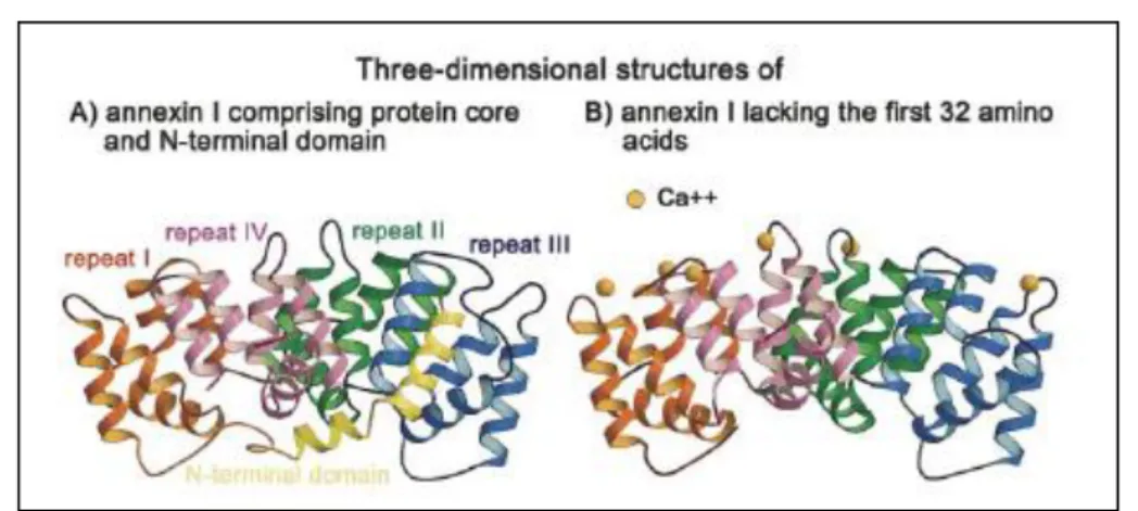
4.2 Annexin A1 structure 49
4.3 ANXA1: an anti-inflammatory protein 51
4.4 ANXA1 post-translational modifications 54
4.5 ANXA1 in cancer 56
4.5.1 ANXA1 in prostate cancer 56
4.5.2 ANXA1 in colon rectal cancer 57
4.5.3 ANXA1 in lung cancer 57
4.5.4 ANXA1 in melanoma 57
4.5.5 ANXA1 in breast cancer 57
4.5.6 ANXA1 in pancreatic cancer 58
4.6 ANXA1 externalization 58
Chapter V. FORMYL PEPTIDE RECEPTORS
5.1 Introduction 60 5.2 FPR mechanism of action 61 5.2.1 FPR1 61 5.2.2 FPR2 63 5.2.3 FPR3 63 5.3 Ligands of FPR family 64 5.3.1 Agonists 64 5.3.2 Antagonists 66 5.4 Regulation of FPRs 67 5.5 FPRs in cancer 68
AIM OF THE WORK
AIM OF THE WORK 69
Chapter VI. MATERIAL AND METHODS
6.1 Cell Cultures 70
6.3 Nuclear extracts 71
6.4 Supernatant analysis 71
6.5 Western blotting analysis 71
6.6 siRNA transfection 72
6.7 Confocal Microscopy 72
6.8 Flow cytometry 73
6.9 PCR 73
6.10 RNA isolation and quantitative RT-PCR assay 73 6.11 Measurement of intracellular Ca2+ signalling 74
6.12 In vitro Wound-Healing Assay 74
6.13 Matrigel Invasion Assay 75
6.14 MTT Assay 75
6.15 Analysis of apoptosis 76
6.16 Cell cycle analysis 76
6.17 Molecular cloning by Gene/CRISPR (clustered, regularly interspaced, short palindromic repeat) -Cas9 (CRISPR associated protein 9) technique
77
6.17.1 Transfection of plasmid DNA and clones selection 80
6.18 Mass spectrometry of protein extracts 80
6.18.1 LC-MS/MS analysis 80
6.18.2 Data analysis 81
6.18.3 Statistical analysis 81
6.18.4 GO analysis 81
6.19 Orthotopic pancreatic cancer xenografts in immunodeficient mice
6.20 H&E tissue staining
82
82
6.21 Statistical analysis 83
Chapter VII. RESULTS
7.1 Expression of ANXA1 in PC cell lines 84
7.2 Localization of ANXA1 in PC cell lines 87
7.3 Effects of ANXA1 knockdown on MIA PaCa-2 and PANC-1 cell migration and invasiveness
88
7.4 90
7.5 Analysis of the secreted forms of ANXA1 92
7.6 Expression of FPRs in MIA PaCa-2 and PANC-1 cells 93 7.7 Activation of FPRs in MIA PaCa-2 and PANC-1 cells 94 7.8 Effects of activation of FPRs on MIA PaCa-2 and PANC-1 cells in migration and invasion assays
7.9 Effects of extracellular ANXA1 on MIA PaCa-2 and PANC-1 cells in migration and invasion assays
98
7.10 Effects of MIA PaCa-2 supernatants on PANC-1 cell migration 99 7.11 Creation of genomic ANXA1 deletions in MIA PaCa-2 cell line
using CRISPR/Cas9 technique
100
7.12 Comparative proteomic analysis of MIA PaCa-2 PGS and ANXA1 KO derived sub-line MIA PaCa-2
102
7.13 Validation of protein identified as differentially expressed in the LC-MS/MS analysis
104
7.14 Effects of ANXA1 knockout on MIA PaCa-2 migration and invasion
107
7.15 MIA PaCa-2 ANXA1 KO answer to the migratory and pro-invasive effects of Ac2-26
108
7.16 Effects of ANXA1 knockout on MIA PaCa-2 proliferation 7.17 ANXA1 is not involved in apoptosis induced by gemcitabine 7.18 KO of ANXA1 decreases the metastatic potential of highly aggressive MIA PaCa-2 cells
110 112 113
DISCUSSION
Discussion
………...……….………
115APPENDIX. COMPUTATIONAL DESIGN OF PROTEIC INHIBITORS OF ANXA1
A.1 Background 122
A.2 Methods and results
A.2.1 Design of new sequences A.2.2 Structure check
A.2.3 Binding energy calculation
125 125 126 127 BIBLIOGRAPHY Bibliography
………...……….………
I-XIV
ACKNOWLEDGEMENTS Acknowledgements LVI- 1 -
The present PhD project belongs to the general theme of scientific investigation relative to the study of biological functions of Annexin A1 both in physiological and in pathological processes. The aim of this work has been to identify and characterize in details the biological mechanisms underlying the protein involvement in tumor progression, with particular attention to pancreatic cancer. Few scientific works have reported information about the correlation of Annexin A1 with pancreatic cancer progression. The study of patients’ biopsies had shown that protein expression was associated to the increase of metastatization degree, a minor cell differentiation and a minor time of survival of patients. To better define the role of Annexin A1 in this model, we analyzed four cell lines of human pancreatic cancer: MIA PaCa-2, PANC-1, BxPC-3 and CAPAN-2. All of them presented very similar levels of Annexin A1 expression but only MIA PaCa-2 and PANC-1 showed a mesenchymal phenotype, as demonstrated by the high levels of vimentin, a typical mesenchymal marker, so only these ones are described as more aggressive cells. For this reason, we continued the investigation of Annexin A1 in MIA PaCa-2 and PANC-1 cells. By immunofluorescence assay we showed that Annexin A1 co-localized with Focal Adhesion Kinases and F-actin, two proteins typically involved in cell migration, so we postulated the hypothesis that Annexin A1 could be involved in cell motility. To identify the functional role of Annexin A1 in these cell lines, a down-modulation of protein expression was performed by transient transfection of specific siRNAs. Through the assays of Wound healing and invasion through a coating of matrigel, we showed that MIA PaCa-2 and PANC-1 with lower Annexin A1 levels migrated and invaded slower than control cells.
Several functions of Annexin A1 are carried out by its extracellular form which interacts with the Formyl Peptide Receptors (FPR) in both autocrine and paracrine manner. The expression of the receptor isoforms FPR-1 and FPR-2 was analyzed by cytofluorimetric assay and PCR. Receptor activation was studied in presence of either agonists such as fMLP and Ac2-26, a mimetic peptide of Annexin A1, or antagonists like Boc-1. To verify if the pathways triggered by the activation of Formyl Peptide Receptors were involved in the processes of cell migration and invasion, we performed the assays of Woung healing and invasion with MIA PaCa-2 and PANC-1 in presence of receptor agonists and antagonists: fMLP and Ac2-26 stimulated migration and invasion in either cell line, while antagonist Boc-1 reverted this effect. Through compartimentalized protein extractions, MIA PaCa-2 cell line, but not PANC-1 cells showed, in addition to the full length form of 37kDa, a shorter form of 33kDa relative to the C-terminal portion, the likely result of a proteolytic cleavage that the protein undergoes when it is phosphorylated.
- 2 -
Moreover only MIA PaCa-2 externalized Annexin A1 in the 37, 33 and 3kDa forms; this last one corresponded to the N-terminal portion which is considered as the sequence with the main biological functions. So we focused our attention on the extracellular form of Annexin A1: following the administration of a specific blocking antibody, MIA PaCa-2 cells lost their capability of migration and invasion. On the other hand, PANC-1 were not affected by the antibody, confirming the absence of the protein in their supernatant. Furthermore, after the addition of the supernatant of MIA PaCa-2, the PANC-1 cell line acquired a greater migration rate, confirming the importance of the protein in the processes of migration and invasion.
To better characterize the role of Annexin A1 in vitro and, above all, in vivo, we generated Annexin A1 knock-out clones of MIA PaCa-2 cells. We chose the technique of Gene-CRISPR/Cas9 with which we created the genomic delection of Annexin A1, compared with wild type cells and cells transfected with PGS, a scrambled vector used as technical control. By the proteomic analysis of the obtained clones, 36 proteins appeared up-regulated and 26 down-modulated in absence of Annexin A1, these proteins could be involved in several cell pathways like cell proliferation and trafficking, metabolism, cytoskeletal organization and others. Based on the previous data, we preferred to better characterize the aspect of the cytoskeletal organization. We confirmed the variation of some proteins that seemed particularly interesting: for example we validated the down-modulation of vimentin and lamin A/C; on the other hand up-regulation of CD44 and cytokeratin 18 was observed. By immunofluorescence analysis, a strong depolimerization of F-actin in MIA PaCa-2 knock-out for Annexin A1 was detected. So we analyzed the processes of migration and invasion showing that MIA PaCa-2 without Annexin A1 migrated and invaded in a significant slower manner compared with MIA PaCa-2 wild type and transfected with PGS. Furthermore, no modifications were observed in the expression of proteins involved in the pathways triggered by Formyl Peptide Receptors. In fact MIA PaCa-2 knock-out for Annexin A1 showed very similar levels of the receptor isoforms 1 and 2. These receptors appeared active since the migration and invasion rate of the MIA PaCa-2 cells knock-out for Annexin A1 increased in the presence of agonist Ac2-26 and decreased with antagonist Boc-1. Moreover, to complete the characterization of clones, we analyzed the cell proliferation, showing that these cells proliferated more rapidly, had higher S/G2 phases and higher levels of proteins as Cyclin A1, phospho-ERK and ALDH7A1.
Finally, MIA PaCa-2 wild type, PGS and Annexin A1 knock-out have been used to create orthotopic xenografts in the pancreas of SCID female
- 3 -
mice. In the absence of Annexin A1, the tumor mass appeared not affected and retained a volume very similar to the tumor generated by MIA PaCa-2 wild type and PGS but the metastatization degree strongly decreased. This phenomenon was analyzed in mice livers which represent the first organ mainly affected by pancreatic cancer metastasis.
- 4 -
Annexin 1 (ANXA1) is a multifunctional protein of 37 kDa, and represents the first characterized member of the annexin superfamily, so called since their main property is to bind (i.e. to annex) cell membranes in Ca2+-dependent manner. ANXA1 is over-expressed in tissues from patients affected by pancreatic carcinoma (PC), where the protein seems to be associated with the malignant transformation and the poor prognosis. In this PhD project, experiments were performed to understand the role of ANXA1 in human PC development with particular attention to migration and invasion processes. We observed in all the analyzed PC cell lines, a huge expression and a localization of ANXA1 mostly on the motility sub-structures. Interestingly, in MIA PaCa-2 cells we found also two cleaved forms of ANXA1 (33 and 3 kDa) that localize at cellular membranes and are secreted outside the cells, as confirmed by MS analysis. MIA PaCa-2 and PANC-1 cell lines express Formyl Peptide Receptors (FPRs) 1 and 2: the treatment of this cells with the ANXA1 mimetic peptide, Ac2-26, induced intracellular calcium release, consistent with nFPR activation, and significantly increased cell migration/invasion rate. ANXA1 effects on MIA PaCa-2 and PANC-1 migration and invasiveness were observed both by down-modulating its expression through siRNAs and by treatment with a blocking antibody. The importance of the secreted form of ANXA1 in cellular motility was confirmed when MIA PaCa-2 were compared with PANC-1 cells that lack both the cleaved and the externalized forms. Moreover, the treatment of PANC-1 cells with MIA PaCa-2 supernatants, significantly increased the migration rate of these cells. To better characterize the functional role of the protein in PC progression, ANXA1 Knock-Out (KO) clones from MIA PaCa-2 cells were obtained. The expression of several proteins was affected by the absence of ANXA1, particularly the cytoskeletal organization was negatively conditioned. In fact, MIA PaCa-2 ANXA1 KO lost their migratory and invasive capabilities, proliferated more rapidly and seemed to acquire a less aggressive phenotype. To confirm this aspect the MIA PaCa-2 wild type, PGS (the scrambled vector) and ANXA1 KO were implanted to create orthotopic xenograft in vivo. The PC mass of ANXA1 KO MIA PaCa-2 was not significantly smaller than the other experimental points, but the metastatization degree appeared particularly reduced as showed on livers of mice with MIA PaCa-2 wild type and PGS which showed a higher degree of metastatic lesions compared to MIA PaCa-2 ANXA1 KO.
This project provides new insights on the role of ANXA1 in PC progression. In in vitro models, the intracellular ANXA1 is involved in the maintenance of the cytoskeleton integrity. When secreted, the protein stimulates PC cells migration and invasion through FPR activation. This is
- 5 -
confirmed by in vivo xenograft experiments where ANXA1 appears to stimulate the metastatization process.
- 6 -
5-FU: 5-FluoruracilαSMA: α Smooth Muscle Actin ABC: ATP-Binding Cassette
Ac2-26: NH2-terminal mimetic peptide Ach: Acetylcholine
ADM: Acinar-Ductal Metaplasia ALDH: Aldehyde Dehydrogenase ALX: lipoxin A4 receptor
AMP: Adenosine Monophosphate ANXA1: Annexin A1
ARF: Alternate Reading Frame ATP: Adenosine Triphosphate BC: Breast Cancer
bHLH: basic Helix-Loop-Helix BMP: Bone Morphogenetic Protein Boc1: t-Boc-Met-Leu-Phe
Boc2: t-Boc-Met-D-Leu-D-Phe bp: base pair
CA: Carbohydrate Antigen CAII: Carbonic Anhydrase II
CAF:Cancer-Associated Fibroblasts Cas9: CRISPR associated protein 9 CCK: Cholecytokinin
CD: Cluster of Differentiation
CFTR: Cystic Fibrosis Transmembrane Receptor Cdc42: Cell division control protein 42
CDK: Cyclin-Dependent Kinase
CDKN2A: Cyclin-Dependent Kinase inhibitor 2A CK: Cytokeratin
COX-2: Cyclooxygenase-2
cPLA2: cytosolic Phospholipase A2 CRC: Colon Rectal Cancer
CRISPR: Clustered, Regularly Interspaced, Short Palindromic Repeat CSC: Cancer Stem Cell
CsH: Cyclosporin H
CT: Computer Tomography
CysLT1: Cysteinyl Leukotriene receptor 1 DAPK: Death Associated Protein Kinase DC: Dendritic Cell
DDR2: Discoidin Domain Receptor tyrosine kinase 2 DPC4: Deleted in Pancreatic Carcinoma 4 gene
- 7 -
DSB: DNA Double-Strand Break DTT: DithiothreitolECM: Extracellular Matrix EGF: Epidermal Growth Factor
EGFR: Epidermal Growth Factor Receptor EMT: Epithelial to Mesenchymal Transition
ErbB2: Epidermal growth factor receptor tyrosine kinase ERK: Extracellular signal-Regulated Protein
FAK: Focal Adhesion Kinase FBS: Fetal Bovine Serum FGF: Fibroblast Growth Factor
fMLP: formylMethionilLeucilPhenylalanine FoxD3: Forkhead box D3
FPR: Formil Peptide Receptor FPRL1: FPR-like 1
FPRL2: FPR-like 2
FSP1: Fibroblast-Specific Protein 1
GEF: Guanine-nucleotide Exchange Factor GLP-1: Glucagon-Like Peptide-1
GO: Gene Ontology
GPCR: G Protein Coupled Receptor GR: Glucocorticoid Receptor
GRE: Glucocorticoid Responsive Element GRK: G protein coupled kinase
GRP: Gastric Releasing Peptide GTP: Guanosine Triphosphate HD: Homozygous Deletion HDR: Homology-Directed Repair HGF: Hepatocyte Growht Factor HIF1α: Hypoxia Induced Factor 1 α HL-60: Human promyelocyitc Leukemia HNF1β: Hepatocyte Nuclear Factor 1 β HNF6: Hepatocyte Nuclear Factor HS: Horse Serum
ID: Inhibitor of DNA binding protein IFN: Interferon
IL: Interleukin
INK4A: Inhibitors of CDK4
iNOS: inducible Nitric Oxide Synthase IP3: Inositol triphosphate
- 8 -
IPMN: Intraductal Papillary Mucinous Neoplasm JNK: Jun N-terminal Kinase
KO: Knock Out
KRAS: Kirsten Rat Sarcoma Oncogene LC-FFA: Long Chain-Free Fatty acid
LC/MS: Liquid Chromatography-Mass Spectrometry LL37: Leucine Leucine 37
LPS: Lipopolysaccharide LXA4: Lipoxin A4
MAPK: Mitogen Activated Protein Kinase MCH: Melanin Concentrating Hormone MCN: Mucinous Cystic Neoplasm
MDM2: Mouse Double Minute 2 homolog MEK: Mitogen-activated ERK Kinase MET: Mesenchymal to Epithelial Transition MMP: Membrane Metallo-Proteases
MRI: Magnetic Resonance Imaging
MT1-MMP: Membrane-Ttethered poteases – Membrane Metallo-Protease MVB: Multivesicular Bodie
nab: albumin nanoparticles
NF-kB: Nuclear Factor kappa-light-chain-enhancer of activated B cells NHEJ: Non-Homologous End Joining
OCN: Ostecalcin
PAM: Protospacer-Adjacent Motif
PanIN: Pancreatic Intraepithelial Neoplasia PBMC: Peripheral Blood leucocytes
PBS: Phosphate Buffer Saline PC: Pancreatic Cancer
PDAC: Pancreatic Ductal Adenocarcinoma PDGF: Platelet Derived Growth Factor Pdx1: Pancreatic and duodenal homeobox 1 PI3K: Phosphoinositide 3-Kinase
PIP2: Phosphatidylinositol 4,5-bisphosphate PKA/C: Protein Kinase A/C
PLCb: Phospholipase C b PRL: Prolactin
PMN: Polymorphonuclear leukocyte RER: Rough Endoplasmic Reticulum Rho: Ras homologue
- 9 -
SBEs: SMAD Binding ElementsSCID: Severe Combined Immunodeficiency SDF: Stroma-Derived Factor
sgRNA: single-guide RNA shRNA: short hairpin RNA siRNA: small interference RNA
SKCO-15: Human colonic ephitelial cells
SPARC: Secreted Protein Acidic and Rich in Cystein SUMO-1:Small Ubiquitin-related Modifier-1
TALEN: Transcription Activator-Like Effector Nuclease TCF: T Cell Factor
TG: Triglyceride
TGF: Trasforming Growth Factor TNF: Tumor Necrosis Factor
uPAR: uroknase Plasminogen Activator Receptor VEFG: Vascular Endothelial Growth Factor
WT: Wild Type
WASP: Wiskott–Aldrich Syndrome Protein Zeb: Zinc finger E-box binding homeobox ZFN: Zinc-Finger Nuclease
ZG: Zymogen Granule ZO-1: Zona Occludens 1
- 10 -
CHAPTER 1
PANCREAS
1.1 Pancreatic anatomy
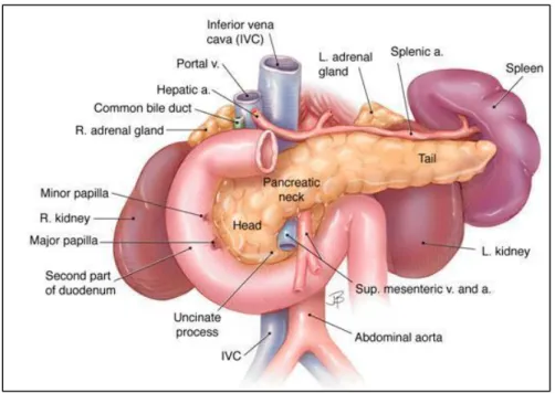
The pancreas is a soft, elongated, flattened gland of 12-20 cm in length and a weight of about 100 grams. The name pancreas derives from the Greek roots ‘pan’ meaning ‘all’ and ‘creas’ meaning ‘flesh’ [1]. It is composed by four parts as head, neck, body and tail in a manner represented in figure 1.1. Furthermore, about its physiological role, it is divided in a exocrine and endocrine portions.
Figure 1.1: Pancreas sections [2]
The tail of the pancreas and the spleen are in the left upper quadrant of the abdomen, instead the head of the pancreas is in the right upper quadrant just to the right of the midline (Fig. 1.2):
The head of the pancreas lies in the loop of the duodenum; The tail of the pancreas lies near the hilum of the spleen;
The body of the pancreas lies posterior to the distal portion of the stomach between the tail and the neck;
The portion of the pancreas that lies anterior to the aorta is thinner than the adjacent portions of the head and body of the pancreas. This
- 11 -
region is sometimes designated as the neck of the pancreas and marks the junction of the head and body;
The close proximity of the neck of the pancreas to major blood vessels posteriorly including the superior mesenteric artery, superior mesenteric-portal vein, inferior vena cava, and aorta limits the option for a wide surgical margin when pancreatectomy is done;
The common bile duct passes through the head of the pancreas to join the main duct of the gland near the duodenum. The portion nearest the liver lies in a groove on the dorsal aspect of the head;
The minor papilla where the accessory pancreatic duct drains into the duodenum and the major papilla (ampulla of Vater) where the main pancreatic duct enters the duodenum are depicted.[3; 4].
Figure 1.2: Anatomic relationships of the pancreas with surrounding organs and structures
The celiac trunk and the superior mesenteric artery both arise from the abdominal aorta and have multiple branches that supply several organs including the pancreas. The anastomosis of their branches around the pancreas provides collateral circulation that generally assures a secure arterial supply to the organ. Most of the arteries are accompanied by veins that drain into the portal and splenic veins as they pass behind the pancreas.
- 12 -
The superior mesenteric vein becomes the portal vein when it joins the splenic vein [3].
1.1.2 The exocrine portion
The exocrine cells are packed with membrane-bound secretory granules which contain digestive enzymes that are exocytosed into the lumen of the acinus. The pancreatic acini are arranged in clusters like grapes at the ends of a branching duct system. Centroacinar cells are typically located at the junction of an acinus or acinar tubule with a small ductule, but they may be interspersed within an acinar tubule.
Figure 1.3: Pancreatic tissue with acinar, centroacinar and ductal cells [5] The acinar cells have several short, slender microvilli about 0.2 µm in length and extend into the lumen of the acinus. The lumen typically contains flocculent electron-dense material, which is the secreted digestive enzymes. Thin filaments form the axis of the microvilli as well as a network beneath the apical plasmalemma. These microfilaments apparently play a structural role because their disruption causes expansion of the acinar lumen and loss of microvilli. In figure 1.3 are shown the acinar cells which are larger than
- 13 -
centroacinar cells and are easily identified because of the darkly stained the zymogen granules (ZG). The basal portion (B), in the opposite site of the luminal one (A), of the canard cells lies next to the interstitial space that contains vessels (V), nerves and connective tissue. Nuclei (N) with nucleoli (n) are in the basal portion of the acinar cells: the nucleus usually is spherical, about 6 µm in diameter, with one or more nucleoli in the interior. Mitochondria (m) appear elongate, cylindrical structures that may appear oval in cross-section and may contain well-developed cristae and many matrix granules and they occur throughout the cytoplasm, among the granular endoplasmic reticulum or zymogen granules and adjacent to the basolateral cell border. The cytoplasmic matrix occupies about 45% of the cell volume. Tight junctions form a belt-like band around the apical end of the cell and are produced by the apposition of the external membrane leaflets of neighboring cells. These junctions prevent the reflux of secreted substances from the duct into the intercellular space. Gap junctions are distributed on the lateral cellular membranes and are formed by the apposition of larger, disk-shaped membrane plaques, they allow communication between cells. Rough endoplasmic reticulum (RER) occupies about 20% of the cell volume and fills most of the basal region of the acinar cells, although small amounts also occur in the apical region adjacent to and among the zymogen granules. This reticulum is composed of numerous parallel cisternal membranes covered with closely spaced attached ribosomes, giving the structures a granular appearance. The Golgi complex (G) is located between the nucleus and the mass of zymogen granules present in the resting gland, it consists of flattened, membranous saccules as well as small vesicles or vacuoles that contain flocculent electron-dense material. The Golgi complex is believed to play an important role in the transport of secretory proteins and the formation of zymogen granules, in fact the precursors of zymogen granules that formed starting from Golgi complex are membrane-bound vesicles slightly larger than zymogen granules and much less numerous, occupying only about 2% of the cytoplasm. Studies of the chemical composition of the zymogen granules, that appear as spherical, membrane-bound vesicles and slightly less than 1 mm in diameter, have shown that they contain about 12 to 15 different digestive enzymes, which make up about 90% of the granule protein. [6; 7].
About the ductal system, the duct of Wirsung is the main pancreatic one from which originates the accessory duct of Santorini, other connections are the interlobular ducts, that drain into the main duct throughout the pancreas and the intralobular ducts (sometimes called intercalated ductules) that link acinar tubules to the interlobular ducts. Enzymes from acinar cells are released into a bicarbonate-rich solution that is secreted by the
- 14 -
centroacinar and ductal cells and flows from the acini and acinar tubules to the intralobular ducts, then into the interlobular ducts and main duct and, finally, into the duodenum at the major or minor papillae. The integrity of the duct system is of key importance in preventing entry of the exocrine enzymes into the interstitial space where they may be activated and cause tissue damage manifest as pancreatitis. The main and interlobular ducts have thick dense collagenous walls. The connective tissue component of the duct wall becomes progressively thinner as the ducts branch and become narrower. Intercellular tight junctions, also called zonula occludens, between duct cells, centroacinar cells and acinar cells play a major role in preventing leakage of the duct system [6].
Ductal cells express markers such as cytokeratin 19 (K19), cystic fibrosis transmembrane receptor (CFTR), carbonic anhydrase II (CAII), DBA lectin and transcriptional factors as HNF1β (Hepatocyte Nuclear Factor 1 β), HNF6 (Hepatocyte Nuclear Factor) and Sox9 [8].
1.1.3 The endocrine portion
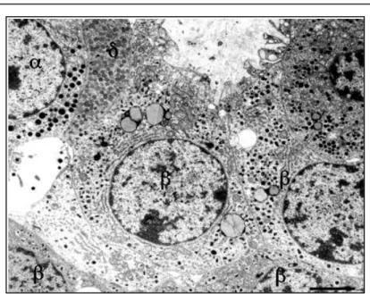
The bulk of the pancreas is composed of pancreatic exocrine cells and their associated ducts, but embedded within this exocrine tissue, there is roughly one million small clusters of cells called the Islets of Langerhans, which are the endocrine cells of the pancreas and secrete insulin, glucagon and several other hormones. Islets vary greatly in size, ~70% are in the size range of 50-250 μm in diameter in humans with an average in the range of 100-150 μm [9]. Smaller islets are dispersed throughout the acinar lobules and most larger islets lie along the main and interlobular ducts of the pancreas. Most islets are spherical or ellipsoid, but they can be irregular in shape, reflecting sometimes the pressure of an adjacent structure, often a duct. In the tail of the pancreas there is a higher population density of islets than in the head and body [10; 11; 12; 13]. In adult humans the number of islets is calculated to be 500000-1 million whereas, they comprise 1-2% of the pancreas in adults of most mammalian species. In addition to the islets, isolated islet cells may be found dispersed in the acinar lobules or in association with ducts [14; 15]. The differences among the islets are detectable through electron microscopy (Fig.1.4).
- 15 -
Figure 1.4: The α-, β-, and δ-cells are labeled. At the ultrastructural level, the cell types are distinguished primarily by differences in their granules. The α-cell granules
are typically slightly larger than β-cell granules. δ-cell granules are typically less densely stained than the granules in α- and β-cells. Scale bar = 4 μm [5]
Each islet is surrounded and penetrated by a rich network of capillaries lined by a fenestrated endothelium, these capillaries are arranged in a portal system that conveys blood from the islets to acinar cells. This insula-acinar portal system consists of afferent arterioles that enter the islet, form a capillary glomerulus and leave the islet as efferent capillaries passing into the exocrine tissue. A parallel arterial system supplies blood directly to the exocrine pancreas and permits the local action of islet hormones on the exocrine pancreas. Acinar cells surrounding islets of Langerhans, termed peri-insular acini, are morphologically and biochemically different from acini situated farther away (tele-insular acini): they appear as have larger cells, with different ratios of specific digestive enzymes [16]. β cells, the most numerous (50% to 80%), secrete insulin, α cells (5% to 20%) secrete glucagon, PP cells (10% to 35%) secrete pancreatic polypeptide, δ cells (5%) secrete somatostatin. Other rare cell types, like ε ones, occur in the islets. In humans, the islets are subdivided into units, each of which exhibits a central aggregation of β cells surrounded by varying numbers of peripherally located cells that secrete the other hormones (Fig.1.5).
- 16 -
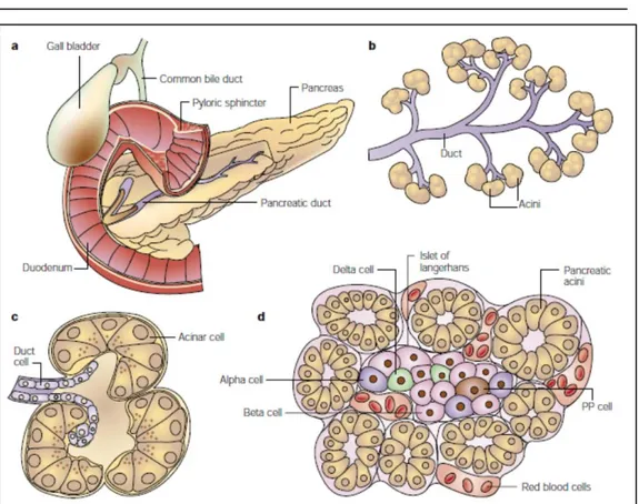
Figure 1.5: a. Gross anatomy of the pancreas; b. The exocrine pancreas; c. a single acinus; d. A pancreatic islet embedded in exocrine tissue [17].
1.2 Pancreatic development
The pancreas appears as a complex organ comprised of three critical cell lineages: islet, acinar and ductal. As is the case for other endodermal organs, the development of the pancreas is thought to result from interactions between the epithelium and its associated mesenchyme. The pancreas is first discernible as the dorsal bud that emerges from the proximal duodenum at four weeks of gestation. Particularly, the pancreatobiliary system appears at gestation week 5 in the human; then the fusion of the dorsal and ventral anlagen occurs during week 7. Full development of acinar tissue extends into the postnatal period. In mice, pancreatic development begins at embryonic day 8.5 (e8.5) and is largely complete by day e14.5 [18; 19]. Its embryological origin has two buds developing on the dorsal and the ventral side of the duodenum. Pancreatic development is a tightly regulated process, with the endocrine and exocrine compartments emerging from a common progenitor population; this process involves the interplay of Hedgehog and Notch
- 17 -
signaling and other cues from the mesenchyme. Notably, Pdx1 is required for the specification of pancreatic lineages [20].
The pancreas develops from two outgrowths of the distal part to the stomach. The ventral diverticulum gives rise to the common bile duct, gallbladder, liver and the ventral pancreatic portion that becomes a part of the head of the pancreas with its duct system including the uncinate portion of this organ. The dorsal pancreatic anlagen gives rise to a portion of the head, the body and tail of the pancreas including a major duct that is continuous through the three regions. Fusion of the duct systems results in the formation of the main pancreatic duct from the ducts of both dorsal and ventral anlagen. The caudal portion of the head of the pancreas (uncinate) and the major papilla (ampulla of Vater) are derived from the ventral anlagen. It becomes apparent that the duct of Santorini is derived from the dorsal part, whereas the duct of Wirsung is derived from the fusion of duct systems of both dorsal and ventral anlagen and drains into the duodenum at the ampulla of Vater. Furthermore, “common channel” refers to the fused portion of the bile and pancreatic ducts proximal to entry into the duodenum. The common channel has received much attention because stones in the biliary tract (gallstones) may lodge in the common channel causing obstruction of both pancreatic and biliary duct systems which is frequently the cause of acute pancreatitis [21].
Of intense interest in the study of ductal cells has been their potential capacity to give rise to islet cells, following the model reported in figure 1.6. If possible, this would be another vehicle to generate islet cells for transplantation as well as a potential treatment of diabetes mellitus, furthermore One study concluded that β cell progenitors can be activated in the injured adult mouse pancreas and are located in the ductal lining [22; 23]. Generally, metaplasia is the word used to define the conversion or replacement of one differentiated cell type with another in the context of a given tissue. In some tissues, metaplasia is associated with an increased risk of cancer.
- 18 -
Figure 1.6: Mature duct cells regress to a less differentiated phenotype and then act as pancreatic progenitors to form new acini, islets and ducts [24].
1.3 Pancreatic functions
1
.3.1 The exocrine portionThe pancreas can be considered as two glands that are intimately mixed together into one organ. The exocrine portion of the pancreas plays a major role in the digestion of food. The stomach slowly releases partially digested food into the duodenum as a thick and acidic liquid called chyme. The acini, the major functional units of the pancreas, secrete pancreatic juice to complete the digestion of chyme in the duodenum. They respond to several intracellular messengers as acetylcholine (Ach), cholecytokinin (CCK), bombesin (GRP –gastric releasing peptide-) and substance P. Pancreatic juice is a mixture of water, salts, bicarbonate and many different digestive enzymes. The pancreatic enzymes are specialize in digesting specific compounds found in chime and it is possible to recognize:
Pancreatic amylase: it breaks large polysaccharides like starches and glycogen into smaller sugars such as maltose, maltotriose and glucose. Maltase, secreted by the small intestine, then breaks maltose into the monosaccharide glucose, which the intestines can directly absorb.
Trypsin, chymotrypsin (endopeptidases) and carboxypeptidase (esopeptidase): they are protein-digesting enzymes that break proteins down into their aminoacid subunits. These aminoacids can then be absorbed by the intestines.
Pancreatic lipase: it is a lipid-digesting enzyme that breaks large triglyceride molecules into fatty acids and monoglycerides. Bile released by the gallbladder emulsifies fats to increase the surface
- 19 -
area of triglycerides that pancreatic lipase can react with. The fatty acids and monoglycerides produced by pancreatic lipase can be absorbed by the intestines.
Ribonuclease and deoxyribonuclease: they digest nucleic acids. Ribonuclease breaks down molecules of RNA into the sugar ribose and the nitrogenous bases adenine, cytosine, guanine and uracil. Deoxyribonuclease digests DNA molecules into the sugar deoxyribose and the nitrogenous bases adenine, cytosine, guanine, and thymine.
The mechanism by which proteins are exported is well characterized: they are first synthesized on polysomes, later they cross the RER (thanks to the SRP –signal-peptide recognition particle– a cytosolic protein which facilitates the binding of mRNA-ribosomal complex to RER) and the Golgi apparatus. Starting from this one, the secretory granules then move by an undefined mechanism to the apical portion of the acinar cells, fuse with it and discharge their contents into the luminal space by an exocytosis process.
If these proteins are secreted inside the pancreatic parenchyma, the consequences can be potentially disastrous with a strong autodigestion. For this reason the enzymes are produced like proenzymes and are packaged in the zymogen granules. They remain inactive until their reach to the duodenal lumen. For example, once in the duodenum, trypsinogen, the major proteolytic enzyme is converted to active trypsin by an enzyme called enterokinase, a brush border enzyme expressed in the duodenal mucosa. The same active trypsin is essential for the activation of other proteolytic and lipolytic pancreatic enzymes. Finally, acinar cells product also the trypsin inhibitor, which is packaged in zymogen granules together with trypsinogen and activates small amounts of trypsin that may form inside the cells or the body of pancreas [25; 26].
The basal volume of pancreatic secretion is estimated to be 0.2/0.3 ml/min, although, when stimulated, pancreatic secretion clan reach 4.0/4.5 ml/min; compressively, the daily output of pancreatic juice is approximatively of 2.5 L [27].
Duct cells secrete a bicarbonate-rich fluid at a considerable variable flow rate of 0.4 ml/min depending of the state of pancreas stimulation. The purpose of alkaline secretion is to neutralize gastric acid that enters the duodenum, an essential process for achieving optimal conditions for pancreatic enzyme activity. In fact, inadequate bicarbonate secretion with failure to reach a neutral pH, as occurs in chronic pancreatitis, contributes to severe maldigestions. As it is elucidated in figure 1.7, carbonic anhydrase catalyzes the production of HCO3- and H+ from carbonic acid. HCO3- is then
- 20 -
transported across the luminal plasma membrane by a HCO3-/Cl- exchanger. The major source of luminal Cl- is now believed to be from the concomitant secretion of the anion via a luminal membrane Cl- channel. This channel is regulated by cAMP-dependent protein kinase or CFTR protein, which is defective just in cystic fibrosis. The recycling of Cl- is, therefore, a major factor in determining HCO3- secretion: the inhibition of Cl- channel activity will decrease HCO3- secretion. This may explain why pancreatic insufficiency develops in some cystic fibrosis patients, as it results from defective ductal secretion. In this condition, proteinaceous acinar secretions become concentrated and their precipitation can cause blockage and destruction of pancreatic ducts. Proton generated during the production of HCO3- must be rapidly transported out of the cells or cell pH would drop precipitously. This occurs at the basolateral membrane through two different mechanisms. One involves Na+/K+ ATPase (proton pump), different from the one found in parietal cells of the stomach, in the basolateral membrane and may provide an alternative and perhaps primary mechanism for rapid proton extrusion. Na+/K+ ATPase is also present in the basolateral membrane, it is necessary for producing favorable electrochemical gradients for Cl- secretion. Na+, some K+ and water accompany HCO3- secretion, mostly entering the duct lumen by passive paracellular diffusion, their rate of transport is determined by prevailing electrochemical and osmotic forces [28].
Figure 1.7: Mechanism of active bicarbonate secretion by pancreatic ductal cells [27].
Therefore, ductal cells of the pancreas have recently been under scrutiny as they may be pancreatic stem cells. When the pancreas is
- 21 -
damaged by duct ligation, cellophane wrapping, pancreatectomy, genetically targeted destruction by IFNγ or when cells are specifically destroyed by streptozotocin, there is some increase in the mitotic activity in the ducts and limited regeneration of the organ. This led to the hypothesis that, during regeneration, duct cells act as progenitors for the generation of new pancreatic cells, however, only cell tracing experiments would make the link between the cells observed in the ducts and new islets [28].
1.3.2 The endocrine portion
The endocrine portion of the pancreas controls the homeostasis of glucose in the bloodstream. In general, the islet is composed of 5 cell types: α, β, δ, ε, and PP that produce glucagon, insulin, somatostatin, ghrelin and pancreatic polypeptide, respectively. Of these hormones, insulin is the primary hormone whose actions on a variety of cell types shifts, on balance, the metabolic flux of nutrients (primarily glucose) toward storage forms of energy (glycogen, protein and fat) and is therefore considered an anabolic hormone. By contrast, glucagon, acting typically in an antagonistic fashion to insulin, functions as a catabolic hormone, causing breakdown of glycogen, protein and fat. The other hormones of the islet appear to have either a secondary or uncertain physiologic role in metabolism: somatostatin functions in the inhibition of insulin and glucagon secretion, whereas the significance of pancreatic polypeptide and ghrelin are unclear [29; 30].
Nutrients in the form of glucose, aminoacids and long chain-free fatty acids (LC-FFAs) are absorbed from the gastrointestinal tract into the portal circulation, where they are detected by β cells via an integrated biochemical-based sensing mechanism (in the case of aminoacids, the β cell primarily responds to valine and arginine). This sensing mechanism is tightly coupled to the production and release of insulin into the blood stream.
Two phases of insulin release are observed: an acute or first phase and a more chronic or second one. First, insulin release is reflective of membrane-docked of insulin granules that are engaged immediately upon stimulus coupling. In the second phase, insulin release represents pre-formed or newly-formed granules recruited to the membrane after the immediate stimulus response. Studies have shown that a positive autocrine/paracrine response of insulin (via its receptor signaling) is important in the maintenance of insulin synthesis in the β cell; the secretion mechanism is described in figure 1.8.
- 22 -
Figure 1.8: The insulin secretion mechanism in detail [31].
Although the insulin receptor is present in a host of key metabolically active organs (for example, liver, muscle, fat and brain) and cells (α cells and β cells of the pancreatic islet). Its actions in each of these organs and cells differ: in the liver, insulin promotes glycolysis, inhibits gluconeogenesis, promotes synthesis of glycogen (glycogenesis) and inhibits the breakdown of glycogen (glycogenolysis); in adipose tissue, insulin promotes glucose uptake and glycolysis, the synthesis and storage of triglycerides (TGs) and the inhibition of lipid breakdown; in skeletal muscle, insulin promotes glucose uptake and glycolysis, the synthesis and storage protein and the inhibition of protein breakdown; in bone, finally, insulin acts primarily on osteoblasts to promote osteoclast activity and enhance production and release of osteocalcin (OCN), a hormone that supports insulin release by the β cell. Insulin actions in the brain are now better appreciated and include the regulation of female fertility, appetite, and overall glucose homeostasis. Within the β cell, paracrine/autocrine effects of insulin sustain β cell growth, survival and function. Taken together, the actions of insulin in these tissues support anabolic pathways that lead to the generation of ATP and the conversion of ingested nutrients into the major storage forms of energy (glycogen, protein and fat) [30].
In contrast, α cells of the pancreatic islets secrete the hormone glucagon, which serves to counterbalance the actions of insulin. Glucagon promotes glycogenolysis and inhibits glycogenesis in liver and skeletal muscle, enhances lipolysis and inhibits triglyceride synthesis in adipose
- 23 -
tissue. α cells also express cell surface insulin receptor and respond locally to secreted insulin by suppressing glucagon release.
The liver is thought to secrete an yet unidentified factor(s) that appears to feed back to support β cell mass. The gut secretes incretin hormones (glucagon-like peptide-1 or GLP-1 and glucose-dependent insulinotropic peptide or GIP), which support glucose-dependent insulin secretion and β cell replication. Both GLP-1 and GIP are proteolytic products of the larger pro-glucagon peptide, which is also produced by α cells of the islet. However, it is unclear whether α cell-derived incretins are a major contributor to β cell function/replication, although at least one study using human tissue has suggested that it may contribute.
In addition, a healthy gut microbiota profile is thought to be essential to maintain leanness and normal β cell function. Furthermore, the cells of the central nervous system are known to secrete multiple peptides (melanin concentrating hormone –MCH-, serotonin and prolactin –PRL- and others) that have been shown both in vitro and in vivo to support β cell function and proliferation. Likewise, bone-derived OCN has been shown to support β cell replication and insulin secretion [32; 33].
- 24 -
CHAPTER 2
PANCREATIC CARCINOMA
2.1 Introduction: epidemiology, etiology and symptoms
Pancreatic cancer (PC) is the fourth leading cause of cancer death in the West World countries. It accounts for 277.000 new cases diagnosed each year in the world, among which approximately 49.000 occur in the USA and Europe [34]. With a 5-year survival rate of only 3% and a median survival of less than 6 months, a diagnosis of PC represents now the true problem for this tumor. Due to a lack of specific symptoms and limitations in diagnostic methods, the disease often eludes detection during its formative stages. Whipple and colleagues reported the first pancreaticoduodenectomy in 1935 and surgery since offers the only possibility of cure, although surgical intervention alone rarely achieves a curative end point but, for the 15/20% of patients who undergo potentially curative resection, the 5-year survival is only 20% [35; 36].
The etiology of PC remains poorly defined, although important clues of disease pathogenesis have emerged from epidemiological and genetic studies. PC is generally associated with advancing age: rare before the age of 40, it gradually culminates in a 40-fold increased risk by the age of 80. The incidence of PC is declining slowly in white men, but it is increasing in other groups, possibly because of changes in smoking patterns. Women account for 57% of new cases. Smoking, diabetes and obesity increase risk, instead a link between alcohol or coffee consumption and PC has not been verified [37; 38; 39; 40]. Physical activity, high fruit and vegetable intake and, possibly, nonsteroidal anti-inflammatory drugs reduce the risk [41]. On the genetic level, numerous studies have documented an increased risk (approximately threefold) in relatives of PC patients, it is estimated that 10% of PCs are due to an inherited predisposition, even if it has a lower penetrance unlike familial cancer syndromes for breast, colon and melanoma [42; 43; 44; 45].
PC often develops without clear early signs or symptoms and the eventual manifestations depends on the tumor location within the gland. Up to 50% of patients presents jaundice, which is more common with patients whose cancers are located in the head of the pancreas where tumors can cause obstruction of the adjacent biliary system [46]. Other common manifestations are vague abdominal discomfort, nausea and weight loss. Large tumors that advance beyond the pancreas can also cause duodenal
- 25 -
obstruction or gastrointestinal bleeding. Steatorrhea can also result from obstruction of the pancreatic duct, whereas hyperglycemia and diabetes have been associated with early manifestation of disease. Patients with advanced disease can also present abdominal and back pain, anorexia, dyspepsia, gallbladder enlargement, migratory thrombosis (Trousseaux syndrome), subcutaneous fat necrosis (panniculitis), hyperglycemia, ascites and depression [45; 47; 48].
Based on the information about the physiologic development of pancreas, it has been found in some tissues that metaplasia can be associated with the increased risk of cancer. Pancreatic acinar cells have the capacity to undergo metaplasia to a ductal cell phenotype in the setting of acute or chronic inflammation, representing an important link to pancreatic ductal adenocarcinoma (PDAC). Acinar-ductal metaplasia (ADM) might represent reprogramming of a progenitor population, direct transdifferentiation of acinar cells to ductal cells, or transdifferentiation via an intermediate cell type (potentially a progenitor cell) [49]. Metaplastic acinar structures are highly proliferative, express Notch target genes, and exhibit mosaic expression patterns for EGFR, ErbB2, and pErk, reminiscent of the PDAC precursors [50; 51; 52]. Spontaneous ADM has been described in vitro, accompanied by the induction of Pdx1 expression during culture of acinar cells [53]. Another relevant transcription factor is Mist1. Mist1 functions as a homodimer, and its loss results in ADM in vitro, with accompanying induction of cytokeratins K19 and K20. Transgenic mice expressing a dominant-negative Mist1 undergo ADM in vivo [54]. Collectively, these studies suggest that loss of Mist1 initiates metaplasia and that Pdx1 expression fosters ADM.
2.2 The genomic landscape of PC
2.2.1 KRAS (Kirsten Rat Sarcoma Oncogene)
The better characterized forms of PC almost universally carries one or more of four genetic defects. Particularly, ninety percent of tumors have activating mutations in the KRAS oncogene. KRAS encodes a small guanine nucleotide transferase, GTPase, that in its active GTP-bound form promotes a wide range of cellular responses including proliferation, survival, migration and metabolism through several effector pathways including the Raf/MEK/ERK (MAPK) and PI3K/AKT kinase cascades (Fig. 2.1) [55]. Transcription of the mutant KRAS gene produces an abnormal Ras protein that is ‘locked’ in its activated form, resulting in the aberrant activation of
- 26 -
proliferative and survival signaling pathways. Furthermore, there is evidences for an important contribution of autocrine epidermal growth factor (EGF)-family signalling. This autocrine loop and resulting stimulation of the phosphatidylinositol 3-kinase (PI3K) pathway is required for transformation of several cell lineages by RAS-family oncogenes. Consistent with the existence of such an autocrine loop, pancreatic ductal adenocarcinomas (PDAC), the more frequent kind of PC, overexpress EGF-family ligands (such as transforming growth factor-α -TGF-α- and EGF) and receptors (EGFR, ERBB2, also known as HER2/neu, and ERBB3) [56; 57; 58]. The main mutation of this gene is KRAS G12D and in several cases it is used also in in vivo models, for example engineered mice in which KRAS is activated develop spontaneous PanINs and later PDAC, above all if they are subjected to pancreatic constant inflammatory insults (patients suffering from chronic pancreatitis have a 16-fold increased risk of developing PC) [59; 60; 61; 62]. Even in vitro fibroblast expressing KRASG12D exhibit elevated Ras-GTP levels with the association of enhanced proliferative properties and escape from premature senescence [63]. The activated mutation of KrasG12D could be one of the earliest genetic abnormalities of pancreatic neoplasia and is sufficient to initiate the transformation of pancreatic ductal cells to PanINs, but, to guarantee tumor development in vivo, it is usual to induce chronic pancreatitis [64]. Several reports show that the most efficient method to induce pancreatitis is the use of caerulein, a CCK analog that binds and activates the CCK receptor. There are two distinct CCK receptor subtypes, namely CCK1 (previously named CCKA) and CCK2 (previously named CCKB) receptors. CCK receptors are G-protein-coupled receptors initiating transient Ca2+ oscillations by activating phospholipase C and induction of inositol triphosphate (IP3)-dependent Ca2+ release from endoplasmic reticulum in pancreatic acinar cells. CCK1R mediates for example the secretion of pancreatic digestive enzymes and may also be involved in the regulation of satiety and feeding behavior, while CCK2R stimulate gastric acid production. CCK1R has a role in the exocrine effects of cerulein such as amylase secretion. However, CCK2R is now recognized to mediate the mitogenic and anti-apoptotic effects of gastrin on gastrointestinal and pancreatic cells. In a pancreatic tumor cell line expressing the endogenous CCK2R, the proliferative effects of the CCK2R have been shown to be induced by the activation of the Jak2/Stat3 pathway by this receptor [65]. Caerulein-induced acute pancreatitis is a well-studied animal model in which this substance causes an edematous pancreatitis. Initiation of acute inflammation is mediated through premature intracellular activation of zymogens in the acinar cells, leading to acinar death and an inflammatory response associated with
- 27 -
mild pancreatic edema [66; 67]. Interestingly, oncogenic K-Ras activation is rarely observed in human endocrine tumors and this may in part explain the concept that tumor mainly arises from the acinar cell compartment of the exocrine pancreas [68].
Figure 2.1: The Ras activation cascade [69]. 2.2.2 CDKN2A (Cyclin-Dependent Kinase inhibitor 2A)
Activation of KRAS alone in the pancreatic epithelium led to the development of PanINs, highlighting the need for additional cooperating events to promote tumor progression to the malignant PDAC stage. In a number of studies, additional genetic alterations have been combined with activation of KRAS to identify cooperating events context of KRAS-dependent pancreatic tumorigenesis [70]. The inactivation of the CDKN2A gene is the resultant loss of the p16 protein, a regulator of the G1-S transition of the cell cycle, and a corresponding increase in cell proliferation. The inheritance of
- 28 -
mutant CDKN2A alleles confers a 13-fold increased risk of pancreatic cancer. Loss of CDKN2A function is brought by mutation, deletion or promoter hypermethylation and occurs in 80/95% of sporadic pancreatic adenocarcinomas. CDKN2A loss is generally seen in moderately advanced lesions that show features of dysplasia. The locus 9q21 encodes two tumour suppressors: INK4A (INhibitors of CDK4) and ARF (the Alternate Reading Frame protein product of the CDKN2A locus), via distinct first exons and alternative reading frames [71; 72; 73]. INK4A inhibits CDK4/CDK6-mediated phosphorylation of RB (retinoblastoma), thereby blocking entry into the S phase of the cell cycle; ARF stabilizes p53 by inhibiting its MDM2 (Mouse double minute 2 homolog)-dependent proteolysis. INK4A seems to be the more important PC suppressor, as germline and sporadic mutations have been identified that target this protein. When primary cells are placed into culture, INK4A-transcript expression is induced and this can be considered a stress response to the inappropriate growth environment that is associated with in vitro culture [74; 75]. This induction by environmental stress and aberrant proliferative signals provides a plausible basis for the tumour-suppression function of INK4A, although the relationship of this phenomenon to cancer suppression in vivo is not established. Other studies have implicated INK4A in the cellular response to DNA damage in vivo, so the absence of INK4A might also contribute to the chemoresistance of pancreatic adenocarcinoma [76]. Notably, while KRAS mutations are often detected in non-neoplastic states, such as chronic pancreatitis and, possibly, in normal pancreas, loss of INK4A usually occurs only in later stages of pancreatic neoplasia [77; 78; 79]. Although loss of INK4A probably facilitates the oncogenicity of activated RAS alleles, as shown in animal models, its occurrence later in pancreatic tumour progression indicates that the intersection of these pathways might require other events, such as disrupted contacts with the extracellular matrix or elevations in the level of activated KRAS [80].
2.2.3 TP53
The TP53 tumor-suppressor gene is mutated, generally by missense alterations of the DNA binding domain, in 50%–75% of tumors. In general, TP53 functions as a heterotetrameric complex that transactivates key target genes in response to a variety of cellular insults, resulting in cell cycle arrest or apoptosis. TP53 mutations arise in later-stage PanINs that have acquired significant features of dysplasia, reflecting the function of TP53 in preventing
- 29 -
malignant progression, permitting, in this way, cells to bypass DNA damage control checkpoints and apoptotic signals and contributing to genomic instability [81]. In contrast to many other cancer types, in PC there does not seem to be a reciprocal relationship in the loss of CDKN2A and TP53. The initiation of pancreatic tumorigenesis by endogenous KRASG12D expression in the context of Trp53R172H greatly hastens the development of locally invasive and widely metastatic PDAC that faithfully recapitulates all of the extant features of the human disease [82]. Cytogenetic studies have provided evidence that telomere dynamics might contribute to this genomic instability [83]. Although reactivation of telomerase is crucial to the emergence of immortal cancer cells, a preceding and transient period of telomere shortening and dysfunction might also contribute to carcinogenesis by leading to the formation of chromosomal rearrangements through breakage–fusion–bridge cycles. The survival of cells with critically short telomeres (crisis), which continue to go through breakage–fusion–bridge events, is enhanced by inactivation of the p53-dependent DNA-damage response, allowing the acquisition of oncogenic chromosomal alterations. Studies in the telomerase-knockout mouse support this model, as telomere dysfunction and p53 loss cooperate to promote the development of carcinomas in multiple tissues. An analysis of a large series of human pancreatic cancer cell lines revealed that telomeres were frequently lost from chromosome ends and that anaphase bridging occurred, indicating that persistent genomic instability is associated with critically short telomeres. Telomere dysfunction was an early step in the pathogenic process. Moreover, studies of PDAC revealed that tumors have shortened telomere length and that the activation of telomerase is a late event [84; 85; 86; 87].
2.2.4 SMAD4
The deleted in pancreatic carcinoma 4 gene (DPC4, also known as SMAD4/MADH4 - mothers against decapentaplegic homolog 4 -) is lost in about 50% of pancreatic cancers, resulting in aberrant signaling by the transforming growth factor-β (TGFβ) cell surface receptor. This gene maps to chromosome 18q21 [88]. The pathogenic role of SMAD4 inactivation is strongly supported by the identification of inactivating intragenic lesions of SMAD4 in a subset of tumors. SMAD4 seems to be a progression allele for pancreatic adenocarcinoma, as its loss occurs only in later-stage PanINs and it has become a predictor of decreased survival in pancreatic adenocarcinoma [89; 90; 91]. In a study using human pancreatic cancer
- 30 -
samples from primary and metastatic lesion, mutations in the DPC4 gene have been associated with higher metastatic potential [92]. The mechanism by which SMAD4 loss contributes to tumorigenesis is likely to involve its role in TFG-β mediated growth inhibition. TGF-β can behave as both a tumor suppressor and a tumor promoter. Its tumor suppressor function can be explained largely by its ability to inhibit proliferation of normal epithelial and lymphoid cells by either blocking the G1–S cell cycle transition (from which most human cancers originate) and to induce apoptosis [93]. However, late-stage human carcinomas often become resistant to TGF-β growth inhibition and, in addition, secrete elevated levels of this growth factor [94]. But, by now, the roles of TGF-β signaling in pancreatic adenocarcinoma pathogenesis are not well defined: its role can be well illustrated as a tumor suppressor pathway by the presence of chromosomal deletions and mutations in DPC4 in 55% of pancreatic tumors, a tumor suppressor that has been implicated in mediating the growth inhibitory and antiangiogenic effects of TGF-β [88; 95; 96]. This cytokine shows inconsistent effects on cultured cell lines with respect to cell proliferation rates and dependency on SMAD4 status for TGF-β responsiveness. Furthermore SMAD4 loss is also likely to contribute to tumor progression through effects on the interaction of tumor with stroma [97; 98; 99; 100]. Particularly, heterotypic microenvironmental cellular interactions seem to be important in the pathogenesis of pancreatic adenocarcinoma. Notably, these tumors show a marked proliferation of stromal fibroblasts and deposition of extracellular matrix components such as matrix metalloproteinases and collagens (desmoplasia) [101].The role of this process in cancer pathogenesis remains uncertain, as it is not well established whether the response is part of the tumorigenic programme or whether it represents a form of host defence against the tumor. Recent evidence indicates that the carcinoma cells direct the desmoplastic response and that TGF-β contributes to this process [102]. There are suggestions that SMAD4 loss might be permissive for these effects, notably, Smad4-deficient tumors show increased growth and invasiveness in this model. Another role for SMAD4 in regulating heterotypic interactions is indicated by experiments in which Smad4 is reintroduced into some pancreatic adenocarcinoma cell lines. In these experiments, Smad4 blocks tumorigenic growth in immunodeficient mice by inhibiting angiogenesis, but does not affect cell sensitivity to TGF-β [96]. These concepts are consistent with recent studies showing that cancers ‘programme’ an oncogenic stroma that, in turn, contributes to tumor growth through paracrine signaling, angiogenesis and protection from immune attack [103; 104].
- 31 -
Generally, transgenic mouse models which are commonly used are reported in the table 2.1:
Table 2.1: Mouse models of pancreatic carcinoma [105] 2.3 Staging of PC
In 1905, S.P.L. Hulst described for the first time small microscopic lesions in the pancreas which are now described as “pancreatic intraepithelial neoplasia (PanIN)”. PanIN lesions are noninvasive epithelial proliferation within the smaller pancreatic ducts and they are graded histologically as PanIN-1 (low-grade), PanIN-2 (intermediate-grade) or PanIN-3 (high-grade) characterized by columnar to cuboidal cells with varying amunts of mucins and based on the degree of architectural and cellular atypia present in the lesion (Fig. 2.2) [106].
- 32 -
Figure 2.2: A normal pancreatic duct (A) and multiple PanIN lesions (B-F). PanIN-1 (B), PanIN with associated lobulocentric atrophy (B and C), PanIN-2 (E), and PanIN-3
(F), all hematoxylin and eosin images [107].
Generally PanIN lesions are not detected macroscopically and are clinically silent. PanIN-1 is characterized by two early stages: PanIN-1A and PanIN-1B which show minimal cytological and architectural atypia. PanIN-2 lesions show mild to moderate cytological atypia with frequent papillary formation and also nuclear abnormalities for example enlargement, some loss of polarity, crowding. PanIN-3 is the most severe lesion with abnormal cell mitosis and budding into lumen [108]. Paralleling this histologic progression is a genetic progression. PanIN-1 and PanIN-2 often harbor genetic alterations in the KRAS and p16/CDKN2A genes, whereas PanIN-3 lesions and invasive adenocarcinomas, in addition to genetic alterations in KRAS and p16/CDKN2A, also often harbor mutations in TP53 and SMAD4 (Fig. 2.3) [109; 110]. High-grade PanINs, however, are rarely found, unless there is an associated invasive pancreatic cancer or the patient which has a strong family history of PC; these observations support the hypothesis that PanIN lesions are precursors to invasive adenocarcinoma [111; 112; 113].
- 33 -
Figure 2.3: Genetic progression model of pancreatic adenocarcinoma [17] The second major precursor lesion to be identified in the pancreas was the intraductal papillary mucinous neoplasm (IPMN). IPMNs arise in the larger pancreatic ducts and, as the name suggests, they are typically papillary and often produce copious amounts of mucins. IPMNs are, by definition, larger than PanINs (>1.0 cm, with respect to <0.5 cm for PanINs) [3]. As is observed with PanINs, low-grade IPMNs often harbor KRAS and p16/CDKN2A gene mutations, high-grade IPMNs harbor further mutations in TP53 and SMAD4. When an adenocarcinoma arises in association with an IPMN, the IPMN and the invasive carcinoma almost always harbor the samegenetic alterations, supporting the hypothesis that IPMNs are a precursor to invasive adenocarcinomas [114]. Far less common than IPMNs, are the Mucinous cystic neoplasms (MCNs), they are large mucin-producing precancerous lesions of the pancreas that almost always arise in the body of tail of the gland and commonly arise in women. In contrast to IPMNs, MCNs do not significantly involve the pancreatic duct system. However, like IPMNs, MCNs can progress to adenocarcinoma. The KRAS, p16/CDKN2A, RNF43, TP53 and SMAD4 genes have all been reported to be mutated in MCNs [3; 114]. Finally, it can be mentioned some small cancer lesions, rarely encountered outside of screening trials. There have been several reports of long-term survival of patients with surgically resected small, lymph node negative, pancreatic cancers [115].
- 34 -
2.4 Biomarkers for detection of PCOne of the main difficulties about PC detection is just the lack of reliable screening tests, either molecular or imaging based. Commonly used imaging studies as abdominal CT (computer tomography) or MRI (Magnetic Resonance Imaging) are still inadequate for diagnosing PC at an early stage since they do not reliably detect tumors smaller than 1-2 cm [116]. The mucin-associated carbohydrate antigen CA 19–9 is a biomarker of PDAC with limited clinical utility in the screening setting. CA 19–9 has demonstrated modest effectiveness in the screening of symptomatic individuals with a range of 70–90% of cases. But the principal limitations of CA 19–9 include its frequent elevation associated with nonmalignant conditions such as pancreatitis and obstructive jaundice and its inability to detect many early stage malignancies [117; 118]. These limitations of CA 19–9 have led investigators to search for alternative biomarkers for use in screening for PDAC. For example, a panel of 7 proteins (ALCAM, ICAM-1, LCN2, TIMP-1, REG1A, REG3 and IGFBP-4) with or without the addition of CA 19–9, selected based on findings in a mouse model, was able to discriminate human PC cases from matched controls in a small group of presymptomatic and prediagnostic blood samples [119]. Biomarker profiles indicative of a specific cancer include not only those factors produced by the tumor itself but also represent the systemic response to the growing tumor including acute phase reactants, inflammatory cytokines, growth and angiogenic factors, etc. An accurate panel of circulating levels of significant biomarkers in patients diagnosed with PC, benign pancreatic disease and healthy control individuals is reported in table 2.2:
![Figure 1.6: Mature duct cells regress to a less differentiated phenotype and then act as pancreatic progenitors to form new acini, islets and ducts [24]](https://thumb-eu.123doks.com/thumbv2/123dokorg/7214977.76839/24.892.156.688.148.381/figure-mature-regress-differentiated-phenotype-pancreatic-progenitors-islets.webp)
![Figure 1.7: Mechanism of active bicarbonate secretion by pancreatic ductal cells [27]](https://thumb-eu.123doks.com/thumbv2/123dokorg/7214977.76839/26.892.199.663.663.927/figure-mechanism-active-bicarbonate-secretion-pancreatic-ductal-cells.webp)
![Figure 1.8: The insulin secretion mechanism in detail [31].](https://thumb-eu.123doks.com/thumbv2/123dokorg/7214977.76839/28.892.237.671.166.500/figure-the-insulin-secretion-mechanism-in-detail.webp)
![Figure 2.3: Genetic progression model of pancreatic adenocarcinoma [17] The second major precursor lesion to be identified in the pancreas was the intraductal papillary mucinous neoplasm (IPMN)](https://thumb-eu.123doks.com/thumbv2/123dokorg/7214977.76839/39.892.196.730.165.473/progression-pancreatic-adenocarcinoma-precursor-identified-intraductal-papillary-neoplasm.webp)
![Table 2.2: Biomarkers, their concentration and significance in healthy individuals and patients with benign lesions or PDAC [120]](https://thumb-eu.123doks.com/thumbv2/123dokorg/7214977.76839/41.892.187.755.183.889/table-biomarkers-concentration-significance-healthy-individuals-patients-lesions.webp)