Image
Global Imaging Insights
Glob Imaging Insights, 2017 doi: 10.15761/GII.1000121 Volume 2(3): 1-1
ISSN: 2399-7397
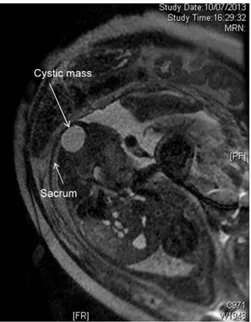
An anterior myelomeningocele had been suspected in a female fetus on the basis of fetal ultrasonography; prenatal MRI at the 24th week of gestation confirmed a cystic mass in the anterior sacrococcygeal
Fetal sacrococcygeal teratoma
Bellieni CV*1, Molinaro F2, Sica M2, Angotti R2, Messina M2, Bocchi C3, Severi F3 and Carbone S4
1Department of Mother and Infant, University Hospital, Siena, Italy
2Division of Pediatric Surgery, Department of Medical, Surgical and Neurological Sciences, University of Siena, Italy 3Division of Obstetrics, University of Siena, Italy
4Institute of radiology, University Hospital, Siena, Italy
Correspondence to: Carlo Valerio Bellieni, Department of Mother and Infant,
University Hospital, Siena, Italy, E-mail: [email protected]
Received: May 04, 2017; Accepted: May 17, 2017; Published: May 20, 2017
area [1]. The mass was surgically removed at one month of age, and hystopathological analysis determined it was a mature teratoma (Figure 1).
Figure 1. Prenatal diagnosis (fetal MRI): a tumor in the fetal sacrococcygeal area.
Copyright: ©2017 Bellieni CV. This is an open-access article distributed
under the terms of the Creative Commons Attribution License, which permits unrestricted use, distribution, and reproduction in any medium, provided the original author and source are credited.
