Received: 09 January 2019 Accepted: 11 March 2019 Published: 20 March 2019 http://www.aimspress.com/journal/Materials
Review
Colloidal stability of liposomes
Domenico Lombardo1,*, Pietro Calandra2, Maria Teresa Caccamo1, Salvatore Magazù3 and Mikhail Alekseyevich Kiselev4
1
Consiglio Nazionale delle Ricerche, Istituto per i Processi Chimico-Fisici, 98158 Messina, Italy
2
Consiglio Nazionale delle Ricerche, Istituto Studio Materiali Nanostrutturati, 00015 Roma, Italy
3
Dipartimento di Fisica e Scienze della Terra, Università di Messina, 98166 Messina, Italy
4
Frank Laboratory of Neutron Physics, Joint Institute for Nuclear Research, Dubna, Moscow 141980, Russia
* Correspondence: Email: [email protected].
Abstract: In recent years, the development of novel approaches for the formation of lipid nano-formulations for efficient transport of drug molecules in living systems offers a wide range of biotechnology applications. However, despite the remarkable progress in recent methodologies of synthesis that provide a wide variety of solutions concerning the liposome surface functionalization and grafting with synthetic targeting ligands, the action of most liposomes is associated with a number of unwanted side effects diminishing their efficient use in nanomedicine and biotechnology. The major limitation in the use of such versatile and smart drug delivery systems is connected with their limited colloidal stability arising from the interaction with the complex environment and multiform interactions established within the specific biological media. Herein, we review the main interactions involved in liposomes used in drug delivery processes. We also analyze relevant strategies that aim at offering possible perspectives for the development of next-generation of liposomes nanocarriers that are able to overcome the critical issues during their action in complex biological media.
Keywords: liposomes; soft interaction; drug delivery; nanomedicine; nanoscience; nanotechnology
1. Introduction
The development of nanocarriers technology for the efficient delivery of therapeutic drugs has experienced considerable expansion in recent years [1–3]. The design and engineer of novel
functional nanomaterials have generated a variety of smart nanocarriers for the efficient encapsulation and controlled delivery of therapeutic drugs [4–6]. In this respect, liposomes, composed of natural or synthetic lipids, represent a versatile nano-platform for the development of enhanced drug delivery systems [6,7]. These nano-platform that offer many benefits connected with versatile self-assembly processes, still maintain the supremacy in the nanomedicine clinic applications and in the biotechnology market [8,9]. More specifically, some studies evidences that liposomal encapsulation of chemotherapeutic drugs may reduce the overall toxicity, and the severity of the side effects relative to free drugs action [10,11]. For example, liposomal amphotericin B (a chemotherapeutic drug) shows a considerably lower number of side effects compared to conventional amphotericin B [11]. Furthermore, cationic liposomes can improve the accumulation process in tumors and the pharmacokinetics of the photosensitizer, and reduce the side effect of skin photosensitization [6]. On the other side, although some recent studies showed that liposomal doxorubicin is less toxic than other second-line chemotherapy regimens for ovarian cancer, it is worth pointing that liposome-specific side effects such as various skin and hypersensitivity reactions were reported in addition to severe myelosuppression [10,11]. Moreover, the lower toxicological profile of liposomal cytarabine compared to conventional cytarabine could not be confirmed [11]. Finally, the treatment with the DOXIL nano-drug, the first FDA-approved liposomal Doxorubicin formulation, evidenced (together with the infusion-related reactions) several adverse reactions including: anemia, neutropenia, thrombocytopenia, hand-foot syndrome, skin rash, asthenia, fatigue, fever, nausea, stomatitis, diarrhea [12]. Those results evidenced that despite the remarkable progress of recent researches, specific toxicity and several unwanted side effects are still present and diminish the efficient use of liposomes in nanomedicine [9–13].
The comprehension of liposomes interaction in the complex environment of the biological systems still represents a big challenge within the nanotechnology field. In this respect, a variety of strategies can be developed for the engineering of liposomes nanocarriers with desired physico-chemical properties that facilitate the transfer of the therapeutic drug through the natural barriers of biological systems [13–16].
Herein, we review the main interactions that regulate the colloidal stability in liposomes. We also analyze relevant strategies that aim at offering possible perspectives for the development of next-generation of liposomes nanocarriers that are able to overcome the critical issues during their action in drug delivery processes within the complex biological media.
2. Structural properties of liposomes
From the structural point of view liposomes are metastable structures consisting of one or more lipid bilayers surrounding aqueous compartments, where the hydrophilic (or polar) head groups are oriented towards the (exterior and interior) aqueous phases. In aqueous solutions their structures and dimensions depend on the conditions of preparation (i.e., sonication, stirring, extrusion, electroformation or microfluidification). Liposomes size ranges mainly between 25 and 2500 nm and, according to the number of bilayers, they can be classified in unilamellar (ULVs), multilamellar or oligolamellar. Multilamellar vesicles (MLVs) have an onion-like structure composed of concentric bilayer (hydrated multilayers), while the unilamellar vesicles can be further classified into small unilamellar vesicles (SUVs, diameter < 100 nm), large unilamellar vesicles (LUVs, diameter 100–1000 nm), or giant unilamellar vesicles (GUVs, diameter > 1 μm) [17]. The size of liposome
nanocarriers employed in nanomedicine applications ranges mainly between 50 and 500 nm [8]. The drug release efficiency sensitively depends on the lipid bilayers characteristics and on drug release process.
3. Types of interactions in liposomes nanocarriers
The relevant (soft) interactions in liposomes nanocarriers ensure an enhanced flexibility of nanocarrier structure and enable the system to withstand the main perturbation while preserving the reversibility of the self-assembly processes. The main (non-covalent) interactions acting on liposomes are the hydrogen bonds, hydrophobic interaction, van der Waals forces, electrostatic and steric forces.
Hydrogen bonding is a fundamental interaction which is present on many relevant functions of biological systems involved in the major macromolecules in biochemistry (such as proteins, nucleic acids and carbohydrates). For example, the hydrogen bond mechanism (solvation-desolvation) is involved in the formation and structural organization of a ligand-protein complex, and the resulting molecular recognition processes in bio-membranes. Apart with protein-ligand interaction, hydrogen bonds are also implicated in many intermolecular interactions, including those involving protein-protein, protein-liposome and protein-nucleic acid complexes [18–20]. Moreover, they can also promote liposome bridging that leads to the formation of large aggregates.
The hydrophobic effect favorites the interaction with of nonpolar (hydrophobic) components and the self-assembly processes in many nanocarrier systems [21,22]. The insertion in water of a hydrophobic (non-polar) component favourites the breaking of the H-bonding water network and the reorganization of the water molecules around the hydrophobic molecules. This favourites the formation of cavities that host an assembly of non-polar (hydrophobic) molecules. Hydrophobic interactions promote intra- and inter-molecular associations of biomolecules in various biological phenomena. It also regulates the inclusion of hydrophobic drugs within the lipid nanocarriers.
The Van der Waals interaction is generated by permanent (or induced) dipoles within the nanocarriers that may result in net attractive forces between biological nano-components. London dispersion forces are short-range attractive interactions generated by a temporary dipole induced by a polarization of the electron distribution, due to the presence of an adjacent atom. For example, Van der Waals forces drive the packing of the (uncharged) hydrophobic alkyl tails of phospholipids molecules inside the bio-membrane bilayers [23].
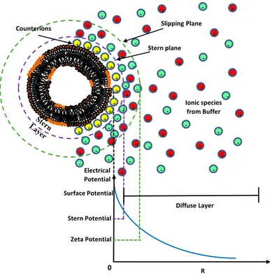
Efficiency of the liposome in drug delivery processes is sensitively influenced by the (density and typology) of their surface charge (electrostatic interaction). In water environments, most liposomes nano-platforms, in fact, exhibit some surface charge due to the dissociation/ionization of the lipid surface groups (such as the anionic dimyristoyl phosphatidylglycerol (DMPG) or cationic 1,2-dioleoyl-sn-glycero-3-phosphoethanolamine (DOPE) orzwitterionic dimyristoylphosphatidylcholine (DMPC)), or the surface adsorption of charged molecules/ions. The formation of a cloud of counterions around the nanoparticles (electrical double layer, EDL) counterbalance the surface nanocarrier charge (Figure 1) [24]. The presence of a sufficiently high electrostatic repulsion prevents, in general, aggregation and flocculation of charged nanocarriers and could promote their interaction with cells [25–27]. An estimate of the effective surface charge of liposomes can be obtained with the measurement of the zeta potential, i.e., the nanocarriers electrostatic potential at the so-called shear plane, (i.e., at the surface where the ions are not bound to the nanoparticle).
Figure 1. Sketch of the distribution of charges around a charged liposome. The electrical
double layer (EDL) is composed of a Stern layer of ions strongly bound to the liposome charged surface and an adjacent diffuse layer of mobile ions (which are loosely associated).
Finally, colloidal stability of liposome can be enhanced through the addition to the solution medium of small amounts of polymer (steric stabilization interaction). The magnitude of the stabilization depends on whether the polymer is adsorbed or irreversibly grafted onto the nanoparticle surface (stealth liposomes), on the polymer grafting regime and on the solvent quality and charging regime (ionic strength) [28].
4. Diseased tissues and biological barriers
The presence of biological (or physical) barriers in the living organisms can affect the accumulation of therapeutic nanoparticles into the diseased tissues. Liposomes interaction with blood proteins has an important role in the tissue distribution and clearance process of liposomes which are intravenously injected.
Nanocarrier clearance process involves the adsorption on the surface of nanocarrier of the plasma opsonins proteins and their recognition by the mononuclear phagocyte system (MPS) (also known as reticulo endothelial system, RES) [11,12]. This process is followed by elimination of the cargo at the hepatic level and by the successive process of metabolism by Kupffer cells (or by splenic macrophages). In a second clearance process, the blood low-density lipoproteins (LDLs) and high-density lipoproteins (HDLs) interact with the liposomes and cause changes on the structure of liposomes surface (lipid transfers/depletion) with the reduction of their colloidal stability. This is followed by the liposome destruction and the drug cargo release process to the plasma [11,12].
+ + + + + + + + + + + + + + + + + + -+ + + + + + + + -+ + Diffuse Layer Stern plane Electrical Potential Surface Potential Stern Potential Slipping Plane Zeta Potential R 0 Ionic species from Buffer + + + + + + + + + + + + + Counterions
-Antifouling surface (protein resistant) ligands such as poly(ethylene glycol) (PEG) and zwitterionic macromolecules are employed in order to avoid nonspecific protein adsorption and cell adhesion before nanocarries reach the target tumor sites [11,12].
The aforementioned processes identify the key role of the physico-chemical properties of nanocarriers during the clearance processes. In this respect the design and engineering of the physico-chemical properties of novel nanocarriers allow a proper control over the structure-function relationship thus minimizing the RES sequestration of therapeutic compounds and unwanted side effects during drug delivery processes. Size, surface charge and colloidal stability are the main factors affecting clearance process (by the MPS) by means of the proteins opsonization process. 5. Interaction of liposome and biological system
When a liposome interacts with a cell, the delivery of the drug and its distribution in the target cell can occur in several ways [8]. In liposomes adsorption to the cells membrane, the nanocarrier lipid bilayer is degraded by mechanical strain (or the enzymes action), thus leading to the active drugs release into the extracellular fluid, where they can diffuse through the cytoplasm and the cell membrane. Alternatively, the liposomal drug content is released directly into the cytoplasm by means of the liposomal membrane fusion with the plasma membrane of the target cell. Finally, the receptor-mediated endocytosis process regards only vesicles of a maximum diameter of 150 nm and active drugs that can endure the acidic lysosomes environment, where liposomes are (enzymatically) processed. In phagocytosis processes, large liposomes (with diameter larger than 150 nm) are phagocytosed by the immune system, through the intervention of specialized cells, such as macrophages, monocytes and Kupffer cells [8].
5.1. Modes of interactions of liposomes with biological systems
Upon their insertion in biological fluids liposomes undergo transformation that may profoundly alter their structural properties and then their functional use. The assessment of the colloidal stability of liposomes is a complex task due to the complex biological environment encountered in diseased tissues. Diseased tissues (and cancer cells) are characterised by a different microenvironment in comparison with the normal cells. In tumour (or inflammatory) tissues the blood vessels present large vascular fenestrations (with diameters between 50 and 200 nm) that allow drug-loaded nanocarriers to diffuse outside the blood vessels region (extravasation) thus entering the tumor interstitial space and concentrating into the target site [6,7]. After the injected into the blood circulation, the nanocarriers interact with the complex biological environment encountered (blood components, cytoplasm, nucleus and intracellular membranes). The clearance process of the circulating liposomes from the bloodstream and the high uptake by the MPS are actual obstacles to any attempt at targeting to tumors. The nanocarriers physico-chemical properties such as the dimension, morphology, surface functionality, are the main properties that can influence their biological clearance [6,7]. To preserve the liposomes efficiency, the defenses of the organism must be circumvented by avoiding the recognition (and neutralization/elimination) of the invading active drugs.
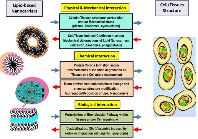
The main interactions of lipid based nanostructured materials with biological systems can be classified into three basic modes: chemical, physico-mechanical and electronic interactions.
5.1.1. Physical and mechanical interactions
Physical interactions such as aggregation/flocculation and fusion/coalescence strongly influence the transport properties (and shelf life) of liposomes as can result in loss of liposome associated drug and changes in dimension/morphology. Furthermore, presence of high-aspect-ratio nanostructures (or sub-components) can mechanically perturb the cellular substructures (mechanical interaction) as they may cause mechanical stress, deformation, and damage of bio-membranes, when cells attempt to engulf the nano-structures into lysosomes structures during cellular uptake. The sharp edges can also favour the spontaneous penetration of cell membranes with low energy barriers thus causing the lipid extraction and membrane damage.
5.1.2. Chemical interaction
Inclusion of nanomaterials in biological environments often leads to high chemical reactivity that creates non-equilibrium systems and/or unwanted phase transformations [29]. Chemical interactions between nanocarriers and biological fluids (phases) include the chemical adsorption of ions, small molecules,ligand and proteins exchange. The biomolecular adsorption, including protein corona formation, plays an important role as the structural transformations such as dispersion, aggregation, and deposition may influence the pathway of the drug delivery process.
Two main types of chemical degradation reactions can affect the performance of phospholipid bilayers:
- peroxidation of unsaturated acyl chains (if present);
- hydrolysis of the ester bonds linking the fatty acids to the glycerol backbone
The hydrolysis and oxidation of lipids may promote the appearance of short-chain lipids and then soluble derivatives will form in the lipid membrane, resulting in the decrease of the quality and performances of lipid nanocarriers. Oxidative and reductive dissolution processes may also cause the release of soluble ionic species that are often the primary factors of adverse biological responses. Further unwanted effects driven by oxidation or hydrolysis include oxide formation, sulfidation, degradation, and dissolution [29].
5.1.3. Biological interactions
Biological stability of liposomes, another important factor that influences their biomedical application, depends on the presence of specific agents (such as proteins) that interact with liposomes during the drug delivery process (biological interaction). For example, cationic liposomes in plasma are prone to aggregation processes and exhibit leakage phenomena. High density lipoproteins (HDLs) are responsible for destabilization of liposomes prior to their interaction with circulating phagocytic cells (such as monocytes) [11,12]. Destabilization of liposomes is caused by the lipid exchange between the liposomes and HDLs. Moreover, possible ion complexation can modify the ionic strength and pH of the biological media thus influencing nanocarriers structural properties and their related functions. For example, redox interactions can be associated with correlated molecular reorganization processes such as liposome assembly/disassembly [7,8], and can perturb some important biochemical pathways (or initiate new pathways) that lead to adverse outcomes , which are mediated by reactive oxygen and nitrogen species. Finally, different strategies can be adopted to
enhance biological stability of liposomes in drug delivery processes and to increase circulation time in the blood stream.
In Figure 2, we report a schematic representation of the main modes of interaction between lipid nanocarriers and biological systems. The arrows highlight the bi-directionality of the interactions, as the lipid-based nanocarriers (and their nanostructure transformation) induce specific responses in biological tissues (and micro-environment) while, conversely, the biological environment induces chemical or physical/mechanical transformations in the reference lipid nanocarrier.
Figure 2. Modes of the synergistic interaction between lipid nanocarriers and biological systems. 6. Strategies to improve nanocarrier stability and circulation time
When injected into the blood circulation nanocarriers rapidly interact with the complex biological environment encountered. The clearance of circulating nanocarriers from the bloodstream, and their high uptake by the MPS, represents an obstacle to any attempt at targeting to diseased tissues (such as tumors or inflammations). In order to preserve the optimal nanocarriers efficiency, the organism defenses must be circumvented by avoiding the nanocarrier recognition and the consequent neutralization.
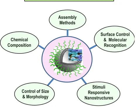
The retention of drug stability depends on the number of factors such as lipid (chemical) composition, method of manufacture (lipid organization and assembly methods), nanocarrier dimension, morphology and surface characteristic, physicochemical properties of drug and method of drug loading [30]. A schematic representation of the factors that influence liposomes drug are reported in Figure 3. We shortly review these inter-correlated factors in the following section.
Cellular/Tissues structures pertubation and /or Mechanical stress (plasma, lisosomes, cytoskeleton) Cell/Tissue induced Confinement and/or Mechanical deformations of Lipid Nanocarriers
(adhesion, lisosomes, phagocytosis)
Physical & Mechanical Interaction
Protein Corona formation and/or ions/molecules dissolution degradation on
Tissues and Cell micro-environment and /or Mechanical stress Micro-environment induced phase change and
chemical structure modification. Aggregation/Deposition of Lipid Nanocarriers
(adhesion, lisosomes, phagocytosis)
Chemical Interaction
Perturbation of Biomolecular Pathway within Tissues and/or Cell membrane Destabilization, (Dis-)Assembly induced by redox or interaction with agents (lipoprotein)
Photonic Properties of Lipid Nanocarriers
Biological Interaction
Lipid-based Nanocarriers
Cell/Tissues Structure
Figure 3. Schematic representation of the factors that influence drug delivery processes with liposomes nanocarriers.
Composition, size and lamellarity of liposomes strongly influence the encapsulation efficiency, the efflux rate of liposomal encapsulated material and the fate of a drug after cellular uptake [31].
Permeability and stability of liposomes are influenced by the rigidity/stiffness of the component lipids bilayer. Selection of lipid in turn depends on the (gel-liquid) phase transition temperature of lipids Tm. This parameter depends on the fatty acid side chains, degree of unsaturation, chain length and type of polar head groups. Lipids with long acyl chain are most commonly used because high phase transition temperature [32].
A first method to prolong the release rate of entrapped drugs is based on the choice of drugs with enhanced hydrophobic character or by incorporating the cholesterol lipids. Due to its hydrophobic character, in fact, the cholesterol lipids preferentially interact with the core region of the neutral membrane of the liposomes, thus inducing a dense packing of phospholipids. (bilayer-tightening effect). This causes a reduction of their permeability and increases in vivo (and in vitro) stability, and inhibits their transfer to the high-density lipoprotein (HDLs) and the low-density lipoprotein (LDLs) [33]. The presence of sterol backbone, however, may promote perturbation of the lipid bilayer structure, as recently evidenced in mixed lipid vesicles systems containing sodium cholate NaDC [34,35].
6.1. Inclusion of charged components (electric stabilization)
Inclusion of charged components creating a sensitive electrostatic surface charge (ζ-potential) that promote the interaction of liposomes with cells and prevents their aggregation and flocculation in solution. Some studies indicate that the negatively charged liposomes are less stable than positive and neutral ones when injected into the blood circulation, as they interact with the biological system
Control Factors in Liposomes Nanocarriers
Assembly Methods Control of Size & Morphology Chemical Composition Surface Control & Molecular Recognition Stimuli Responsive Nanostructures
subsequently to their opsonization process with circulating proteins. This induces a rapid uptake by the MPS and possible toxic effects. Liposomes with neutral charge containing phosphatidylcholine evidenced a more stable character and were bound the lowest amount of protein. Moreover, liposomes that contain only one class of negatively charged phospholipids bound a high amount of protein and were very unstable, with respect of liposomes containing phosphatidylcholine, that evidenced a lower amount of bound proteins and a better stability [36,37]. A recent investigation evidenced also that cationic liposomes exhibited a preferential uptake in angiogenic tumor vessels, providing an efficient selective delivery of diagnostic (and therapeutic) agents to angiogenic blood vessels of solid tumors. On the other hand, anionic (or neutral) liposomes may be used as drugs carriers to the extravascular compartment of tumors due to their extravasation [38].
6.2. Surface conjugation with polymers (steric stabilization)
Liposome surface conjugation with polymers represent another important approach that allows to overcome most of the challenges in drug delivery processes, such as toxicity, the low blood circulation half-life, interception by the immune system, biocompatibility and antigenicity issues [7,8]. For this purpose, both synthetic (e.g., poly(ethylene glycol) PEG; poly(vinyl alcohol), PVA) and natural (e.g., alginate, dextran, chitosan) polymers can be employed [8]. More specifically, PEG polymers creates, in fact, a concentration of highly hydrated polymer brushes that sterically inhibits both electrostatic and hydrophobic interactions with the plasma proteins or cells, and reduce the liposomal uptake process by the MPS. PEGylated liposomes, in fact, are not opsonized and are able then to escape the capture by the cells phagocytic systems (so called “stealth liposomes” effect). Many investigations evidenced that PEGylated liposomes are able to ameliorate the blood-circulation time and the colloidal stability, together with the low plasma clearance and the low volume of distribution [39].
It is worth pointing that combined electrostatic and steric interaction generated by drug inclusion may induce phase transitions in lipid-based nanocarriers that sensitively influence the structural stability of the nanocarriers, as demonstrated by different studies [40–44]. A detailed study of the interactions occurring between drug nanocarriers and biological systems should become a prominent task of the design and characterization of new drug delivery systems. In this respect, different scattering techniques can be applied by employing artificial membranes as simplified models for cell membranes [45–47]. Those studies have given a strong input to the understanding of the complex combination of soft interactions that a biomolecule can develop toward biological systems.
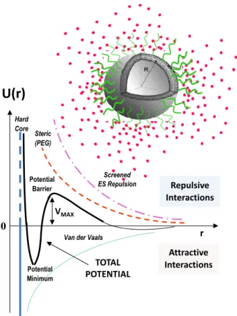
In Figure 4, the interaction potential of the main soft interaction expressed by a liposome nanocarrier system is reported. The presence of an energy barrier resulting from the balance between repulsive and attractive forces prevents the adhesion of two nanocarriers while approaching one another. Control over the nanocarriers soft interactions represents, then, a fundamental stage for the engineering of the colloidal stability and biocompatibility of the therapeutic compounds that are able to overcome obstacles and biological barriers to cellular and tissue uptake.
Figure 4. Example of the main soft interaction expressed by a liposome nanocarrier system. 7. Stimuli-responsive nanocarriers and targeted drug delivery: perspectives and critical issues
Successful therapeutic application of smart lipid nanocarriers can be achieved through innovative technologies that allow a (locally activated) drug release action limited to selective sites within the diseased tissues. A promising method to increase the specificity of the drug delivery process is obtained by surface conjugation of a specific ligand which is able to selectively bind to a given receptor of the target cell (active targeting). A large variety of ligands (including proteins, peptides, antibodies, aptamers, lectins, and vitamins) which can bind to specific overexpressed receptors on tumour tissues, have been used as biomarkers for targeted drug delivery [48,49]. For example, the folic acid (FA) targeting ligand, has a high binding affinity to folate receptors (FR), which are overexpressed in a wide range of FR-positive tumour types, including lung, ovarian, brain, breast, colon, and kidney cancers [8,48]. Despite the increasing interest in these novel approaches, several recent investigations evidenced that active targeting through the tumor-targeting ligand does not always result in increased accumulation of the nanoparticles in tumors [49–51].
Advanced liposomes nanoplatforms can be obtained also by exploiting a number of external/internal triggerable properties for the development of stimuli-responsive liposomes, whereby the liposome stability (and/or permeability) is controlled by a triggerable component which performs a task when activated by internal stimuli from target tissues (such as redox potential or pH) or by external stimuli (such as temperature, hyperthermia, light, ultrasound, and [electro] magnetic fields) [52]. For example, a pH-sensitive liposome which is stable at the (regular tissues) physiological pH, may acquire fusogenic properties under the acidic environment which is present in
U(r)
VMAX Potential Minimum TOTAL POTENTIAL 0 r Screened ES Repulsion Attractive Interactions Repulsive Interactions Potential BarrierVan der Vaals Steric
(PEG) Hard Core
pathological tissues, and release its active drugs nearby tumors and infection tissues [52]. Moreover, recent developments has led to significant interest in utilizing liposome nanocarriers for simultaneous detection (by using fluorescence, magnetic resonance, ultrasound and nuclear imaging) of specific bio-molecules (such as proteins, DNA and small molecule targets) and treatment of heavy metal toxicity and cancers (theranostic nanocarriers) [53].
Finally, although several innovative liposome nano-formulations for targeted and stimuli responsive delivery of drugs have been proposed at a preclinical level, up to now only few of them have been admitted in the clinical trials, while none of them have been admitted in the market yet [9,54]. Therefore, a deeper knowledge and understanding of the real interactions involved in the diseased tissues is fundamental for the development of novel therapeutic approaches and protocols based on the employment of smart liposome nanoplatform.
8. Conclusions
The use of liposomes in the drug delivery technologies provides many benefits associated with its capacity for a versatile self-assembly. Despite the success of several liposomal formulations in vivo and the progressive achievements in nanomedicine applications, a clear understanding of their interactions with bio-membrane is still far to be attained.
In this review, we concisely analyze the main factors influencing liposomes colloidal stability during their interaction with cells and in drug delivery applications. We also discussed some strategies that can be developed to overcome the biological barriers, and how these approaches have stimulated the development of advanced drug delivery systems. The complex microenvironment in living systems strongly affects the functionality of nanomaterials, and may compromise the design goals of lipid nanocarriers. Therefore, a deeper knowledge and understanding of the real interactions involved in the diseased tissues is fundamental for the development of novel therapeutic approaches and protocols based on the employment of smart liposome nano-platform.
Acknowledgments
D.L. acknowledges funding from Marie Curie Actions under EU FP7 Initial Training Network SNAL 608184. The work of M.A.K. was financed by the Russian Scientific Foundation (Project no. 14-12-00516). The authors acknowledge the CNR-IPCF technical staff (G. Spinella, G. Lupo and R. Caruso) for the technical support and the various upgrades of experimental set-up performed.
Conflict of interest
The authors declare no conflict of interest. References
1. Chen G, Roy I, Yang C, et al. (2016) Nanochemistry and nanomedicine for nanoparticle-based dagnostics and therapy. Chem Rev 116: 2826–2885.
2. Ali I, Lone MN, Suhail M, et al. (2016) Advances in nanocarriers for anticancer drugs delivery. Curr Med Chem 23: 2159–2187.
3. Pasqua L, Leggio A, Sisci D, et al. (2016) Mesoporous silica nanoparticles in cancer therapy: relevance of the targeting function. Mini Rev Med Chem 16: 743–753.
4. Chow EKH, Ho D (2013) Cancer nanomedicine: from drug delivery to imaging. Sci Transl Med 5: 216rv4.
5. Lee BK, Yun YH, Park K (2015) Smart nanoparticles for drug delivery: boundaries and opportunities. Chem Eng Sci 125: 158–164.
6. Bozzuto G, Molinari A (2015) Liposomes as nanomedical devices. Int J Nanomed 10: 975–999. 7. Allen TM, Cullis PR (2013) Liposomal drug delivery systems: from concept to clinical
applications. Adv Drug Deliver Rev 65: 36–48.
8. Bobo D, Robinson KJ, Islam J, et al. (2016) Nanoparticle-based medicines: a review of FDA-approved materials and clinical trials to date. Pharm Res 33: 2373–2387.
9. Wilhelm S, Tavares AJ, Dai Q, et al. (2016) Analysis of nanoparticle delivery to tumours. Nat Rev Mater 1: 16014.
10. Iwamoto T (2013) Clinical application of drug delivery systems in cancer chemotherapy: review of the efficacy and side effects of approved drugs. Biol Pharm Bull 36: 715–718.
11. Brand W, Noorlander CW, Giannakou C, et al. (2017) Nanomedicinal products: a survey on specific toxicity and side effects. Int J Nanomed 12: 6107–6129.
12. Janssen Products Expert Committee, DOXIL (doxorubicin HCl liposome injection), 2018. Available from: https://www.doxil.com.
13. Ishida T, Harashima H, Kiwada H (2001) Interactions of liposomes with cells in vitro and in vivo: opsonins and receptors. Curr Drug Metab 2: 397–409.
14. Ishida T, Harashima H, Kiwada H, et al. (2002) Liposome clearance. Bioscience Rep 22: 197– 224.
15. Lombardo D, Calandra P, Barreca D, et al. (2016) Soft interaction in liposome nanocarriers for therapeutic drug delivery. Nanomaterials 6: E125.
16. Dai Y, Xu C, Sun X, et al. (2017) Nanoparticle design strategies for enhanced anticancer therapy by exploiting the tumour microenvironment. Chem Soc Rev 46: 3830–3852.
17. Sackmann E (1995) Physical basis of self-organization and function of membranes: physics of vesicles, In: Lipowsky R, Sackmann E, Handbook of Biological Physics, Elsevier, 213–303. 18. Israelachvili J, Wennerström H (1996) Role of hydration and water structure in biological and
colloidal interactions. Nature 379: 219–225.
19. Franks F (1972) Water—a comprehensive treatise, New York, NY, USA: Plenum.
20. Magazù S, Migliardo F, Telling MT (2007) Study of the dynamical properties of water in disaccharide solutions. Eur Biophys J 36: 163–171.
21. Degiorgio V, Corti M (1985) Physics of amphiphiles: micelles, vesicles and microemulsions, Amsterdam: North-Holland.
22. Tanford C (1980) The hydrophobic effect: formation of micelles and biological membranes, 2 Eds., New York: Wiley.
23. Parsegian VA (2006) Van der Waals forces: a handbook for biologists, chemists, engineers, and physicists, Cambridge University Press.
24. Hunter RJ (1986) Foundations of Colloid Science, Oxford University Press.
25. Cevc G (1993) Electrostatic characterization of liposomes. Chem Phys Lipids 64: 163–186. 26. Dan N (2002) Effect of liposome charge and PEG polymer layer thickness on cell-liposome
27. Lombardo D (2014) Modeling dendrimers charge interaction in solution: relevance in biosystems. Biochem Res Int 2014: 837651.
28. Akpinar B, Fielding LA, Cunningham VJ, et al. (2016) Determining the effective density and stabilizer layer thickness of sterically stabilized nanoparticles. Macromolecules 49: 5160–5171. 29. Wang Z, Zhu W, Qiu Y, et al. (2016) Biological and environmental interactions of emerging
two-dimensional nanomaterials. Chem Soc Rev 45: 1750–1780.
30. Moore TL, Rodriguez-Lorenzo L, Hirsch V, et al. (2015) Nanoparticle colloidal stability in cell culture media and impact on cellular interactions. Chem Soc Rev 44: 6287–6305.
31. Plessis JD, Ramachandran C, Weiner N (1996) The influence of lipid composition and lamellarity of liposomes on the physical stability of liposomes upon storage. Int J Pharm 127: 273–278.
32. Ceh B, Lasic DD (1995) A rigorous theory of remote loading of drugs into liposomes. Langmuir 11: 3356–3368.
33. Geng S, Yang B, Wang G, et al. (2014) Two cholesterol derivative-based PEGylated liposomes as drug delivery system, study on pharmacokinetics and drug delivery to retina. Nanotechnology 25: 275103.
34. Kiselev MA, Janich M, Hildebrand A, et al. (2013) Structural transition in aqueous lipid/bile salt [DPPC/NaDC] supramolecular aggregates: SANS and DLS study. Chem Phys 424: 93–99. 35. Kiselev MA, Lombardo D, Lesieur P, et al. (2008) Membrane self assembly in mixed
DMPC/NaC systems by SANS. Chem Phys 345: 173–180.
36. Hernández-Caselles T, Villalaín J, Gómez-Fernández JC (1993) Influence of liposome charge and composition on their interaction with human blood serum proteins. Mol Cell Biochem 120: 119–126.
37. Narenji M, Talae MR, Moghimi HR (2017) Effect of Charge on Separation of Liposomes upon Stagnation. Iran J Pharm Res 16: 423–431.
38. Krasnici S, Werner A, Eichhorn ME, et al. (2003) Effect of the surface charge of liposomes on their uptake by angiogenic tumor vessels. Int J Cancer 105: 561–567.
39. Jain NK, Nahar M (2010) PEGylated nanocarriers for systemic delivery. Methods Mol Biol 624: 221–234.
40. Dan N (2014) Nanostructured lipid carriers: effect of solid phase fraction and distribution on the release of encapsulated materials. Langmuir 30: 13809–13814.
41. Bourgaux C, Couvreur P (2014) Interactions of anticancer drugs with biomembranes: what can we learn from model membranes? J Control Release 190: 127–138.
42. Lombardo D, Calandra P, Magazù S, et al. (2018) Soft nanoparticles charge expression within lipid membranes: The case of amino terminated dendrimers in bilayers vesicles. Colloid Surface B 170: 609–616.
43. Dan N (2016) Membrane-induced interactions between curvature-generating protein domains: the role of area perturbation. AIMS Biophys 4: 107–120.
44. Lombardo D, Calandra P, Bellocco E, et al. (2016) Effect of anionic and cationic polyamidoamine (PAMAM) dendrimers on a model lipid membrane. BBA-Biomembranes 1858: 2769–2777.
45. Katsaras J, Gutberlet T (2000) Lipid bilayers: Structure and Interactions, Springer Science & Business Media.
46. Wanderlingh U, D’Angelo G, Branca C (2014) Multi-component modeling of quasielastic neutron scattering from phospholipid membranes. J Chem Phys 140: 05B602.
47. Kiselev MA, Lombardo D (2017) Structural characterization in mixed lipid membrane systems by neutron and X-ray scattering. BBA-Gen Subjects 1861: 3700–3717.
48. Kiselev MA, Lesieur P, Kisselev AM, et al. (2001) A sucrose solutions application to the study of model biological membranes. Nucl Instrum Meth A 470: 409–416.
49. Blanco E, Shen H, Ferrari M (2015) Principles of nanoparticle design for overcoming biological barriers to drug delivery. Nat Biotechnol 33: 941–951.
50. Pirollo KF, Chang EH (2008) Does a targeting ligand influence nanoparticle tumor localization or uptake? Trends Biotechnol 26: 552–558.
51. Bae YK, Park K (2011) Targeted drug delivery to tumors: myths, reality and possibility. J Control Release 153: 198–205.
52. Mura S, Nicolas J, Couvreur P (2013) Stimuli-responsive nanocarriers for drug delivery. Nat Mater 12: 991–1003.
53. Xing H, Hwang K, Lu Y (2016) Recent developments of liposomes as nanocarriers for theranostic applications. Theranostics 6: 1336–1352.
54. Lombardo D, Kiselev AM, Caccamo MT (2019) Smart nanoparticles for drug delivery application: development of versatile nanocarrier platforms in biotechnology and nanomedicine. J Nanomater 2019: 3702518.
© 2019 the Author(s), licensee AIMS Press. This is an open access article distributed under the terms of the Creative Commons Attribution License (http://creativecommons.org/licenses/by/4.0)