Published Ahead of Print 18 January 2013.
2013, 195(7):1436. DOI: 10.1128/JB.01994-12.
J. Bacteriol.
Susana Merino
Markus Wilhelms, Victor Gonzalez, Juan M. Tomás and
Gene Transcriptional Hierarchy
http://jb.asm.org/content/195/7/1436
Updated information and services can be found at:
These include:
REFERENCEShttp://jb.asm.org/content/195/7/1436#ref-list-1
at:
This article cites 45 articles, 29 of which can be accessed free
CONTENT ALERTS
more»
articles cite this article),
Receive: RSS Feeds, eTOCs, free email alerts (when new
http://journals.asm.org/site/misc/reprints.xhtml
Information about commercial reprint orders:
http://journals.asm.org/site/subscriptions/
To subscribe to to another ASM Journal go to:
on April 29, 2013 by UNIVERSITAT DE BARCELONA
http://jb.asm.org/
Aeromonas hydrophila Lateral Flagellar Gene Transcriptional
Hierarchy
Markus Wilhelms, Victor Gonzalez, Juan M. Tomás, Susana Merino
Departamento de Microbiología, Facultad de Biología, Universidad de Barcelona, Barcelona, Spain
Aeromonas hydrophila AH-3 lateral flagella are not assembled when bacteria grow in liquid media; however, lateral flagellar
genes are transcribed. Our results indicate that A. hydrophila lateral flagellar genes are transcribed at three levels (class I to III
genes) and share some similarities with, but have many important differences from, genes of Vibrio parahaemolyticus. A.
hydro-phila lateral flagellum class I gene transcription is
70dependent, which is consistent with the fact that lateral flagellum is
consti-tutively transcribed, in contrast to the characteristics of V. parahaemolyticus. The fact that multiple genes are included in class I
highlights that lateral flagellar genes are less hierarchically transcribed than polar flagellum genes. The A. hydrophila lafK-fliEJ
Lgene cluster (where the subscript L distinguishes genes for lateral flagella from those for polar flagella) is exclusively from class I
and is in V. parahaemolyticus class I and II. Furthermore, the A. hydrophila flgAMN
Lcluster is not transcribed from the
54/
LafK-dependent promoter and does not contain class II genes. Here, we propose a gene transcriptional hierarchy for the A.
hy-drophila lateral flagella.
S
warming motility is defined as a rapid multicellular movement
of bacteria across a surface that is powered by rotating flagella.
Most bacteria that swarm have multiple constitutive flagella
dis-tributed randomly on the cell surface (peritrichous flagella) and
increase the flagellum number per cell on contact with surfaces (
1
,
2
). On the other hand, polar flagellated bacteria have developed
two different strategies to swarm: some bacteria, such as
Pseu-domonas aeruginosa, synthesize an alternative polar flagellar
mo-tor that can propel bacteria on surfaces (
3
,
4
), and others, such as
Vibrio parahaemolyticus, Aeromonas spp., and Rhodospirillum
cen-tenum, have developed lateral flagella distributed randomly on the
cell surface which are induced when grown on solid surfaces or in
viscous environments (
5
,
6
).
Phylogenetic analysis and organization of lateral flagellar genes
suggest that this flagellar system originated in Betaproteobacteria
and Gammaproteobacteria from a duplication of the entire
flagel-lar gene complex in the nonenteric gammaproteobacterial lineage,
which was then horizontally transferred to the Betaproteobacteria
and the enteric bacteria (
7
). In contrast to polar or peritrichous
flagella systems (primary systems), lateral flagellar systems lack
fliO, and the fliEFGHIJKLMNPQR gene cluster is split into two
gene clusters (fliEFGHIJL
and fliMNPQRL
[where the subscript L
distinguishes genes for lateral flagella from those for polar
fla-gella]). The fliKL
L(lafEF) genes are arranged in the fliD
L-motB
L(lafB-lafU) cluster (
5
,
8
).
The best-studied functional lateral flagellar systems are
those of V. parahaemolyticus and A. hydrophila. Both are
en-coded by 38 genes distributed in six clusters, while V.
parahae-molyticus genes are distributed in two discontinuous regions
on chromosome II (
9
); A. hydrophila genes are distributed in a
unique chromosomal region (
10
,
11
). V. parahaemolyticus
flgAMN
Land motY
L-lafK-fliEFGHIJ
Lclusters are transcribed
divergently from flgBCDEFGHIJKLL, fliMNPQRL-flhBAL, lafA,
and fliDSTKLA
L-motAB
L. A. hydrophila orthologous genes
ex-hibit the same distribution, although only flgAMN
Lgenes are
transcribed divergently. Furthermore, A. hydrophila does not
contain any gene orthologous to V. parahaemolyticus motY
L(
12
), and a modification accessory factor gene, maf-5 (
13
),
which is independently transcribed, is present between flgLL
and lafA.
Synthesis and assembly of any flagellar system is regulated
co-ordinately by a transcriptional cascade composed of three or four
levels of hierarchy: class I to III or I to IV (
14
,
15
). In polar
flagel-lated Gammaproteobacteria, such as Vibrio, mesophilic
Aeromo-nas, and Pseudomonas species, four levels of hierarchy have been
described. Transcription of class II and III is
54dependent, and
transcription of class IV is
28dependent (
16
,
17
,
18
). At the top of
the hierarchy is a
54-associated transcriptional activator (FlrA in
A. hydrophila) which activates
54-dependent promoters
preced-ing the class II clusters. One of the class II clusters encodes a
two-component signal-transducing system (FlrBC in A. hydrophila)
whose regulator (FlrC) activates class III genes. The class
III-tran-scribed A. hydrophila
28factor, which activates transcription of
class IV genes, is class II transcribed in Vibrio spp., and flagellar
hierarchy is independently transcribed in P. aeruginosa (
16
,
17
,
18
).
Inducible peritrichous flagella (lateral flagella) of V.
parahae-molyticus and A. hydrophila do not posses an FlhDC master
regu-lator and are
54dependent (
9
,
19
) as polar flagella. In this work,
we investigated the A. hydrophila lateral flagellar transcriptional
hierarchy by two techniques: promoter-lacZ fusion assays (used to
measure
-galactosidase activity in several mutant backgrounds)
and reverse transcription-PCR (RT-PCR) assays. Until now, little
was known about A. hydrophila’s transcriptional hierarchy,
trans-lational and posttranstrans-lational regulatory mechanisms that ensure
careful regulation, proper assembly of several different proteins,
and whether or not there was a large enough quantity of individual
lateral flagellar subunits.
Received 17 October 2012 Accepted 11 January 2013 Published ahead of print 18 January 2013
Address correspondence to Juan M. Tomás, [email protected].
Copyright © 2013, American Society for Microbiology. All Rights Reserved.
doi:10.1128/JB.01994-12
1436 jb.asm.org Journal of Bacteriology p. 1436 –1445 April 2013 Volume 195 Number 7
on April 29, 2013 by UNIVERSITAT DE BARCELONA
http://jb.asm.org/
MATERIALS AND METHODS
Bacterial strains, plasmids, and growth conditions. The bacterial strains
and plasmids used in this study are listed inTable 1. Escherichia coli strains were grown on Luria-Bertani (LB) Miller broth and LB Miller agar at 37°C, while Aeromonas strains were grown either in tryptic soy broth (TSB) or tryptic soy agar (TSA) at 30°C. When required, ampicillin (50 g/ml), kanamycin (50 g/ml), rifampin (100 g/ml), spectinomycin (50 g/ml), chloramphenicol (25 g/ml), and tetracycline (20 g/ml) were added to the media.
Motility assays (swarming and swimming). Freshly grown bacterial
colonies were transferred with a sterile toothpick into the center of swarm agar (1% tryptone, 0.5% NaCl, 0.5% agar) or swim agar (1% tryptone, 0.5% NaCl, 0.25% agar). The plates were incubated face up for 16 to 24 h at 25°C, and motility was assessed by examining the migration of bacteria through the agar from the center toward the periphery of the plate. More-over, swimming motility was assessed by light microscopy observations in liquid media.
DNA techniques. DNA manipulations were carried out essentially
according to standard procedures (20). DNA restriction endonucleases
and E. coli DNA polymerase Klenow fragment were obtained from Pro-mega. T4 DNA ligase and alkaline phosphatase were obtained from Invit-rogen and GE Healthcare, respectively. PCR was performed using BioTaq DNA polymerase (Ecogen) in a Gene Amplifier PCR system and a PerkinElmer 2400 thermal cycler.
Nucleotide sequencing and computer sequence analysis. Plasmid
DNA for sequencing was isolated by a Qiagen plasmid purification kit (Qiagen, Inc., Ltd.) as recommended by the suppliers. Double-stranded DNA sequencing was performed by using the Sanger dideoxy-chain ter-mination method (21) with the BigDye Terminator v3.1 cycle sequencing kit (Applied Biosystems). Custom-designed primers used for DNA se-quencing were purchased from Sigma-Aldrich. The DNA sequences were inspected in the GenBank and EMBL databases at the National Center for Biotechnology Information (NCBI) (22). The Terminator search pro-gram in the GCG Wisconsin package was used to search for factor-inde-pendent transcriptional terminators. Neural Network Promoter Predic-tion, PromScan (23), and PRODORIC (24) were used to search promoter sequences.
Total RNA extraction and RT-PCR. Total RNA was isolated, by RNA
Protect bacterial reagent (Qiagen) and an RNeasy Minikit (Qiagen), from
A. hydrophila AH-3 and rpoN, lafK, and lafS mutant strains grown in
liquid medium (TSB), viscous medium (TSB plus 18% [wt/vol] Ficoll), or solid agar (TSA). To ensure that RNA was devoid of contaminating DNA, the preparation was treated with RNase-free TurboDNase I (Ambion). First-strand cDNA synthesis was carried out with Moloney-murine leu-kemia virus (M-MuLV) reverse transcriptase (New England BioLabs) and random oligonucleotides (Promega) on 5g of total DNase-digested RNA. The reaction mixtures were incubated at 25°C for 10 min, 37°C for 120 min, and 75°C for 15 min. Control reaction mixtures lacking reverse transcriptase were used to confirm that RNA samples were not contami-nated with genomic DNA (RT negative controls). PCR, second-strand synthesis, and subsequent DNA amplification were carried out using the Accuprime TaqDNA polymerase (Invitrogene), specific oligonucleotides, and 30 PCR cycles. Amplicons were analyzed by agarose gel electropho-resis with ethidium bromide staining. A. hydrophila ribosomal 16S prim-ers were used as a control for the cDNA template. RT-PCR amplifications were performed at least twice with total RNA preparations obtained from a minimum of two independent extractions.
Mapping the A. hydrophila AH-3 fliML, lfgAL, lafK, lafB, and lafT transcription start sites by RACE PCR. Amplifications of the A. hydro-phila AH-3 fliML, lfgAL, lafK, lafB, and lafT cDNA 5= ends were performed
using the 5= random amplification of cDNA ends (RACE) system, version 2.0 (Invitrogen). Total RNA extraction from A. hydrophila AH-3 was per-formed as mentioned above. First-strand cDNA was synthesized using the entire volume of DNase-digested total RNA (5g), the fliML, lfgAL, lafK,
lafB, and lafT internal primers GSP1-FLIML(5=-ATCTTGCAAGGTGT
G-3=), GSP1-LfgAL(5=-GAGCTTGGAACAAATC-3=), GSP1-LafK (5=-G
ATATAACGAGCCAGTC-3=), GSP1-LafB (5=-TTTCGACAAACTTCTT G-3=), and GSP1-LafT (5=-AATTATCGATGATGAAAC-3=), respectively, and the Thermoscript RT-PCR system (Invitrogen) at 45°C for 45 min. Reverse transcriptase was deactivated at 85°C for 5 min, and 1l of RNase H was then added and incubated at 37°C for 20 min. Purification of cDNA with S.N.A.P. columns, as well as tailing of purified cDNA using terminal deoxynucleotidyl transferase and dCTP, was done according to the 5= RACE system, version 2.0, instructions. Confirmation of cDNA was per-formed after each step by PCR with nested primers. Tailed cDNA was amplified by primary PCR using 10M each primer, the 5= RACE abridged anchor primer (AAP) that binds to the tailed cDNA sequence, and GSP2-FLIML(5=-AGATGTCGACCTGATATTGG-3=), GSP2-FLGAL (5=-ATCTCCGGTACGAATGGT-3=), GSP2-LafK (5=-TTCATTAACCA GGATGAC G-3=), GSP2-LafB (5=-GCATTCTCCAACCCACTAT-3=), and GSP2-LafT (5=-TTCAT CGAGTGCCTTCAT-3=), which bind to the respective internal gene sequences. The PCR program applied was 94°C for 1 min and then 35 cycles of 94°C for 45 s, 55°C for 30 s, and 72°C for 1 min, followed by an extension at 72°C for 5 min. PCR products were
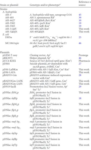
TABLE 1 Bacterial strains and plasmid used in this study
Strain or plasmid Genotype and/or phenotypea Reference or source Strains
A. hydrophila
AH-3 A. hydrophila wild type, serogroup O:34 45
AH-405 AH-3, spontaneous Rifr 39
AH-4427 AH-405⌬flaB; flaA::Kmr 10
AH-5502 AH-405; rpoN::Kmr
19
AH-5503 AH-405; lafK::Kmr
19
AH-3::lafS AH-405; lafS::Kmr This work
AH-3⌬lafA AH-405⌬lafA This work
E. coli
DH5␣ F⫺endA hdsR17(rk⫺mk⫹) supE44 thi-1
recA1 gyr-A9680lacZ
25
MC1061pir thi thr1 leu6 proA2 his4 argE2 lacY1
galK2 ara14 xyl5 supE44pir 46 Plasmids
pGEM-T easy Cloning vector, Apr Promega
pRK2073 Helper plasmid, Spr 46
pUC4-KIXX Source of Tn5-derived nptII gene (Kmr
) Pharmacia pDM4 Suicide plasmid, pir dependent with
sacAB genes, oriR6K, Cmr 27
pDM-LafSKm pDM4 with AH-3 lafS::Km, CmrKmr This work
pDM-LAFA pDM4 with AH-3⌬lafA, Cmr This work
pBAD33-Gm pBAD33 arabinose-induced expression vector with Gmr 28
pBAD33Gm-LAFK pBAD33 with AH-3 lafK gene, Gmr
This work pBAD33Gm-LAFS pBAD33 with AH-3 lafS gene, Gmr This work
pDN19 lac⍀ Promoterless lacZ fusion vector, Spr
SmrTcr
29
pDNlac-fliMLp fliMLpromoter-lacZ fusion in
pDN19lac⍀, Tcr This work
pDNlac-lafKp lafK promoter-lacZ fusion in
pDN19lac⍀, Tcr 18
pDNlac-flgMLp flgMLpromoter-lacZ fusion in
pDN19lac⍀, Tcr
This work pDNlac-flgALp flgALpromoter-lacZ fusion in
pDN19lac⍀, Tcr
This work pDNlac-flgBLp flgBLpromoter-lacZ fusion in
pDN19lac⍀, Tcr This work
pDNlac-maf-5p1 maf-5 promoter-lacZ fusion in
pDN19lac⍀, Tcr This work
pDNlac-maf-5p2 maf-5 promoter-lacZ fusion in
pDN19lac⍀, Tcr
This work pDNlac-lafAp lafA promoter-lacZ fusion in
pDN19lac⍀, Tcr
This work pDNlac-lafBp lafB promoter-lacZ fusion in
pDN19lac⍀, Tcr This work
pDNlac-lafTp lafT promoter-lacZ fusion in
pDN19lac⍀, Tcr This work
aTcr, tetracycline resistant; Kmr, kanamycin resistant; Apr, ampicillin resistant; Rifr,
rifampin resistant; Cmr
, chloramphenicol resistant; Spr
, spectinomycin resistant; Smr
, streptomycin resistant; Gmr, gentamicin resistant.
on April 29, 2013 by UNIVERSITAT DE BARCELONA
http://jb.asm.org/
analyzed by agarose gel electrophoresis, and amplified bands were excised from the gel, purified, and sequenced with GSP2-FLIML, GSP2-FLGAL, GSP2 LafK, GSP2-LafB, or GSP2-LafT primer.
Construction of defined mutants. To obtain the A. hydrophila AH-3:: lafS mutant, lafS was amplified by PCR with 5=-CGCGGATCCAACCCA
AGCCAGAGTTGAG-3= and 5=-CGCGGATCCATGAAACACCAGGAC ACA-3= (the BamHI site is underlined), ligated into vector pGEMTeasy (Promega), and transformed into E. coli DH5␣ (25). The Tn5-derived kanamycin resistance cartridge (nptll) from pUC4-KIXX was obtained by SmaI digestion, and the cassette was inserted into the XbaI blunt-ended restriction internal site of lafS. The cartridge contains an outward-reading promoter that ensures the expression of downstream genes when inserted in the correct orientation; however, such insertion will alter the regulation of those genes (26). The presence of a single BglII site in the SmaI-digested cassette allowed its orientation to be determined. Constructs containing the mutated genes were ligated to suicide vector pDM4 (27), electropo-rated into E. coli MC1061pir, and plated on chloramphenicol-kanamy-cin plates at 30°C to obtain the pDM-LafSKm plasmid.
The chromosomal in-frame lafA deletion mutant A. hydrophila AH-3⌬lafA was constructed by allelic exchange as described by Milton et al. (27). Briefly, DNA regions flanking the lafA gene were amplified using the primers A (5=-CGCGGATCCTTTGGTGTCGACTTCTCCT-3=), B (5=-C CCATCCACTAAACTTAAACAAGAGTTCAGCTGGTTCTGG-3=), C (5=-TGTTTAAGTTTAGTGGATGGGAGCACCAATATGACCAAGAA-3=), and D (CGCGGATCCCAGCACCATGTTGACCTT-3=) in two sets of asymmetric PCRs to amplify DNA fragments of 779 (pair AB) and 733 (pair CD) bp, respectively. DNA fragments AB and CD were annealed at their overlapping regions (double-underlined letters in primers B and C) and amplified as a single fragment using primers A and D. The fusion product was purified, BamHI digested (the BamHI site is underlined in primers A and D), ligated into BglII-digested and phosphatase-treated pDM4 vector (27), electroporated into E. coli MC1061pir, and plated on chloramphenicol plates at 30°C to obtain pDM-LAFA plasmid. Plasmid pDM-LafSKm or pDM-LAFA was transferred into an A. hydrophila AH-405 rifampin-resistant (Rifr) strain by triparental matings using E. coli
MC1061pir containing the insertion constructs and the mobilizing strain HB101/pRK2073. Transconjugants were selected on plates contain-ing chloramphenicol, kanamycin, and rifampin or containcontain-ing chloram-phenicol and rifampin. PCR analysis confirmed that the vector had inte-grated correctly into the chromosomal DNA. After sucrose treatment, transconjugants that were Rifr, Kmr, and Cmsor were Rifrand Cmswere
chosen and confirmed by PCR.
Plasmid constructions. Plasmids LAFK and
pBAD33Gm-LAFS, containing the complete lafK and lafS genes of A. hydrophila AH-3, respectively, under the arabinose promoter (PBAD) on pBAD33-Gm (28)
were obtained. Oligonucleotides 5=-GGATATCTAATGATAGCGGGGT TAC-3= and 5=-CCCAAGCTTCATCAGCTTGTTTCGCACCT-3= gener-ate a band of 1,841 bp containing the lafK gene, and oligonucleotides 5=-TCCCCCGGGAACCCAAGCCAGAGTTGAG-3= and 5=-CCCAAGC TTATGAAACACCAGCACACA-3 generate a band of 1,162 bp contain-ing the lafS gene (the EcoRV site is in italics, the SmaI site is double underlined, and the HindIII site is underlined). The amplified bands were digested with EcoRV or SmaI and HindIII and ligated into SmaI- and HindIII-digested pBAD33-Gm vector (19) to construct the pBAD33Gm-LAFK and pBAD33Gm-LAFS recombinant plasmids. Plasmids were in-dependently introduced into E. coli DH5␣ (25) and sequenced.
Construction of flagellar promoter-lacZ fusions. Oligonucleotide
primer pairs for the A. hydrophila AH-3 promoter regions of the fliML,
flgML, flgAL, flgBL, maf-5, lafA, lafB, and lafT genes (19) are listed inTable 2. Primers were designed to amplify fragments of 493 to 1,560 bp that encompassed regions both upstream and downstream of the predicted start codon. Restriction sites were added to some primers for cloning purposes. Promoter fragments were PCR amplified from A. hydrophila AH-3 genomic DNA, ligated into pGEM-T Easy (Promega), and trans-formed into E. coli DH5␣ (25). DNA inserts containing fliML, flgML, flgAL,
maf-5, and lafA promoters were recovered by EcoRI/BamHI restriction
digestion, inserts containing lafB and flgBLpromoters were recovered by
EcoRI/BglII restriction digestion, and the insert containing lafT promoter was recovered by SmaI/BamHI restriction digestion. The BglII restriction site in the lafB insert is 158 bp downstream from the lafB start codon. The EcoRI restriction sites come from the pGEM-T Easy plasmid. Digested fragments were ligated into plasmid pDN19lac⍀ EcoRI/BamHI-digested or EcoRI blunt-ended BamHI (29), transformed into E. coli DH5␣ (25), and selected for tetracycline resistance (Tcr). The final constructs were
confirmed by DNA sequencing.
Transmission electron microscopy (TEM). Three independent
sam-ples of bacterial suspensions grown in TSB or TSB medium with 18% (wt/vol) Ficoll at 25°C were placed on Formvar-coated grids and nega-tively stained with a 2% solution of uranyl acetate (pH 4.1). Preparations were observed on a Hitachi 600 transmission electron microscope.
Immunoblotting assays. A. hydrophila grown on plates (TSA), in
vis-cous medium (TSB plus 18% [wt/vol] Ficoll), or in liquid cultures (TSB) at 25°C was used to analyze lateral flagellins by Western blotting. For analysis of cytoplasmic fractions, cells grown on plates were collected with 20 mM MgCl2in 100 mM Tris (pH 8.0) and harvested by centrifugation
(5,000⫻ g). Cells grown in liquid or viscous medium were collected by centrifugation (5,000⫻ g). Both were suspended in 20 mM MgCl2in 100
mM Tris (pH 8.0) and diluted to an optical density at 600 nm of 0.8. Flagella were removed from the cells by shearing in a vortex with a glass bar for 3 to 4 min and then passing repetitively (minimum of six times) through a syringe. Cells without flagella on their surface were collected by centrifugation at 8,000⫻ g for 30 min, resuspended in the same cold buffer, and subjected to French press cell lysis. After shearing, the super-natants were also collected for analysis. The lysates were centrifuged at 5,000⫻ g to remove unbroken cells. After centrifugation at 4°C for 1 h at 115,000⫻ g, the soluble fraction (cytoplasmic fraction) remained in the supernatant while the insoluble fraction (membrane-enriched fraction) was retained in the pellet. The cytoplasmic fraction was analyzed by SDS-PAGE and transferred to nitrocellulose membranes, and the membranes were blocked with bovine serum albumin (3 mg/ml) and probed with
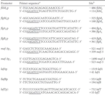
TABLE 2 Primers used for lateral flagellar promoter-lacZ fusion
construction
Promoter Primer sequencea Siteb
fliMLp 5=-TGCAACAGAGAGCAAACCG-3= ⫺486 fliML 5=-CGGGATCCTGAGTTGTTCTCGGTCTG-3= ⫹114 fliML
flgMLp 5=-AGCAAGAGCAATCGGAATC-3= ⫺325 flgML
5=-CGGGATCCCATCCGATGTAGTTGCCAAT-3= ⫹168 flgML
flgALp 5=-GGAAGATCTTCCCGGTGATTTTCATATTC-3= ⫺494 flgAL 5=-CGGGATCCCGTGCATTCAGCCAGATAG-3= ⫹386 flgAL
flgBLp 5=-CGGGATCCCGTGCATTCAGCCAGATAG-3= ⫺419 flgBL
5=-GGAAGATCTTCCCGGTGATTTTCATATTC-3= ⫹361 flgBL
maf-5p1 5=-GAGCTCTGCGCAAAGAAA-3= ⫺521 maf-5
5=-CGGGATCCTCAAGTGCAAGACCAGAGC-3= ⫹339 maf-5
maf-5p2 5=-CGTTGACCCGAGAAGTCA-3= ⫺1490 maf-5
5=-CGGGATCCTCGATCCAGCCTTGAAA-3= ⫺523 maf-5
lafAp 5=-TGTATGGCACTGGGTTGG-3= ⫺1519 lafA
5=-GCGGATCCGGTGGTCATGGAAGCAAA-3= ⫹41 lafA
llafBp 5=-TCTGCTGAAAACCGGTGG-3= ⫺553 lafB
5=-TTCTGCGCCTGTAAATTG-3= ⫹194 lafB
lafTp) 5=-TCCCCCGGGTGAGTTTGACACATCACCC-3= ⫺732 lafT 5=-CGCGGATCCATGAAACACCAGGACACA-3= ⫹53 lafT aUnderlined letters show the BamHI restriction site. Double-underlined letters show the BglII restriction site. Italicized letters show the SmaI restriction site.
bNumbers are numbers of nucleotides upstream (⫺) or downstream (⫹) from the start site for the indicated gene.
Wilhelms et al.
1438 jb.asm.org Journal of Bacteriology
on April 29, 2013 by UNIVERSITAT DE BARCELONA
http://jb.asm.org/
polyclonal rabbit anti-polar or anti-lateral flagellin antibodies (1:1,000) that were previously obtained (11). The unbound antibody was removed by three washes in phosphate-buffered saline (PBS), and a goat anti-rabbit alkaline phosphatase-conjugated secondary antibody (1:1000) was added. The unbound secondary antibody was removed by three washes in PBS. The bound conjugate was then detected by the addition of 5-bromo-4-chloroindolylphosphate disodium-nitroblue tetrazolium. Incubations were carried out for 1 h, and washing steps with 0.05% Tween 20 in phosphate-buffered saline were included after each incubation step.
For analysis of whole cells and bacterial supernatants before shearing, cells grown in liquid or viscous medium were collected at an optical den-sity at 600 nm of 0.8, and cells grown in plates were collected with 20 mM MgCl2in 100 mM Tris (pH 8.0) and diluted to an identical optical density
at 600 nm. Both were harvested by centrifugation (5,000⫻ g), and cell suspensions as well as supernatants were analyzed by Western blotting with anti-lateral flagellin antibodies (1:1,000).
-Galactosidase assays. The promoter-lacZ fusion plasmids de-scribed above were introduced into several A. hydrophila strains (Table 1). The cultures were grown in TSB or TSB medium with 18% (wt/vol) Ficoll at 25°C to an optical density at 600 nm of 0.6 or 0.8. Bacterial cells were permeabilized with chloroform and sodium dodecyl sulfate (SDS) over-night and assayed for-galactosidase activity as described by Miller (30). All experiments were performed at least 3 separate times.
Statistical analysis. The data obtained from the-galactosidase assays
were analyzed by the t test using Microsoft Excel software.
RESULTS
Lateral flagellar genes of A. hydrophila AH-3 are transcribed,
but lateral flagella are not expressed in liquid media. A.
hydro-phila and V. parahaemolyticus both have dual flagellar systems
(polar and lateral flagella) but do not share structural or
regula-tory genes, and both contribute to motility in semisolid plates (
9
,
18
,
19
). A. hydrophila polar flagellum is constitutive; however,
lateral flagella are induced in highly viscous media or on surfaces.
Both flagellar types have
54-dependent response regulators, FlrA
and LafK, which are essential for polar and lateral flagellum
gen-eration, respectively (
10
,
19
). Furthermore, despite A. hydrophila
FlrA and LafK showing 57% similarity to each other, their
C-ter-minal domains might recognize different DNA binding regions,
and LafK is unable to compensate for the FlrA mutation and vice
versa (
18
,
19
). In order to know whether viscosity conditions
reg-ulate transcription of lateral flagella, we measured the
-galacto-sidase activity of pDNlac-lafKp (lafKp-lacZ) after growth in liquid
(TSB) and viscous media (TSB with 18% [wt/vol] Ficoll or 3%
[wt/vol] gelatin) as well as on solid plates (TSA) (
Fig. 1B
). Data
showed similar
-galactosidase values after growth in liquid and
viscous media or on solid plates. Lateral flagellin (LafA)
transcrip-tion was analyzed by measuring
-galactosidase activity of
pDNlac-lafAp (lafAp-lacZ). Lateral flagellin transcription in
liq-uid media shows a very slight reduction compared to
transcrip-tion in viscous media or on solid plates, although lateral flagella
are not produced in liquid media (
Fig. 1A
and
B
). However, the
TEM assays showed lateral flagella in 85% of bacterial cells grown
in TSB with Ficoll and in 80% of cells grown in TSA. Levels of
-galactosidase activity from other lateral flagella promoters, such
as flgAL, flgBL, and lafB promoters, were also analyzed, with all of
them being similar in liquid and viscous media (
Fig. 1B
). In
addi-tion, RT-PCR assays showed that lafK, lafA, flgAL, flgBL, and lafB
transcription are viscosity/surface independent, since they are
transcribed in liquid and viscous media as well as on solid plates
(
Fig. 1C
). No RT-PCR product was obtained with primer pairs for
flgA
Land flgBL, which are divergently transcribed genes,
eliminat-ing the possibility of residual DNA in the RNA samples from
liq-uid cultures.
The presence of lateral flagellin (LafA) in the cytoplasmic
frac-tion of A. hydrophila AH-3 and the mutants AH-4427 (without
polar flagellin), AH-3
⌬lafA (without lateral flagellin), and
AH-5502 (rpoN) grown in liquid or viscous medium or on solid plates
was analyzed by Western blotting with anti-lateral flagellin (1:
1,000) polyclonal antibodies (
Fig. 2A
). We analyzed the presence
of polar flagellins by Western blotting with anti-polar flagellin
(1:1,000) polyclonal antibodies in these strains (
Fig. 2B
). AH-3
and AH-4427 mutant cytoplasmic fractions obtained after growth
in viscous media or on solid plates showed positive reactions with
anti-lateral flagellin serum but were unable to react with similar
fractions grown in liquid media. AH-3⌬lafA and AH-5502 (rpoN)
cytoplasmic fractions were unable to react with anti-lateral
flagel-lin serum. AH-3 and AH-3⌬lafA cytoplasmic fractions reacted
positively with polar flagellin serum and were unable to react with
AH-4427 and AH-5502 (rpoN) cytoplasmic fractions in all growth
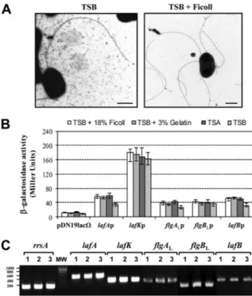
FIG 1 (A) Transmission electron microscopy of A. hydrophila AH-3 (wild
type) grown at 25°C in TSB and TSB with 18% (wt/vol) Ficoll. Bacteria were gently placed onto Formvar-coated copper grids and negatively stained using a 2% solution of uranyl acetate. Bar, 0.5m. (B) Analysis of -galactosidase activity of pDNlac-lafKp, pDNlac-lafAp, pDNlac-flgALp, pDNlac-flgBLp, and pDNlac-lafBp plasmids in A. hydrophila AH-405 after growth in liquid (TSB) medium, viscous medium (TSB plus 18% [wt/vol] Ficoll or 3% [wt/vol] gela-tin), and solid agar (TSA) at 25°C. As a control, we also measured the pDN19lac⍀ promoterless plasmid. The results shown are representative of three independent experiments. (C) RT-PCR amplification of lafK, lafA, flgAL,
flgBL, and lafB from cDNA of AH-3 after growth in liquid medium (TSB), viscous medium (TSB plus 18% [wt/vol] Ficoll), or solid agar (TSA) (lanes 1, 2, and 3, respectively). A. hydrophila ribosomal 16S (rrsA) amplification was used as a control for cDNA template. RT-PCR amplifications were performed at least twice with total RNA preparations obtained from a minimum of two independent extractions. The Ecogen molecular weight marker (MW) was used.
on April 29, 2013 by UNIVERSITAT DE BARCELONA
http://jb.asm.org/
conditions tested. Furthermore, we analyzed the presence of
lat-eral flagellin by Western blotting in whole cells of AH-3 before
shearing, as well as in the supernatants before and after shearing,
when grown in liquid or viscous medium or on solid plates.
Non-lateral flagellin was detected in whole cells or supernatants from
liquid cultures before and after shearing (
Fig. 2C
). These data
suggest that lateral flagellin is not translated in liquid media,
al-though it is transcribed.
The A. hydrophila AH-3
54factor (RpoN) is not involved in
lafK transcription. A. hydrophila AH-3 encodes an alternative
54sigma factor (RpoN) which is essential for both polar and lateral
flagellum expression (
10
,
19
). Lateral flagellar clusters contain
only one gene that encodes a
54-dependent response regulator,
LafK (
10
,
38
,
43
). In silico analysis of the A. hydrophila upstream
lafK sequence did not shown putative
54promoter sequences. In
order to establish if there was any relationship between the
54factor (RpoN) and lafK transcription, we measured the
-galac-tosidase activity of A. hydrophila wild-type and rpoN mutant
(AH-5502) strains carrying the lateral flagella gene promoter-lacZ
fu-sion plasmid pDNlac-lafKp. Similar
-galactosidase activities
were detected in both strains (
Fig. 3B
). In addition, RT-PCR
as-says showed lafK transcription in both the wild type and the rpoN
mutant (
Fig. 3C
).
Given that lafK transcription was
54independent, we
per-formed 5= RACE, as described in the Materials and Methods, to
further analyze the lafK promoter region. Primary PCR of tailed
cDNA using primers AAP and GSP2-LafK gave a unique DNA
band of 620 bp (
Fig. 4
). Sequence of the amplified band indicates
that it was tailed with G residues. The lafK transcription start was
located 335 nucleotides (nt) upstream from the lafK translation
start site, and DNA sequence upstream of the transcription start
contains a
70promoter sequence (TTGAAT-N16-TATGAT)
(
Fig. 4
). Furthermore, in silico analysis of the lateral flagellar
re-gion of A. caviae Sch3N (also showing dual flagellar systems)
al-lowed us to identify a
70promoter sequence 331 bp upstream of
the lafK start codon (5=-TTGAAT-N16-TATCAT-3=).
Identification of A. hydrophila lateral flagellum
28-depen-dent promoters. Transcriptions of polar and peritrichous flagellar
late genes are
28dependent (
16
,
17
,
18
,
31
). The lafB-U cluster of
the A. hydrophila lateral flagellar chromosomal region contains a
gene, lafS, which encodes a sigma factor orthologous to the V.
parahaemolyticus
28factor FliAL
and homologous to the A.
hy-drophila
28factor FliA (38/54 and 34/54% identity/similarity,
respectively) (
10
,
11
,
23
). In A. hydrophila, mutation of LafS
abol-ishes lateral flagellum formation (
11
), and lateral flagella were
restored by complementation with the pBAD33Gm-LAFS
plas-mid in the presence of 0.2%
L-arabinose. In silico sequence analysis
of A. hydrophila AH-3 lateral flagellum genes show a putative
28promoter sequence upstream of the anti-
28factor flgM
L
(
19
),
putative
54promoter sequences upstream of fliML, flgAL, flgBL,
maf-5, lafB, and the lateral flagellin gene, lafA (
11
,
19
), and several
putative promoter sequences upstream of the motor gene lafT. In
order to study which of these lateral flagella genes were
28depen-dent, we independently transferred the promoter-lacZ fusion
plasmids pDNlac-fliM
Lp (fliM
Lp-lacZ), pDNlac-flgM
Lp (flgM
Lp-lacZ), pDNlac-flgA
Lp (flgALp-lacZ), pDNlac-flgBLp (flgBLp-lacZ),pDNlac-maf-5p
1(maf-5p
1-lacZ), pDNlac-lafAp (lafAp-lacZ),
pDNlac-lafBp (lafBp-lacZ), and pDNlac-lafTp (lafTp-lacZ) into
A. hydrophila AH-405 (AH-3 with rifampin resistance) and the
lafS mutant (AH-3::lafS). Transconjugants were chosen and
-ga-lactosidase activity measured. Transcription from the flgM
Land
lafA promoters appeared to be highly affected by the lafS
muta-tion, since 90 and 98% reduction of
-galactosidase activity,
re-spectively, was found in the AH-3::lafS mutant compared to that
of the wild-type AH-405. However, the activity of the lafB
pro-moter was only slightly affected in the AH-3::lafS mutant (31%
reduction), and activities from fliM
L, flgA
LflgB
L, lafB, and lafT
promoters exhibited comparable values for both strains (
Fig. 3A
).
The maf-5p
1-lacZ cluster does not contain any promoter region,
since
-galactosidase activity in the wild-type AH-405 was similar
to that obtained with the pDN19lac
⍀ promoterless plasmid (data
not shown). Sequence analysis of the maf-5 upstream region
found a pseudogene which encodes an incomplete flagellin
frag-ment homologous (77/79% identity/similarity) to the C-terminal
region of ASA_0374 of A. salmonicida A449 (
32
). We amplified the
region both upstream and downstream of the predicted
pseudo-gene start codon and cloned it into the plasmid pDN19lac
⍀ to
generate the promoter-lacZ fusion plasmid pDNlac-maf-5p2
(maf-5p
2-lacZ). This plasmid was transferred by triparental
con-jugation into A. hydrophila AH-405 and the AH-3::lafS mutant.
The measurement of
-galactosidase activity from the maf-5p2
promoter is significant only for wild-type AH-405 and was similar
to the one obtained with the pDN19lac
⍀ promoterless plasmid in
the lafS mutant (
Fig. 3A
).
Total RNAs from A. hydrophila AH-3 and the lafS mutant were
used to amplify internal fragments of fliML, flgML, flgAL, flgBL,
maf-5, lafA, lafB, and lafT transcripts, but no lafA and maf-5
am-plicons were obtained from the lafS mutant (
Fig. 3C
).
Further-more, analysis of flgM
L-flgA
Ltranscription in the wild type by
RT-PCR showed that these two genes were cotranscribed. The
results suggest that flgM
L, maf-5, and lafA transcription are
28dependent, and flgML
is also transcribed from the flgAL
promoter.
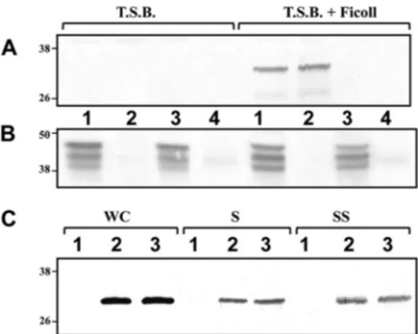
FIG 2 (A) Western blot analysis with anti-lateral flagellin (1:1,000) polyclonal
antibodies of cytoplasmic fractions of A. hydrophila AH-3 (wild type), mutant AH-4427 (without polar flagellin), AH-3⌬lafA (without lateral flagellin), and AH-5502 (rpoN) (lanes 1, 2, 3, and 4, respectively) of bacteria grown in liquid (TSB) or viscous medium (TSB plus 18% [wt/vol] Ficoll) at 25°C in TSB. (B) Western blot analysis with anti-polar flagellin (1:1,000) polyclonal antibodies of cytoplasmic fractions of the same strains and grown conditions as those described for panel A. (C) Western blot analysis with anti-lateral flagellin (1:1,000) polyclonal antibodies of whole cells (WC) and supernatant before shearing (S) and after shearing (SS) of A. hydrophila AH-3 grown in liquid medium (TSB), viscous medium (TSB plus 18% [wt/vol] Ficoll), or an agar plate (TSA) (lanes 1, 2, and 3, respectively).
Wilhelms et al.
1440 jb.asm.org Journal of Bacteriology
on April 29, 2013 by UNIVERSITAT DE BARCELONA
http://jb.asm.org/
Identification of A. hydrophila LafK-dependent
54lateral
flagellar promoters. Promoters recognized by the
54holoen-zyme require specialized enhancer-binding proteins, which bind
specific sequences located in a relatively remote position from the
transcription start site (
33
). Lateral flagellar clusters only contain
one gene that encodes a
54enhancer-binding protein, LafK (
34
),
which is required for V. parahaemolyticus lateral flagellum
tran-scription (
9
). In A. hydrophila the LafK mutation abolishes lateral
flagellum formation and swarming motility (
19
), as the wild-type
phenotype is restored by complementation with the
pBAD33Gm-LAFK plasmid in the presence of 0.2%
L-arabinose. To investigate
which of the A. hydrophila lateral flagellar clusters are
54and LafK
dependent,
-galactosidase activities of the A. hydrophila wild
type and the lafK (AH-5503) and rpoN (AH-5502) mutants
car-rying the promoter-lacZ fusion plasmids pDNlac-fliM
Lp (fliM
Lp-lacZ), pDNlac-lafKp (lafKp-p-lacZ), pDNlac-flgA
Lp (flgALp-lacZ),pDNlac-flgB
Lp (flgB
Lp-lacZ), pDNlac-lafBp (lafBp-lacZ), and
pDNlac-lafTp (lafTp-lacZ) were measured. Activities from flgBL
and lafB promoters appeared to be affected in both LafK and
RpoN mutant strains. The flgBL
promoter showed a reduction of
93% in both mutants, and the lafB promoter show a reduction of
75 and 73% in the RpoN and LafK mutants, respectively (
Fig. 3B
).
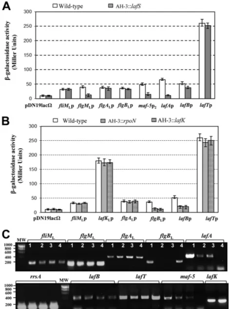
FIG 3 Analysis of-galactosidase activity after growth in TSB with 18% Ficoll at 25°C. (A) pDNlac-fliMLp, pDNlac-flgMLp, pDNlac-flgALp, pDNlac-flgBLp, pDNlac-maf-5p2, pDNlac-lafAp, pDNlac-lafBp, and pDNlac-lafTp plasmids in A. hydrophila wild-type (AH-405) and lafS mutant (AH-3::lafS) strains. (B) pDNlac-fliMLp, pDNlac-lafKp, pDNlac-flgALp, pDNlac-flgBLp, pDNlac-lafBp, and pDNlac-lafTp plasmids in the A. hydrophila wild type (AH-405) and rpoN (AH-5502) and lafK (AH-5503) mutant strains. pDNlac-lafKp was not analyzed in the lafK mutant. As a control, we measured the pDN19lac⍀ promoterless plasmid. The results shown are representative of three independent experiments. Bars represent standard deviations. (C) RT-PCR amplification of fliML, lafK,
flgML, flgAL, flgBL, maf-5, lafA, lafB, and lafT from cDNA of AH-3 (lane 1), AH-3::rpoN (2), AH-3::lafK (3), and AH-3::lafS (4) mutants. A. hydrophila ribosomal 16S (rrsA) amplification was used as a control for the cDNA template. RT-PCR amplifications were performed at least twice with total RNA preparations obtained from a minimum of two independent extractions. The Ecogen molecular weight marker (MW) was used.
on April 29, 2013 by UNIVERSITAT DE BARCELONA
http://jb.asm.org/
No significant variations were obtained from fliM
L, lafK, flgA
L,
and lafT promoters in any of these mutants. Furthermore,
RT-PCRs to compare fliM
L, flgA
L, flgB
L, lafB, and lafT gene
transcrip-tion in the wild type and the RpoN and LafK mutants showed
fliM
L, flgA
L, lafB, and lafT amplicons in the wild type and both
mutants, whereas no flgBL
amplicon was found in the RpoN and
LafK mutants (
Fig. 3C
). These results suggest that the flgB
Lpro-moter is
54and LafK dependent.
To identify the fliM
L, flgA
L, lafB, and lafT promoter regions,
amplification of the A. hydrophila AH-3 fliML, flgAL, lafB, and lafT
cDNA 5= ends was performed using 5= RACE as described in
Ma-terials and Methods. Primary PCR of tailed cDNA using primers
AAP and GSP2-FLIM
Lor GSP2-LafT give amplicons of 537 and
428 bp, respectively. However, primers AAP and GSP2-FLGAL
or
GSP2-LAFB render two amplicons of 650 and 827 bp and 700 and
588 bp, respectively. DNA sequence of the amplified bands
indi-cates that amplicons were tailed with G residues. The fliM
Land
lafT transcription starts were located 85 and 12 nt upstream from
the fliM
Land lafT translation start sites, respectively. The flgA
Ltranscription starts were located 158 and 332 nt upstream from
the flgA
Lstart site. The lafB transcription starts were located 39
and 158 nt upstream from the lafB start site. We were able to
identify
70promoter sequences in DNA regions upstream of the
transcription starts of fliML, flgAL, and lafT. Both
28and
54promoter sequences were found upstream of the lafB
transcrip-tion start (
Fig. 4A
).
DISCUSSION
Functional lateral flagellar systems, whose flagella are randomly
distributed over the cell surface in a manner similar to that of the
peritrichous flagella of Enterobacteriaceae, were reported in polar
flagellated bacteria with dual flagellar systems, such as A.
hydro-phila and V. parahaemolyticus (
5
). Lateral flagellar systems of these
two species are encoded by 38 genes, 37 of which are orthologous
and do not share either structural or regulatory genes with polar
flagellar systems. Despite these similarities, A. hydrophila lateral
FIG 4 (A) Amplification of the A. hydrophila AH-3 lafK, fliML, flgAL, lafB, and lafT cDNA 5= end was performed using the 5= RACE system, version 2.0 (Invitrogen). Amplicons were obtained by PCR using primers AAP and GSP2-LafK (lafKp), GSP2-FLIML(fliMLp), GSP2-FLGAL(flgALp), GSP2-LAFB (lafBp), and GSP2-LAFT (lafTp). Lanes: 1, PCR negative control; 2, primary PCR template; MW, molecular weight standard (Ecogen). Underlined sequences show start codons, asterisks show locations of the transcriptional start sites, and boldface nucleotides show potential consensus sequences. (B) Alignment in silico of28and 54promoter elements in A. hydrophila lateral flagellar promoters. The consensus28sequence is from Kutsukake (47). The consensus54sequence is from Barrios (48).
Wilhelms et al.
1442 jb.asm.org Journal of Bacteriology
on April 29, 2013 by UNIVERSITAT DE BARCELONA
http://jb.asm.org/
flagellum genes are distributed in a unique chromosomal region,
whereas V. parahaemolyticus lateral flagellar genes are distributed
in two discontinuous chromosomal regions (
9
,
11
,
19
). The
pres-ence of two active flagellar systems implies a high energetic cost for
a bacterium, therefore lateral flagellar synthesis should be
care-fully regulated in response to different environmental conditions.
Different environmental conditions have been associated with
lat-eral flagellar induction (
35
,
36
), but the most extensive association
is growth in viscous media or on a solid surface, which reduces
polar flagellum motility. However, while V. parahaemolyticus and
Azospirillium brasilense defects in polar flagellum formation or
motility allow lateral flagellum expression (
37
,
38
,
39
), Aeromonas
sp. polar flagellum defects do not induce constitutive lateral
fla-gella (
10
,
40
). This difference suggests that Aeromonas polar
fla-gella do not act as mechanosensors and that lateral flafla-gellar
regu-lation is not linked to polar flagella.
The V. parahaemolyticus lateral flagellar system is the best
stud-ied at the regulatory level, and it has been demonstrated that its
viscosity/surface-dependent expression is transcriptionally
regu-lated (
41
). Transcription of V. parahaemolyticus lateral flagellar
genes is organized into 3 levels (class I to III), where the first level
contains the unique lateral flagellum
54-associated
transcrip-tional activator, lafK, and the third level contains the lateral
flagel-lin gene, lafA. Lateral flagella of A. hydrophila are also expressed in
viscous media or on solid surfaces (
Fig. 1A
), but their regulatory
mechanisms have not been determined yet. Our data show that A.
hydrophila lateral flagellar genes are transcribed in liquid and
vis-cous media or on solid surfaces, although lateral flagellin is not
transduced in liquid media. All of the data suggest that lateral
flagella expression should be regulated by transductional
mecha-nisms.
Two transcriptional hierarchy models in Gamma- and
Alpha-proteobacteria lateral flagella have been described (
7
,
9
,
42
). Until
now, V. parahaemolyticus lateral flagellar transcriptional
hierar-chy has represented the Gammaproteobacteria model. In this
model, the
54-associated transcriptional activator, LafK, controls
transcription of class II lateral flagellum genes which contain the
28factor (fliA
L
) involved in transcription of class III lateral
fla-gellum genes (
9
). In order to establish the A. hydrophila lateral
flagellar cluster transcription hierarchy, promoter-lacZ fusions
with lateral flagellum promoters were analyzed in defined A.
hy-drophila lafK, lafS, and rpoN mutants. Furthermore, transcription
analysis of genes in different lateral flagellar clusters in these A.
hydrophila mutants were tested by RT-PCR assays.
Class III. In V. parahaemolyticus the
28factor (FliA
L) is
in-volved in transcription of late genes, such as lateral flagellin, the
anti-sigma factor flgML, and motor components (motABL), but
also some middle genes, such as flgKLL
and fliDSTKLAL
(
9
). Our
data show that A. hydrophila flgM
L, maf-5, and lafA are transcribed
from
28-dependent promoters, with the maf-5 promoter being
upstream of the pseudoflagelin gene, and fliML, flgBL, flgAL, and
lafT are transcribed from
28-independent promoters (
Fig. 3A
and
C
). Furthermore, the slight reduction of
-galactosidase
ac-tivity of lafB promoter fusion and the presence of the lafB
tran-script in the LafS mutant suggest that the lafBCXEFSTU cluster is
transcribed from different promoters, with one of them being
characterized as
28dependent. A similar situation has been
re-ported in the V. parahaemolyticus lateral flagellar orthologous
cluster fliDSTKLAL-motABL
(
9
) (
Fig. 4
and
5
).
Class II. Transcription of the V. parahaemolyticus lateral
fla-gellar genes included in the class II level are
54and LafK
depen-dent (
9
). A. hydrophila LafK is essential for lateral flagella
forma-tion; however, in contrast to the case for V. parahaemolyticus, lafK,
flgA
Land lafT are transcribed from
54/LafK-independent
pro-moters and are not class II genes (
Fig. 5
). Our results also suggest
that fliML
is transcribed from
54/LafK-independent promoters
and flgB is transcribed from a
54/LafK-dependent promoter, the
sequence of which was predicted in silico. Furthermore, assays
indicate that lafB promoter activity was only 73 to 75% reduced in
the LafK and RpoN mutants (
Fig. 3B
), and the amplification of the
lafB cDNA 5= ends by 5= RACE shows two promoter sequences, a
28promoter sequence (TAAGGG-N17-GTCGAAA) and a
54promoter sequence (TGGCAT-N5-TTCTG), with the latter being
more active (
Fig. 4
). These results indicate that lafBCXEFSTU is
transcribed from two promoters. A similar situation is
de-scribed for V. parahaemolyticus, although it has not been
stud-ied at the transcriptional level. The
28factor lafS is contained
in the lafBCXEFSTU cluster, but the lack of LafK does not
prevent lafS transcription in Aeromonas. RT-PCR assays
showed transcription of maf-5 and lafA in LafK and RpoN
mutants (
Fig. 3C
). Data suggest that lafS is transcribed from a
second promoter in a
54/LafK-independent manner and is less
active than the promoter upstream of lafB. Amplification of the
lafS cDNA 5= ends by 5= RACE allowed us to obtain an
ampli-con (data not shown).
Class I. As previously indicated, A. hydrophila lateral flagellum
FIG 5 Comparative proposed A. hydrophila and Vibrio parahaemolyticus (9) lateral flagellar gene transcription hierarchies. Diagrams show the three levels of lateral flagellar hierarchy, class I to III. Class I genes are at the top of the hierarchy, being70dependent (dep.) in Aeromonas. One of the class I genes encodes a54-associated transcriptional activator (LafK) that activates54 -dependent promoters preceding the class II clusters. One of the class II genes encodes the28factor (LafS), which activates the transcription of class III genes. In Aeromonas, the28factor might also be transcribed from another promoter.
on April 29, 2013 by UNIVERSITAT DE BARCELONA
http://jb.asm.org/
regulatory cascade class I seems to include more that one gene.
Although previous in silico analysis showed putative
54promoter
sequences upstream of lafK, fliM
L, and flgA
L(
19
), the data
ob-tained now show that upstream regions of fliML, flgAL, lafT, and
lafK contain
70promoter sequences (
Fig. 4
). A. hydrophila lateral
flagellum class I gene transcription is
70dependent, as has been
reported for A. hydrophila polar flagellar class I genes. Data are
consistent with the fact that A. hydrophila lateral flagellar genes are
transcribed in liquid and viscous media and on solid medium, in
contrast to V. parahaemolyticus (
41
). A. hydrophila lateral flagella
are not induced by mutation of polar flagellum genes, as happens
in V. parahaemolyticus. In V. parahaemolyticus, the lafK promoter
is located upstream of motY
L, which encodes a lateral motor
pro-tein that does not possess an orthologue in A. hydrophila, and
genes of the motY
L-lafK-fliEFGHIJ
Lcluster are classified as class I
and II. In addition, the V. parahaemolyticus fliMNPQRL-flhABL
lateral flagellar cluster is classified as class I (
43
), as is the case in A.
hydrophila. In A. hydrophila, lafS transcription feeds into class I,
since LafK mutation does not abolish transcription of
28-depen-dent promoters (
Fig. 3C
). The A. hydrophila lateral flagellar
tran-scriptional hierarchy is complex, since many clusters of genes are
transcribed independently of LafK. LafK is not strictly the master
lateral flagellar regulator in A. hydrophila.
Our results indicate that despite A. hydrophila lateral flagella
only being expressed in viscous media or on solid surfaces, their
genes are transcribed in liquid, although lateral flagellin was not
detected in liquid media. Recently, it has been described that the
A. hydrophila AH-3 lateral flagellin is glycosylated (
44
), although
it is nonglycosylated in V. parahaemolyticus, and this fact could
contribute to the complexity of their lateral flagella transcription
hierarchy and the important differences between these two
bacte-ria. An A. hydrophila AH-3 in-frame deletion mutant of the
pseu-daminic acid biosynthetic gene pseB homologue resulted in the
abolition of lateral flagella formation by posttranscriptional
regu-lation of the flagellin, which was restored by complementation
with the wild-type pseB homologue or Campylobacter pseB (data
not shown).
ACKNOWLEDGMENTS
This work was supported by Plan Nacional de I⫹ D (Ministerio de Edu-cación, Ciencia y Deporte and Ministerio de Sanidad, Spain) and Gener-alitat de Catalunya (Centre de Referència en Biotecnologia).
We thank Maite Polo for her technical assistance and the Servicios Científico-Técnicos from the University of Barcelona.
REFERENCES
1. Harshey RM, Matsuyama T. 1994. Dimorphic transition in Escherichia
coli and Salmonella typhimurium: surface-induced differentiation into
hy-perflagellate swarmer cells. Proc. Natl. Acad. Sci. U. S. A. 91:8631– 8635. 2. Kearns DB. 2010. A field guide to bacterial swarming motility. Nat. Rev.
Microbiol. 8:634 – 644.
3. Doyle TB, Hawkins AC, McCarter LL. 2004. The complex flagellar torque generator of Pseudomonas aeruginosa. J. Bacteriol. 186:6341– 6350. 4. Toutain CM, Zegans ME, O’Toole GA. 2005. Evidence for two flagellar stators and their role in the motility of Pseudomonas aeruginosa. J. Bacte-riol. 187:771–777.
5. Merino S, Shaw JG, Tomás JM. 2006. Bacterial lateral flagella: an induc-ible flagella system. FEMS Microbiol. Lett. 263:127–135.
6. Shinoda S, Okamoto K. 1977. Formation and function of Vibrio
parahaemolyticus lateral flagella. J. Bacteriol. 129:1266 –1271.
7. Liu R, Ochman H. 2007. Origins of flagellar gene operons and secondary flagellar systems. J. Bacteriol. 189:7098 –7104.
8. Ren CP, Beatson SA, Parkhill J, Pallen MJ. 2005. The Flag-2 locus, an
ancestral gene cluster, is potentially associated with a novel flagellar system from Escherichia coli. J. Bacteriol. 187:1430 –1440.
9. Stewart BJ, McCarter LL. 2003. Lateral flagellar gene system of Vibrio
parahaemolyticus. J. Bacteriol. 185:4508 – 4518.
10. Canals R, Ramirez S, Vilches S, Horsburgh G, Shaw JG, Tomás JM,
Merino S. 2006. Polar flagellum biogenesis in Aeromonas hydrophila. J.
Bacteriol. 188:542–555.
11. Gavín R, Rabaan AA, Merino S, Tomás JM, Gryllos I, Shaw JG. 2002. Lateral flagella of Aeromonas species are essential for epithelial cell adher-ence and biofilm formation. Mol. Microbiol. 43:383–397.
12. Okabe M, Yakushi T, Kojima M, Homma M. 2002. MotX and MotY, specific components of the sodium-driven flagellar motor, colocalize to the outer membrane in Vibrio alginolyticus. Mol. Microbiol. 46:125–134. 13. Karlyshev AV, Linton D, Gregson NA, Wren BW. 2002. A novel paralo-gous gene family involved in phase-variable flagella-mediated motility in
Campylobacter jejuni. Microbiology 148:473– 480.
14. Chilcott GS, Hughes KT. 2000. Coupling of flagellar gene expression to flagellar assembly in Salmonella enterica serovar Typhimurium and
Esch-erichia coli. Microbiol. Mol. Biol. Rev. 64:694 –708.
15. McCarter LL. 2001. Polar flagellar motility of the Vibrionaceae. Microbiol. Mol. Biol. Rev. 65:445– 462.
16. Dasgupta N, Wolfgang MC, Goodman AL, Arora SK, Jyot J, Lory S,
Ramphal R. 2003. A four-tiered transcriptional regulatory circuit controls
flagellar biogenesis in Pseudomonas aeruginosa. Mol. Microbiol. 50:809 – 824.
17. Prouty MG, Correa NE, Klose KE. 2001. The novel sigma54 and sigma28 dependent flagellar gene transcription hierarchy of Vibrio cholerae. Mol. Microbiol. 39:1595–1609.
18. Wilhelms M, Molero R, Shaw JG, Tomas JM, Merino S. 2011. Tran-scriptional hierarchy of Aeromonas hydrophila polar-flagellum genes. J. Bacteriol. 193:5179 –5190.
19. Canals R, Altarriba M, Vilches S, Horsburgh G, Shaw JG, Tomás JM,
Merino S. 2006b. Analysis of the lateral flagellar gene system of Aeromonas hydrophila AH-3. J. Bacteriol. 188:852– 862.
20. Sambrook J, Fritsch EF, Maniatis T. 1989. Molecular cloning: a labora-tory manual, 2nd ed. Cold Spring Harbor Laboralabora-tory, Cold Spring Har-bor, NY.
21. Sanger F, Nicklen S, Coulson AR. 1977. DNA sequencing with chain-terminating inhibitors. Proc. Natl. Acad. Sci. U. S. A. 74:5463–5467. 22. Altschul FS, Madden TL, Schaffer AA, Zhang J, Zang Z, Miller W,
Lipman J. 1997. Gapped BLAST and PSI-BLAST: a new generation of
protein database search programs. Nucleic Acid Res. 25:3389 –3402. 23. Studholme DJ, Dixon R. 2003. Domain architectures of
sigma54-dependent transcriptional activators. J. Bacteriol. 185:1757–1767. 24. Münch R, Hiller K, Grote A, Scheer M, Klein J, Schobert M, Jahn D.
2005. Virtual footprint and PRODORIC: an integrative framework for regulon prediction in prokaryotes. Bioinformatics 21:4187– 4189. 25. Hanahan D. 1983. Studies on transformation of Escherichia coli with
plasmids. J. Mol. Biol. 166:557–580.
26. Bott M, Meyer M, Dimroth P. 1995. Regulation of anaerobic citrate metabolism in Klebsiella pneumoniae. Mol. Microbiol. 18:533–546. 27. Milton DL, O’Toole R, Horstedt P, Wolf-Watz H. 1996. Flagellin A is
essential for the virulence of Vibrio anguillarum. J. Bacteriol. 178:1310 – 1319.
28. Jimenez N, Lacasta A, Vilches S, Reyes M, Vazquez J, Aquillini E,
Merino S, Regué M, Tomás JN. 2009 Genetics and proteomics of Aero-monas salmonicida lipopolysaccharide core biosynthesis. J. Bacteriol. 191:
2228 –2236.
29. Totten PAA, Lory S. 1990. Characterization of the type a flagellin gene from Pseudomonas aeruginosa PAK. J. Bacteriol. 172:7188 –7199. 30. Miller JH. 1992. A short course in bacterial genetics, 2nd ed. Cold Spring
Harbor Laboratory Press, Plainview, NY.
31. Ohnishi K, Kutsukake K, Suzuki H, Iino T. 1990. Gene fliA encodes an alternative sigma factor specific for flagellar operons in Salmonella
typhi-murium. Mol. Gen. Genet. 221:139 –147.
32. Reith ME, Singh RK, Curtis B, Boyd JM, Bouevitch A, Kimball J,
Munholland J, Murphy C, Sarty D, Williams J, Nash JH, Johnson SC, Brown LL. 2008. The genome of Aeromonas salmonicida subsp.
salmoni-cida A449: insights into the evolution of a fish pathogen. BMC Genomics
18:9 – 427. doi:10.1186/1471-2164-9-427.
33. Buck M, Gallegos MT, Studholme DJ, Guo Y, Gralla JD. 2000. The bacterial enhancer-dependent sigma 54 transcription factor. J. Bacteriol.
182:4129 – 4136.
Wilhelms et al.
1444 jb.asm.org Journal of Bacteriology
on April 29, 2013 by UNIVERSITAT DE BARCELONA
http://jb.asm.org/
34. Merino S, Tomás JM. 2009. Lateral flagella systems. In Jarrell K (ed.), Pili and flagella: current research and future trends. Caister Academic Press, Norfolk, United Kingdom.
35. Berleman JE, Bauer CE. 2005. A che-like signal transduction cascade involved in controlling flagella biosynthesis in Rhodospirillum centenum. Mol. Microbiol. 55:1390 –1402.
36. Madi L, Kessel M, Sadovnik E, Henis Y. 1988. Electron microscopic studies of aggregation and pellicle formation in Azospirillum spp. Plant Soil 109:115–121.
37. Alexandre G, Rohr R, Bally R. 1999. A phase variant of Azospirillum
lipoferum lacks a polar flagellum and constitutively expresses
mechano-sensing lateral flagella. Appl. Environ. Microbiol. 65:4701– 4704. 38. Kawagishi I, Maekawa Y, Atsumi T, Homma M, Imae Y. 1995. Isolation
of the polar and lateral flagellum-defective mutants in Vibrio alginolyticus and identification of their flagellar driving energy sources. J. Bacteriol.
177:5158 –5160.
39. McCarter LL, Hilmen M, Silvermanm M. 1988. Flagellar dynamometer controls swarmer cell differentiation of V. parahaemolyticus. Cell 54:345–351. 40. Altarriba A, Merino S, Gavín R, Canals R, Rabaan A, Shaw JG, Tomas
JM. 2003. A polar flagella operon (flg) of Aeromonas hydrophila contains
genes required for lateral flagella expression. Microb. Pathog. 34:249 –259. 41. Belas R, Simon M, Silverman M. 1986. Regulation of lateral flagella gene
transcription in Vibrio parahaemolyticus. J. Bacteriol. 167:210 –218.
42. Chang Y, Tang T, Li J-L. 2007. Isolation of a flagellar operon in
Azospi-rillum brasilense and functional analysis of FlbD. Res. Microbiol. 158:521–
528.
43. Gode-Potratz CJ, Kustusch RJ, Breheny PJ, Weiss DS, McCarter LL. 2011. Surface sensing in Vibrio parahaemolyticus triggers a program of gene expression that promotes colonization and virulence. Mol. Micro-biol. 79:240 –263.
44. Wilhelms M, Fulton KM, Twine SM, Tomás JM, Merino S. 2012. Differential glycosylation of polar and lateral flagellins in Aeromonas
hy-drophila AH-3. J. Biol. Chem. 287:27851–27862.
45. Merino S, Camprubi S, Tomas JM. 1991. The role of lipopolysaccharide in complement-killing of Aeromonas hydrophila strains of serotype O:34. J. Gen. Microbiol. 137:1583–1590.
46. Rubires X, Saigi F, Piqué N, Climent N, Merino S, Albertí S, Tomás JM,
Regué M. 1997. A gene (wbbL) from Serratia marcescens N28b (O4)
com-plements the rfb-50 mutation of Escherichia coli K-12 derivatives. J. Bac-teriol. 179:7581–7586.
47. Kutsukake K, Ohya Y, Iino T. 1990. Transcriptional analysis of the flagellar regulon of Salmonella typhimurium. J. Bacteriol. 172:741–747. 48. Barrios H, Valderrama B, Morett E. 1999. Compilation and analysis
of54-dependent promoter sequences. Nucleic Acid Res. 27:4305– 4313.