Table of contents
Abstract
1
Chapter 1. Introduction
3
1.1 Ageing as public health priority
3
1.2 Inflammageing and its potential contribution to age-associated diseases
4
1.3 Oxidative stress and antioxidant defence mechanisms
6
1.4 The oxidative stress theory of ageing and risk of diseases
9
1.5 CKD as age related disease
11
Chapter 2. Summary of key results
17
2.1 Introduction to the experimental results
17
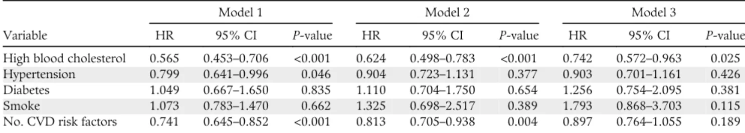
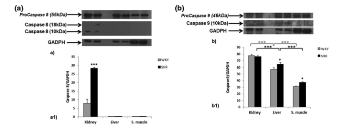
2.2 CKD and hypertension
18
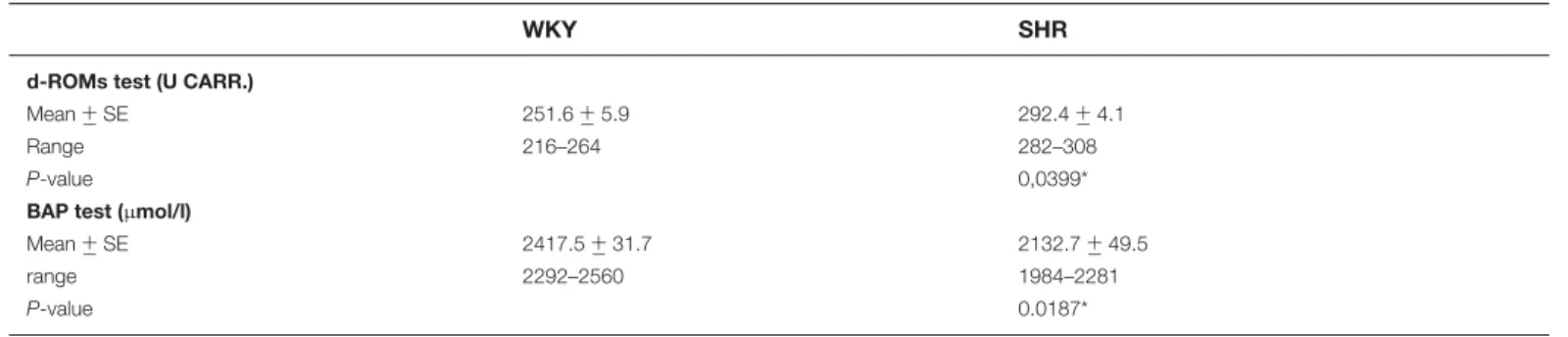
2.3 Oxidative stress and inflammatory status in CKD
22
2.4 CKD and obesity
26
2.5 CKD and hyperuricemia
29
References
35
1
Abstract
Chronic kidney disease (CKD) is a major public health problem worldwide and its
main consequences include the loss of renal function leading to end-stage renal
disease (ESRD), an increased risk of cardiovascular disease (CVD), a significant
increase in morbidity and mortality, and a decrease in health related quality of life.
The risk of CKD increases with age, though there seems to be a complex relationship
between ageing and this disease: elderly patients are overrepresented in the dialysis
population and geriatric complications are highly detectable in younger patients with
ESRD. This has led to the hypothesis of a premature biological ageing process of
different organ systems associated with CKD.
The present research work was based on translational approach to study the role of
many CKD risk factors such as hypertension, oxidative stress/inflammation, obesity,
and hyperuricemia with the aim of identifying new molecular mechanisms of kidney
damage to prevent it by successful behaviour modifications. For this purpose, both
human and animal models were used.
Human pathological models: in both ESRD and obese patients, the role of oxidative
stress, inflammation and hyperuricemia in progression and complications of CKD
was investigated.
Human physiological models: in a consistent healthy population, the oxidative status
and its correlation with traditional cardiovascular risk factors were examined. In
addition, the health history data of centenarian subjects was utilized to study the
clinical and prognostic value of traditional cardiovascular risk factors in relation to
mortality.
Animal models: the mechanisms renal damage, induced by hypertension
(Spontaneously Hypertensive Rat) and obesity (Cafeteria diet rats), were verified. In
this context, the antioxidant and cytoprotective effects of a nutraceutical (Bergamot
extract) on obesity was also tested.
This multilevel approach has allowed us to individually and synergistically analyze
some aspects of the complex pathogenic mechanism of CKD, in order to clarify the
2
role of the new amplifying risk factors for CKD and to prepare an effective
personalized prevention plan by acting on both modifiable and non-modifiable risk
factors.
3
Chapter 1. Introduction
1.1 Ageing as public health priority
With the extension of life expectancy, the percentage of elderly individuals in the
general population has considerably increased, whereby understanding why ageing
results in progressively higher vulnerability to chronic morbidity, disability, and
frailty has become a public health priority
[
Bektas et al., 2018
]
.
The population ageing is a worldwide burden, and the number of older adults is
increasing at an accelerating rate: it is estimated that in the next 30 years at least 20%
of the population will be aged >60 years and the most substantial increase will be
observed in the oldest-age group (aged>85 years) [Kinsella and He, 2009].
Population ageing occurs at various rates in different geographic regions (Figure 1)
wherein Europe currently includes the most aged population but, in the next years, it
is anticipated that Asia, South America, and Africa will experience the most rapid
rate of increase in population ageing [
Bektas et al., 2018
]. These demographic
changes in age composition will affect needs and demand for health and social care in
many countries worldwide and urgently require the adaptation of health policies to
tackle complex chronic diseases and disabilities and to improve elders' quality of life
[Christensen et al., 2009].
Fig. 1. The Speed of Population Ageing: time required or expected for percentage of population
aged 65 and over torise from 7 to 14 % [Kinsella and He, 2009]
4
1.2 Inflammageing and its potential contribution to age-associated
diseases
Ageing is a ubiquitous and physiological phenomenon influenced by a complex
interaction between genetic and environmental factors.
The intracellular and cellular
processes that contribute to ageing include genomic instability, mitochondrial
dysfunction, telomere attrition, epigenetic alterations, loss of proteostasis,
deregulated nutrient sensing, cellular senescence, stem cell exhaustion and altered
intercellular communication [Rebelo-Marques et al., 2018]. This phenomenon can
alter cell population and damage cell functionality, thereby compromising the
function of physiological systems (e.g. immune system, musculoskeletal system,
cardiovascular system, endocrine system, and nervous systems), increasing the risk of
organism failure (Figure 2).
Fig. 2. Ageing hallmarks [Rebelo-Marques et al., 2018]
A salient feature of ageing tissues is chronic inflammation, characterized by high
levels of pro-inflammatory markers, a condition defined “inflammageing”, a term
first coined in 2000 by Claudio Franceschi [Franceschi et al., 2000]. Inflammageing
describes the low-grade, chronic, systemic inflammation in ageing, in the absence of
overt infection (“sterile” inflammation), and represents a highly significant risk factor
5
for both morbidity and mortality in elderly people [Franceschi et al., 2000]. This
pro-inflammatory state is characterized by high levels of circulating pro-pro-inflammatory
mediators, including interleukines, C-reactive protein (CRP), interferon (IFNα) and
IFNβ, transforming growth factor-β (TGFβ), tumour necrosis factor-α (TNF) and its
soluble receptors, and serum amyloid A. Under physiological conditions, these
plasmatic inflammatory mediators are involved in defense mechanisms against
infections or extraneous molecules but when their expression is exacerbated and
prolonged, they become detrimental. Epidemiological studies have demonstrated that
inflammageing is a risk factor for many age-related diseases such as: cardiovascular
diseases (CVD), cancer, chronic kidney disease (CKD), dementia, and depression as
well as for global indicators of poor health status, such as multimorbidity, mobility
and disability in daily activities, sarcopenia, frailty and premature death (Figure 3)
[Salimi et al., 2018; Leonardi et al., 2018]. On the basis of these findings,
inflammageing should be considered one of the pillars of the biology of ageing and a
marker of accelerated ageing. However, it is unclear if inflammation causes the
associated pathology directly or is instead a biomarker for the rate of biological
ageing.
6
1.3 Oxidative stress and antioxidant defence mechanisms
Oxidative stress occurs when there is an imbalance between the production of free
radical species and the antioxidant ability to neutralize their harmful effects
[Salisbury et al., 2015]. Free radicals can be defined as higly reactive molecular
species (atoms or molecules) that contain one or more unpaired electrons in their
external shell or outer orbit and are capable of independent existence
[Chandrasekaran et al., 2017]. In cells, these radicals can act as oxidants or reductants
by losing or accepting a single electron and are continuously produced by the
organism’s normal use of oxygen [Lobo et al., 2010]. Free radicals include reactive
radical and non-radical derivatives of oxygen (ROS) and nitrogen (RNS) that are
collectively called reactive oxygen nitrogen species (RONS) [Powers et al., 2011].
The generation of RONS is a physiological process and, at moderate or low levels,
RONS are important molecules involved in a number of cellular signaling pathways,
in the extraction of energy from organic molecules, in immune defense, in mitogenic
response, and in redox regulation [Genestra et al., 2007]. An excess production or a
decreased scavenging of RONS has been implicated in ageing and age-related
diseases [Venkataraman et al., 2013]. Both endogenous and exogenous sources of
RONS have been described. The endogenous sources of RONS include different
subcellular organelles such as mitochondria, peroxisomes and endoplasmic reticulum,
where oxygen consumption is high [Phaniendra et al., 2015]. NADPH oxidase is the
prevalent source of the superoxide radical (•O
2
-), which is formed by the addition of
one electron leak from
the
electron transport system during cellular respiration to the
molecular oxygen [Miller et al., 1990]. Most of the superoxide is dismutated into
hydrogen peroxide (H
2O
2) by superoxide dismutase (SOD) [Genestra et al., 2007].
H
2O
2is a neutral molecule because it has no unpaired electrons, but it is able to form
the most reactive and dangerous radical, the hydroxyl radical (•OH), through the
Fenton or Haber–Weiss reaction. Hydroxyl radicals mainly react with phospholipids
in cell membranes and proteins. In activated neutrophils, in the presence of chloride
and myeloperoxidase, H
2O
2can be converted to hypochlorous acid that can react with
7
DNA and produce pyrimidine oxidation products and add chloride to DNA bases
[Kulcharyk et al., 2001]. Another important determinant in the cellular redox
equilibrium is nitric oxide (NO). In mammals, NO can be generated by three main
isoforms of nitric oxide synthase (NOS): endothelial NOS, related to vasodilation and
vascular regulation, neuronal NOS, linked to cellular signaling, and inducible NOS,
activated in response to various endotoxin or cytokine signals [Adams et al., 2015].
All isoforms of NOS utilize arginine as the substrate, and molecular oxygen and
reduced nicotinamide-adenine-dinucleotide phosphate (NADPH) as co-substrates.
The reaction of NO with superoxide radical (•O
2
![Fig. 1. The Speed of Population Ageing: time required or expected for percentage of population aged 65 and over torise from 7 to 14 % [Kinsella and He, 2009]](https://thumb-eu.123doks.com/thumbv2/123dokorg/2870315.9368/5.892.194.701.834.1063/speed-population-ageing-required-expected-percentage-population-kinsella.webp)
![Fig. 4. Oxidative stress and tissue damage [Rao et al., 2011]](https://thumb-eu.123doks.com/thumbv2/123dokorg/2870315.9368/10.892.204.737.364.904/fig-oxidative-stress-and-tissue-damage-rao-et.webp)